Embed Size (px)
Citation preview

Oxygen Delivery, Bronchial Hygiene and Airway Clearance
Dana Evans, BHS, RRT-NPS, AE-CShawna Strickland, MEd, RRT-NPS
University of Missouri-ColumbiaRespiratory Therapy Clinical Instructors

Oxygen Cylinders
Made of steel or aluminum– Remember that steel is magnetic…don’t take a
steel tank into the MRI suite!– The aluminum tank is more suited to portability
Sizes– Typically found in the hospital: E and H– Typically found in the home: D and smaller

Oxygen Cylinders
Identifiers– Color (in the US: oxygen is green, air is yellow)
• Aluminum tanks have a color strip at the top and silver on the bottom
• Steel tanks are solid colors (unless it’s a gas mix)
– Identification label with contents• Medical oxygen is 99.5% pure

How do I get oxygen out of the tank?
Equipment necessary:– Regulator – Tank key– Tank – Oxygen delivery device
Things to remember:– “crack and bleed”

How long does the tank last?
Every size tank holds a different amount of gas (obviously, bigger tanks last longer than smaller tanks)
What do I need to figure out the duration?– Cylinder factor
• E cylinder factor = 0.28
– Flow rate of oxygen to the patient– How full is the tank?

Cylinder Duration Equation
Your patient is wearing a nasal cannula with oxygen flowing at 2 LPM. He is using an E cylinder and it is full (2200 psig).Equation:0.28 x 2200 2 LPMThis tank will last 308 minutes…– 5 hours and 8 minutes

Try one on your own…
Your patient is wearing a nasal cannula with oxygen flowing at 5 LPM. He is using an E cylinder and it is half full (1100 psig).
How long will this tank last?

Oxygen Orders
Remember that oxygen is a drug…– It must be prescribed by a physician.
PRN
Oxygen saturations via pulse oximeter– SpO2

Suctioning
Definition:– The removal of tracheobronchial and upper airway
secretions
Purpose:– To clear the airways of obstructions for improved gas
exchange and prevent aspiration
Important to remember:– This is always a sterile procedure when the patient has
an endotracheal tube or tracheostomy tube

One-Use Sterile Catheters
Sized in French (typically 6-14 Fr)Most catheters are 56 cm longCommon features:– Thumb port to apply suction– Side holes in the distal tip for plugging– Distal tip is blunt and open– Flexible– Some have markings for length (cm)

Closed-Circuit Catheters
Common features:– Endotracheal or tracheostomy tube adaptor
– Suction catheter inside sterile sheath
– Thumb port
– Lavage port
Popular because:– No disconnection from the ventilator (decreased VAP)
– Reduced cost
– Reduced exposure of HCP to infectious materials

Complications of Suctioning
Hypoxemia
Cardiac arrhythmias
Trauma to airway mucosa
Atelectasis
Contamination of lower airway
Contamination of caregivers
Increased intracranial pressure

Suction Catheters

Manual Ventilation
Purpose:– To provide positive pressure ventilation and
supplemental oxygen to a patient who is• Apneic
• Bradycardic
• Intubated or trached
• Unable to expand all lung areas due to weakness

Spontaneous Ventilation
Ribs expand and diaphragm drops to create a negative pressure inside the thoracic cavity
The lungs fill with air because the atmospheric pressure greater than the intrathoracic pressure
Exhalation is passive (relying on chest recoil)

Positive Pressure Ventilation
Concept:– External pressure applied to the lung to move air – Exhalation is still passive
Advantages:– Provide ventilation and oxygen for those who can’t (for
whatever reason) do it themselves
Disadvantages:– Over-inflation can cause many pulmonary and
hemodynamic complications– Under-inflation doesn’t allow adequate ventilation and
oxygenation

Manual Resuscitators
Three sizes:
•Adult (25 kg and larger)
•Pediatric (10-25 kg)
•Neonatal (less than 10 kg)

Features of Manual Ventilators
Oxygen tubing
Oxygen reservoir (to provide more than 0.40 FiO2)
Body of bag
Lots of one-way valves to direct air flow
Patient adaptor (to mask or tube)
Exhalation port (do not occlude this!)
Optional PEEP valve

How to provide breaths with a manual ventilator…
Breath rate: 12 per minute– That works out to one every five seconds
Volume:– Watch the chest– It should gently rise while you squeeze the bag
with two hands• Too little volume: atelectasis and ↓oxygenation
• Too much volume: pneumothorax and ↓oxygenation

What questions do I need to ask before choosing a bronchial hygiene therapy?
1. Does the patient have excessive mucus production?2. Does the patient have a weak, ineffective cough?3. Is the patient able to follow directions?4. Does the patient have a caregiver that can help administer
therapy?5. Is the patient able to ambulate and/or change positions easily?6. What outcomes will be used to assess effectiveness of
therapy?7. If the patient is currently receiving bronchial hygiene, when
was the last time the appropriateness of the therapy was evaluated?
8. Has anything in the patient’s condition changed since the last evaluation?
Traditional Bronchial Hygiene
Directed Cough
Postural Drainage
External manipulation of the thorax– Chest wall percussion– Chest wall vibration

Four Phases of Cough

Postural Drainage Positioning
Use gravity to move secretions to the large airways so the patient can cough them out.

New Methods of Bronchial Hygiene
Positive expiratory pressure (PEP)– Acapella
Flutter valve therapy
Intrapulmonary percussive ventilation (IPV)
High frequency chest wall oscillation (HFCWO)

PEP Therapy
This can be used with or without regular nebulizer therapy
•Using it with nebulizer therapy achieves two goals at once
When the patient exhales, positive pressure is created in the lungs.
This pressure allows air to enter behind areas of mucus obstruction and keeps the airways open during exhalation.
During exhalation, mucus is now able to move the mucus toward the larger airways and the patient can cough it out.

Contraindications to PEP
Patients who are unable to tolerate the ↑ in work of breathingICP > 20 mm HgHemodynamic instabilityEpistaxisUntreated pneumothorax
Recent facial, oral or skull surgery or traumaEsophageal surgeryActive hemoptysisNauseaKnown or suspected tympanic rupture or other middle ear problem

Flutter Valve
Cost of device: $50-60

Flutter Valve Therapy
When correctly, the effect is 3-fold:– Vibrations applied to the airway facilitate the
loosening of secretions– The increase in bronchial pressure helps avoid
air trapping– Expiratory air flows are accelerated and
facilitate the upward movement of mucus

2 Stages of Flutter Technique
Stage 1– Loosening and
mobilizing mucus
– Using flutter will increase the pressure on exhalation and recruit lung units similar to the PEP device
Stage 2– Eliminating mucus
– Cough or huff maneuver follows the flutter to help expel the secretions

Flutter “Tips”
Tilt is important– With the mouthpiece horizontal to the floor:
• Tilt cone up or down to get maximal effect
– Feel the patient’s chest and back for vibrations
Clean the device on a regular basis by disassembling and soaking

IPV
Delivers rapid, high-flow bursts of air (or oxygen) into the lungs.
At the same time, it delivers therapeutic aerosols (medications that might open the airways like Albuterol).
Requires compressed gas to work.

Acapella
Similar to PEP but adds vibration therapy as well.
Can be delivered with aerosol therapy.

Who can use the IPV?
Patients who can breathe on their own with a mouthpiece or mask
Patients who are intubated and on a mechanical ventilator.
Patients who have a tracheostomy and may or may not be on a ventilator.

IPV
Clinical Indications– Bronchiolitis
– Cystic fibrosis
– Chronic bronchitis
– Bronchiectasis
– Neuromuscular disorders
– Emphysema
Treatments typically last for about 15-20 minutes, depending on the individual patient and the medications that need to be given.
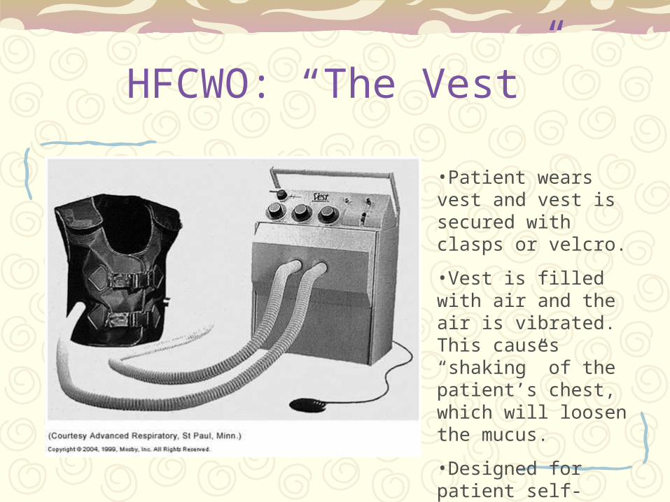
HFCWO: “The Vest”
•Patient wears vest and vest is secured with clasps or velcro.
•Vest is filled with air and the air is vibrated. This causes “shaking” of the patient’s chest, which will loosen the mucus.
•Designed for patient self-administration (home use).

HFCWO: “The Vest”
Pieces and parts:– Foot pedal (makes it
go)
– Patient vest is chosen based on patient size
– Air pulse generator • We can adjust ventilator
flow and speed of vibrations
Treatments are usually about 30 minutes long.
Most aerosolized medications can be administered at the same time.

How do we know that this worked?
Increased sputum production
Improved breath sounds
Improved chest x-ray
Improved arterial blood gases
Improved oxygenation (SpO2 or SaO2)
Patient subjective response– Do you feel better?