Embed Size (px)
Citation preview
antioxidants
Review
Oxidative Stress, Antioxidant Capabilities, andBioavailability: Ellagic Acid or Urolithins?
Silvana Alfei 1,* , Barbara Marengo 2,† and Guendalina Zuccari 1,†
1 Department of Pharmacy, University of Genoa, Viale Cembrano, 4, I-16148 Genoa, Italy;[email protected]
2 Department of Experimental Medicine—DIMES, Via Alberti L.B. 2, I-16132 Genoa, Italy;[email protected]
* Correspondence: [email protected]; Tel.: +39-010-3552296† These authors contributed equally to this paper.
Received: 12 July 2020; Accepted: 3 August 2020; Published: 4 August 2020�����������������
Abstract: Oxidative stress (OS), triggered by overproduction of reactive oxygen and nitrogenspecies, is the main mechanism responsible for several human diseases. The available one-targetdrugs often face such illnesses, by softening symptoms without eradicating the cause. Differently,natural polyphenols from fruits and vegetables possess multi-target abilities for counteracting OS,thus representing promising therapeutic alternatives and adjuvants. Although in several in vitroexperiments, ellagitannins (ETs), ellagic acid (EA), and its metabolites urolithins (UROs) haveshown similar great potential for the treatment of OS-mediated human diseases, only UROs havedemonstrated in vivo the ability to reach tissues to a greater extent, thus appearing as the mainmolecules responsible for beneficial activities. Unfortunately, UROs production depends on individualmetabotypes, and the consequent extreme variability limits their potentiality as novel therapeutics, aswell as dietary assumption of EA, EA-enriched functional foods, and food supplements. This reviewfocuses on the pathophysiology of OS; on EA and UROs chemical features and on the mechanisms oftheir antioxidant activity. A discussion on the clinical applicability of the debated UROs in place ofEA and on the effectiveness of EA-enriched products is also included.
Keywords: oxidative stress (OS); reactive oxygen and nitrogen species; antioxidant effects;ellagitannins (ETs); ellagic acid (EA); urolithins (UROs); human metabotype; pro-oxidant effects;EA-enriched food supplements
1. Introduction
The health effects of many fruits, fruit juices, nuts, and seeds have been associated with their highcontent of antioxidant polyphenols and particularly in ellagitannins (ETs) able to provide ellagic acid(EA), one of the most powerful antioxidant molecules [1–3]. Food chemists consider both ETs andEA as nutraceuticals, because in addition to possessing the basic nutritional values, they are giftedwith several extra health benefits, therefore the dietary intake of foods containing these componentsoften translates in relevant biological effects. For example, documented findings assert a correlationamong the consumption of ET-rich foods and greater cardiovascular health [1–4] or among theconsumption of fruits and vegetables and minor incidence of coronary heart disease [5]. Moreover,much empirical data led to the hypothesis that both EA and ETs might be exploited to prevent chronicand degenerative diseases such as cancer, diabetes, cardiovascular diseases, and central nervous system(CNS) disorders [6].
The first evidence concerning the pharmacological properties of some fruits, vegetables, herbs,seeds, and nuts, comes from folk medicine and ethno-pharmacology. In most cases, this knowledge
Antioxidants 2020, 9, 707; doi:10.3390/antiox9080707 www.mdpi.com/journal/antioxidants

Antioxidants 2020, 9, 707 2 of 31
was largely improved because of the interest in research studies focused on the comprehension of themechanisms and constituents accounting for the therapeutic effects.
Since the late 90s, EA benefits have been reviewed by many authors [1,2,7,8] and both in vitroand in vivo studies have been carried out in an attempt to define the molecular and cellularevents rationalizing the biological activities that EA and/or its derivatives exert in pathologicalconditions. In vitro, EA showed considerable health effects including anti-carcinogenic [1,2,9–12],anti-atherogenic [13], anti-thrombotic [14,15], anti-angiogenic [16], anti-neurodegenerative [9,17–20]properties, as well as the capability to prevent obesity [21].
In this context, EA attracted the interest of researchers for its potential health benefits in pathologicalconditions and nowadays, several literature data support such EA ability [22,23].
The mechanisms at the basis of the EA multifaceted bioactivity relies mainly on its antioxidantand anti-ageing power and on its ability in counteracting the detrimental reactive oxygen and nitrogenspecies (RONS), which are a byproduct of physiologic aerobic metabolism. In normal conditions,RONS generation is kept under control by cellular antioxidant defenses and repair systems and isinvolved in the production of energy from organic molecules, in immune defense, and in the signalingprocess [24]. When overproduced, the detoxification systems of cells fail and RONS accumulate playingan important role in the onset of oxidative stress (OS) and inflammation, in causing irreversible damageto DNA, lipids, and proteins, thus promoting aging, age-related diseases and several degenerativedisorders [25].
For more clarity, OS refers to a cascade of events that frequently triggers and accompaniesthe molecular/cellular pathogenic events, responsible for several human disorders, includingcarcinogenesis [26,27], atherosclerosis, cardiovascular, and neurodegenerative diseases [28,29].Differently, inflammation, being both the cause and the effect of RONS accumulation, is considered apathological hallmark of the most part of human diseases including those that develop in the CNS.
Anyway, the relevance of EA and ETs efficacy is hampered by the low oral and blood-availabilityof these constituents, following their in vivo administration. Although ETs are water-solublemacromolecules [30], they are not bioavailable and rarely are detected in human plasma or intissues after normal consumption of ET-rich foods. Many factors dictate for ETs poor bioavailability,such as their large size ranging between 634 and 3740 Da, their relatively high polarity, the presence of aC-C linkage, and their probable binding to some proteins in saliva, that limits their further metabolismand causes astringency [31]. While some ETs are even resistant to acid and basic hydrolysis and canreach the large intestine almost intact [9,32], most are sensitive to acidic and basic pH and are promptlymetabolized in stomach and duodenum, respectively, releasing free EA [1,2,9,33].
Concerning EA, although it is a smaller molecule, it has a much lower water solubility thanETs and therefore it is in turn poorly bioavailable [9,34], not absorbable, both in the stomach and insmall intestine, and passes unmodified to the grand intestine tract, where it undergoes an extensivemetabolism by the gut microbiota providing urolithins (UROs). A recent study investigating theabsorption of a standardized extract from pomegranate in healthy human volunteers, after the acuteconsumption of 800 mg of extract, indicated that EA maximum concentration (Cmax) in plasma was33.8 ± 12.7 ng/mL at 1 h (tmax) [35]. In another work about dietary ETs consumption, the amount ofEA metabolic derivatives, such as methyl and dimethyl ethers or glucuronic acid conjugates detectedin plasma and urine at 1 and 5 h after ingestion, corresponded to very low concentrations as well,not sufficient to produce significant beneficial effects [35–37]. A further explanation for EA poorbioavailability relies on the EA aptitude to bind irreversibly to cellular DNA and proteins, or toform poorly soluble complexes with calcium and magnesium ions, which strongly limit transcellularabsorption [37–39].
Differently, UROs formed for gut microbiota action, since being dibenzopyran-6-one derivativeswith different hydroxyl substitutions and a more lipophilic character, are 25–80-fold more bioavailableand are much better absorbed than EA. Indeed, after ET-rich foods intake UROs are the most abundant

Antioxidants 2020, 9, 707 3 of 31
phenolic compounds detected in blood, urine, and tissues as reported in Section 7 and therefore it waseasier to evaluate their in vivo activity [9].
Nowadays, among researchers, there is a widespread tendency to think that UROs, rather thanEA, could be the actual bioactive molecules responsible for benefits coming from ETs and EA richfoods [9,36]. This hypothesis is supported by the awareness that, although in vitro findings haveshown that EA and UROs are equally active, studies in vivo have been able to provide assessmentsonly with regard to UROs, since only these metabolites have been found in fluids, cells, and tissues.
As for the knowledge acquired so far, EA would exercise its therapeutic activities only by itsmetabolite UROs.
In order to be able to obtain certainty about EA’s behavior in vivo, scientists have been increasinglyand are incessantly focused on preparing water soluble and absorbable EA formulations, able to protectEA and to reduce or nullify EA metabolism to UROs [40]. The formulation of drug delivery systems,capable of transporting and releasing EA to the target site, represents a valid approach for bypassingthe bad biopharmaceutical features of the polyphenol, thus allowing a better evaluation of its potentialapplication as radical scavenger antioxidant therapeutic.
In addition, UROs, even if endowed with healthy properties similar to those of EA, are notadvisable for safer therapeutic purposes. Actually, UROs are double faceted molecules, able tobe beneficial but, depending on their structure, environmental conditions, the cells under study,age, and health state of the individuals, could result also harmful (see details in Section 6) [41,42].The amount and typology of UROs produced in the gut of individuals depends on the metabolic activityof the microbiota, that is typified by a highly inter-individual heterogeneity [9]. UROs absorption,blood and tissue concentration, and inter-subject variability in the responses to UROs exposure,are unpredictable variables, which lead to heterogeneous comebacks that, paradoxically, could promoteadverse effects (see details in Section 6).
In addition, human microbiota activity is difficult to reproduce in animal models, and cannot beeasily studied and controlled [38,43–47].
Leaving long and detailed dissertations on the ETs and EA sources and on their health effects tothe already existing reviews, the present one focuses preferentially on the physio-pathologic role of OS,on the chemical aspects of EA and UROs, on the mechanisms of their antioxidant effects, and on EApoor solubility and metabolic faith to UROs.
In view of a possible use of EA and/or UROs as template molecules for the development of newantioxidant drugs or as therapeutics, an objective discussion on the lights and shadows of URO systemsand on the current trend that considers UROs as the actual substances responsible for beneficial effectsderiving from ingestion of ET-rich food, and as more bioavailable alternatives to EA, was provided.
Finally, the author’s critical opinion about the production and administration of functional foodsand food supplements enriched with free insoluble EA, to increase its daily intake, versus the morerational approach, based on preparing soluble and absorbable EA formulation, has also been included.
2. Oxidative Stress
2.1. Cell Dysfunctions and Pathological Conditions Associated with Reactive Oxygen Species (ROS) andNitrogen Species (RNS) Overproduction and OS
Reactive oxygen species (ROS) and nitrogen species (RNS), together RONS, include both radicalsand not radical species able to provide radicals in suitable conditions.
These reactive species originate as byproducts of respiration and oxidative metabolism and insmall amounts from normal cellular processes. In absence of mitochondrial dysfunction and underphysiological conditions, a balance between RONS production and their detoxification by endogenousantioxidant defense mechanisms (enzymes and antioxidant vitamins) ensures controlled levels ofRONS, from which cells homeostasis depends [48].
When RONS concentrations overcome the basal and physiological threshold, OS occurs, and ahuge increase in blood flow resistance, paralleled by decreased nitric oxide (NO) bioavailability, as well

Antioxidants 2020, 9, 707 4 of 31
as reduced immune responses happen, which are responsible for cell senescence and aberrant apoptoticevents. All these effects, together with oxidative damage to DNA, lipids, and proteins, contribute tocell dysfunction and senescence, and promote aging and age-related pathological conditions.
Cardiovascular diseases (CVDs), chronic obstructive pulmonary diseases (COPDs), chronic kidneydiseases (CKDs), neurodegenerative diseases (NDs), macular degeneration (MD), biliary diseases, andcancer are several acute and chronic age-related pathologies due to OS, and show different types of OSbiomarkers (Figure 1).
Figure 1. The most reported diseases interconnected to oxidative stress (OS) and to OS-relatedaging, with evidence of the relative species detected and considered OS biomarkers (red explosions).Abbreviations: 4-HNE, trans-4-hydroxy-2-nonenal; 8oxodG, 7,8-dihydro-8-oxo-2′-deoxyguanosine;8oxoGuo, 7,8-dihydro-8-oxoguanosine; ADMA, asymmetric dimethyl l-arginine; AGEs, advancedglycation end products; F2-IsoPs, F2-isoprostanes; MDA, malondialdehyde; MPO, myeloperoxidase;NT, nitrotyrosine; oxLDL, oxidized low-density lipoprotein; PC, protein carbonyl; Prx, peroxiredoxins;P-VASP, phosphorylated vasodilator-stimulated phosphoprotein; Trx, thioredoxin.
RONS-induced senescent cells are subject to irreversible mutations, that lead to the secretionof soluble factors (interleukins, chemokines, and growth factors), of degradative enzymes likematrix metalloproteases (MMPs), and of insoluble proteins/extracellular matrix (ECM) components,that contribute to determine inflammation, hypertension, and endothelial dysfunction, thus favoringpathological settings.
Moreover, OS is also a key player in cancer development. Consequently, to a metabolic adaptation,cancer cells produce high amounts of ROS, potentially harmful for healthy cells, but simultaneously,they are equipped with increased levels of antioxidants able to efficiently counteract OS and to defendthemselves from impairments evoked by OS [27,49].
In fact, in cancer cells, several redox-sensitive transcription factors [protein p53, nuclear erythroidrelated factor 2 (Nrf2), nuclear factor kappa-B (NFκB)] are hyper-activated and are able to modulatethe expression of both antioxidant genes and of signal transduction proteins [protein kinase C (PKC),mitogen-activated protein kinase (MAPK), serine-protein kinase (ATM), etc.].
Through to the constitutive activation of these redox-sensitive pathways, cancer cells ensurethemselves the ability to proliferate and reduce death.
To induce age-related pathologies, RONS act on various pathways affecting several cellularprocesses as reported in Table 1.

Antioxidants 2020, 9, 707 5 of 31
Table 1. Components influenced by reactive oxygen and nitrogen species (RONS) and effects provoked[50]. ↑Means increase; ↓means decrease/reduction.
Components Influenced by RONS Effects
Regulation of mTORC1 Impairments in cell growth and metabolism
Production of IL-1α
Pro-inflammatory state↑NFκB
↑Epithelial–mesenchymal transition↑Tumor metastatic progression
Induction of MMPs expression Cancer, Alzheimer’s, atherosclerosis, osteoarthritis, lung emphysema
↓FOXO proteins activity ↓IGF-1-mediated protection from OS
↓Sarco/endoplasmic reticulum Ca2+-ATPase activity Cardiac senescence
↓Sirtuins activity
↑RONS by SOD inhibitionPro-inflammatory state, tumorigenic effect
↑TNFα and NFκB↑Proto-oncogene c-Jun and c-Myc
Regulation of p16INK4a/pRB and proteins p53/p21 Senescence
Abbreviations: mTORC1, mammalian target of rapamycin complex 1; FOXO, forkhead box O proteins; IGF-1,insulin-like growth factor 1; SOD, superoxide dismutase; TNFα, tumor necrosis factor alpha; p16INK4a,cyclin-dependent kinase inhibitor; pRB, retinoblastoma protein.
2.2. RONS Origin
RONS can derive from both endogenous and exogenous sources (Figure 2, Table 2) [50].
Antioxidants 2020, 9, x FOR PEER REVIEW 6 of 36
Table 1. Components influenced by reactive oxygen and nitrogen species (RONS) and effects
provoked [50]. ↑ Means increase; ↓ means decrease/reduction.
Components Influenced by RONS Effects
Regulation of mTORC1 Impairments in cell growth and metabolism
Production of IL-1α
Pro-inflammatory state
↑NFκB
↑Epithelial–mesenchymal transition
↑Tumor metastatic progression
Induction of MMPs expression Cancer, Alzheimer’s, atherosclerosis, osteoarthritis, lung
emphysema
↓FOXO proteins activity ↓IGF-1-mediated protection from OS
↓Sarco/endoplasmic reticulum Ca2+-ATPase
activity Cardiac senescence
↓Sirtuins activity
↑RONS by SOD inhibition
Pro-inflammatory state, tumorigenic effect
↑TNFα and NFκB
↑Proto-oncogene c-Jun and c-Myc
Regulation of p16INK4a/pRB and proteins
p53/p21 Senescence
Abbreviations: mTORC1, mammalian target of rapamycin complex 1; FOXO, forkhead box O
proteins; IGF-1, insulin-like growth factor 1; SOD, superoxide dismutase; TNFα, tumor necrosis factor
alpha; p16INK4a, cyclin-dependent kinase inhibitor; pRB, retinoblastoma protein.
2.2. RONS Origin
RONS can derive from both endogenous and exogenous sources (Figure 2, Table 2) [50].
Figure 2. Pathways of reactive oxygen species (ROS) production.
Figure 2. Pathways of reactive oxygen species (ROS) production.
Phagocytic cells (neutrophils, monocytes, or macrophages) use NOX for one-electron reduction ofmolecular oxygen to the radical superoxide anion (O2
•−), during cellular respiration. O2•− is considered
the primary ROS, and when reacting with other molecules through enzymatic or non-enzymaticprocesses, the latter catalyzed by metals, generates secondary ROS.
O2•− is mainly dismutated by SOD into the hydrogen peroxide (H2O2), which is able to form
the highly reactive ROS hydroxyl ion (•OH) and radical HOO•, through the Fenton or Haber–Weissreactions. O2
•− is produced also from the irradiation of molecular oxygen with UV rays, photolysisof water, and by exposure of O2 to organic radicals formed in aerobic cells such as NAD•, FpH•,semiquinone radicals, cation radical pyridinium, or by hemoproteins.

Antioxidants 2020, 9, 707 6 of 31
Table 2. Endogenous and exogenous sources of RONS and possible ROS and reactive nitrogen species(RNS).
Endogenous SourcesExogenous Sources Reactive Species
Enzymatic Non-Enzymatic
NOX Respiratory chain Air O2•−
H2O2OH•
OOH•
ONOO•
NO2•
CO3•−
NO•
ONOOCO2−
NO2+
ONOOHN2O3
ONOO−
ONOOCO2−
MPO Glucose auto-oxidation Water pollution
Lipoxygenase NAD• Tobacco
Angiotensin II Semiquinone radicals Alcohol
NOS Radical pyridinium Heavy/transition metals
Xanthene oxidaseHemoproteins
Drugs
Cyclooxygenase Industrial solvents
FpH•Cooking
Radiation
Abbreviations: NADPH, nicotinamide adenine dinucleotide phosphate; MPO, myeloperoxidase; NOX,NADPH oxidase; NAD, nicotinammide adenina dinucleotide; Fp, flavoprotein enzymes.
While the radical O2•− does not react directly with lipids, polypeptides, sugars, or nucleic acids,
•OH reacts especially with phospholipids in cell membranes and proteins.Furthermore, H2O2 can be converted by chloride and MPO to hypochlorous acid, particularly hazardous
for cellular proteins.As a defense mechanism, by using three different kinds of NOS, i.e., epithelial NOS, neuronal NOS,
and inducible NOS, cells generate NO from intracellular L-arginine, which is converted to NO• due toNADPH as an electron source. NO• and ONOO− are produced by reaction of NO with ROS, while NO•
in combination with O2, provides ONOO•, which induces lipid peroxidation in lipoproteins [24,51,52].Table 3 summarizes the most representative radicals and the reactive species ONOOCO2
−
produced in biological aerobic systems.
Table 3. The most representative radicals and the reactive specie ONOOCO2− produced in biological
aerobic systems with sources and functions.
Radical Source Function
O2•− Enzymatic process Autoxidation reactions
Non-enzymatic electron transfer reactions
Reducing agent of iron complexes such ascytochrome-c
Oxidizing agent to oxidize ascorbic acid andα-tocopherol
HO2• Protonation of O2
•− HOO• initiates fatty acid peroxidation
HO•H2O2 generates HO• through the
metal-catalyzed Fenton reaction and theHaber Weiss recombination (HWR)
HO• reacts with both organic and inorganicmolecules (DNA, proteins, lipids,
carbohydrates)
NO•L-arginine (substrate)
NADPH (electron source)Nitric oxide-synthase
Intracellular second messengerStimulates guanylate cyclase and protein kinasesCauses smooth muscle relaxation in blood vessels
NO2• Protonation of ONOO− Homolytic
fragmentation of ONOOCO2−
Acts on the antioxidant mechanismDecreases ascorbate and α-tocopherol in plasma
ONOO• Reaction of O2 with NO•Oxidizes and nitrates methionine and L-tyrosine
residues in proteinsOxidizes DNA to form nitroguanine
CO3•− (SOD)-Cu2+-•OH reacts with HCO3
- togenerate CO3
•− Oxidizes proteins and nucleic acids
Reactive specie Source Function
ONOOCO2− Reaction of ONOO− with CO2
Promotes nitration of tyrosine of the oxyhemoglobinvia free radicals

Antioxidants 2020, 9, 707 7 of 31
Whatever their origin, RONS cause indifferently detrimental oxidative modifications ofcellular macromolecules such as carbohydrates, lipids, proteins, and DNA, producing molecules,considered also markers of OS (Table 4).
Table 4. Oxidative modification of cellular macromolecules: reactions involved and markers of OSproduced in such way.
CellularMacromolecules Reactions OS Biomarkers Ref
ProteinsRNS with free or within polypeptide
sequences L-tyrosine Nitrotyrosine (NT) [53]
Fenton reaction of oxidants withL-lysine, L-arginine, L-proline,
L-threoninePC [54]
Proteins and lipidsMichael-addition of aldehydic lipid
oxidation products to L-lysine,L-cysteine, L-histidine
PC [54]
Proteins and lipids Complex oxidative process oxLDL [55]
Proteins andcarbohydrates
Glyco-oxidation between L-lysineamino groups and L-arginine carbonyl
groups linked to carbohydrates
AGEs(N-ε-carboxymethyl-lysinepentosidine glucosepane)
[56]
Lipids
Hydroxyl and peroxylradicals-mediated lipid peroxidation
of poly-unsaturated fatty acids(linoleic, arachidonic acids)
4-HNEMDA
F2-IsoPs[53]
DNA Mutagenic oxidation
2-hydroxy adenine8-oxoadenine
5-hydroxycytosineCytosine glycol
ThymineGlycol
8-oxoGuo8-oxodG
[57]
In order to counteract these detrimental effects, cells have developed several repair systems ableto repair or eliminate those lipids, proteins, and DNA damaged by the action of RONS. In particular,cytosolic and mitochondrial DNA repair enzymes include polymerases, glycosylases, and nucleases,while proteinases, proteases, and peptidases make up part of the proteolytic enzymes, which take careof removing damaged proteins.
In addition, biological systems have developed both physiological and biochemical mechanismsin order to minimize free radicals (FRs) production and reactive species toxicity. At physiological level,the microvascular system exerts the function of maintaining the levels of O2 in the tissues, while atbiochemical level a protective activity is exerted both by endogenous (enzymatic and non-enzymatic)and exogenous molecules, as reported in Table 5.
In this regard, GSH-Px, glutathione reductase (GR), and methionine sulfoxide reductase (MSR)act as intermediaries in the repair process of oxidative damage.

Antioxidants 2020, 9, 707 8 of 31
Table 5. Endogenous and exogenous molecules for counteracting free radicals (FRs) and reactive species toxicity. ↑Means increase; ↓means decrease/reduction.
Endogenous Molecules Actions Exogenous Molecules Effect
Not enzymatic
Vitamin EVitamin C carotenes Ferritin
Ceruloplasmin seleniumGSH manganese Ubiquinone zinc
Flavonoids coenzyme Q MelatoninBilirubin taurine cysteine
AlbuminUric acid
Interact with RONS and terminate the free radicalchain reactions
Vitamin C ↓O2•
↓•OH
Vitamin E ↓Lipid peroxidationResveratrol
Phenolic acidsFlavonoids
↓O2•
↓•OH
↓Lipid peroxidation
Oillecithin
↓O2•
↓•OH
↓Lipid peroxidationSelenium
Zinc Antioxidant
Acetylcysteine Antioxidant
Enzymatic
SOD Converts O2 to H2O2↓hydroxyl radical production
Catalase (CAT) Decomposes H2O2 to H2O+O2
↓hydroxyl radical production
GSH-PxConverts peroxides and hydroxyl radicals into
nontoxic forms by the oxidation of reducedglutathione (GSH) into glutathione disulphide
GR Converts glutathione disulphide to GSH
GSTs Catalyze the conjugation of GSH to xenobioticsubstrate detoxification
G6PDCatalyzes the dehydrogenation of
glucose-6-phosphate to6-phosphoglucono-∆-lactone
Nrf2 Regulates the expression of antioxidant proteins
ARE Encodes for detoxification enzymes andcytoprotective proteins
NQO1 Catalyzes the two-electron reduction of quinonesand quinonoid compounds to hydroquinones
MSR Carries out the enzymatic reduction of theoxidized form of methionine to methionine
Abbreviations: GSH, reduced glutathione; ARE, antioxidant response element; NQO1, NAD(P)H quinone acceptor oxidoreductase (DT-diaphorase); GSTs, glutathione S-transferases;G6PD, glucosio-6-fosfato dehydrogenase.

Antioxidants 2020, 9, 707 9 of 31
3. Chemical Insights of EA
3.1. EA Chemical Structure and Physical Properties
EA is a chromene-dione derivative whose chemical name is 2, 3, 7, 8-tetrahydroxy-chromeno[5,4,3-cde]chromene-5,10-dione, which can also be seen as a dimeric derivative of gallic acid (GA)(Figure 3) and whose physicochemical properties are summarized in Table 6 [40,58].
Antioxidants 2020, 9, x FOR PEER REVIEW 11 of 36
3. Chemical Insights of EA
3.1. EA Chemical Structure and Physical Properties
EA is a chromene-dione derivative whose chemical name is 2, 3, 7, 8-tetrahydroxy-chromeno[5
,4,3-cde]chromene-5,10-dione, which can also be seen as a dimeric derivative of gallic acid (GA)
(Figure 3) and whose physicochemical properties are summarized in Table 6 [40,58].
(a) (b)
Figure 3. Structure of two metabolites of ETs: (a) gallic acid (GA); (b) ellagic acid (EA).
Table 6. Chemical and physical properties of EA [40,58].
Physicochemical Identifiers Descriptive Data
Chemical Name 1 Ellagic Acid
CAS number 476-66-4
Molecular formula C14H6O8
Molecular weight 302.194 g/mol
Hydrogen bond donor count 4
Hydrogen bond acceptor count 8
Covalently bonded unit count 1
Form/color Cream colored needles from pyridine
Yellow powder
Melting point > 360 °C
Density 1.667 at 18 °C
Dissociation constants
pKa1 = 6.69 (phenol)
pKa2 = 7.45 (phenol)
pKa3 = 9.61 (phenol)
pKa4 = 11.50 (phenol)
Solubility
Slightly soluble in alcohol and water
Insoluble in ether
Soluble in alkalis and pyridine
Vapor pressure 2.81×10⁻¹⁵ mm Hg at 25 °C
Spectral properties UV max (ethanol): 366, 255 nm
1 traditional IUPAC name.
EA appears as cream-colored needles (from pyridine) or yellow powder, odorless, and
incompatible with strong reducing agents. EA possesses a highly thermostable structure with a
melting point of about 450 °C and a boiling point of 796.5 °C [34]. Due to the weak acidic nature of
its four phenolic groups (pKa1 = 5.6 at 37 °C), around neutral pH it is mainly deprotonated on
positions 8 and 8′, while above pH 9.6 lactone rings open to give a carboxyl derivative [40].
EA structure includes both a hydrophilic moiety composed of four hydroxyl and two lactone
groups and a lipophilic planar fragment, consisting of two hydrocarbon phenyl rings. Consequently,
EA is equipped with a high degree of crystallinity, deriving from its planar and symmetrical structure
and from the extensive hydrogen-bonding network, which can form within the crystals. These
structural characteristics, far from being an advantage, lead the acid to be poorly soluble both in
Figure 3. Structure of two metabolites of ETs: (a) gallic acid (GA); (b) ellagic acid (EA).
Table 6. Chemical and physical properties of EA [40,58].
Physicochemical Identifiers Descriptive Data
Chemical Name 1 Ellagic Acid
CAS number 476-66-4Molecular formula C14H6O8Molecular weight 302.194 g/mol
Hydrogen bond donor count 4Hydrogen bond acceptor count 8Covalently bonded unit count 1
Form/color Cream colored needles from pyridineYellow powder
Melting point >360 ◦CDensity 1.667 at 18 ◦C
Dissociation constants
pKa1 = 6.69 (phenol)pKa2 = 7.45 (phenol)pKa3 = 9.61 (phenol)
pKa4 = 11.50 (phenol)
SolubilitySlightly soluble in alcohol and water
Insoluble in etherSoluble in alkalis and pyridine
Vapor pressure 2.81×10⁻¹⁵ mm Hg at 25 ◦CSpectral properties UV max (ethanol): 366, 255 nm
1 traditional IUPAC name.
EA appears as cream-colored needles (from pyridine) or yellow powder, odorless, and incompatiblewith strong reducing agents. EA possesses a highly thermostable structure with a melting point ofabout 450 ◦C and a boiling point of 796.5 ◦C [34]. Due to the weak acidic nature of its four phenolicgroups (pKa1 = 5.6 at 37 ◦C), around neutral pH it is mainly deprotonated on positions 8 and 8′,while above pH 9.6 lactone rings open to give a carboxyl derivative [40].
EA structure includes both a hydrophilic moiety composed of four hydroxyl and two lactonegroups and a lipophilic planar fragment, consisting of two hydrocarbon phenyl rings. Consequently,EA is equipped with a high degree of crystallinity, deriving from its planar and symmetrical structureand from the extensive hydrogen-bonding network, which can form within the crystals. These structuralcharacteristics, far from being an advantage, lead the acid to be poorly soluble both in aqueous or inorganic solvents. EA possesses an insignificant water solubility of about 9.3–9.7 µg/mL at pH 7.4 and21 ◦C [8] and a very poor solubility in alcohol [33], which concretize in a very poor bioavailability andtrivial absorption in gastrointestinal tract (GIT). EA water solubility increases with pH, as well as theantioxidant action [40]. EA is almost insoluble in acidic media and distilled water, while its watersolubility is significantly improved by basic pH. However, in basic solutions, phenolic compounds lack

Antioxidants 2020, 9, 707 10 of 31
stability and these molecules, under ionic form, undergo extensive transformations or are converted toquinones, as a result of oxidation. A stability study on pomegranate fruit peel extract demonstratedthat EA content significantly decreases in a few weeks regardless of the pH of the solution, due to thehydrolysis of the ester group with hexahydroxydiphenic acid formation, suggesting that EA shouldnot be stored in aqueous medium [40].
Concerning organic solvents, EA is slightly soluble in methanol, more soluble in ethanol (EtOH)and dimethyl sulfoxide (DMSO), and shows maximum solubility in N-methyl-2-pyrrolidone (NMP),confirming the effect of basic pH on EA dissolution [40].
In this regard, high concentrations of EtOH (80% or greater) could be a suggestion to solubilize EA,but such solutions are not advisable for clinical purposes. Similarly, highly diluted DMSO solutions ofEA could be achievable, but DMSO is very harmful to humans. One of the most exploited vehicles forEA is polyethylene glycol (PEG) 400, as it is endowed with satisfactory biocompatibility and, at thesame time, is miscible with both aqueous and organic solvents [59]. EA solubility in oils and surfactantsis also provided, helpful for developing emulsifying-based techniques [40]. EA poor solubility notonly prevents it from reaching cells in vivo, but also causes several difficulties in developing any EApharmaceutical formulations [60].
3.2. EA In Vivo Formation and Metabolism
As already reported in previous sections, except for a marginal amount (e.g., 0.7–4.7 mg/100 g ofberries, fresh weight), free form of EA is produced mostly in vivo, essentially upon physiological massiveETs hydrolysis in the stomach and by gut microbiota action in the small intestine. In particular, the ETshydrolysis leads to the production of GA and hexahydroxydiphenoic acid (HHDP), that spontaneouslylactonizes to EA also known as 4,4,’5,5’,6,6’-hexahydroxydiphenic acid 2,6,2’-6-dilactone [1,2,9].Once produced, EA, except for an insignificant fraction, reaches the small and then the large intestineundamaged, where, together with the EA produced by gut microbiota from ETs, is metabolized toUROs, which, in turn, are converted to their conjugates as schematized in Figure 4.Antioxidants 2020, 9, x FOR PEER REVIEW 13 of 36
Figure 4. In vivo EA formation and metabolism.
3.3. EA Chemical Reactivity
EA easily undergoes exothermic acid-base reaction and can be effortlessly sulfonated and
nitrated by the corresponding acids [8].
As to the chemical reactivity, EA can undergo three general reaction types:
(a) nitrosation (electrophilic aromatic substitution, non in vivo) of the electron rich aromatic
rings, by reaction with sodium nitrite, mineral acid, and pyridine [29], to produce a red
quinone oxime (λ max = 538 nm). This reaction is the basis for a spectrophotometric analysis
of EA (Figure 5).
Figure 5. A hypothesized EA non in vivo nitrosation.
(b) in vivo oxidation reactions i.e.,
(i) EA oxidation by reactive FRs. Through this reaction, EA inhibits the dangerous effects
of ROS and lipid peroxidation [29].
Figure 4. In vivo EA formation and metabolism.

Antioxidants 2020, 9, 707 11 of 31
3.3. EA Chemical Reactivity
EA easily undergoes exothermic acid-base reaction and can be effortlessly sulfonated and nitratedby the corresponding acids [8].
As to the chemical reactivity, EA can undergo three general reaction types:
(a) nitrosation (electrophilic aromatic substitution, non in vivo) of the electron rich aromatic rings,by reaction with sodium nitrite, mineral acid, and pyridine [29], to produce a red quinone oxime(λmax = 538 nm). This reaction is the basis for a spectrophotometric analysis of EA (Figure 6).
(b) in vivo oxidation reactions i.e.,
(i) EA oxidation by reactive FRs. Through this reaction, EA inhibits the dangerous effects ofROS and lipid peroxidation [29].
(ii) EA oxidation by nucleophilic addition to the electrophilic epoxide named benzo[a]pyrene-7,8-diol-9, 10-epoxide (BPDE) (Figure 5).
Antioxidants 2020, 9, x FOR PEER REVIEW 14 of 36
(ii) EA oxidation by nucleophilic addition to the electrophilic epoxide named
benzo[a]pyrene-7, 8-diol-9, 10-epoxide (BPDE) (Figure 6).
Figure 6. A hypothesized EA in vivo reaction.
BPDE is a benzo[a]pyrene-derived carcinogen able to promote an electrophilic alkylation of
genetic materials (DNA, RNA) with consequent genetic mutation of the cell. The reaction of EA with
this electrophilic DNA-damaging agent has been proposed as possible mechanism for EA anti-
carcinogenic effects [61].
(c) in vivo other reactions:
(i) EA is able to interact with several important biological macromolecules such as DNA,
exercising anti-mutagenic and anti-carcinogenic activity [62–64].
(ii) EA can act as selective estrogen receptor modulator (SERM) with the possibility to work
both as estrogenic and anti-estrogenic [65].
(iii) as nuclear hormone receptor, working as a so called “Endocrine Disruptome” with
antagonist or agonist activity [66].
(iv) EA by interacting with polyphenol oxidase enzymes [29,67] can be oxidized to produce
a 1,2-quinone able to develop acute cytotoxicity inside unhealthy cells, causing their
death and ameliorating many not curable pathologies such as cancer [68].
4. The Common Sources of EA
EA is a common secondary metabolite in many medicinal plants and vegetables, where its free
form is present at very low concentrations. Mainly, EA is present in glycoside forms, i.e., conjugated
with a saccharide unit, such as glucose, rhamnose, arabinose, or in complex derivatives, as
component of ETs. EA is a sub-fraction of ETs, in many fruits (pomegranates, persimmons,
raspberries, black raspberries, wild strawberries, peaches, plums), in some nuts (walnuts, almonds),
in seeds such as berry seeds, in vegetables [7,8], and in many species of medicinal plants, associated
with health benefits and commonly ingested with a diet. An updated list of plants (43 species), where
EA was isolated or only identified, is available in Table 7.
Figure 5. A hypothesized EA in vivo reaction.
Antioxidants 2020, 9, x FOR PEER REVIEW 13 of 36
Figure 4. In vivo EA formation and metabolism.
3.3. EA Chemical Reactivity
EA easily undergoes exothermic acid-base reaction and can be effortlessly sulfonated and
nitrated by the corresponding acids [8].
As to the chemical reactivity, EA can undergo three general reaction types:
(a) nitrosation (electrophilic aromatic substitution, non in vivo) of the electron rich aromatic
rings, by reaction with sodium nitrite, mineral acid, and pyridine [29], to produce a red
quinone oxime (λ max = 538 nm). This reaction is the basis for a spectrophotometric analysis
of EA (Figure 5).
Figure 5. A hypothesized EA non in vivo nitrosation.
(b) in vivo oxidation reactions i.e.,
(i) EA oxidation by reactive FRs. Through this reaction, EA inhibits the dangerous effects
of ROS and lipid peroxidation [29].
Figure 6. A hypothesized EA non in vivo nitrosation.
BPDE is a benzo[a]pyrene-derived carcinogen able to promote an electrophilic alkylation of geneticmaterials (DNA, RNA) with consequent genetic mutation of the cell. The reaction of EA with thiselectrophilic DNA-damaging agent has been proposed as possible mechanism for EA anti-carcinogeniceffects [61].
(c) in vivo other reactions:
(i) EA is able to interact with several important biological macromolecules such as DNA,exercising anti-mutagenic and anti-carcinogenic activity [62–64].
(ii) EA can act as selective estrogen receptor modulator (SERM) with the possibility to workboth as estrogenic and anti-estrogenic [65].
(iii) as nuclear hormone receptor, working as a so called “Endocrine Disruptome” withantagonist or agonist activity [66].

Antioxidants 2020, 9, 707 12 of 31
(iv) EA by interacting with polyphenol oxidase enzymes [29,67] can be oxidized to produce a1,2-quinone able to develop acute cytotoxicity inside unhealthy cells, causing their deathand ameliorating many not curable pathologies such as cancer [68].
4. The Common Sources of EA
EA is a common secondary metabolite in many medicinal plants and vegetables, where its free formis present at very low concentrations. Mainly, EA is present in glycoside forms, i.e., conjugated with asaccharide unit, such as glucose, rhamnose, arabinose, or in complex derivatives, as component of ETs.EA is a sub-fraction of ETs, in many fruits (pomegranates, persimmons, raspberries, black raspberries,wild strawberries, peaches, plums), in some nuts (walnuts, almonds), in seeds such as berry seeds,in vegetables [7,8], and in many species of medicinal plants, associated with health benefits andcommonly ingested with a diet. An updated list of plants (43 species), where EA was isolated or onlyidentified, is available in Table 7.
Table 7. Presence of ellagic acid in several medicinal plants.
Plant Species Family Ref.
Acalypha hispida Burm.f. Euphorbiaceae [69]Acca sellowiana (O.Berg) Burret 1 Myrtaceae [70]
Baccharis inamoena Gardner 1 Compositae [71]Camellia nitidissima C.W.Chi 1 Theaceae [72]
Campomanesia adamantium (Cambess.) O.Berg Myrtaceae [73]Canarium album (Lour.) DC. 1 Burseraceae [74]Carpobrotus edulis (L.) N. E.Br. Aizoaceae [75]
Castanea crenata Sieb. and Zucc. Fagaceae [76]Clematis ispahanica Boiss. Ranunculaceae [77]
Clematis orientalis L. Ranunculaceae [77]Clerodendrum infortunatum L. 1 Lamiaceae [78]
Cornus officinalis Siebold and Zucc. Cornaceae [79]Elaeagnus rhamnoides (L.) A.Nelson 1 Elaeagnaceae [80]
Euterpe edulis Mart. Arecaceae [81]Eugenia uniflora L. Myrtaceae [82]
Euphorbia pekinensis Rupr. Euphorbiaceae [83]Geum urbanum L. Rosaceae [84]
Gymnanthes lucida Sw. 1 Euphorbiaceae [85]Juglans regia L. Juglandaceae [86]
Lafoensia pacari A. St.-Hil. Lythraceae [87]Myrciaria floribunda (H.West exWilld.) O.Berg Myrtaceae [88]
Myrtus communis L. Myrtaceae [89]Nephelium lappaceum L. Sapindaceae [90]
Pandiaka angustifolia (Vahl) Hepper Amaranthaceae [91]Phyllanthus acuminatus Vahl Phyllanthaceae [92]
Pleurotus eryngii (DC. ex Fr.) Quel Pleurotaceae [93]Plinia cauliflora (Mart.) Kausel 1 Myrtaceae [94]Plinia coronata (Mattos) Mattos 1 Myrtaceae [95]Plinia peruviana (Poir.) Govaerts Myrtaceae [96]
Potentilla anserina L. Rosaceae [97]Psidium brownianum Myrtaceae [98]
Quassia undulata (Guill. and Perr.) D.Dietr. Simaroubaceae [99]Tetrapleura tetraptera (Schum. and Thonn.) Taub. Leguminosae [99]
Salacia chinensis L. Celastraceae [100]Sambucus lanceolata R.Br. Adoxaceae [101]Sanguisorba officinalis L. Rosaceae [102]
Sedum roseum (L.) Scop. 1 Crassulaceae [103]Sterculia striata A. St.-Hil. and Naudin Malvaceae [104]
Syzygium calophyllifolium (Wight) Walp. Myrtaceae [105]Syzygium cumini (L.) Skeels Myrtaceae [106]
Terminalia chebula Retz. Combretaceae [107]1 These plants are cited with the present name according to “The plant list. A working list of all plant species”.Available at http://www.theplantlist.org/.

Antioxidants 2020, 9, 707 13 of 31
In the following Table (Table 8) fruits, vegetables, nuts, fresh or minimally processed food andbeverages are listed, in which EA was found and frequently quantified.
Table 8. Contents of ellagitannins (EA) in fruits, nuts (mg/100 g of fresh weight), in beverages (mg/L),and in seeds (mg/g seed). The total EA content is estimated after acid hydrolysis so as to include ETs inthe estimation [33].
Food/Fruits EA Content Ref.
Red raspberry (Ottawa) 70.8 ± 2.8 mg/100 g [108]Blackberries 150.0 ± 12.0 mg/100 g [109]Cranberry 12.0 ± 0.4 mg/100 g [109]
Blackberries 150.0 ± 12.0 mg/100 g [109]Arctic bramble 390 mg/100 g [110]
Raspberry (wild) 270 mg/100 g [110]Yellow raspberry 1900 mg/100 g [110]Black raspberries 90 mg/100 g [111]
Boysenberries 70 mg/100 g [111]Evergreen blackberries 60 mg/100 g [111]
Marionberries 73 mg/100 g [111]
Cloudberry1090–1423 mg/100 g [108]
360 mg/100 g [110]315.1 mg/100 g [112]
Raspberry1692–1794 mg/100 g [108]
150.0 ± 10.0 mg/100 g [109]263.7 mg/100 g [112]
Rose hip 109.6 mg/100 g [112]Sea buckthorn 1.0 mg/100 g [112]
Strawberry, Honeoye 77.6 mg/100 g [112]Strawberry, Polka 68.3 mg/100 g [112]
KakaduPuree–2014 (month 0) 1496 ± 76 mg/100 g [113]Puree–2014 (month 3) 1165 ± 24 mg/100 g [113]
Whole fruit–2014 (month 0) 1726 ± 334 mg/100 g [113]Whole fruit–2014 (month 3) 1214 ± 192 mg/100 g [113]Average whole fruit (indiv.) 976 ± 223 mg/100 g [113]
Average leaves (indiv.) 5848 ± 1046 mg/100 g [113]Pomegranate seeds pulp 980–2960 mg/100 g [114]
Pomegranate dry peels 8300–26,300 mg/100 g (pressurized water extraction) [115]137–6310 mg/100 g (different solvents) [116]
Pomegranate whole fruit 26–2497 mg/100 g (different solvents) [116]Pomegranate marcs 1 216–352 mg/100 g (different methods) [117]
Red raspberry103.0 ± 3.3 mg/100 g [109]
47 mg/100 g [111]160 mg/100 g [110]
Strawberries
81–184 mg/100 g [108]63.0 ± 9.0 mg/100 g [109]
65–85 mg/100 g [110]31 mg/100 g [118]
Strawberry, Jonsok 79.9 mg/100 g [112]40.3 ± 7.5 mg/100 g [119]
Strawberry jam 24.5 mg/100 g [112]17–29.5 mg/100 g [120]
NutsPecan 330.3 mg/100 g [109]
Walnuts 590.1 mg/100 g [109]Beverages
Cognac 31–55 mg/L [121]Oak-age red wine 0.0094 mg/L [122]
Whiskey 1.2 mg/L [122]Seeds
Marionvblackberry 3200 mg/100 g [123]Red raspberries 870 mg/100 g [123]
Black raspberries 670 mg/100 g [123]Evergreen blackberry 2100 mg/100 g [123]
Boysenberries 3000 mg/100 g [123]Longan 160 mg/100 g [124]Mango 120 mg/100 g [124]
1 byproduct after pomegranate juice processing.

Antioxidants 2020, 9, 707 14 of 31
Discrepancies are not unexpected as EA content values (even for the same plant material)can vary markedly depending on different extracting conditions [111,123,125], genetic diversity,climate, ripening stage soil, and storage conditions [126]. Kakadu (Australian indigenous Kakaduplum, Terminalia ferdinandiana) as well as other two Australian vegetables (i.e., Anise myrtle,Syzygium anisatum and Lemon myrtle, Backhousia citriodora) encompass a great amount of EA inthe free form, thus representing an exception to the rule. Kakadu showed the highest free EAconcentration [(228–14,020 mg/100 g of dried weight (DW)] [113,125–127].
5. EA Antioxidant Power: The Proposed Mechanisms of Action
A large variety of natural antioxidants are present in food and phenolic compounds encompassmore than 8000 molecules, sharing the structural feature of presenting a phenol moiety.
EA hydroxyl groups and the lactone systems are home to hydrogen bonds, but can also act aselectron acceptors and hydrogen donors. Consequently, EA is endowed with the capacity to acceptelectrons from different substrates and with the possibility to participate in antioxidant redox reactions,thus resulting a very efficient FRs scavenger [8].
Antioxidants can be considered chemical protectors classified on the basis of their mode ofaction [128]. Primary antioxidants (Type I, or chain breaking) are chemical species able to preventoxidation by acting as free-radical scavengers, while secondary antioxidants (Type II, or preventive)act through indirect pathways and by retarding the oxidation process. In this case, antioxidantswork through metal chelation, decomposition of hydroperoxides to non-radical species, by repairingof primary antioxidants with hydrogen or electron donation, by deactivating of singlet oxygen,impounding of triplet oxygen, and by absorbing UV radiation [128]. Since EA exerts its protectiveantioxidant effects through both primary and secondary ways of action, it can be classified as amultiple-function antioxidant.
Concerning the primary way of action, the reactions between EA and FRs are second orderreactions and depend both on the concentration of EA and FRs, and on factors influencing theirchemical structures such as the medium pH, polarity, and the reaction conditions. In general,the antioxidant capacity of EA is strongly influenced by the reaction medium and, in particular, it isreduced in solvents able to form hydrogen bonds with EA and it is improved in solvents favoring EAionization to anion phenoxide [129]. In general, the antiradical properties of different natural andsynthetic primary antioxidants with OH groups, derive from their capacity to donate hydrogen atomto a FR. Due to this transfer, non-radical species or a new radical, more stable and less reactive than theprevious ones, form and possess the ability to exert antioxidant effects by several mechanisms.
The possible action mechanisms proposed for Type I antioxidants, including EA, include hydrogenatom transfer (HAT), proton coupled electron transfer (PCET), single electron transfer (SET),single electron transfer followed by proton transfer (SET-PT), sequential proton loss electron transfer(SPLET), radical adduct formation (RAF), and sequential proton loss hydrogen atom transfer(SPLHAT) [128–133]. Table 9 reports the chemical equations associated to these proposed mechanisms.
EA can exert antioxidant effects mainly through three of the above-mentioned reaction mechanisms.The first is based on the SET reactions, while the second and third are based on the HAT and
SPLHAT reactions, respectively. Although the result is the same, i.e., the inactivation of FRs to neutral,cationic, or anionic species, the kinetics and secondary reactions involved in the processes are different.The SET reactions involve the transmission from EA of single electrons with formation of a morestable EA• and charged species. While, the HAT and SPLHAT reactions involve the inactivationof FRs through the donation of a hydrogen atom with formation of EA• and uncharged species.In this regard, EA can react with RONS through HAT reaction, breaking the chain leading to thegeneration of new reactive toxic species, thanks to its different hydroxyl groups, which are goodhydrogen donors [134–138]. In particular, the reaction mechanism of EA with ROO• consists of atransfer of the hydrogen cation from the EA hydroxyls to the radical species, forming a transitionstate of an H-O bond with one electron. On the other hand, the hydroxyl groups can interact with

Antioxidants 2020, 9, 707 15 of 31
the π-electrons of the benzene ring providing molecules endowed with the ability to generate freelong-living radicals stabilized by delocalization, able to interfere and modify radical-mediated oxidationprocesses, by SET reactions.
Table 9. Possible action mechanisms of the Type I antioxidants and related equations.
Action MechanismChemical Equation Features Natural Compounds
[128]Type I
HAT HnAntiox + •R→Hn−1Antiox• + HR
A key reactionmechanism
PolyphenolsEA
PCETHnAntiox + •R→
Hn−1Antiox• + H+ + •
→ HR
Exactly the sameproducts as HAT
FlavonoidsQuinone-hydroquinone
RAF HnAntiox + •R→[HnAntiox-R]•
Presence of multiplebonds peculiar of
electrophilic radicals
CarotenoidsGentisic acidRebamipide
Hydroxybenzyl alcohols
SETHnAntiox + •R→HnAntiox+• + R− Primary pathway
EACurcumin
CarotenoidsCatechinsEdaravoneResveratrol
HnAntiox + •R→HnAntiox+ −• + R+ Secondary pathway
XanthonesCarotenoids
TroloxCaffeic acidGenistein
SPLET
HnAntiox→Hn−1Antiox− + H+
Hn−1Antiox− + •R→Hn−1Antiox• + R−
Crucial mechanism inthe scavenging activityin polar environments
TroloxCurcuminVitamin EQuercetin
EpicatechinPiceatannolResveratrolKaempferol
EsculetinFraxetinMorin
HydroxybenzoicDihydroxybenzoic
FlavonoidsIsoflavonoids
XanthonesProcyanidins
EdaravoneGA
Erodiol
SEPT
(1) HnAntiox + •R→Hn−1Antiox•+ + R−
(2) Hn−1Antiox•+ →Hn−1Antiox• + H+
A two-step mechanisminvolving electron
transfer anddeprotonation as in
SPLET but in adifferent order
Vitamin EGalvinoxylα-tocopherol
BaicaleinAstaxanthin
Quercetin
SPLHAT
(1) HnAntiox→Hn−1Antiox− + H+
(2) Hn−1Antiox− + •R→Hn−2Antiox•− + HR
Deprotonation of theantioxidant and an H
transfer reaction
EAAnthocyanidins
GAEsculetin
α-MangostinPropyl gallate

Antioxidants 2020, 9, 707 16 of 31
In vitro tests have shown that the type and polarity of solvents strongly influence the antioxidantcapacity and the mechanism of action of EA.
The alcohols may act either as acceptors of hydrogen bonds, thus reducing EA antiradical effectsby hydrogen atom transfer (HAT) reactions, or favoring the ionization of the EA to anion phenoxides,which can react rapidly with the peroxyl radicals, through an electron transfer, thus improving EAradical scavenging activity by SET reactions [129].
Since EA is also a Type II antioxidant, it exerts its effects against FRs thanks to its ability ininhibiting the endogenous production of oxidants and in particular of radical hydroxyl (•OH), which isthe most reactive and electrophilic of the oxygen-based radicals.
•OH is the factor main responsible for tissue and DNA damage and therefore, its inhibition isof primary importance for reducing OS generated from the metal-catalyzed Fenton reaction and theHWR (Table 3), according to Equations (1)–(4), respectively, involving the reduced forms of Fe and Cu.
Fe(II) + H2O2→ Fe(III) + OH− + •OH (1)
Cu(I) + H2O2→ Cu(II) + H− + •OH (2)
Fe(III) + O2•−→ Fe(II) + O2 (3)
Cu(II) + O2•−→ Cu(I) + O2 (Fenton) (4)
EA together with melatonin and its metabolites, N1-acetyl-N2-formyl-5-methoxykynuramine(AFMK), cyclic 3-hydroxymelatonin (3OHM), quercetin, and luteolin are exemplary antioxidants ableto exert their protection by chelating and subtracting metal as Fe2+, Fe3+, and copper ions involved inthe production of FRs, thus preventing the oxidation of LDL [128,139,140].
Phenolic structures as that of EA, for the presence of the hydrophobic benzenoid rings and for thecapability of the phenolic hydroxyl groups to form hydrogen-bonding interactions, could also interactwith enzymes involved in radical generation, such as various cytochrome P450 isoforms, lipoxygenases,cyclooxygenase, and xanthine oxidase, thus inhibiting RONS over production [134]. Additionally,EA can work synergistically with other endogenous and exogenous antioxidants, such as ascorbicacid, β-carotene, and β-tocopherol, thus increasing their effectiveness and can regulate intracellularglutathione levels [134].
Regardless, it is necessary to remember that due to some of their hydroxyl groups, phenolic antioxidantsincluding EA, under certain conditions, including high dosage, high concentrations of transition metal ions,alkali pH, and the presence of oxygen molecules, can act as pro-oxidants [141].
These groups may sometimes induce significant DNA damage in the presence of Cu (II) ormay create ROS through the reduction of Cu (II)→Cu. The pro-oxidant activity, peculiar of smallpolyphenols, can trigger apoptosis in cancer cells [142]. In contrast, large molecular-weight phenols,such as ETs, have little or no pro-oxidant properties [143].
6. The Urolithins (UROs) System
6.1. Structure and Chemistry
The term urolithins (UROs) refers to a very large family of metabolites produced from free EAnot absorbed in the stomach and to a lesser extent by the mammalian gut-microbiota. UROs aredibenzopyran-6-one derivatives with different hydroxyl substitutions, which can be considered acombination of benzocoumarin and iso-benzocoumarin, including mainly urolithin A (URO A),urolithin B (URO B), and their isomeric forms Iso URO A and Iso URO B (Figure 4, Section 3.2).
The chemical and physical computed properties of URO A and URO B are reported in Table 10.

Antioxidants 2020, 9, 707 17 of 31
Table 10. Physicochemical computed properties of urolithin (URO) A and URO B.
Property Name URO A URO B
Molecular weight 228.2 g/mol 212.2 g/molXLogP3-AA 2.3 2.7
Hydrogen bond donor count 2 1Hydrogen bond acceptor count 4 3
Rotatable bond count 0 0Exact mass 228.0 g/mol 212.0 g/mol
Monoisotopic mass 228.0 g/mol 212.0 g/molTopological polar surface area 66.8 Å2 46.5 Å2
Heavy atom count 17 16Formal charge 0 0
Complexity 317 289Isotope atom amount 0 0
Defined atom stereocenter count 0 0Undefined atom stereocenter count 0 0
Defined bond stereocenter count 0 0Undefined bond stereocenter count 0 0
Covalently bonded unit count 1 1Compound is canonicalized Yes Yes
6.2. Formation Pathway and Metabolism of UROs
From a chemical point of view, UROs form through the opening and decarboxylation of one ofEA lactone rings and the sequential and progressive removal of hydroxyls from different positions(dehydroxylase activities). The gut microbiota metabolic activity is responsible for these in vivotransformations. In humans, the complete metabolism of EA leads to the production of two UROssubtype, i.e., URO A and B and of their structural isomers Iso URO A and Iso URO B (Figure 4,Section 3.2).
In particular, after the opening of one of the EA lactone rings, catalyzed by a lactonase to giveLuteic Acid (LA), a first decarboxylation occurs thanks to a decarboxylase which provides the firstmetabolite urolithin M-5 (pentahydroxy-urolithin). From this, in the small intestine by removal ofone hydroxyl group from different positions tetrahydroxy-urolithin isomers (URO D, URO M-6) areproduced, subsequently trihydroxy-urolithins (URO C, URO M-7) are synthetized thanks to the removalof a second hydroxyl and finally dihydrox-urolithins (URO A and Iso URO A) are obtained after theremoval of a third one. In addition, monohydroxy-urolithins (URO B and Iso URO B) are also detectedin the large intestine, particularly in those cases in which Iso URO A is produced. Further degradationof UROs to remove the second lactone ring has not been reported so far [43] although it should not bediscarded [38].
Thanks to their chemical structure, UROs can be easily absorbed, circulate in plasma, reach tissues,including the central nervous system [9], exert their activities and then, they are incorporated intoenterohepatic circulation or are conjugated with methyl, glucuronic acid, or sulfate and are eliminatedwith feces and urine. In this regard, results obtained in animals treated with ETs-rich diet (food andbeverages) lead to the identification and quantification of the metabolites in plasma, urine, feces,and that enter tissues. The prevalent metabolites detected in plasma and urine correspond to URO-A,Iso URO-A, and URO-B, mainly conjugated with glucuronic acid [43]. The available data regardingUROs concentrations in plasma, urine, and different tissues are sometimes very different and affectedby an overestimation, depending on the analytical methods adopted. Regardless, the glucuronideconjugated of URO A, B, and Iso URO A has been detected in plasma in the range of 0.045–35 µM andin the urine up to 100 µM, even if concentrations of 5330 and 6185 µM have been also reported forURO A and URO B, respectively.

Antioxidants 2020, 9, 707 18 of 31
Studies for evaluating UROs concentration in the tissues were mainly performed on rats and pigsand showed a strong dependence on the tissue target and a very large range of 100–1050 ng/g withdecreasing accumulation from prostate, intestine, liver, kidney, to lung [43].
6.3. Influence of Individual Metabotype on UROs Production
As described for isoflavones, whose metabolism varies among individuals which may beeither O-desmethylangolensin (ODMA) ‘producers’ or ‘non-producers’ [46], the EA metabolismand consequently the type and quantity of UROs produced depends on the gut microbiota compositionthat in turn depends on the individual state of health and age, on the environmental and life conditions,and on human metabotype (UM) [44,47].
Three different UMs exist: UM 0 (no UROs producers), UM A (producers of URO A), and UMB (producers of URO B and/or Iso URO A). UM dictates gut microbiota composition and, therefore,UROs production. In addition, due to the influence of age, health status, and obesity, UM can changealso across the lifespan, consequently UROs type production and the amount of UROs producedfor a single individual can change across the lifespan and with age [9,47]. In this regard, in vitrostudies concerning metabolism of EA by Gordonibacter urolithinfaciens and G. pamelaea (Gordonibactergenera belonging to Coriobacteriaceae family) showed the production of tri-hydroxyl derivative URO Conly [45]. Monitoring this metabolism by HPLC-MS, it was observed that such bacteria are able toproduce only species of UROs not completely transformed to the final metabolites URO-A, Iso URO-A,and URO-B. In particular, the analysis showed the sequential production of URO-M5, URO-M6 up toURO-C from which, also after longer incubation periods, no further hydroxyl was removed by thesemicrobes [43]. These findings demonstrated that the type of UROs produced depends on the type ofbacteria which form the gut microbiota and that for the complete transformation of EA into URO-Aand URO-B, other bacteria, still unknown, are necessary.
Regardless, it is also possible that Gordonibacter species manage the complete catabolism of EA tofinal UROs only at physiological conditions found in vivo, which might be critical for their functioning.Differently, other genera from Coriobacteriaceae family tested by Selma et al. in 2014, were not able toproduce any kind of UROs [45].
In a study on healthy human volunteers subjected to an acute consumption of 800 mg of pomegranateextract, it was reported that URO A and B were detected after 8 and 24 h in, altogether, three of the11 subjects, whereas URO A-glucuronide was detected in six of the 11 subjects. Hydroxyl-URO A wasfound in three subjects at several time points from 2 to 24 h and URO A-glucuronide was found overa period of 2–24 h in six of the subjects, stating that UROs type production varies from individual toindividual and that they can circulate in plasma up to 24 h after the intake [144].
The dependence of EA metabolism and types of UROs produced by the human gut microbiotacomposition and UM was confirmed also by studies on ex vivo cultures [145].
In order to establish the impact of aging on the distribution of UMs and the potential correlationwith obesity, lifestyle, and health status, a study was performed on a large Caucasian cohort (n = 839),aged from 5 to 90 years. The findings from this study confirmed, for the first time, that aging is themain factor, followed by health status and weight, that determinates the gut microbiota compositionand the type of UROs produced, with potential consequences for human health [47].
6.4. Not All Living Species Produce UROs
The metabolism, which transforms EA in UROs, does not occur in all living species, due tothe incapability to perform such metabolic pathway characterizing the microbiota of some species.Regardless, the UROs production from ETs has been reported for different animals (Table 11).

Antioxidants 2020, 9, 707 19 of 31
Table 11. Production of UROs in different mammalian species.
Mammalian Source URO Type
Rat (Rattus norvegicus) 1 Pomegranate husk 1 A, B, C 1
Rat (Rattus norvegicus) 1 Ellagic acid 1 A 1
Rat (Rattus norvegicus) 1 Oak-flavored milk 1 A, B, C 1
Rat (Rattus norvegicus) 1 Pomegranate extract 1 A, M-6, M-7 1
Rat (Rattus norvegicus) 1 Geraniin (Geranium thunbergii) 1 M-5 1
Mouse (Mus musculos) 1 Pomegranate extract 1 A 1
Mouse (Mus musculos) 1 Pomegranate husk 1 A 1
Baver (Castor canadensis) 1 Wood 1 A, B 1
Complex toothed squirrel (Trogopterus xanthipes) 1 Unknown 1 A 1
Sheep (Ovis Aries) 1 Trifoleum Subterraneum1 A, B 1
Sheep (Ovis Aries) Quebracho 1 A 1
Cattle (Bos primigenius) 1 Young oak leaves 1 A, Iso A, BPig (Sus scrofa domesticus) 1 Acorns 1 A, C, D, BHumans (Homo Sapiens) 1 Pomegranate juice 1 A, C, Iso A, BHumans (Homo Sapiens) 1 Pomegranate extract 1 A, B, CHumans (Homo Sapiens) 1 Walnuts 1 A, B, CHumans (Homo Sapiens) 1 Strawberry 1 A, C, Iso A, BHumans (Homo Sapiens) 1 Raspberry 1 A, C, Iso A, BHumans (Homo Sapiens) 2 Blackberry 2 A, C 2
Humans (Homo Sapiens) 3 Cloudberry 3 A 3
Humans (Homo Sapiens) 1 Oak-aged red wine 1 A 1
Humans (Homo Sapiens) 1 Tea 1 A 1
Humans (Homo Sapiens) 1 Nuts 1 A, Iso A, B 1
1 [38]; 2 [146]; 3 [44].
Concerning UROs production in rats, ETs from strawberries with different degrees ofpolymerization, showed different metabolisms and, in particular, ETs with low degree of polymerizationwere metabolized to URO A and considerable amounts of nasutins, while ETs with high degrees ofpolymerization were converted, probably by the action of Eubacterium only to nausitins [43,147].
Regardless, UROs are metabolites present in humans [38,44,148] pigs [38], in beavers, mice, sheep,and cows [146]. Additionally, in castoreum and in pigs fed oak acorns the removal of EA phenolichydroxyls without opening the lactone ring gives rise to a relevant number of nasutin metabolites [146].
6.5. Mechanisms of Antioxidant Effects of UROs
Among UROs, URO A, Iso URO A, and URO B are the most common UROs found in humans andanimals. Differently from EA, that is both a type I and a type II antioxidant able to exert antioxidanteffects both by SET reactions and HAT ones, UROs have been identified as potent antioxidants onlyby the oxygen radical absorbance capacity (ORAC) chemical in vitro assay [149]. ORAC measuresantioxidant inhibition of oxidation induced by peroxyl radicals and therefore reflects classical radicalchain-breaking antioxidant activity by HAT mechanism [150].
In other assays based on the SET mechanism, such as 2,2-diphenysl-1-picrylhydrazyl (DPPH),ferric reducing ability of plasma (FRAP) and 2,2’-azinobis-(3-ethylbenzothiazoline-6-sulfonic acid)(ABTS•+) assays, UROs proved to possess questionable antioxidant activity. In this regard, URO A,tested as direct radical scavenger (type I antioxidant), showed an IC50 of 152.66 µM in the DPPHtest [150], i.e., 23-fold less active than EA which proved an IC50 of 6.6 µM [151].
In effect, unconjugated UROs do not have other functional groups in addition to two (URO A) oreven one (URO B) phenolic hydroxyl groups, that preferentially act as hydrogen donors, favoring HATreactions and the electron-withdrawing carboxyl group, being part of a lactone ring, cannot promotethe SET mechanism. All this evidence suggests that the antioxidant activity of UROs is mediatedexclusively by the HAT mechanisms [149].
Antioxidants 2020, 9, 707 20 of 31
Although UROs play a minimal role as a direct radical scavenger, their cytoprotective action hasto be attributed mainly to the improvement of the cellular antioxidant defenses and to their activity asoxidases inhibitors [150].
7. EA or UROs: An Open Debate
The question about who really is responsible between EA and UROs for the health benefitsderiving from the intake of ETs-rich foods, has not been clarified at all, and it is still much debated andunder investigation so far. Regardless, the hypothesis that UROs are the actual bioactive compounds,firstly launched by Cerda et al. in 2005 [152], it is now the most consolidated and trustworthy one.This assumption finds justifications in the fact that practically only UROs and their phase II conjugates,are adsorbed and are able to circulate in the blood and to reach the different target tissues, where theymay interact with the cell machinery and trigger different molecular and cell responses. Differently,the rather non-sense blood concentrations achieved by EA are not enough for justifying benefits comingfrom ETs-rich plants intake.
On the other hand, the knowledge of the distribution of UROs in human tissues is still too limitedand incomplete to elect UROs as promising molecules for clinical applications devoted to prevent and/ortreat OS and ageing-related disorders, due to the complexity of performing such investigations [38].The current knowledge concerning the in vivo concentrations of UROs and their conjugates induce toreflect whether the in vitro reported UROs biological effects may be of real relevance in vivo [43].
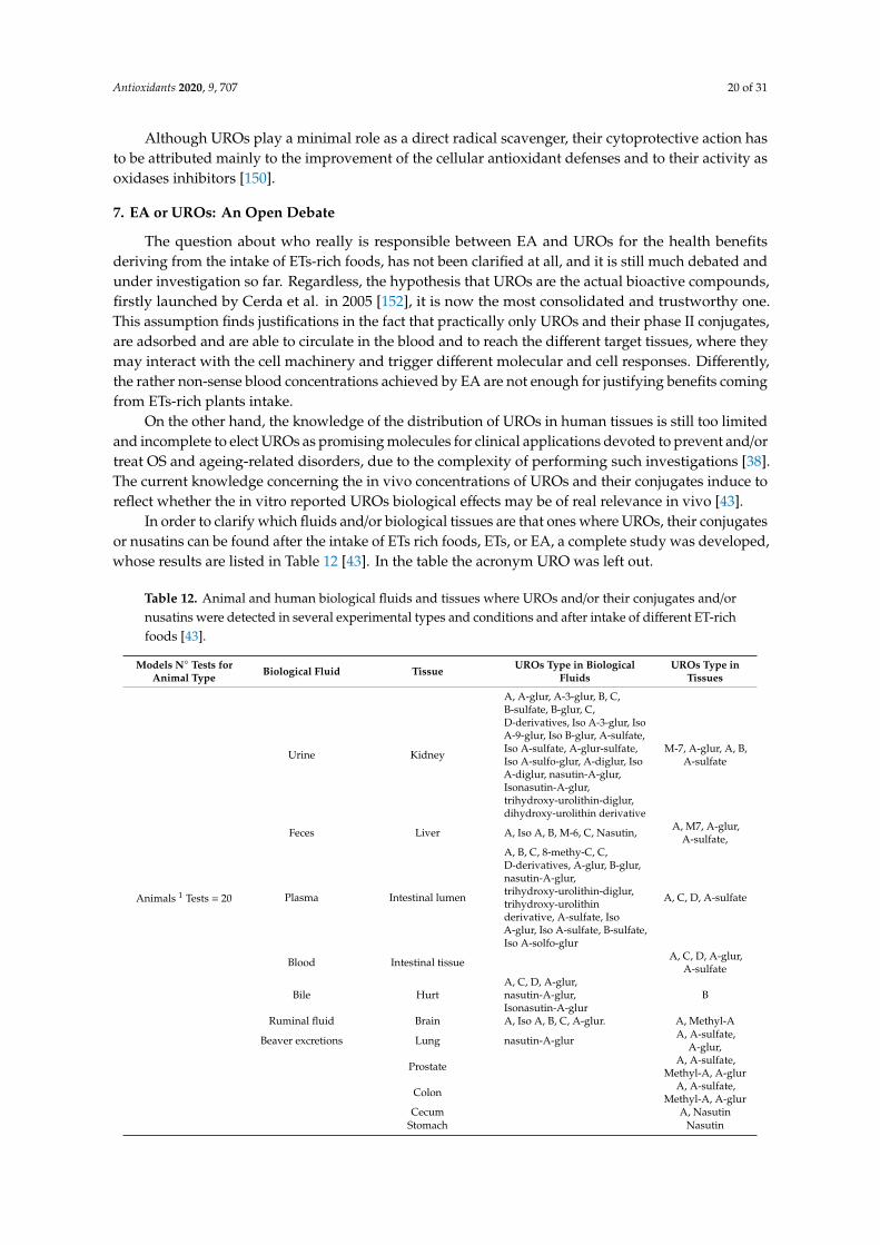
In order to clarify which fluids and/or biological tissues are that ones where UROs, their conjugatesor nusatins can be found after the intake of ETs rich foods, ETs, or EA, a complete study was developed,whose results are listed in Table 12 [43]. In the table the acronym URO was left out.
Table 12. Animal and human biological fluids and tissues where UROs and/or their conjugates and/ornusatins were detected in several experimental types and conditions and after intake of different ET-richfoods [43].
Models N◦ Tests forAnimal Type Biological Fluid Tissue UROs Type in Biological
FluidsUROs Type in
Tissues
Animals 1 Tests = 20
Urine Kidney
A, A-glur, A-3-glur, B, C,B-sulfate, B-glur, C,D-derivatives, Iso A-3-glur, IsoA-9-glur, Iso B-glur, A-sulfate,Iso A-sulfate, A-glur-sulfate,Iso A-sulfo-glur, A-diglur, IsoA-diglur, nasutin-A-glur,Isonasutin-A-glur,trihydroxy-urolithin-diglur,dihydroxy-urolithin derivative
M-7, A-glur, A, B,A-sulfate
Feces Liver A, Iso A, B, M-6, C, Nasutin, A, M7, A-glur,A-sulfate,
Plasma Intestinal lumen
A, B, C, 8-methy-C, C,D-derivatives, A-glur, B-glur,nasutin-A-glur,trihydroxy-urolithin-diglur,trihydroxy-urolithinderivative, A-sulfate, IsoA-glur, Iso A-sulfate, B-sulfate,Iso A-solfo-glur
A, C, D, A-sulfate
Blood Intestinal tissue A, C, D, A-glur,A-sulfate
Bile HurtA, C, D, A-glur,nasutin-A-glur,Isonasutin-A-glur
B
Ruminal fluid Brain A, Iso A, B, C, A-glur. A, Methyl-A
Beaver excretions Lung nasutin-A-glur A, A-sulfate,A-glur,
Prostate A, A-sulfate,Methyl-A, A-glur
Colon A, A-sulfate,Methyl-A, A-glur
Cecum A, NasutinStomach Nasutin

Antioxidants 2020, 9, 707 21 of 31
Table 12. Cont.
Models N◦ Tests forAnimal Type Biological Fluid Tissue UROs Type in Biological
FluidsUROs Type in
Tissues
Humans 2 Tests = 27 3
Urine Prostate
A, B, C, M-5, D, B-sulfate,A-glur, Iso A, Iso A-glur,B-glur, A-3-glur, A-8-glur,trihydroxyurolithin, A-sulfate,Methyl-A, A-sulfo-glur,D-methyletherglur, A-diglur,Dimethy-C, C-glur, D-glur,C-methylethersulfoglur,Methyl-C-glur, Methyl-D-glur,C-sulfate,
A, A-glur, B-glur,
Feces Normal and malignantcolon tissue
A, B, C, D, E, M-5, M-6, M-7,Iso A
A-glur, D, IsoA-glur, A-sulfate,
C, B-glur, A, Iso A,B-sulfate, B, M-5,
M-6
Plasma
A, B, D, A-sulfate, B-sulfate,trihydroxyurolithin, A-glur,Iso A-glur, B-glur, C,Methyl-A, C-glur,Methyl-C-methyletherglur,C-diglur, D-glur, A-sulfo-glur,C-sulfate, IsoA-sulfate,
1 Rats, Iberian pigs, bull, sheep, mice, greenfinches, beetle, beaver, squirrel, pig; 2 Healthy or non- healthy volunteers,patients with COPD, ileostomy, PBH, PCa, MetS, CRC; 3 Number of replicates for test. Abbreviations: COPD,chronic obstructive pulmonary disease; PBH, benign prostatic hyperplasia; PCa, prostate cancer; MetS, metastaticdisease; CRC, colorectal cancer or colorectal carcinoma.
Hazardous Implications Related to UROs Exposure
Results obtained from a randomized clinical trial showed that the inter-individual variabilityin the improvement of cardiovascular risk biomarkers, in overweight-obese individuals consumingpomegranate, depends on different UMs and type of UROs produced.
In particular, a high cardiovascular risk was associated to the UMB that dictates for a gut microbiotacomposition that determinates a higher production URO B and Iso URO A, rather than URO A or noproduction [47]. In fact, in this regard, the positive effects of UROs are obscured by a negative sideeffect Iso URO A and URO B [47].
In recent years, the dual redox behavior of natural polyphenols has increasingly captured theinterest of researchers and the need to investigate their pro-oxidant capabilities in addition to theantioxidant effects has become urgent [149]. In this regard, UROs have received contradictory reportson their antioxidant capacity, and their pro-oxidant properties have been recently studied.
The redox properties of URO A and URO B, have been investigated by using more than one assaymethod including ORAC assay, three cell-based assays, copper-initiated pro-oxidant activity (CIPA)assay, and cyclic voltammetry, and the findings unveiled that UROs, although strong antioxidants inthe ORAC assay, are mostly pro-oxidants in cell-based assays [149].
Citing Rahal et al., 2014 [153]: “Pro-oxidant refers to any endobiotic or xenobiotic that inducesoxidative stress either by generation of ROS or by inhibiting antioxidant systems”. As reported,the pro-oxidant activity of natural polyphenols depends on their dosage and on the presence of properamounts of metal ions [154], but while small polyphenols can exhibit considerable pro-oxidant activity,large molecular-weight phenols, such as ETs, have little or no pro-oxidant properties [144].
In this context, the tendency of small polyphenols to exert pro-oxidants effects is considerablein the presence of high concentrations of transition metal ions, such as Cu2+ or Fe2+. A proposedmechanism suggests that, firstly, UROs reduce Cu2+ to Cu+ and, subsequently, Cu+ is re-oxidized in aFenton-like reaction by the action of H2O2 or O2, leading to the production of oxygen radicals [149].Considering that in living cells, a small amount of hydrogen peroxide is produced as a result ofcellular metabolism, and that transition metal ions are available, the proposed scenario is without adoubt feasible.

Antioxidants 2020, 9, 707 22 of 31
8. Authors Opinions, Future Perspectives and Conclusions
Hippocrates, over 2500 years ago, coined the phrase “Let food be the medicine and medicine be thefood” and in the past, folk medicine, that made use of spices, plants, herbs, fruits, and seeds for thetreatment of several diseases, gave it a considerable applicative importance.
Some foods are more than fuel for the organism, and could contain nutrients as ETs and EA,that proved to possess activities essential to preclude diseases and to own high potentials for beingeffectively used as a sort of medicine to prevent and/or enhance illnesses.
Nowadays, an extensive literature, mainly based on in vitro tests, reports the several EA healthproperties, based on its multi-target antioxidant effects, able to counteract OS and aging-relateddiseases. As a consequence, a worldwide market concerning the production of plants concentratedextracts, EA-enriched foods, EA-based functional foods or food supplements, rapidly has beendeveloped, with the claim to provide tools to increase the daily intake of EA and with a globalconspicuous involvement of large capitals. In this regard, the actual potential, effectiveness, and safetyof EA-enriched products should be discussed and a more wide-ranging debate is needed.
Since EA is practically not soluble and only an insignificant fraction of the EA contained inthese products will actually be absorbed in GIT, essentially, all EA ingested will be metabolized bygut microbiota to UROs, which, easily absorbed, will circulate in blood and reach cells and tissues.Ironically, by the intake of EA-enriched products, in place of improving EA in vivo concentration thatof UROs is increased, both in plasma and in tissues.
Nevertheless, depending on the age of individuals, their health conditions, and the composition oftheir gut microbiota, the types of UROs produced and their concentration can vary and the individualvariability in the responses to UROs exposure is unpredictable and may lead to heterogeneouscomebacks that could promote also adverse effects, such as cardiovascular disorders. Furthermore,by exerting both antioxidant and pro-oxidant activities, high concentration of UROs may causeDNA damage and apoptosis, leading to the development of OS-related pathologies such as cancer.Consequently, EA-enriched products’ commercialization should be subject to more careful control andregulation, and after their intake, URO concentrations and tissues distributions, should be monitoredand kept under continuous control, individual by individual.
An uncontrolled large production and distribution of EA-enriched foods and food supplementsrepresent an outstanding problem, which could have heavy negative repercussions on human healthrather than positive.
Regardless, further detailed and scrupulous toxicity evaluations are necessary both for EA andUROs, before their therapeutic application in humans could be possible. Clinical investigations needto verify the interesting preclinical findings and to validate if the very good results observed in animalsare confirmed in humans.
It is relevant to understand also the inter-individual variability of the population in response toEA and ETs-containing foods, for a better organization of a healthy diet and for establishing the mostsuitable dosages of polyphenolic nutraceuticals and foods rich in ETs and EA for each person.
As far as the authors’ opinion is concerned, it seems anyway conceivable to propose that EAremains the robust compound worthy of further extensive investigations.
In relation to its chemical structure and antioxidant mechanisms, EA might be either a safenutraceutical, an innovative therapeutic, or a template molecule for the development of novel drugsable to fight RONS for promoting human health.
Finally, although attractive, the hypothesis that declares UROs as the actual substances responsibleof beneficial effects coming from the ingestion of EA-rich foods is far from being clearly demonstrated,because studies investigating the in vivo EA effects are limited or even missing.
There is no evidence for attributing with certainty also to EA an in vivo activity, but it is equallyincorrect to affirm the opposite and attribute in vivo activity to UROs only.

Antioxidants 2020, 9, 707 23 of 31
Author Contributions: Conceptualization, methodology, investigation, data curation, writing—original draftpreparation, S.A.; writing—review and editing, visualization, S.A., B.M. and G.Z. All authors have read andagreed to the published version of the manuscript.
Funding: This research received no external funding.
Acknowledgments: The authors thank Deirdre Kantz, for English language help.
Conflicts of Interest: The authors declare no conflict of interest.
References
1. Larrosa, M.; García-Conesa, M.T.; Espín, J.C.; Tomás-Barberán, F.A. Ellagitannins, ellagic acid and vascularhealth. Mol. Asp. Med. 2010, 31, 513–539. [CrossRef] [PubMed]
2. Larrosa, M.; González-Sarrías, A.; Yáñez-Gascón, M.J.; Selma, M.V.; Azorín-Ortuño, M.; Toti, S.;Tomás-Barberán, F.; Dolara, P.; Espín, J.C. Anti-inflammatory properties of a pomegranate extract andits metabolite urolithin-A in a colitis rat model and the effect of colon inflammation on phenolic metabolism.J. Nutr. Biochem. 2010, 21, 717–725. [CrossRef] [PubMed]
3. Tomás-Barberan, F.A.; Espín, J.C.; García-Conesa, M.T. Bioavailability and Metabolism of Ellagic Acid andEllagitannins. In Chemistry and Biology of Ellagitannins; Quideau, S., Ed.; World Scientific Publishing Co PteLtd.: Singapore, 2009; pp. 273–297.
4. Beretta, G.; Rossoni, G.; Santagati, N.A.; Facino, R.M. Anti-Ischemic Activity and Endothelium-DependentVasorelaxant Effect of Hydrolysable Tannins from the Leaves of Rhus coriaria (Sumac) in Isolated RabbitHeart and Thoracic Aorta. Planta Med. 2009, 75, 1482–1488. [CrossRef] [PubMed]
5. Mente, A.; de Koning, L.; Shannon, H.S.; Anand, S.S. A Systematic Review of the Evidence Supporting aCausal Link Between Dietary Factors and Coronary Heart Disease. Arch. Intern. Med. 2009, 169, 659–669.[CrossRef] [PubMed]
6. Fukushima, Y.; Ohie, T.; Yonekawa, Y.; Yonemoto, K.; Aizawa, H.; Mori, Y.; Watanabe, M.; Takeuchi, M.;Hasegawa, M.; Taguchi, C.; et al. Coffee and green tea as a large source of antioxidant polyphenols in theJapanese population. J. Agric. Food Chem. 2009, 57, 1253–1259. [CrossRef] [PubMed]
7. Derosa, G.; Maffioli, P.; Sahebkar, A. Ellagic Acid and Its Role in Chronic Diseases. Adv. Exp. Med. Biol.2016, 928, 473–479. [CrossRef]
8. Ríos, J.-L.; Giner, R.M.; Marín, M.; Recio, M.C. A Pharmacological Update of Ellagic Acid. Planta Med.2018, 84, 1068–1093. [CrossRef]
9. Alfei, S.; Turrini, F.; Catena, S.; Zunin, P.; Grilli, M.; Pittaluga, A.M.; Boggia, R. Ellagic acid a multi-targetbioactive compound for drug discovery in CNS? A narrative review. Eur. J. Med. Chem. 2019, 183, 111724.[CrossRef]
10. Hseu, Y.-C.; Chou, C.-W.; Kumar, K.J.; Senthil Fu, K.-T.; Wang, H.-M.; Hsu, L.-S.; Kuo, Y.-H.; Wu, C.-R.;Chen, S.-C.; Yang, H.-L. Ellagic acid protects human keratinocyte (HaCaT) cells against UVA-induced oxidativestress and apoptosis through the upregulation of the HO-1 and Nrf-2 antioxidant genes. Food Chem. Toxicol.2012, 50, 1245–1255. [CrossRef]
11. BenSaad, L.A.; Kim, K.H.; Quah, C.C.; Kim, W.R.; Shahimi, M. Anti-inflammatory potential of ellagic acid,gallic acid and punicalagin A&B isolated from Punica granatum. BMC Compl. Altern. Med. 2017, 17, 47.[CrossRef]
12. Heber, D. Multitargeted therapy of cancer by ellagitannins. Cancer Lett. 2008, 269, 262–268. [CrossRef][PubMed]
13. Khateeb, J.; Gantman, A.; Kreitenberg, A.J.; Aviram, M.; Fuhrman, B. Paraoxonase 1 (PON1) expressionin hepatocytes is upregulated by pomegranate polyphenols: A role for PPAR-γ pathway. Atherosclerosis2010, 208, 119–125. [CrossRef]
14. Crescente, M.; Jessen, G.; Momi, S.; Höltje, H.-D.; Gresele, P.; Cerletti, C.; de Gaetano, G. Interactions ofgallic acid, resveratrol, quercetin and aspirin at the platelet cyclooxygenase-1 level Functional and modellingstudies. Thromb. Haemost. 2017, 102, 336–346. [CrossRef] [PubMed]
15. Trifirò, G.; Calogero, G.; Ippolito, F.M.; Cosentino, M.; Giuliani, R.; Conforti, A.; Venegoni, M.; Mazzaglia, G.;Caputi, A.P. Adverse drug events in emergency department population: A prospective Italian study.Pharmacoepidemiol. Drug Saf. 2005, 14, 333–340. [CrossRef] [PubMed]

Antioxidants 2020, 9, 707 24 of 31
16. Vanella, L.; Di Giacomo, C.; Acquaviva, R.; Barbagallo, I.; Li Volti, G.; Cardile, V.; Abraham, N.G.; Sorrenti, V.Effects of Ellagic Acid on Angiogenic Factors in Prostate Cancer Cells. Cancers 2013, 5, 726–738. [CrossRef]
17. Nejad, K.H.; Gharib-Naseri, M.K.; Sarkaki, A.; Dianat, M.; Badavi, M.; Farbood, Y. Effects of ellagic acidpretreatment on renal functions disturbances induced by global cerebral ischemic-reperfusion in rat. Iran J.Basic Med. Sci. 2017, 20, 75–82. [CrossRef] [PubMed]
18. Firdaus, F.; Zafeer Mohd, F.; Waseem, M.; Anis, E.; Hossain, M.M.; Afzal, M. Ellagic acid mitigatesarsenic-trioxide-induced mitochondrial dysfunction and cytotoxicity in SH-SY5Y cells. J. Biochem. Mol. Toxicol.2018, 32, e22024. [CrossRef]
19. González-Sarrías, A.; Núñez-Sánchez, M.Á.; Tomé-Carneiro, J.; Tomás-Barberán, F.A.; García-Conesa, M.T.;Espín, J.C. Comprehensive characterization of the effects of ellagic acid and urolithins on colorectal cancerand key-associated molecular hallmarks: MicroRNA cell specific induction of CDKN1A (p21) as a commonmechanism involved. Mol. Nutr. Food Res. 2016, 60, 701–716. [CrossRef]
20. Boggia, R.; Turrini, F.; Roggeri, A.; Olivero, G.; Cisani, F.; Bonfiglio, T.; Summa, M.; Grilli, M.; Caviglioli, G.;Alfei, S.; et al. Neuroinflammation in Aged Brain: Impact of the Oral Administration of Ellagic AcidMicrodispersion. Int. J. Mol. Sci. 2020, 21, 3631. [CrossRef]
21. Wu, S.; Tian, L. A new flavone glucoside together with known ellagitannins and flavones with anti-diabeticand anti-obesity activities from the flowers of pomegranate (Punica granatum). Nat. Prod. Res. 2019, 33,252–257. [CrossRef]
22. García-Niño, W.R.; Zazueta, C. Ellagic acid: Pharmacological activities and molecular mechanisms involvedin liver protection. Pharmacol. Res. 2015, 97, 84–103. [CrossRef] [PubMed]
23. De Oliveira, M.R. The Effects of Ellagic Acid upon Brain Cells: A Mechanistic View and Future Directions.Neurochem. Res. 2016, 41, 1219–1228. [CrossRef] [PubMed]
24. Genestra, M. Oxyl radicals, redox-sensitive signalling cascades and antioxidants. Cell. Sign. 2007, 19,1807–1819. [CrossRef] [PubMed]
25. Venkataraman, K.; Khurana, S.; Tai, T.C. Oxidative stress in aging—Matters of the heart and mind. Int. J.Mol. Sci. 2013, 14, 17897–17925. [CrossRef] [PubMed]
26. Marengo, B.; Raffaghello, L.; Pistoia, V.; Cottalasso, D.; Pronzato, M.A.; Marinari, U.M.; Domenicotti, C.Reactive oxygen species: Biological stimuli of neuroblastoma cell response. Cancer Lett. 2005, 228, 111–116.[CrossRef] [PubMed]
27. Marengo, B.; Nitti, M.; Furfaro, A.L.; Colla, R.; De Ciucis, C.; Marinari, U.M.; Pronzato, M.A.; Traverso, N.;Domenicotti, C. Redox Homeostasis and Cellular Antioxidant Systems: Crucial Players in Cancer Growthand Therapy. Oxid. Med. Cell. Longev. 2016, 2016, 6235641. [CrossRef] [PubMed]
28. Kaneto, H.; Katakami, N.; Matsuhisa, M.; Matsuoka, T. Role of reactive oxygen species in the progression oftype 2 diabetes and atherosclerosis. Mediat. Inflamm. 2010, 2010, 453892. [CrossRef]
29. Touqeer, A.; William, S.N.; Nabavi, S.F.; Ilkay, E.O.; Nady, B.; Eduardo, S.; Nabavi, S.M. Insights into effectsof ellagic acid on the nervous system: A mini review. Cur. Pharm. Des. 2016, 22, 1350–1360.
30. Akhtar, S.; Ismail, T.; Fraternale, D.; Sestili, P. Pomegranate peel and peel extracts: Chemistry and foodfeatures. Food Chem. 2015, 174, 417–425. [CrossRef]
31. Mena, P.; Calani, L.; Bruni, R.; Del Rio, D. Bioactivation of High-Molecular-Weight Polyphenols by the GutMicrobiome. In Diet-Microbe Interactions in the Gut; Tuohy, K., Del Rio, D., Eds.; Academic Press: Cambridge,MA, USA, 2015; pp. 73–101.
32. Sandhu, A.K.; Miller, M.G.; Thangthaeng, N.; Scott, T.M.; Shukitt-Hale, B.; Edirisinghe, I.; Burton-Freeman, B.Metabolic fate of strawberry polyphenols after chronic intake in healthy older adults. Food Funct. 2018, 9,96–106. [CrossRef]
33. Landete, J.M. Ellagitannins, ellagic acid and their derived metabolites: A review about source, metabolism,functions and health. Food Res. Int. 2011, 44, 1150–1160. [CrossRef]
34. Muthukumaran, S.; Tranchant, C.; Shi, J.; Ye, X.; Xue, S.J. Ellagic acid in strawberry (Fragaria spp.): Biological,technological, stability, and human health aspects. Food Qual. Saf. 2017, 1, 227–252. [CrossRef]
35. Garcia-Muñoz, C.; Vaillant, F. Metabolic Fate of Ellagitannins: Implications for Health, and ResearchPerspectives for Innovative Functional Foods. Critic. Rev. Food Sci. Nutr. 2014, 54, 1584–1598. [CrossRef][PubMed]
Antioxidants 2020, 9, 707 25 of 31
36. Andrade, M.A.; Lima, V.; Silva, A.; Sanches Vilarinho, F.; Castilho, M.C.; Khwaldia, K.; Ramos, F. Pomegranateand grape by-products and their active compounds: Are they a valuable source for food applications?Trends Food Sci. Technol. 2019, 86, 68–84. [CrossRef]
37. Nuñez-Sánchez, M.A.; García-Villalba, R.; Monedero-Saiz, T.; García-Talavera, N.V.; Gómez-Sánchez, M.B.;Sánchez-Álvarez, C.; García-Albert, A.M.; Rodríguez-Gil, F.J.; Ruiz-Marín, M.; Pastor-Quirante, F.A.; et al.Targeted metabolic profiling of pomegranate polyphenols and urolithins in plasma, urine and colon tissuesfrom colorectal cancer patients. Mol. Nutr. Food Res. 2014, 58, 1199–1211. [CrossRef]
38. Smeriglio, A.; Barreca, D.; Bellocco, E.; Trombetta, D. Proanthocyanidins and hydrolysable tannins:Occurrence, dietary intake and pharmacological effects. Br. J. Pharmacol. 2017, 174, 1244–1262. [CrossRef]
39. Serrano, J.; Puupponen-Pimiä, R.; Dauer, A.; Aura, A.-M.; Saura-Calixto, F. Tannins: Current knowledgeof food sources, intake, bioavailability and biological effects. Mol. Nutr. Food Res. 2009, 53, S310–S329.[CrossRef]
40. Zuccari, G.; Baldassari, S.; Ailuno, G.; Turrini, F.; Alfei, S.; Caviglioli, G. Formulation Strategies to ImproveOral Bioavailability of Ellagic Acid. Appl. Sci. 2020, 10, 3353. [CrossRef]
41. Mileo, A.M.; Miccadei, S. Polyphenols as Modulator of Oxidative Stress in Cancer Disease: New TherapeuticStrategies. Oxid. Med. Cell. Longev. 2015, 2016, 6475624. [CrossRef]
42. Stanisławska, I.J.; Piwowarski, J.P.; Granica, S.; Kiss, A.K. The effects of urolithins on the response of prostatecancer cells to non-steroidal antiandrogen bicalutamide. Phytomedicine 2018, 46, 176–183. [CrossRef]
43. Tomás-Barberán, F.A.; González-Sarrías, A.; García-Villalba, R.; Núñez-Sánchez, M.A.; Selma, M.V.;García-Conesa, M.T.; Espín, J.C. Urolithins, the rescue of “old” metabolites to understand a “new” concept:Metabotypes as a nexus among phenolic metabolism, microbiota dysbiosis, and host health status. Mol. Nutr.Food Res. 2017, 61, 1500901. [CrossRef]
44. Tomás-Barberán, F.A.; García-Villalba, R.; González-Sarrías, A.; Selma, M.V.; Espín, J.C. Ellagic AcidMetabolism by Human Gut Microbiota: Consistent Observation of Three Urolithin Phenotypes in InterventionTrials, Independent of Food Source, Age, and Health Status. J. Agric. Food Chem. 2014, 62, 6535–6538.[CrossRef] [PubMed]
45. Selma, M.V.; Beltrán, D.; García-Villalba, R.; Espín, J.C.; Tomás-Barberán, F.A. Description of urolithinproduction capacity from ellagic acid of two human intestinal Gordonibacter species. Food Funct. 2014, 5,1779–1784. [CrossRef] [PubMed]
46. Selma, M.V.; Tomás-Barberán, F.A.; Beltrán, D.; García-Villalba, R.; Espín, J.C. Gordonibacter urolithinfacienssp. nov., a urolithin-producing bacterium isolated from the human gut. Int. J. Syst. Evol. Microbiol. 2014, 64,2346–2352. [CrossRef]
47. Cortés-Martín, A.; García-Villalba, R.; González-Sarrías, A.; Romo-Vaquero, M.; Loria-Kohen, V.;Ramírez-de-Molina, A.; Tomás-Barberán, F.A.; Selma, M.V.; Espín, J.C. The gut microbiota urolithinmetabotypes revisited: The human metabolism of ellagic acid is mainly determined by aging. Food Funct.2018, 9, 4100–4106. [CrossRef]
48. Carvalho, C.; Moreira, P.I. Oxidative Stress: A Major Player in Cerebrovascular Alterations Associated toNeurodegenerative Events. Front. Physiol. 2018, 9, 806. [CrossRef]
49. Traverso, N.; Ricciarelli, R.; Nitti, M.; Marengo, B.; Furfaro, A.L.; Pronzato, M.A.; Marinari, U.M.;Domenicotti, C. Role of glutathione in cancer progression and chemoresistance. Oxid. Med. Cell Longev.2013, 2013, 972913. [CrossRef] [PubMed]
50. Liguori, I.; Russo, G.; Curcio, F.; Bulli, G.; Aran, L.; Della-Morte, D.; Gargiulo, G.; Testa, G.; Cacciatore, F.;Bonaduce, D.; et al. Oxidative stress, aging, and diseases. Clin. Interv. Aging 2018, 13, 757–772. [CrossRef]
51. Salisbury, D.; Bronas, U. Reactive Oxygen and Nitrogen Species: Impact on Endothelial Dysfunction.Nurs. Res. 2015, 64, 53–66. [CrossRef] [PubMed]
52. Adams, L.; Franco, M.C.; Estevez, A.G. Reactive nitrogen species in cellular signaling. Exp. Biol. Med.2015, 240, 711–717. [CrossRef]
53. Frijhoff, J.; Winyard, P.G.; Zarkovic, N.; Davies, S.S.; Stocker, R.; Cheng, D.; Knight, A.R.; Taylor, E.L.;Oettrich, J.; Ruskovska, T.; et al. Clinical Relevance of Biomarkers of Oxidative Stress. Antiox. Red. Sign.2015, 23, 1144–1170. [CrossRef] [PubMed]
54. Barreiro, E. Role of Protein Carbonylation in Skeletal Muscle Mass Loss Associated with Chronic Conditions.Proteomes 2016, 4, 18. [CrossRef] [PubMed]

Antioxidants 2020, 9, 707 26 of 31
55. Trpkovic, A.; Resanovic, I.; Stanimirovic, J.; Radak, D.; Mousa, S.A.; Cenic-Milosevic, D.; Jevremovic, D.;Isenovic, E.R. Oxidized low-density lipoprotein as a biomarker of cardiovascular diseases. Crit. Rev. Clin.Lab. Sci. 2015, 52, 70–85. [CrossRef] [PubMed]
56. Reynaert, N.L.; Gopal, P.; Rutten, E.P.A.; Wouters, E.F.M.; Schalkwijk, C.G. Advanced glycation end productsand their receptor in age-related, non-communicable chronic inflammatory diseases; Overview of clinicalevidence and potential contributions to disease. Int. J. Biochem. Cell Biol. 2016, 81, 403–418. [CrossRef][PubMed]
57. Jacob, K.D.; Noren Hooten, N.; Trzeciak, A.R.; Evans, M.K. Markers of oxidant stress that are clinicallyrelevant in aging and age-related disease. Mech. Ageing Dev. 2013, 134, 139–157. [CrossRef]
58. PubChem. Available online: https://pubchem.ncbi.nlm.nih.gov (accessed on 4 August 2020).59. Strickley, R.G. Solubilizing excipients in oral and injectable formulations. Pharm. Res. 2004, 21, 201–230.
[CrossRef]60. Behl, G.; Sharma, M.; Dahiya, S.; Chhikara, A.; Chopra, M. Synthesis, Characterization, and Evaluation of
Radical Scavenging Ability of Ellagic Acid-Loaded Nanogels. J. Nanomat. 2011, 2011, 695138. [CrossRef]61. Huetz, P.; Mavaddat, N.; Mavri, J. Reaction between ellagic acid and an ultimate carcinogen. J. Chem.
Inf. Model. 2005, 45, 1564–1570. [CrossRef]62. Ahire, V.; Kumar, A.; Mishra, K.P.; Kulkarni, G. Ellagic Acid Enhances Apoptotic Sensitivity of Breast Cancer
Cells to γ-Radiation. Nutr. Cancer 2017, 69, 904–910. [CrossRef]63. Cheng, H.; Lu, C.; Tang, R.; Pan, Y.; Bao, S.; Qiu, Y.; Xie, M. Ellagic acid inhibits the proliferation of human
pancreatic carcinoma PANC-1 cells in vitro and in vivo. Oncotarget 2017, 8, 12301–12310. [CrossRef]64. Zhao, J.; Li, G.; Bo, W.; Zhou, Y.; Dang, S.; Wei, J.; Li, X.; Liu, M. Multiple effects of ellagic acid on human
colorectal carcinoma cells identified by gene expression profile analysis. Int. J. Oncol. 2017, 50, 613–621.[CrossRef] [PubMed]
65. Pang, X.; Fu, W.; Wang, J.; Kang, D.; Xu, L.; Zhao, Y.; Liu, A.L.; Du, G.H. Identification of Estrogen Receptorα Antagonists from Natural Products via In Vitro and In Silico Approaches. Oxid. Med. Cell Longev.2018, 2018, 6040149. [CrossRef]
66. Kolšek, K.; Mavri, J.; Sollner Dolenc, M.; Gobec, S.; Turk, S. Endocrine Disruptome—An Open SourcePrediction Tool for Assessing Endocrine Disruption Potential through Nuclear Receptor Binding. J. Chem.Inf. Model. 2014, 54, 1254–1267. [CrossRef] [PubMed]
67. Muñoz-Muñoz, J.L.; Garcia-Molina, F.; Garcia-Molina, M.; Tudela, J.; García-Cánovas, F.;Rodriguez-Lopez, J.N. Ellagic acid: Characterization as substrate of polyphenol oxidase. IUBMB Life2009, 61, 171–177. [CrossRef] [PubMed]
68. Morina, F.; Takahama, U.; Yamauchi, R.; Hirota, S.; Veljovic-Jovanovic, S. Quercetin 7-O-glucoside suppressesnitrite-induced formation of dinitrosocatechins and their quinones in catechin/nitrite systems under stomachsimulating conditions. Food Funct. 2015, 6, 218–228. [CrossRef]
69. Siraj, M.A.; Shilpi, J.A.; Hossain, M.G.; Uddin, S.J.; Islam, M.K.; Jahan, I.A.; Hossain, H. Anti-Inflammatoryand Antioxidant Activity of Acalypha hispida Leaf and Analysis of its Major Bioactive Polyphenols by HPLC.Adv. Pharm. Bull. 2016, 6, 275–283. [CrossRef]
70. Aoyama, H.; Sakagami, H.; Hatano, T. Three new flavonoids, proanthocyanidin, and accompanying phenolicconstituents from Feijoa sellowiana. Biosci. Biotechnol. Biochem. 2018, 82, 31–41. [CrossRef]
71. Jaramillo-García, V.; Trindade, C.; Lima, E.; Guecheva, T.N.; Villela, I.; Martinez-Lopez, W.; Corrêa, D.S.;Ferraz, A.d.B.F.; Moura, S.; Sosa, M.Q.; et al. Chemical characterization and cytotoxic, genotoxic,and mutagenic properties of Baccharis trinervis (Lam, Persoon) from Colombia and Brazil. J. Ethnopharmacol.2018, 213, 210–220. [CrossRef]
72. Yang, R.; Guan, Y.; Zhou, J.; Sun, B.; Wang, Z.; Chen, H.; He, Z.; Jia, A. Phytochemicals from Camellianitidissima Chi Flowers Reduce the Pyocyanin Production and Motility of Pseudomonas aeruginosa PAO1.Front. Microbiol. 2018, 8, 2640. [CrossRef]
73. Campos, J.F.; Espindola, P.P.d.T.; Torquato, H.F.V.; Vital, W.D.; Justo, G.Z.; Silva, D.B.; Carollo, C.A.;de Picoli Souza, K.; Paredes-Gamero, E.J.; dos Santos, E.L. Leaf and Root Extracts from Campomanesiaadamantium (Myrtaceae) Promote Apoptotic Death of Leukemic Cells via Activation of Intracellular Calciumand Caspase-3. Front. Pharmacol. 2017, 8, 466. [CrossRef]
74. Yang, L.-P.; Gu, X.-L.; Chen, J.-X.; Yang, J.; Tan, S.-Y.; Duan, W.-J. Chemical constituents from Canariumalbum Raeusch and their anti-influenza A virus activities. J. Nat. Med. 2018, 72, 808–815. [CrossRef]

Antioxidants 2020, 9, 707 27 of 31
75. Hafsa, J.; Hammi, K.M.; Khedher, M.R.B.; Smach, M.A.; Charfeddine, B.; Limem, K.; Majdoub, H.Inhibition of protein glycation, antioxidant and antiproliferative activities of Carpobrotus edulis extracts.Biomed. Pharmacother. 2016, 84, 1496–1503. [CrossRef] [PubMed]
76. Tuyen, P.T.; Xuan, T.D.; Tu Anh, T.T.; Mai Van, T.; Ahmad, A.; Elzaawely, A.A.; Khanh, T.D. Weed SuppressingPotential and Isolation of Potent Plant Growth Inhibitors from Castanea crenata Sieb. et Zucc. Molecules2018, 23, 345. [CrossRef] [PubMed]
77. Karimi, E.; Nohooji, M.G.; Habibi, M.; Ebrahimi, M.; Mehrafarin, A.; Khalighi-Sigaroodi, F.Antioxidant potential assessment of phenolic and flavonoid rich fractions of Clematis orientalis andClematis ispahanica (Ranunculaceae). Nat. Prod. Res. 2018, 32, 1991–1995. [CrossRef] [PubMed]
78. Shendge, A.K.; Basu, T.; Chaudhuri, D.; Panja, S.; Mandal, N. In vitro Antioxidant and AntiproliferativeActivities of Various Solvent Fractions from Clerodendrum viscosum Leaves. Pharmacogn. Mag. 2017, 13,S344–S353. [CrossRef]
79. Huang, J.; Zhang, Y.; Dong, L.; Gao, Q.; Yin, L.; Quan, H.; Chen, R.; Fu, X.; Lin, D. Ethnopharmacology,phytochemistry, and pharmacology of Cornus officinalis Sieb. et Zucc. J. Ethnopharmacol. 2018, 213, 280–301.[CrossRef]
80. Cho, C.H.; Jang, H.; Lee, M.; Kang, H.; Heo, H.J.; Kim, D.-O. Sea Buckthorn (Hippophae rhamnoides L.) LeafExtracts Protect Neuronal PC-12 Cells from Oxidative Stress. J. Microbiol. Biotechnol. 2017, 27, 1257–1265.[CrossRef]
81. Vieira, G.S.; Marques, A.S.F.; Machado, M.T.C.; Silva, V.M.; Hubinger, M.D. Determination of anthocyaninsand non-anthocyanin polyphenols by ultra performance liquid chromatography/electrospray ionization massspectrometry (UPLC/ESI–MS) in jussara (Euterpe edulis) extracts. J. Food Sci. Technol. 2017, 54, 2135–2144.[CrossRef]
82. Falcão, T.R.; de Araújo, A.A.; Soares, L.A.L.; de Moraes Ramos, R.T.; Bezerra, I.C.F.; Ferreira, M.R.A.;de Souza Neto, M.A.; Melo, M.C.N.; de Araújo, R.F.; de Aguiar Guerra, A.C.V.; et al. Crude extract andfractions from Eugenia uniflora Linn leaves showed anti-inflammatory, antioxidant, and antibacterialactivities. BMC Complem. Altern. Med. 2018, 18, 84. [CrossRef]
83. Lee, I.-S.; Jung, S.-H.; Kim, J.S. Polyphenols from Euphorbia pekinensis Inhibit AGEs Formation In Vitro andVessel Dilation in Larval Zebrafish In Vivo. Planta Med. 2018, 84, 176–181. [CrossRef]
84. Ton That, Q.; Nguyen Thien, T.V.; Dang, H.P.; Le Hoan, N.; Vo, L.K.T.; Nguyen, M.H.D.; Ngu, N.T.;Nguyen, T.S.; Hansen, P.E. Chemical constituents of Geum urbanum L. roots. Nat. Prod. Res. 2018, 32,2529–2534. [CrossRef] [PubMed]
85. Ochoa-Pacheco, A.; Escalona Arranz, J.C.; Beaven, M.; Peres-Roses, R.; Gámez, Y.M.; Camacho-Pozo, M.I.;Maury, G.L.; de Macedo, M.B.; Cos, P.; Tavares, J.F.; et al. Bioassay-guided In vitro Study of the Antimicrobialand Cytotoxic Properties of the Leaves from Excoecaria Lucida Sw. Pharmacogn. Res. 2017, 9, 396–400.[CrossRef]
86. Vu, D.C.; Vo, P.H.; Coggeshall, M.V.; Lin, C.-H. Identification and Characterization of Phenolic Compoundsin Black Walnut Kernels. J. Agric. Food Chem. 2018, 66, 4503–4511. [CrossRef]
87. Pereira, L.O.M.; Vilegas, W.; Tangerina, M.M.P.; Arunachalam, K.; Balogun, S.O.; Orlandi-Mattos, P.E.;Colodel, E.M.; Martins, D.T.d.O. Lafoensia pacari A. St.-Hil.: Wound healing activity and mechanism ofaction of standardized hydroethanolic leaves extract. J. Ethnopharmacol. 2018, 219, 337–350. [CrossRef]
88. De Oliveira, L.M.; Porte, A.; de Oliveira Godoy, R.L.; da Costa Souza, M.; Pacheco, S.;de Araujo Santiago, M.C.P.; Gouvêa, A.C.M.S.; da Silva de Mattos do Nascimento, L.; Galhardo Borguini, R.Chemical characterization of Myrciaria floribunda (H. West ex Willd) fruit. Food Chem. 2018, 248, 247–252.[CrossRef] [PubMed]
89. Díaz-de-Cerio, E.; Arráez-Román, D.; Segura-Carretero, A.; Ferranti, P.; Nicoletti, R.; Perrotta, G.M.;Gómez-Caravaca, A.M. Establishment of pressurized-liquid extraction by response surface methodologyapproach coupled to HPLC-DAD-TOF-MS for the determination of phenolic compounds of myrtle leaves.Ana. Bioanal. Chem. 2018, 410, 3547–3557. [CrossRef]
90. Hernández, C.; Ascacio-Valdés, J.; De la Garza, H.; Wong-Paz, J.; Aguilar, C.N.; Martínez-Ávila, G.C.;Castro-López, C.; Aguilera-Carbó, A. Polyphenolic content, in vitro antioxidant activity and chemicalcomposition of extract from Nephelium lappaceum L. (Mexican rambutan) husk. Asian Pac. J. Trop. Med.2017, 10, 1201–1205. [CrossRef]

Antioxidants 2020, 9, 707 28 of 31
91. Ifeanacho, M.O.; Ikewuchi, C.C.; Ikewuchi, J.C. Investigation of the profile of phenolic compounds in theleaves and stems of Pandiaka heudelotii using gas chromatography coupled with flame ionization detector.Food Sci. Nutr. 2017, 5, 646–652. [CrossRef]
92. Navarro, M.; Moreira, I.; Arnaez, E.; Quesada, S.; Azofeifa, G.; Vargas, F.; Alvarado, D.; Chen, P. Flavonoidsand Ellagitannins Characterization, Antioxidant and Cytotoxic Activities of Phyllanthus acuminatus Vahl.Plants 2017, 6, 62. [CrossRef]
93. Hu, Q.; Yuan, B.; Xiao, H.; Zhao, L.; Wu, X.; Rakariyatham, K.; Zhong, L.; Han, Y.; Muinde Kimatu, B.;Yang, W. Polyphenols-rich extract from Pleurotus eryngii with growth inhibitory of HCT116 colon cancercells and anti-inflammatory function in RAW264.7 cells. Food Funct. 2018, 9, 1601–1611. [CrossRef]
94. Inada, K.O.P.; Duarte, P.A.; Lapa, J.; Miguel, M.A.L.; Monteiro, M. Jabuticaba (Myrciaria jaboticaba) juiceobtained by steam-extraction: Phenolic compound profile, antioxidant capacity, microbiological stability,and sensory acceptability. J. Food Sci. Technol. 2018, 55, 52–61. [CrossRef] [PubMed]
95. Neves, N.d.A.; Stringheta, P.C.; Gómez-Alonso, S.; Hermosín-Gutiérrez, I. Flavonols and ellagic acidderivatives in peels of different species of jabuticaba (Plinia spp.) identified by HPLC-DAD-ESI/MSn.Food Chem. 2018, 252, 61–71. [CrossRef] [PubMed]
96. Mazzarino, L.; Pitz, H.d.S.; Voytena, A.P.L.; Trevisan, A.C.D.; Ribeiro-Do-Valle, R.M.; Maraschin, M.Jaboticaba (Plinia peruviana) extract nanoemulsions: Development, stability, and in vitro antioxidant activity.Drug Develop. Ind. Pharm. 2018, 44, 643–651. [CrossRef] [PubMed]
97. Morikawa, T.; Imura, K.; Akagi, Y.; Muraoka, O.; Ninomiya, K. Ellagic acid glycosides with hepatoprotectiveactivity from traditional Tibetan medicine Potentilla anserina. J. Nat. Med. 2018, 72, 317–325. [CrossRef][PubMed]
98. Sobral-Souza, C.E.; Silva, A.R.P.; Leite, N.F.; Costa, J.G.M.; Menezes, I.R.A.; Cunha, F.A.B.; Rolim, L.A.;Coutinho, H.D.M. LC–MS analysis and cytoprotective effect against the mercurium and aluminium toxicityby bioactive products of Psidium brownianum Mart. ex DC. J. Hazard. Mater. 2019, 370, 54–62. [CrossRef]
99. Odubanjo, V.O.; Ibukun, E.O.; Oboh, G.; Adefegha, S.A. Aqueous extracts of two tropical ethnobotanicals(Tetrapleura tetraptera and Quassia undulata) improved spatial and non-spatial working memoriesin scopolamine-induced amnesic rats: Influence of neuronal cholinergic and antioxidant systems.Biomed. Pharmacother. 2018, 99, 198–204. [CrossRef]
100. Ghadage, D.M.; Kshirsagar, P.R.; Pai, S.R.; Chavan, J.J. Extraction efficiency, phytochemical profiles andantioxidative properties of different parts of Saptarangi (Salacia chinensis L.)—An important underutilizedplant. Biochem. Biophys. Rep. 2017, 12, 79–90. [CrossRef]
101. Pinto, J.; Spínola, V.; Llorent-Martínez, E.J.; Fernández-de Córdova, M.L.; Molina-García, L.; Castilho, P.C.Polyphenolic profile and antioxidant activities of Madeiran elderberry (Sambucus lanceolata) as affected bysimulated in vitro digestion. Food Res. Int. 2017, 100, 404–410. [CrossRef]
102. Im, S.H.; Wang, Z.; Lim, S.S.; Lee, O.-H.; Kang, I.-J. Bioactivity-guided isolation and identification ofanti-adipogenic compounds from Sanguisorba officinalis. Pharm. Biol. 2017, 55, 2057–2064. [CrossRef]
103. Cui, Q.; Du, R.; Anantpadma, M.; Schafer, A.; Hou, L.; Tian, J.; Davey, R.A.; Cheng, H.; Rong, L. Identification ofEllagic Acid from Plant Rhodiola rosea L. as an Anti-Ebola Virus Entry Inhibitor. Viruses 2018, 10, 152. [CrossRef]
104. De Britto Policarpi, P.; Turcatto, L.; Demoliner, F.; Ferrari, R.A.; Bascuñan, V.L.A.F.; Ramos, J.C.; Jachmanián, I.;Vitali, L.; Micke, G.A.; Block, J.M. Nutritional potential, chemical profile and antioxidant activity of Chichá(Sterculia striata) nuts and its by-products. Food Res. Int. 2018, 106, 736–744. [CrossRef] [PubMed]
105. Sathyanarayanan, S.; Chandran, R.; Thankarajan, S.; Abrahamse, H.; Thangaraj, P. Phytochemical composition,antioxidant and anti-bacterial activity of Syzygium calophyllifolium Walp. fruit. J. Food Sci. Technol. 2018, 55,341–350. [CrossRef] [PubMed]
106. Gajera, H.P.; Gevariya, S.N.; Hirpara, D.G.; Patel, S.V.; Golakiya, B.A. Antidiabetic and antioxidantfunctionality associated with phenolic constituents from fruit parts of indigenous black jamun(Syzygium cumini L.) landraces. J. Food Sci. Technol. 2017, 54, 3180–3191. [CrossRef] [PubMed]
107. Kalra, P.; Karwasra, R.; Gupta, Y.K.; Ray, S.B.; Singh, S. Terminalia chebula supplementation attenuatescisplatin-induced nephrotoxicity in Wistar rats through modulation of apoptotic pathway. Nat. Prod. Res.2019, 33, 1641–1645. [CrossRef] [PubMed]
108. Kähkönen, M.P.; Hopia, A.I.; Heinonen, M. Berry Phenolics and Their Antioxidant Activity. J. Agric.Food Chem. 2001, 49, 4076–4082. [CrossRef]

Antioxidants 2020, 9, 707 29 of 31
109. Daniel, E.M.; Krupnick, A.S.; Heur, Y.-H.; Blinzler, J.A.; Nims, R.W.; Stoner, G.D. Extraction, stability,and quantitation of ellagic acid in various fruits and nuts. J. Food Compos. Anal. 1989, 2, 338–349. [CrossRef]
110. Määttä-Riihinen, K.R.; Kamal-Eldin, A.; Törrönen, A.R. Identification and Quantification of PhenolicCompounds in Berries of Fragaria and Rubus Species (Family Rosaceae). J. Agric. Food Chem. 2004, 52,6178–6187. [CrossRef]
111. Wada, L.; Ou, B. Antioxidant Activity and Phenolic Content of Oregon Caneberries. J. Agric. Food Chem.2002, 50, 3495–3500. [CrossRef]
112. Koponen, J.M.; Happonen, A.M.; Mattila, P.H.; Törrönen, A.R. Contents of Anthocyanins and Ellagitanninsin Selected Foods Consumed in Finland. J. Agric. Food Chem. 2007, 55, 1612–1619. [CrossRef]
113. Williams, D.J.; Edwards, D.; Pun, S.; Chaliha, M.; Burren, B.; Tinggi, U.; Sultanbawa, Y. Organic acids inKakadu plum (Terminalia ferdinandiana): The good (ellagic), the bad (oxalic) and the uncertain (ascorbic).Food Res. Int. 2016, 89, 237–244. [CrossRef]
114. Khosravi, F.; Nasri, M.H.F.; Farhangfar, H.; Modaresi, J. Nutritive value and polyphenol content ofpomegranate seed pulp ensiled with different tannin-inactivating agents. Anim. Feed Sci. Technol. 2015, 207,262–266. [CrossRef]
115. Çam, M.; Hısıl, Y. Pressurised water extraction of polyphenols from pomegranate peels. Food Chem.2010, 123, 878–885. [CrossRef]
116. Masci, A.; Coccia, A.; Lendaro, E.; Mosca, L.; Paolicelli, P.; Cesa, S. Evaluation of different extraction methodsfrom pomegranate whole fruit or peels and the antioxidant and antiproliferative activity of the polyphenolicfraction. Food Chem. 2016, 202, 59–69. [CrossRef]
117. Boggia, R.; Turrini, F.; Villa, C.; Lacapra, C.; Zunin, P.; Parodi, B. Green Extraction from Pomegranate Marcsfor the Production of Functional Foods and Cosmetics. Pharmaceuticals 2016, 9, 63. [CrossRef] [PubMed]
118. Mattila, P.; Kumpulainen, J. Determination of Free and Total Phenolic Acids in Plant-Derived Foods by HPLCwith Diode-Array Detection. J. Agric. Food Chem. 2002, 50, 3660–3667. [CrossRef] [PubMed]
119. Häkkinen, S.H.; Kärenlampi, S.O.; Mykkänen, H.M.; Heinonen, I.M.; Törrönen, A.R. Ellagic acid content inberries: Influence of domestic processing and storage. Eur. Food Res. Technol. 2000, 212, 75–80. [CrossRef]
120. Da Silva Pinto, M.; Lajolo, F.M.; Genovese, M.I. Bioactive Compounds and Antioxidant Capacity of StrawberryJams. Plant Food Hum. Nutr. 2007, 62, 127–131. [CrossRef]
121. Clifford, M.N.; Scalbert, A. Ellagitannins—Nature, occurrence and dietary burden. J. Sci. Food Agric.2000, 80, 1118–1125. [CrossRef]
122. Glabasnia, A.; Hofmann, T. Sensory-Directed Identification of Taste-Active Ellagitannins in American(Quercus alba L.) and European Oak Wood (Quercus robur L.) and Quantitative Analysis in Bourbon Whiskeyand Oak-Matured Red Wines. J. Agric. Food Chem. 2006, 54, 3380–3390. [CrossRef]
123. Bushman, B.S.; Phillips, B.; Isbell, T.; Ou, B.; Crane, J.M.; Knapp, S.J. Chemical Composition of Caneberry(Rubus spp.) Seeds and Oils and Their Antioxidant Potential. J. Agric. Food Chem. 2004, 52, 7982–7987.[CrossRef] [PubMed]
124. Soong, Y.-Y.; Barlow, P.J. Quantification of gallic acid and ellagic acid from longan (Dimocarpus longan Lour.)seed and mango (Mangifera indica L.) kernel and their effects on antioxidant activity. Food Chem. 2006, 97,524–530. [CrossRef]
125. Williams, D.J.; Edwards, D.; Pun, S.; Chaliha, M.; Sultanbawa, Y. Profiling ellagic acid content: The importanceof form and ascorbic acid levels. Food Res. Int. 2014, 66, 100–106. [CrossRef]
126. Konczak, I.; Maillot, F.; Dalar, A. Phytochemical divergence in 45 accessions of Terminalia ferdinandiana(Kakadu plum). Food Chem. 2014, 151, 248–256. [CrossRef] [PubMed]
127. Pfundstein, B.; El Desouky, S.K.; Hull, W.E.; Haubner, R.; Erben, G.; Owen, R.W. Polyphenolic compounds inthe fruits of Egyptian medicinal plants (Terminalia bellerica, Terminalia chebula and Terminalia horrida):Characterization, quantitation and determination of antioxidant capacities. Phytochemistry 2010, 71, 1132–1148.[CrossRef]
128. Galano, A.; Mazzone, G.; Alvarez-Diduk, R.; Marino, T.; Alvarez-Idaboy, J.R.; Russo, N. Food Antioxidants:Chemical Insights at the Molecular Level. Annu. Rev. Food Sci. Technol. 2016, 7, 335–352. [CrossRef]
129. Santos Sánchez, N.; Salas-Coronado, R.; Villanueva, C.; Hernandez-Carlos, B. Antioxidant Compounds andTheir Antioxidant Mechanism. In Antioxidants; Intechopen: London, UK, 2019; pp. 1–28.
130. Klein, E.; Lukeš, V.; Ilcin, M. DFT/B3LYP study of tocopherols and chromans antioxidant action energetics.Chem. Phys. 2007, 336, 51–57. [CrossRef]

Antioxidants 2020, 9, 707 30 of 31
131. Litwinienko, G.; Ingold, K.U. Solvent Effects on the Rates and Mechanisms of Reaction of Phenols with FreeRadicals. Acc. Chem. Res. 2007, 40, 222–230. [CrossRef]
132. Galano, A.; Medina, M.E.; Tan, D.X.; Reiter, R.J. Melatonin and its metabolites as copper chelating agentsand their role in inhibiting oxidative stress: A physicochemical analysis. J. Pineal Res. 2015, 58, 107–116.[CrossRef]
133. Mazzone, G.; Galano, A.; Alvarez-Idaboy, J.R.; Russo, N. Coumarin–Chalcone Hybrids as Peroxyl RadicalScavengers: Kinetics and Mechanisms. J. Chem. Inf. Model. 2016, 56, 662–670. [CrossRef]
134. Pereira, D.M.; Valentão, P.; Pereira, J.A.; Andrade, P.B. Phenolics: From Chemistry to Biology. Molecules2009, 14, 2202–2211. [CrossRef]
135. Valentão, P.; Fernandes, E.; Carvalho, F.; Andrade, P.B.; Seabra, R.M.; Bastos, M.L. Hydroxyl radical andhypochlorous acid scavenging activity of small Centaury (Centaurium erythraea) infusion. A comparativestudy with green tea (Camellia sinensis). Phytomedicine 2003, 10, 517–522. [CrossRef]
136. Valentão, P.; Fernandes, E.; Carvalho, F.; Andrade, P.B.; Seabra, R.M.; Bastos, M.L. Antioxidative Properties ofCardoon (Cynara cardunculus L.) Infusion Against Superoxide Radical, Hydroxyl Radical, and HypochlorousAcid. J. Agric. Food Chem. 2002, 50, 4989–4993. [CrossRef] [PubMed]
137. Valentão, P.; Fernandes, E.; Carvalho, F.; Andrade, P.B.; Seabra, R.M.; Bastos, M.d.L. Antioxidant Activity ofHypericum androsaemum Infusion: Scavenging Activity against Superoxide Radical, Hydroxyl Radical andHypochlorous Acid. Biol. Pharm. Bull. 2002, 25, 1320–1323. [CrossRef] [PubMed]
138. Valentão, P.; Fernandes, E.; Carvalho, F.; Andrade, P.B.; Seabra, R.M.; Bastos, M.d.L. Studies on the AntioxidantActivity of Lippia citriodora Infusion: Scavenging Effect on Superoxide Radical, Hydroxyl Radical andHypochlorous Acid. Biol. Pharm. Bull. 2002, 25, 1324–1327. [CrossRef] [PubMed]
139. Galano, A.; Francisco Marquez, M.; Pérez-González, A. Ellagic Acid: An Unusually Versatile Protectoragainst Oxidative Stress. Chem. Res. Toxicol. 2014, 27, 904–918. [CrossRef] [PubMed]
140. Álvarez-Diduk, R.; Galano, A.; Tan, D.X.; Reiter, R.J. N-Acetylserotonin and 6-Hydroxymelatonin againstOxidative Stress: Implications for the Overall Protection Exerted by Melatonin. J. Phys. Chem. B 2015, 119,8535–8543. [CrossRef]
141. Eghbaliferiz, S.; Iranshahi, M. Prooxidant Activity of Polyphenols, Flavonoids, Anthocyanins and Carotenoids:Updated Review of Mechanisms and Catalyzing Metals. Phytother. Res. 2016, 30, 1379–1391. [CrossRef]
142. Alfei, S.; Marengo, B.; Domenicotti, C. Polyester-Based Dendrimer Nanoparticles Combined with EtoposideHave an Improved Cytotoxic and Pro-Oxidant Effect on Human Neuroblastoma Cells. Antioxidants 2020, 9, 50.[CrossRef]
143. Olas, B. Berry Phenolic Antioxidants—Implications for Human Health? Front. Pharmacol. 2018, 9, 78.[CrossRef]
144. Mertens-Talcott, S.U.; Jilma-Stohlawetz, P.; Rios, J.; Hingorani, L.; Derendorf, H. Absorption, Metabolism,and Antioxidant Effects of Pomegranate (Punica granatum L.) Polyphenols after Ingestion of a StandardizedExtract in Healthy Human Volunteers. J. Agric. Food Chem. 2006, 54, 8956–8961. [CrossRef]
145. Piwowarski, J.P.; Granica, S.; Stefanska, J.; Kiss, A.K. Differences in Metabolism of Ellagitannins by HumanGut Microbiota ex Vivo Cultures. J. Nat. Prod. 2016, 79, 3022–3030. [CrossRef] [PubMed]
146. González-Barrio, R.; Truchado, P.; Ito, H.; Espín, J.C.; Tomás-Barberán, F.A. UV and MS Identification ofUrolithins and Nasutins, the Bioavailable Metabolites of Ellagitannins and Ellagic Acid in Different Mammals.J. Agric. Food Chem. 2011, 59, 1152–1162. [CrossRef] [PubMed]
147. Milala, J.; Kosmala, M.; Karlinska, E.; Juskiewicz, J.; Zdunczyk, Z.; Fotschki, B. Ellagitannins from Strawberrieswith Different Degrees of Polymerization Showed Different Metabolism through Gastrointestinal Tract ofRats. J. Agric. Food Chem. 2017, 65, 10738–10748. [CrossRef] [PubMed]
148. Garcia, M.; Hernández, L.; Pérez, A.; Vaillant, F. Diversity of urinary excretion patterns of main ellagitannins’colonic metabolites after ingestion of tropical highland blackberry (Rubus adenotrichus) juice. Food Res. Int.2014, 55, 161–169. [CrossRef]
149. Kallio, T.; Kallio, J.; Jaakkola, M.; Mäki, M.; Kilpeläinen, P.; Virtanen, V. Urolithins Display both Antioxidantand Pro-oxidant Activities Depending on Assay System and Conditions. J. Agric. Food Chem. 2013, 61,10720–10729. [CrossRef] [PubMed]
150. Cásedas, G.; Les, F.; Choya-Foces, C.; Hugo, M.; López, V. The Metabolite Urolithin-A Ameliorates OxidativeStress in Neuro-2a Cells, Becoming a Potential Neuroprotective Agent. Antioxidants 2020, 9, 177. [CrossRef][PubMed]

Antioxidants 2020, 9, 707 31 of 31
151. Alfei, S.; Turrini, F.; Catena, S.; Zunin, P.; Parodi, B.; Zuccari, G.; Pittaluga, A.M.; Boggia, R. Preparation ofellagic acid micro and nano formulations with amazingly increased water solubility by its entrapment inpectin or non-PAMAM dendrimers suitable for clinical applications. New J. Chem. 2019, 43, 2438–2448.[CrossRef]
152. Cerdá, B.; Tomás-Barberán, F.A.; Espín, J.C. Metabolism of Antioxidant and Chemopreventive Ellagitanninsfrom Strawberries, Raspberries, Walnuts, and Oak-Aged Wine in Humans: Identification of Biomarkers andIndividual Variability. J. Agric. Food Chem. 2005, 53, 227–235. [CrossRef]
153. Rahal, A.; Kumar, A.; Singh, V.; Yadav, B.; Tiwari, R.; Chakraborty, S.; Dhama, K. Oxidative Stress, Prooxidants,and Antioxidants: The Interplay. BioMed Res. Int. 2014, 2014, 761264. [CrossRef]
154. Yordi, E.; Molina Pérez, E.; Matos, M.J.; Uriarte, E. Antioxidant and Pro-Oxidant Effects of PolyphenolicCompounds and Structure-Activity Relationship Evidence. In Nutrition, Well-Being and Health; Bouayed, J.,Bohn, T., Eds.; InTech: Rijeka, Croatia, 2012; pp. 23–48.
© 2020 by the authors. Licensee MDPI, Basel, Switzerland. This article is an open accessarticle distributed under the terms and conditions of the Creative Commons Attribution(CC BY) license (http://creativecommons.org/licenses/by/4.0/).