Embed Size (px)
Citation preview

Oxidation of PKGIα mediates an endogenousadaptation to pulmonary hypertensionOlena Rudyka,1, Alice Rowana, Oleksandra Prysyazhnaa, Susanne Krasemannb, Kristin Hartmannb, Min Zhanga,Ajay M. Shaha, Clemens Ruppertc, Astrid Weissd, Ralph T. Schermulyd, Tomoaki Idae, Takaaki Akaikee, Lan Zhaof,and Philip Eatona,1
aSchool of Cardiovascular Medicine & Sciences, British Heart Foundation Centre of Excellence, King’s College London, London, United Kingdom; bInstituteof Neuropathology, University Medical Centre Hamburg-Eppendorf, Hamburg, Germany; cUniversities of Giessen & Marburg Lung Center Giessen Biobank,Justus-Liebig-University Giessen, Giessen, Germany; dExcellence Cluster Cardio-Pulmonary System, Justus-Liebig-University Giessen, Giessen, Germany;eDepartment of Environmental Medicine and Molecular Toxicology, Tohoku University Graduate School of Medicine, Sendai, Japan; and fFaculty ofMedicine, Department of Medicine, Imperial College London, London, United Kingdom
Edited by Gregg L. Semenza, Johns Hopkins University, Baltimore, MD, and approved May 21, 2019 (received for review March 14, 2019)
Chronic hypoxia causes pulmonary hypertension (PH), vascularremodeling, right ventricular (RV) hypertrophy, and cardiac failure.Protein kinase G Iα (PKGIα) is susceptible to oxidation, forming aninterprotein disulfide homodimer associated with kinase targetinginvolved in vasodilation. Here we report increased disulfide PKGIαin pulmonary arteries from mice with hypoxic PH or lungs frompatients with pulmonary arterial hypertension. This oxidation islikely caused by oxidants derived from NADPH oxidase-4, super-oxide dismutase 3, and cystathionine γ-lyase, enzymes that wereconcomitantly increased in these samples. Indeed, products thatmay arise from these enzymes, including hydrogen peroxide, glu-tathione disulfide, and protein-bound persulfides, were increasedin the plasma of hypoxic mice. Furthermore, low-molecular-weighthydropersulfides, which can serve as “superreductants” were at-tenuated in hypoxic tissues, consistent with systemic oxidativestress and the oxidation of PKGIα observed. Inhibiting cystathio-nine γ-lyase resulted in decreased hypoxia-induced disulfide PKGIαand more severe PH phenotype in wild-type mice, but not inCys42Ser PKGIα knock-in (KI) mice that are resistant to oxidation.In addition, KI mice also developed potentiated PH during hypoxiaalone. Thus, oxidation of PKGIα is an adaptive mechanism that limitsPH, a concept further supported by polysulfide treatment abrogat-ing hypoxia-induced RV hypertrophy in wild-type, but not in the KI,mice. Unbiased transcriptomic analysis of hypoxic lungs beforestructural remodeling identified up-regulation of endothelial-to-mesenchymal transition pathways in the KI compared with wild-type mice. Thus, disulfide PKGIα is an intrinsic adaptive mechanismthat attenuates PH progression not only by promoting vasodilationbut also by limiting maladaptive growth and fibrosis signaling.
pulmonary hypertension | protein kinase G | hypoxia | redox |oxidative stress
Hypoxic pulmonary vasoconstriction is a physiological re-sponse that enhances blood oxygenation during localized
alveolar hypoxia. When larger territories become hypoxic, asoccurs with high-altitude living or with lung disease, this pro-motes sustained pulmonary hypertension (PH) and vascularremodeling involving right ventricular (RV) hypertrophy, car-diac failure, and premature death (1–3). Prolonged hypoxia isassociated with production of oxidants (3, 4), which historicallyhave been considered pathogenic. However, oxidants can par-ticipate in regulatory and adaptive redox signaling by reversiblemodification of proteins (5–7). PKG is a serine/threonine pro-tein kinase, which phosphorylates biologically important targets,including those that regulate smooth muscle relaxation, plateletfunction, and cell growth and division. PKGIα is susceptible tooxidation, forming an interprotein disulfide homodimer associatedwith kinase targeting and activation resulting in vasodilation andcardiac diastolic relaxation (8–10). During acute hypoxia, pulmo-nary cells become proreducing, which may explain the conversion ofdisulfide PKGIα to its reduced form under such conditions (11) and
so its potential contribution to hypoxic pulmonary vasoconstriction.In contrast, chronic hypoxia, a time when production of reactiveoxygen species (ROS) is elevated (4, 12–15), is paradoxically asso-ciated with increased PKGI expression (16), arguably serving anadaptive mechanism to limit PH. Consistent with up-regulation ofthe kinase being adaptive, PKGI knock-out mice develop sponta-neous PH even during normoxia (17). In addition, PKGIα over-expression reduces migration of pulmonary arterial smooth musclecells (PASMCs) subjected to hypoxia in vitro (18), suggesting apotential therapeutic value for PKGIα in hypoxia-associated pul-monary arterial remodeling.Posttranslational regulation of PKG has recently emerged as a
topic of interest in PH. Decreased PKG activity due to tyrosinenitration has been reported (19), with this nitrative stress-mediated PKG dysfunction being associated with poorer out-comes in caveolin 1-deficient mice during PH (19). A role fordisulfide PKGIα in the acute vascular responses to hypoxia hasbeen suggested (11); however, the occurrence and role of thisoxidative modification during sustained low levels of oxygen re-main to be elucidated. Although oxidants have been proposed asmediators of the adverse vascular remodeling that accompaniesPH (20–22), of course this mirrors a generic paradigm proposed
Significance
This study demonstrates that oxidation of protein kinase G Iα(PKGIα) to its disulfide-activated state occurs in pulmonary ar-teries during chronic hypoxia, and that this is a protectiveevent that limits progression of pulmonary hypertension by atleast two mechanisms. Firstly, it induces pulmonary vasodila-tion that counters and offsets maladaptive vasoconstrictionduring chronic hypoxia, and, secondly, disulfide PKGIα is pro-tective by preventing maladaptive growth and fibrosis signal-ing. Consistent with oxidation of PKGIα being protective,administration of polysulfides to mice during hypoxia, whichincreased the abundance of the disulfide form of the kinase,was therapeutic and limited disease progression.
Author contributions: O.R. and P.E. designed research; O.R., A.R., O.P., S.K., K.H., C.R.,A.W., R.T.S., T.I., T.A., and L.Z. performed research; M.Z. and A.M.S. contributed newreagents/analytic tools; O.R., A.R., O.P., S.K., K.H., C.R., A.W., R.T.S., T.I., T.A., and L.Z.analyzed data; O.R., L.Z., and P.E. wrote the paper; and M.Z. and A.M.S. discussedthe data.
The authors declare no conflict of interest.
This article is a PNAS Direct Submission.
This open access article is distributed under Creative Commons Attribution License 4.0(CC BY).1To whom correspondence may be addressed. Email: [email protected] or [email protected].
This article contains supporting information online at www.pnas.org/lookup/suppl/doi:10.1073/pnas.1904064116/-/DCSupplemental.
Published online June 11, 2019.
13016–13025 | PNAS | June 25, 2019 | vol. 116 | no. 26 www.pnas.org/cgi/doi/10.1073/pnas.1904064116
Dow
nloa
ded
by g
uest
on
Janu
ary
6, 2
020

for most diseases. This has led to antioxidant supplements beingadvocated as a panacea, as they have for PH (13, 23), but, ingeneral, they have failed in large-scale clinical trials and oftenhave proven harmful when administered to humans with variousdiseases (24, 25). This may be because antioxidants prevent in-trinsic cellular responses (25, 26), for example by neutralizingROS species that may otherwise react with redox sensor pro-teins, such as PKGIα, to initiate adaptive signaling.The role of redox-regulated PKGIα in controlling the tone of
systemic vessels has been widely reported (8–10), whereas theimportance of these events in the pulmonary circulation is lesswell defined. In the present study, the redox state of pulmonaryPKGIα and its potential role in the pathogenesis of hypoxia-induced PH was investigated. The novelty of this study is thatdisulfide PKGIα accumulates during chronic hypoxia and thatthis serves an endogenous, adaptive redox mechanism bypromoting vasodilation that limits PH and the associatedadverse pulmonary arterial and right heart remodeling thatotherwise ensues. Furthermore, disulfide PKGIα accumula-tion during chronic hypoxia was associated, likely causatively,with a loss of low-molecular-weight hydropersulfides that canserve as “superreductants” that otherwise maintain the kinasein the reduced state. Pharmacological agents that inducedisulfide PKGIα have therapeutic potential in PH, anotheroriginal finding of this work.
Results and DiscussionDisulfide PKGIα Level Is Elevated in Pulmonary Tissues from HypoxicMice and Lungs from Pulmonary Hypertensive Patients. Mousemodels of hypoxic PH reproduce the pulmonary vessel con-striction and muscularization (27, 28), observed in humans inGroup 3 of the World Health Organization PH classificationsystem (3, 29). For this reason, we established and validated thisPH model in C57BL/6 mice by subjecting them to chronic hyp-oxia (10% O2) for 28 d (SI Appendix, Fig. S1 A–D). Sub-sequently, wild-type (WT) alone, or WT together with “redox-dead” Cys42Ser PKGIα knock-in (KI) mice that cannot formPKGIα disulfide dimer (8, 10) were compared in their responsesto 3, 14, or 28 d of hypoxia; 3 or 14 d of hypoxia increased theamount of total, as well as disulfide, PKGIα in WT pulmonaryvessels compared with those maintained in normoxic room air(Fig. 1 A and B). Similarly, elevated disulfide PKGIα levels, aswell as a trend toward increased total PKGIα, were evident inwhole lungs of mice subjected to hypoxia for 28 d (Fig. 1C), orthose from human pulmonary arterial hypertension (PAH) pa-tients (Fig. 1D). This elevation in total PKGI expression was asobserved by others (16), and was considered an adaptive process.However, it was unclear whether the increase in disulfide PKGIαcontributes to pulmonary adaptation to hypoxia. PKGIα oxida-tion was not altered in the RV and was slightly reduced in the leftventricle and septum of hypoxic WT mice (SI Appendix, Fig.S2A). As anticipated, disulfide PKGIα was not evident in tissuesfrom the KI mice regardless of the experimental intervention(Fig. 1 B and C and SI Appendix, Fig. S2A), although totalPKGI was up-regulated in pulmonary vessels (Fig. 1 A and B)and lungs (Fig. 1C) of the KI mice in response to hypoxia, asoccurred in WTs.Next, we went on to investigate the molecular basis for pul-
monary PKGIα oxidation during chronic hypoxia. Our attentionturned to the ROS that might mediate oxidation of PKGIα, andtheir potential enzymatic sources. Previous studies showed thathydrogen peroxide (H2O2) (8, 30), persulfides (9), or nitricoxide-related species (10) induced oxidation of PKGIα. In ad-dition, expression of the H2O2-producing superoxide dismutase(SOD) enzyme was altered in hypoxic PH (12, 31). The H2O2-generating enzyme nonphagocytic NADPH oxidase-4 (Nox4),that can also increase cystathionine γ-lyase (CSE) (32), was up-regulated in pulmonary vascular cells and pulmonary vessels
during chronic hypoxia in vitro (33) and in vivo (20). For thesereasons, expression of SOD, Nox4, and CSE enzymes was com-pared in normoxic or hypoxic tissues. Hypoxia increased Nox4,SOD3, and CSE protein expression comparably in WT or KIpulmonary arteries (Fig. 1 A and B) and lungs (Fig. 1C).SOD1 or SOD2 expression was not significantly altered in thelungs of mice subjected to hypoxia (SI Appendix, Fig. S2B), al-though the latter was previously observed to decrease in PHlungs (34, 35). To ensure accurate measurement of Nox4 proteinexpression, the custom-made Nox4 antibodies were validated byusing Nox4-knockout lung tissue (SI Appendix, Fig. S2C).Up-regulation of oxidant-producing proteins may reflect an
adaptive role for oxidants whereby they lower pulmonary bloodpressure by disulfide PKGIα-dependent vasodilation. Indeed,increased SOD3 expression is consistent with an established rolefor this enzyme in protecting the lung from hypoxia-induced PH(36–38). Consistent with such a protective role for the dismutase,SOD3 depletion from smooth muscle cells potentiated the se-verity of phenotypic responses to PH in mice (39), whereas itsoverexpression in lung attenuated PH-induced arterial remod-eling and pressure (37). Thus, the increased SOD3 observedherein was likely an adaptive response that limits dysfunctionduring PH, with the rational likelihood that the increasedNox4 that also generates H2O2 is likewise beneficial. In line withthis, Fawn-Hooded rats that produce less H2O2 develop spon-taneous PH, compared with Sprague Dawley controls. However,treatment of Fawn-Hooded rats with an SOD2 mimetic, which isanticipated to enhance H2O2 derived from superoxide, abro-gated the PH phenotype (27), consistent with a crucial role foroxidants in the regulation of pulmonary vasotone.Nox4 up-regulation during PH has been observed multiply at
both the messenger RNA (mRNA) and protein levels (4, 20, 21, 33,40), but little is known about the downstream targets of the H2O2generated by this enzyme (21). A large body of literature describesdetrimental effects of Nox4 in PH (21, 22, 41, 42). For example,inhibition of Nox4 by VCC588646, VCC202273, or GKT136901reduced PASMC proliferation in vitro, and vascular remodelingtogether with RV hypertrophy in monocrotaline-treated rats (22),consistent with a causative role for Nox4 in PH. However, thespecificity and isoform selectivity for many of these inhibitors havebeen questioned (43), and thus the protection observed may resultfrom a widespread inhibition of superoxide-producing Nox iso-forms, rather than Nox4 alone. Indeed, a recent report demon-strated that constitutive or inducible Nox4 knock-out mice developa similar PH phenotype to WTs when subjected to chronic hypoxiafor 21 d (44). Therefore, Nox4 up-regulation may serve as a pro-tective rather than a detrimental mechanism. Nox4-mediated cys-teine oxidation of KV1.5 channels leads to the inhibition of thechannel activity, sustained depolarization, and pulmonary vaso-constriction observed during the pathogenesis of hypoxia-inducedPH (21). In contrast, another group showed that KV1.5 oxidationinduces activation of these channels, which couples to vasodilation(rather than vasoconstriction) in response to H2O2 (45). Oxidantsalso induce disulfide PKGIα (8, 10, 30), which significantly mediatesvasodilation by H2O2, including via phosphoactivation of large-conductance potassium channels (46). It is likely that chronic hyp-oxia results in oxidation of multiple other proteins, but studying the“redox-dead” PKGI KI allowed a specific role for oxidation of thiskinase in adaptation to PH to be defined (7, 47).The expression of CSE enzyme was also increased during
hypoxia. CSE is known for its generation of the vasorelaxant H2S(48), while this enzyme may also directly generate cysteine per-sulfides from cystine (49). The term “persulfide” is defined hereas all molecular species containing more than one sulfur atom ineach low-molecular-weight and protein/peptidyl thiol moiety.H2S can additionally be oxidized to the persulfides by theproduct of SOD3 and Nox4, namely H2O2 (9, 50), or directly byCu/Zn SOD (51). These persulfide species cause vasodilation
Rudyk et al. PNAS | June 25, 2019 | vol. 116 | no. 26 | 13017
MED
ICALSC
IENCE
S
Dow
nloa
ded
by g
uest
on
Janu
ary
6, 2
020

and blood pressure lowering by promoting disulfide PKGIα (9).Thus, the increased expression of SOD3, Nox4, and CSE wouldprovide an integrated mechanism that limits dysfunction duringPH by generating oxidant species that couple to vasodilation byincreasing disulfide PKGIα. Such vasodilation would reducepulmonary and RV pressure to limit the progressive adverseremodeling, consistent with the disease-limiting effects of ther-apies that enhance cGMP-dependent activation of PKGI (52–54). Therefore, protein oxidation, as observed in idiopathic PAH(55), may provide beneficial, adaptive mechanisms, as opposedto the solely deleterious role it has historically been associatedwith. It is notable that the increased disulfide PKGIα, as well asthe Nox4 and CSE enzymes that likely contribute to oxidation of
the kinase, are observed in samples from humans with PAH(Fig. 1D).
Alteration of Reactive Oxygen and Sulfur Species Metabolome DuringChronic Hypoxia. To further explore the molecular nature of thespecies that mediate PKGIα oxidation, reactive oxygen and sul-fur metabolites were analyzed in lungs or plasma of mice sub-jected to chronic hypoxia for 3 and 14 d. Hypoxia increasedpulmonary disulfide PKGIα levels (Fig. 2A), substantiating theobservations shown in Fig. 1. A one-step fluorescence-basedAmplex Red assay was employed to assess H2O2 abundance inthese samples, as it is a product of both Nox4 and SOD3. Weobserved a time-dependent increase of H2O2 in lungs of mice
Fig. 1. Disulfide PKGIα is increased in pulmonary arteries and lungs of mice subjected to chronic hypoxia and in lungs of PAH patients. (A) Disulfide PKGIα,total PKGI, CSE, Nox4, and SOD3 protein expression in vessels of WT mice subjected to either normoxia or chronic hypoxia for 3 d. (B) Disulfide PKGIα, totalPKGI, CSE, Nox4, and SOD3 protein expression in vessels of WT or Cys42Ser PKGIα KI mice subjected to either normoxia or chronic hypoxia for 14 d. (C)Disulfide PKGIα, total PKGI, CSE, Nox4, and SOD3 protein expression in lungs of WT or Cys42Ser PKGIα KI mice subjected to either normoxia or chronic hypoxiafor 28 d. (D) Disulfide PKGIα, total PKGI, CSE, and Nox4 protein expression in lungs of PAH patients. *P < 0.05, **P < 0.01 versus normoxia or respective WT;n = 6 to 8 per group; monomer, PKGIα monomer; dimer, PKGIα dimer; Nox4, NADPH oxidase 4; SOD, extracellular SOD; WT, WT mice; PKG KI, “redox-dead”Cys42Ser PKGIα KI mice. In some cases, the aspect ratio of the original immunoblots was altered to enable a concise multipanel figure with a consistentpresentation style; the original uncropped representative images of these immunoblots are also available in SI Appendix, Figs. S8 and S9.
13018 | www.pnas.org/cgi/doi/10.1073/pnas.1904064116 Rudyk et al.
Dow
nloa
ded
by g
uest
on
Janu
ary
6, 2
020

subjected to hypoxia, whereas plasma H2O2 was only elevated after3 d (Fig. 2A). This was anticipated and rationalized by the concomi-tantly increased Nox4 and SOD3 expression in pulmonary arteriesafter 3 d or 14 d of hypoxia (Fig. 1 A and B). The ROS-generatingactivity of Nox4 is regulated primarily by its expression (56–58); inaddition, both mRNA and protein levels of Nox4 can be up-regulated
via hypoxia-inducible factor 1α (33). Therefore, it is likely that H2O2
elevation in hypoxic lungs is, at least partially, a result of increasedprotein abundance and activity of this oxidase. Overall, the elevatedamounts of H2O2 observed in hypoxic tissues are consistent with in-creased expression and activity of both Nox4 and SOD3, but otherROS-producing enzymes may also contribute.
Fig. 2. Reactive oxygen and sulfur species level in lung and plasma of mice subjected to chronic hypoxia. (A) Representative disulfide PKGIα increase in lungsof WTmice subjected to either normoxia or chronic hypoxia for 3 and 14 d. (B) Low-molecular-weight persulfides and oxidized forms of sulfur metabolism in lung andplasma of WTmice subjected to either normoxia or chronic hypoxia for 3 and 14 d. Thiol and hydropersulfide-containing compound were alkylated with tyrosine anda hydroxyphenyl-containing derivative, β-(4-hydroxyphenyl)ethyl iodoacetamide (HPE-IAM), and their HPE-IAM adducts were quantified by liquid chromatography-electrospray ionization-tandem mass spectrometry (LC-ESI-MS/MS). (C) Protein persulfidation in lung and plasma of WT mice subjected to either normoxia or chronichypoxia for 3 and 14 d. Protein-bound hydropersulfides were alkylated with HPE-IAM, and pronase digest samples were quantitatively analyzed by LC-ESI-MS/MS. *P <0.05, **P < 0.01 versus normoxia; n = 6 to 8 per group. In some cases, the aspect ratio of the original immunoblots was altered to enable a concise multipanel figurewith a consistent presentation style; the original uncropped representative images of these immunoblots are also available in SI Appendix, Fig. S10.
Rudyk et al. PNAS | June 25, 2019 | vol. 116 | no. 26 | 13019
MED
ICALSC
IENCE
S
Dow
nloa
ded
by g
uest
on
Janu
ary
6, 2
020

Next, the sulfur metabolites were analyzed by mass spec-trometry, as before (49, 59). Hypoxia induced a time-dependentreduction in plasma homocysteine (Fig. 2A), which may reflect theincreased expression and activity of pulmonary CSE, consumingthis as a substrate. Another H2S producing enzyme, namely cys-tathionine β-synthase, can also metabolize homocysteine (60);however, we could not detect this protein in the lung. Reactivesulfur intermediates, such as low-molecular-weight persulfides(RSnH and RSSnR, n > 1), as well as total protein persulfidationwere assessed (Fig. 2 B and C and SI Appendix, Fig. S3). Thesesulfur intermediates have inherent chemical properties that conferreactivity to different biological targets (49, 59), and are antici-pated to alter the thiol redox state of proteins, including PKGIα.Cysteine (CysSSH) and glutathione persulfides (GSSH) weretime-dependently decreased in both lungs and plasma, althoughthe change in GSSH levels failed to reach statistical significance inplasma (Fig. 2B). This is notable, as these hydropersulfide speciesare superior reductants that are capable of rapid neutralizing re-actions with ROS, as well as the reduction of disulfide-containingmolecules (49, 61), including those on PKGI. Other sulfur deriv-atives such as HS−, HSS−, and thiosulfates (HS2O3
−) were alsodecreased in the plasma and lung in hypoxia (SI Appendix, Fig.S3). Reduction of such reducing equivalents will favor accumu-lation of oxidized products, and is consistent with the increaseddisulfide PKGIα present in the pulmonary system during hypoxia.It was notable that lung cystine was decreased in a time-dependentmanner during hypoxia, while its plasma level was hardly detect-able at 14 d of hypoxia, after a transient increase at 3 d (Fig. 2B).Cystine is an alternative substrate for CSE that can be utilized bythis enzyme specifically during oxidative stress (49). Therefore,these data further corroborate an increase in systemic CSE ac-tivity, and are consistent with increased oxidative stress and higheramounts of disulfide PKGIα during hypoxia.Thus, the abundance of some reactive sulfur species is attenuated
in hypoxia-induced PH. Interestingly, a decrease in reactive persul-fide species accompanied by increased CSE expression was recentlyreported in the lungs of patients with chronic obstructive pulmonarydisease (62). Such lower amounts of hydropersulfides may appearcounterintuitive to increased CSE activity (61). A likely explanationof this is that the sulfur species produced by CSE are eliminated byH2O2 or other endogenous electrophiles accumulating in the hyp-oxic tissue. Indeed, oxidized glutathione was increased in plasma(Fig. 2B), while no increase in plasma H2O2 was observed after 14 dof hypoxic exposure (Fig. 2A), further supporting this suggestion.Furthermore, protein-bound persulfides (CysSSH/CysSH) were in-creased in plasma after 14 d of hypoxia (Fig. 2C). Enhanced proteinpersulfidation in plasma may represent a compensatory induction ofpersulfide biosynthesis in response to the sulfide consumption dur-ing hypoxia. Although the exact underlying mechanisms still remainunclear, these observations are consistent with systemic oxidativestress, including within the pulmonary system.
Effect of CSE Inhibition and Polysulfide Donors on Hypoxia-InducedPH. To experimentally test the importance of CSE in limiting PHduring chronic hypoxia, C57BL/6 mice were subjected to chronichypoxia for 14 d with or without L-propargylglycine (L-PPG)—apharmacological inhibitor of this enzyme; 14 d of hypoxia, albeitnot when L-PPG was also present, increased pulmonary disulfidePKGIα levels compared with normoxia (Fig. 3A). Moreover, L-PPG treatment of mice subjected to hypoxia increased RVpressure and hypertrophy compared with vehicle-treated hypoxicmice (Fig. 3A). CSE expression was decreased in WT or KI miceby administering small interfering RNA (siRNA) to them in vivo.This silencing approach, which was initially validated in mousePASMCs (SI Appendix, Fig. S4A), decreased lung CSE proteinby nearly 50%, with a trend toward decreased disulfide PKGIα inthe same samples (Fig. 3B). As with inhibition of CSE by L-PPG,siRNA-induced knockdown of this enzyme increased RV pres-
sure and hypertrophy in WT mice compared with vehicle-treatedcontrols subjected to normoxia or hypoxia (Fig. 3B). There wasno effect of CSE siRNA on RV systolic pressure (RVSP) or RVhypertrophic remodeling in the KI mice (SI Appendix, Fig. S4B).These observations are consistent with the causal role of CSE-derived persulfide species that facilitate disulfide PKGIα andlimit PH and the consequent dysfunction.To test this concept further, mice were exposed to potassium
polysulfides (persulfides) or sodium hydrosulfide, interventionsthat previously induced disulfide PKGIα (9). Once again, po-tassium polysulfide induced PKGIα oxidation, this time in hu-man PASMCs (Fig. 3C), and either compound limited RVpressure increases and hypertrophy in C57BL/6 mice subjectedto hypoxia (Fig. 3D and SI Appendix, Fig. S4C), consistent withits vasodilatory role. Notably, the protection provided to WTmice by polysulfides was less evident in the KI mice (Fig. 3D),further strengthening the rationale that disulfide PKGIα for-mation in the lung during hypoxia serves as a protective mech-anism. These observations strike a chord with the inversecorrelation between plasma H2S level and pulmonary arterialpressure observed in humans with PAH (63).
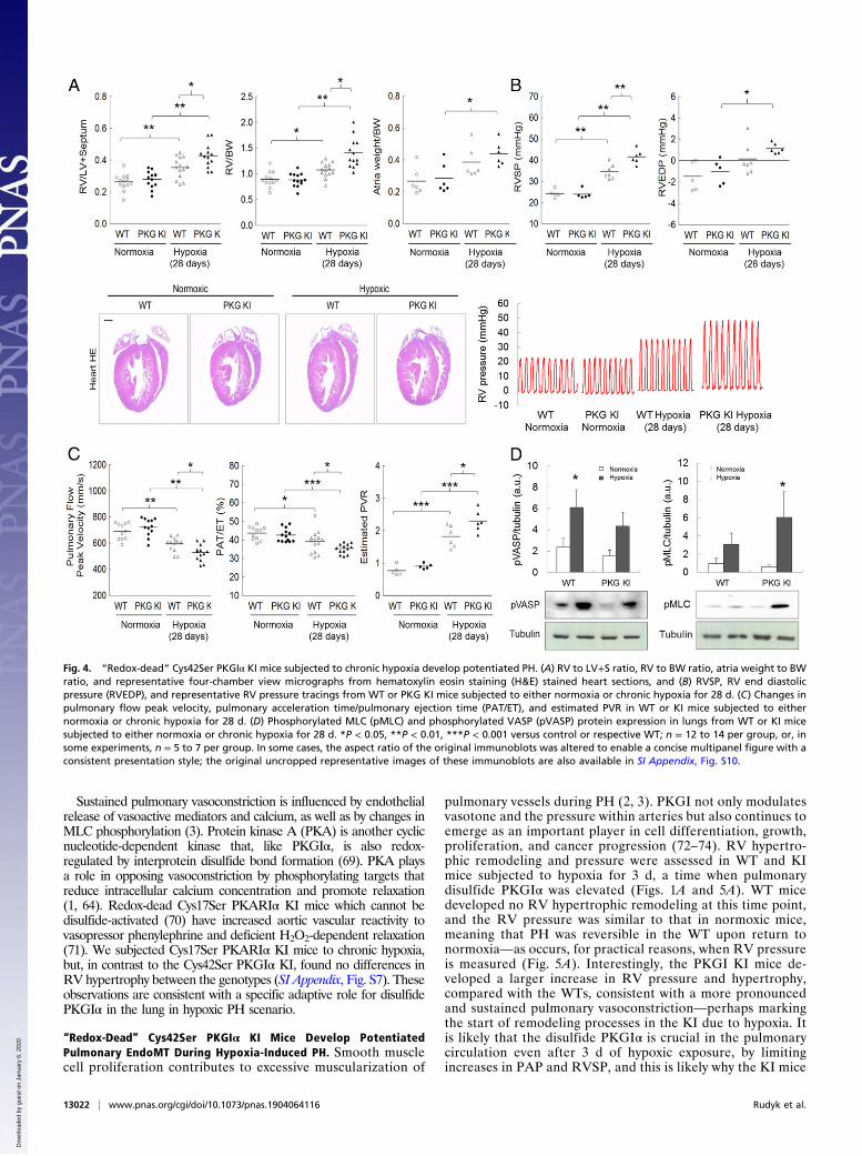
“Redox-Dead” Cys42Ser PKGIα KI Mice Develop More SevereHypoxia-Induced PH Phenotype. The role of disulfide PKGIα asan adaptive mechanism during hypoxic PH was examined inmore depth by comparing disease progression in WT versus KImice. KI mice showed potentiated increases in RV hypertrophyand pressure (Fig. 4 A and B) compared with WT during hyp-oxia. This potentiated dysfunction in the KIs that cannot formthe targeting and activating disulfide PKGIα during hypoxia wasfurther evidenced by an exacerbated decline in pulmonary vas-cular blood flow indexes, together with a higher pulmonaryvascular resistance (PVR) (Fig. 4C). Cardiac function declinewas moderate in both genotypes (SI Appendix, Fig. S5).Phosphorylation of vasodilator-stimulated phosphoprotein
(VASP) was decreased, while myosin light chain (MLC)phosphorylation was increased in lungs (Fig. 4D) and pul-monary arteries (SI Appendix, Fig. S6A) of the KI comparedwith WT during hypoxia. Lack of VASP phosphorylation inthe KIs is consistent with deficient disulfide PKGIα targeting,and was observed previously (10). Phosphorylated MLC statusis modulated by MLC kinase and by MLC phosphatase, theactivity of which is phosphoregulated by PKGIα (64, 65).Disulfide PKGIα is anticipated to activate MLC phosphatasewhich, in turn, dephosphorylates MLC and enables smooth musclerelaxation (64). The Cys42Ser PKGIα KIs are likely deficient in thisactivity, resulting in higher MLC phosphorylation. This may beconsistent with Cys42 oxidation targeting PKGIα, which is plausi-ble given the disulfide occurs in the middle of the N-terminalleucine zipper of the kinase that binds a similar zipper in MLCphosphatase (47, 64). The enhanced phosphorylation of MLCin the KI during hypoxia is in agreement with enhanced vas-oconstrictory kinase activation (66, 67), with an inability tosuitably activate the disulfide PKGIα-dependent activation ofMLC phosphatase that enables compensatory vasodilationin WT.
H2O2-dependent Vasodilation Is Impaired in “Redox-Dead” Cys42SerPKGIα KI Pulmonary Arteries. Disulfide PKGIα contributes toblood pressure homeostasis, being a component of endothelium-derived hyperpolarizing factor-dependent vasodilation (8, 47). Itdecreases calcium concentration in vascular smooth muscle cell,and mediates oxidant-induced vasodilation (68). Since PASMCsare abundant in PKGIα and its disulfide-dimerized form, it wasrational to test whether this mechanism is preserved in pulmo-nary vessels. First- or second-order pulmonary arteries from theKIs demonstrated impaired vasodilatory responses to H2O2compared with the WT, despite equal constriction to the pressor
13020 | www.pnas.org/cgi/doi/10.1073/pnas.1904064116 Rudyk et al.
Dow
nloa
ded
by g
uest
on
Janu
ary
6, 2
020

agonist U-46619 in each genotype (SI Appendix, Fig. S6B). Iso-lated perfused mouse lung was next employed to test vascularresponses in pulmonary resistance vessels. Potentiated pressoragonist-induced constriction and deficient pulmonary vaso-dilatory responses to H2O2 were observed in the perfused lungsfrom the KI mice, compared with the WT (SI Appendix, Fig.S6C), further supporting the crucial vasodilatory role of disulfidePKGIα in pulmonary circulation. This is consistent with a globalrole for PKGIα oxidation in vasodilation (8, 11).Disulfide PKGIα formation transduces increased abundance
of oxidants to vasodilation in the systemic circulation (8, 10),
with the same basic events clearly in operation in the pul-monary system. This oxidative activation of PKGIα occurs inthe pulmonary tissues of WT mice during chronic hypoxia for28 d, and the evidence presented thus far indicates this off-sets the hypertension in airway blood vessels that occurs atthis time. This adaptive mechanism that enhances PKGIαactivity to trigger vasodilation lowers RV pressure, thus re-ducing the associated RV hypertrophic remodeling. It isplausible that interventions that increase disulfide PKGIαmay be therapeutic, limiting progression to heart failure inPAH patients.
Fig. 3. Effect of CSE inhibition and polysulfides in hypoxia-induced PH. (A) Pulmonary disulfide PKGIα expression, RV pressure, and RV to left ventricle +septum (LV+S) ratio in C57BL/6 mice subjected to either normoxia or chronic hypoxia for 14 d with or without CSE inhibitor L-PPG (50 mg/kg/d). (B) CSE proteinexpression and disulfide PKGIα level in lungs of mice treated with CSE siRNA (1.3 mg·kg−1·d−1); RV pressure and RV to LV+S ratio in WT mice subjected toeither normoxia or chronic hypoxia for 14 d with or without CSE siRNA (1.3 mg·kg−1·d−1). (C) Disulfide PKGIα formation in response to persulfides donor K2Sxtreatment in human pulmonary artery smooth muscle cells. (D) RV pressure and RV to LV+septum ratio in C57BL/6 mice subjected to either normoxia orchronic hypoxia for 14 d with or without persulfides donor K2Sx (2 mg·kg−1·d−1); RV to LV+S ratio in WT or KI mice subjected to chronic hypoxia for 14 d withor without K2Sx (2 mg·kg−1·d−1). *P < 0.05, **P < 0.01 versus control; n = 5 to 7 per group. In some cases, the aspect ratio of the original immunoblots wasaltered to enable a concise multipanel figure with a consistent presentation style; the original uncropped representative images of these immunoblots arealso available in SI Appendix, Fig. S10.
Rudyk et al. PNAS | June 25, 2019 | vol. 116 | no. 26 | 13021
MED
ICALSC
IENCE
S
Dow
nloa
ded
by g
uest
on
Janu
ary
6, 2
020
Sustained pulmonary vasoconstriction is influenced by endothelialrelease of vasoactive mediators and calcium, as well as by changes inMLC phosphorylation (3). Protein kinase A (PKA) is another cyclicnucleotide-dependent kinase that, like PKGIα, is also redox-regulated by interprotein disulfide bond formation (69). PKA playsa role in opposing vasoconstriction by phosphorylating targets thatreduce intracellular calcium concentration and promote relaxation(1, 64). Redox-dead Cys17Ser PKARIα KI mice which cannot bedisulfide-activated (70) have increased aortic vascular reactivity tovasopressor phenylephrine and deficient H2O2-dependent relaxation(71). We subjected Cys17Ser PKARIα KI mice to chronic hypoxia,but, in contrast to the Cys42Ser PKGIα KI, found no differences inRV hypertrophy between the genotypes (SI Appendix, Fig. S7). Theseobservations are consistent with a specific adaptive role for disulfidePKGIα in the lung in hypoxic PH scenario.
“Redox-Dead” Cys42Ser PKGIα KI Mice Develop PotentiatedPulmonary EndoMT During Hypoxia-Induced PH. Smooth musclecell proliferation contributes to excessive muscularization of
pulmonary vessels during PH (2, 3). PKGI not only modulatesvasotone and the pressure within arteries but also continues toemerge as an important player in cell differentiation, growth,proliferation, and cancer progression (72–74). RV hypertro-phic remodeling and pressure were assessed in WT and KImice subjected to hypoxia for 3 d, a time when pulmonarydisulfide PKGIα was elevated (Figs. 1A and 5A). WT micedeveloped no RV hypertrophic remodeling at this time point,and the RV pressure was similar to that in normoxic mice,meaning that PH was reversible in the WT upon return tonormoxia—as occurs, for practical reasons, when RV pressureis measured (Fig. 5A). Interestingly, the PKGI KI mice de-veloped a larger increase in RV pressure and hypertrophy,compared with the WTs, consistent with a more pronouncedand sustained pulmonary vasoconstriction—perhaps markingthe start of remodeling processes in the KI due to hypoxia. Itis likely that the disulfide PKGIα is crucial in the pulmonarycirculation even after 3 d of hypoxic exposure, by limitingincreases in PAP and RVSP, and this is likely why the KI mice
Fig. 4. “Redox-dead” Cys42Ser PKGIα KI mice subjected to chronic hypoxia develop potentiated PH. (A) RV to LV+S ratio, RV to BW ratio, atria weight to BWratio, and representative four-chamber view micrographs from hematoxylin eosin staining (H&E) stained heart sections, and (B) RVSP, RV end diastolicpressure (RVEDP), and representative RV pressure tracings from WT or PKG KI mice subjected to either normoxia or chronic hypoxia for 28 d. (C) Changes inpulmonary flow peak velocity, pulmonary acceleration time/pulmonary ejection time (PAT/ET), and estimated PVR in WT or KI mice subjected to eithernormoxia or chronic hypoxia for 28 d. (D) Phosphorylated MLC (pMLC) and phosphorylated VASP (pVASP) protein expression in lungs from WT or KI micesubjected to either normoxia or chronic hypoxia for 28 d. *P < 0.05, **P < 0.01, ***P < 0.001 versus control or respective WT; n = 12 to 14 per group, or, insome experiments, n = 5 to 7 per group. In some cases, the aspect ratio of the original immunoblots was altered to enable a concise multipanel figure with aconsistent presentation style; the original uncropped representative images of these immunoblots are also available in SI Appendix, Fig. S10.
13022 | www.pnas.org/cgi/doi/10.1073/pnas.1904064116 Rudyk et al.
Dow
nloa
ded
by g
uest
on
Janu
ary
6, 2
020

that cannot form the disulfide in PKGI develop a larger ormore sustained pressure increase.Three days of hypoxia was therefore considered a logical time
point to monitor changes in gene expression, as this is beforestructural remodeling in the WT had occurred, in an attempt to
define additional events that are important in the pathogenesisof hypoxic pulmonary disease. Thus, a transcriptomic screenusing an Affymetrix microarray was performed on lungs fromWT or KI mice subjected to normoxia or hypoxia for 3 d.Pathway analysis of these mRNA expression abundance data
Fig. 5. Enhanced pulmonary vascular growth signaling and EndoMT in redox-dead Cys42Ser PKGIα KI mice subjected to chronic hypoxia. (A) Pulmonary disulfide PKGIαlevel in C57BL/6mice subjected to hypoxia for 1, 3, and 7 d; RVSP and RV to LV+S ratio fromWT or PKG KI mice subjected to short-time chronic hypoxia for 3 d. *P < 0.05,**P < 0.001 versus control or WT; n = 6 to 8 per group. (B) Unbiased pathway analysis of processes with the largest number of alterations in gene expression in lungs ofWT or KI mice subjected to 3 d of hypoxia comparedwith normoxic animals, listed in descending order. (C) Representative confocal images in lung sections fromWT or KImice subjected to chronic hypoxia for 28 d, stained simultaneously with nuclear (DAPI, blue), smooth muscle (α-SMA, red), and endothelial (CD31, green) markers. (Scalebar, 50 μm.) (D) Desmin, α-SMA, Twist-1, and phospho-vimentin protein expression in lungs from WT or KI mice subjected to chronic hypoxia for 28 d. *P < 0.05 versuscontrol; n = 6 to 8 per group. ECM, extracellular matrix; TGF, transforming growth factor; BMP, bone morphogenetic protein; JAK/STAT; janus kinase/signal transducerand activator of transcription proteins; MAPK, mitogen-activated protein kinase; α-SMA, α-smooth muscle actin; CD31, cluster of differentiation 31; WT, wild type mice;PKG KI, “redox-dead” Cys42Ser PKGIα KI mice. Unique effect is gene changes which are unique to either theWT or KI group. Common effects are gene changes that arecommon to both theWT aswell as the KI group. Similar effects are gene changeswhich are neither common nor unique toWTor KI groups. This terminology reflects thedefinitions in the Metalcore Training Manual (Version 5.0). In some cases, the aspect ratio of the original immunoblots was altered to enable a concise multipanel figurewith a consistent presentation style; the original uncropped representative images of these immunoblots are also available in SI Appendix, Fig. S11.
Rudyk et al. PNAS | June 25, 2019 | vol. 116 | no. 26 | 13023
MED
ICALSC
IENCE
S
Dow
nloa
ded
by g
uest
on
Janu
ary
6, 2
020
revealed an up-regulation of progrowth, extracellular matrixremodeling and endothelial-to-mesenchymal transition (EndoMT)cellular signaling pathways in the KI compared with the WT after3 d of hypoxia (Fig. 5B). This was notable, as EndoMT recentlyemerged as an important regulator of pulmonary vascular remod-eling in rodent models of PH and human disease (75).Affymetrix microarray mRNA analysis was performed on the
whole lung; therefore, it was necessary to establish whether theincreased growth and EndoMT were evident in pulmonary bloodvessels. Increased coexpression of α-smooth muscle actin (α-SMA)and cluster of differentiation 31 (CD31) in lung endothelial cells ofthe KI mice subjected to hypoxia was prominent compared withthat measured in WT (Fig. 5C). Protein expressions of α-SMA anddesmin, as well as the EndoMT transcriptional regulator Twist-1 and phosphorylated Vimentin (75) (Fig. 5D), were increased inthe lungs of the KI to a greater extent than those of WT followinghypoxia. The KI mice subjected to 28 d of hypoxia demonstratedsignificantly exacerbated pulmonary vascular muscularizationcompared with WT exposed to the same intervention, as evi-denced by a greater accumulation of α-SMA expressing cells inpulmonary vessels (SI Appendix, Fig. S6D). It is plausible that in-creased disulfide PKGIα during hypoxia may prevent pulmonaryvascular muscularization, possibly by impairing EndoMT. Whetherthis mechanism serves to alleviate pressure and PVR, perhapsindependently of the disulfide PKGIα pressure-lowering role, re-mains to be definitively elucidated.In summary, disulfide PKGIα accumulates during chronic
hypoxia in mouse and man, likely due to the accumulation ofH2O2, glutathione disulfide, and protein-bound persulfidesunder these conditions. Depletion of superreducing persulfidespecies in mouse hypoxic tissues may also contribute todisulfide PKGIα abundance, which serves as an endogenous,adaptive redox signaling mechanism that limits PH to atten-uate RV hypertrophy and disease progression. DisulfidePKGIα may also prevent the progression of EndoMT and solimit adverse pulmonary vascular remodeling. Pharmacologi-cal interventions that enhance disulfide PKGIα levels, such as
polysulfides as demonstrated herein, may provide a noveltherapeutic strategy to combat disease resulting from PH.
Materials and MethodsAnimals, Induction of Hypoxic Pulmonary Hypertension, and Treatment. Allanimal procedures were performed in accordance with the Home OfficeGuidance on the Operation of the Animals (Scientific Procedures) Act 1986 inthe United Kingdom and were approved by the King’s College AnimalWelfare and Ethical Review Body. Mice constitutively expressing PKGIαCys42Ser were produced on a pure C57BL/6 background by Taconic Artemisas described (8, 76) and bred on-site. Age- and body weight-matched WT orPKGIα Cys42Ser KI male offspring were used in most of the studies. In someexperiments, age- and body weight-matched adult C57BL/6 male mice werepurchased from Charles River, as highlighted in more detail in Results andDiscussion. Animals had ad libitum access to standard chow and water andwere kept in specific pathogen-free conditions under a 12-h day/night cycleat 20 °C and 60% humidity before hypoxic exposure. Hypoxic PH was in-duced by exposing mice to normobaric hypoxia (10% of inspired O2) in alarge ventilated chamber (Biospherix, Ltd) (SI Appendix, Fig. S1). The CO2
level was monitored continuously with CO2 meter and soda lime. Fresh cage,water, and food changes were performed once every 7 d to 10 d for all ofthe animals. Additional materials and procedures can be found in SI Ap-pendix, SI Materials and Methods.
Study Approval. All animal procedures were performed in accordance withthe Home Office Guidance on the Operation of the Animals (ScientificProcedures) Act 1986 in the United Kingdomandwere approved by the King’sCollege Animal Welfare and Ethical Review Body. The protocol of the studyusing human samples was approved by the Ethics Committee of the Justus-Liebig-University School of Medicine (No. 111/08 and 58/15). Informed con-sent was obtained in written form from each subject.
ACKNOWLEDGMENTS. O.R. is a British Heart Foundation Intermediate BasicScience Research Fellow (Sponsor Reference FS/14/57/31138) and a recipientof a Butrous Foundation Young Investigator Award (2018). P.E. is supportedby the European Research Council (ERC Advanced Award) and the MedicalResearch Council. We thank Dr. James Clark for excellent technical help withthe Scisense ADVantage Admittance PV Systems use, and Dr. Rob Haworthfor indispensable help with hypoxic chamber maintenance.
1. P. I. Aaronson et al., Hypoxic pulmonary vasoconstriction: Mechanisms and contro-
versies. J. Physiol. 570, 53–58 (2006).2. R. T. Schermuly, H. A. Ghofrani, M. R. Wilkins, F. Grimminger, Mechanisms of disease:
Pulmonary arterial hypertension. Nat. Rev. Cardiol. 8, 443–455 (2011).3. M. R. Wilkins, H. A. Ghofrani, N. Weissmann, A. Aldashev, L. Zhao, Pathophysiology
and treatment of high-altitude pulmonary vascular disease. Circulation 131, 582–590
(2015).4. D. M. Tabima, S. Frizzell, M. T. Gladwin, Reactive oxygen and nitrogen species in
pulmonary hypertension. Free Radic. Biol. Med. 52, 1970–1986 (2012).5. J. R. Burgoyne, H. Mongue-Din, P. Eaton, A. M. Shah, Redox signaling in cardiac
physiology and pathology. Circ. Res. 111, 1091–1106 (2012).6. A. M. Shah, Parsing the role of NADPH oxidase enzymes and reactive oxygen species
in heart failure. Circulation 131, 602–604 (2015).7. O. Rudyk, P. Eaton, Biochemical methods for monitoring protein thiol redox states in
biological systems. Redox Biol. 2, 803–813 (2014).8. O. Prysyazhna, O. Rudyk, P. Eaton, Single atom substitution in mouse protein kinase G
eliminates oxidant sensing to cause hypertension. Nat. Med. 18, 286–290 (2012).9. D. Stubbert et al., Protein kinase G Iα oxidation paradoxically underlies blood pressure
lowering by the reductant hydrogen sulfide. Hypertension 64, 1344–1351 (2014).10. O. Rudyk, O. Prysyazhna, J. R. Burgoyne, P. Eaton, Nitroglycerin fails to lower blood
pressure in redox-dead Cys42Ser PKG1α knock-in mouse. Circulation 126, 287–295
(2012).11. B. H. Neo, S. Kandhi, M. S. Wolin, Roles for redox mechanisms controlling protein
kinase G in pulmonary and coronary artery responses to hypoxia. Am. J. Physiol. Heart
Circ. Physiol. 301, H2295–H2304 (2011).12. D. Patel, R. Alhawaj, M. S. Wolin, Exposure of mice to chronic hypoxia attenuates
pulmonary arterial contractile responses to acute hypoxia by increases in extracellular
hydrogen peroxide. Am. J. Physiol. Regul. Integr. Comp. Physiol. 307, R426–R433
(2014).13. C. M. Wong, G. Bansal, L. Pavlickova, L. Marcocci, Y. J. Suzuki, Reactive oxygen species
and antioxidants in pulmonary hypertension. Antioxid. Redox Signal. 18, 1789–1796
(2013).14. E. Nozik-Grayck, K. R. Stenmark, Role of reactive oxygen species in chronic hypoxia-
induced pulmonary hypertension and vascular remodeling. Adv. Exp. Med. Biol. 618,
101–112 (2007).
15. P. Siques, J. Brito, E. Pena, Reactive oxygen species and pulmonary vasculature during
hypobaric hypoxia. Front. Physiol. 9, 865 (2018).16. N. L. Jernigan, B. R. Walker, T. C. Resta, Pulmonary PKG-1 is upregulated following
chronic hypoxia. Am. J. Physiol. Lung Cell. Mol. Physiol. 285, L634–L642 (2003).17. Y. D. D. Zhao et al., Protein kinase G-I deficiency induces pulmonary hypertension
through Rho A/Rho kinase activation. Am. J. Pathol. 180, 2268–2275 (2012).18. B. Yi et al., cGMP-dependent protein kinase Iα transfection inhibits hypoxia-induced
migration, phenotype modulation and annexins A1 expression in human pulmonary
artery smooth muscle cells. Biochem. Biophys. Res. Commun. 418, 598–602 (2012).19. Y. Y. Zhao et al., Persistent eNOS activation secondary to caveolin-1 deficiency induces
pulmonary hypertension in mice and humans through PKG nitration. J. Clin. Invest.
119, 2009–2018 (2009).20. M. Mittal et al., Hypoxia-dependent regulation of nonphagocytic NADPH oxidase
subunit NOX4 in the pulmonary vasculature. Circ. Res. 101, 258–267 (2007).21. M. Mittal et al., Hypoxia induces Kv channel current inhibition by increased NADPH
oxidase-derived reactive oxygen species. Free Radic. Biol. Med. 52, 1033–1042 (2012).22. S. A. Barman et al., NADPH oxidase 4 is expressed in pulmonary artery adventitia and
contributes to hypertensive vascular remodeling. Arterioscler. Thromb. Vasc. Biol. 34,
1704–1715 (2014).23. Y. J. Suzuki, R. H. Steinhorn, M. T. Gladwin, Antioxidant therapy for the treatment of
pulmonary hypertension. Antioxid. Redox Signal. 18, 1723–1726 (2013).24. A. M. R. Salles, T. F. Galvao, M. T. Silva, L. C. D. Motta, M. G. Pereira, Antioxidants for
preventing preeclampsia: A systematic review. ScientificWorldJournal 2012, 243476
(2012).25. G. Bjelakovic, D. Nikolova, L. L. Gluud, R. G. Simonetti, C. Gluud, Antioxidant sup-
plements for prevention of mortality in healthy participants and patients with various
diseases. Cochrane Database Syst. Rev. 3, CD007176 (2012).26. G. Bjelakovic, D. Nikolova, L. L. Gluud, R. G. Simonetti, C. Gluud, Antioxidant sup-
plements for prevention of mortality in healthy participants and patients with various
diseases. Cochrane Database Syst. Rev. 2, CD007176 (2008).27. A. L. Firth, J. Mandel, J. X. Yuan, Idiopathic pulmonary arterial hypertension. Dis.
Model. Mech. 3, 268–273 (2010).28. K. L. Colvin, M. E. Yeager, Animal models of pulmonary hypertension: Matching
disease mechanisms to etiology of the human disease. J. Pulm. Respir. Med. 4, 198
(2014).
13024 | www.pnas.org/cgi/doi/10.1073/pnas.1904064116 Rudyk et al.
Dow
nloa
ded
by g
uest
on
Janu
ary
6, 2
020

29. G. Simonneau et al., Haemodynamic definitions and updated clinical classification ofpulmonary hypertension. Eur. Respir. J. 53, 1801913 (2019).
30. J. R. Burgoyne et al., Cysteine redox sensor in PKGIa enables oxidant-induced acti-vation. Science 317, 1393–1397 (2007).
31. D. Hernandez-Saavedra, K. Swain, R. Tuder, S. V. Petersen, E. Nozik-Grayck, Redoxregulation of the superoxide dismutases SOD3 and SOD2 in the pulmonary circula-tion. Adv. Exp. Med. Biol. 967, 57–70 (2017).
32. R. K. Mistry et al., Transcriptional regulation of cystathionine-γ-lyase in endothelialcells by NADPH oxidase 4-dependent signaling. J. Biol. Chem. 291, 1774–1788 (2016).
33. I. Diebold, A. Petry, J. Hess, A. Görlach, The NADPH oxidase subunit NOX4 is a newtarget gene of the hypoxia-inducible factor-1. Mol. Biol. Cell 21, 2087–2096 (2010).
34. S. L. Archer et al., Epigenetic attenuation of mitochondrial superoxide dismutase 2 inpulmonary arterial hypertension: A basis for excessive cell proliferation and a newtherapeutic target. Circulation 121, 2661–2671 (2010).
35. S. L. Archer, Acquired mitochondrial abnormalities, including epigenetic inhibition ofsuperoxide dismutase 2, in pulmonary hypertension and cancer: Therapeutic impli-cations. Adv. Exp. Med. Biol. 903, 29–53 (2016).
36. D. Xu et al., Exacerbated pulmonary arterial hypertension and right ventricular hy-pertrophy in animals with loss of function of extracellular superoxide dismutase.Hypertension 58, 303–309 (2011).
37. E. Nozik-Grayck et al., Lung EC-SOD overexpression attenuates hypoxic induction ofEgr-1 and chronic hypoxic pulmonary vascular remodeling. Am. J. Physiol. Lung Cell.Mol. Physiol. 295, L422–L430 (2008).
38. F. Kamezaki et al., Gene transfer of extracellular superoxide dismutase amelioratespulmonary hypertension in rats. Am. J. Respir. Crit. Care Med. 177, 219–226 (2008).
39. E. Nozik-Grayck et al., Selective depletion of vascular EC-SOD augments chronichypoxic pulmonary hypertension. Am. J. Physiol. Lung Cell. Mol. Physiol. 307, L868–L876 (2014).
40. S. Ismail et al., NOX4 mediates hypoxia-induced proliferation of human pulmonaryartery smooth muscle cells: The role of autocrine production of transforming growthfactor-beta1 and insulin-like growth factor binding protein-3. Am. J. Physiol. LungCell. Mol. Physiol. 296, L489–L499 (2009).
41. X. Lu et al., Hypoxia downregulates PPARγ via an ERK1/2-NF-κB-Nox4-dependentmechanism in human pulmonary artery smooth muscle cells. Free Radic. Biol. Med.63, 151–160 (2013).
42. D. E. Green et al., The Nox4 inhibitor GKT137831 attenuates hypoxia-induced pul-monary vascular cell proliferation. Am. J. Respir. Cell Mol. Biol. 47, 718–726 (2012).
43. S. Altenhöfer, K. A. Radermacher, P. W. Kleikers, K. Wingler, H. H. Schmidt, Evolutionof NADPH oxidase inhibitors: Selectivity and mechanisms for target engagement.Antioxid. Redox Signal. 23, 406–427 (2015).
44. C. Veith et al., NADPH oxidase 4 is not involved in hypoxia-induced pulmonary hy-pertension. Pulm. Circ. 6, 397–400 (2016).
45. Y. Nishijima et al., Contribution of KV1.5 channel to hydrogen peroxide-Inducedhuman arteriolar dilation and its modulation by coronary artery disease. Circ. Res.120, 658–669 (2017).
46. K. Khavandi et al., Pressure-induced oxidative activation of PKG enables vaso-regulation by Ca2+ sparks and BK channels. Sci. Signal. 9, ra100 (2016).
47. O. Prysyazhna, P. Eaton, Redox regulation of cGMP-dependent protein kinase Iα inthe cardiovascular system. Front. Pharmacol. 6, 139 (2015).
48. G. Yang et al., H2S as a physiologic vasorelaxant: Hypertension in mice with deletionof cystathionine gamma-lyase. Science 322, 587–590 (2008).
49. T. Ida et al., Reactive cysteine persulfides and S-polythiolation regulate oxidativestress and redox signaling. Proc. Natl. Acad. Sci. U.S.A. 111, 7606–7611 (2014).
50. R. Greiner et al., Polysulfides link H2S to protein thiol oxidation. Antioxid. RedoxSignal. 19, 1749–1765 (2013).
51. K. R. Olson et al., Metabolism of hydrogen sulfide (H2S) and production of reactivesulfur species (RSS) by superoxide dismutase. Redox Biol. 15, 74–85 (2018).
52. K. Semen et al., Sildenafil reduces signs of oxidative stress in pulmonary arterial hy-pertension: Evaluation by fatty acid composition, level of hydroxynonenal and heartrate variability. Redox Biol. 7, 48–57 (2016).
53. K. J. Bubb et al., Inhibition of phosphodiesterase 2 augments cGMP and cAMP sig-naling to ameliorate pulmonary hypertension. Circulation 130, 496–507 (2014).
54. H. A. Ghofrani et al.; PATENT-1 Study Group, Riociguat for the treatment of pul-monary arterial hypertension. N. Engl. J. Med. 369, 330–340 (2013).
55. R. Bowers et al., Oxidative stress in severe pulmonary hypertension. Am. J. Respir. Crit.Care Med. 169, 764–769 (2004).
56. K. H. Krause, Tissue distribution and putative physiological function of NOX familyNADPH oxidases. Jpn. J. Infect. Dis. 57, S28–S29 (2004).
57. J. D. Lambeth, T. Kawahara, B. Diebold, Regulation of Nox and Duox enzymatic ac-tivity and expression. Free Radic. Biol. Med. 43, 319–331 (2007).
58. Y. Nisimoto, B. A. Diebold, D. Cosentino-Gomes, J. D. Lambeth, Nox4: A hydrogenperoxide-generating oxygen sensor. Biochemistry 53, 5111–5120 (2014). Correction in:Biochemistry 53, 5472 (2014).
59. T. Akaike et al., Cysteinyl-tRNA synthetase governs cysteine polysulfidation and mi-tochondrial bioenergetics. Nat. Commun. 8, 1177 (2017).
60. M. Koutmos, O. Kabil, J. L. Smith, R. Banerjee, Structural basis for substrate activationand regulation by cystathionine beta-synthase (CBS) domains in cystathionine beta-synthase. Proc. Natl. Acad. Sci. U.S.A. 107, 20958–20963 (2010).
61. J. M. Fukuto et al., Biological hydropersulfides and related polysulfides–A new con-cept and perspective in redox biology. FEBS Lett. 592, 2140–2152 (2018).
62. T. Numakura et al., Production of reactive persulfide species in chronic obstructivepulmonary disease. Thorax 72, 1074–1083 (2017).
63. Y. H. Chen et al., Endogenous hydrogen sulfide in patients with COPD. Chest 128,3205–3211 (2005).
64. J. R. Burgoyne, P. Eaton, Oxidant sensing by protein kinases a and g enables in-tegration of cell redox state with phosphoregulation. Sensors (Basel) 10, 2731–2751(2010).
65. H. K. Surks et al., Regulation of myosin phosphatase by a specific interaction withcGMP- dependent protein kinase Ialpha. Science 286, 1583–1587 (1999).
66. J. Wang, L. Weigand, J. Foxson, L. A. Shimoda, J. T. Sylvester, Ca2+ signaling inhypoxic pulmonary vasoconstriction: Effects of myosin light chain and Rho kinaseantagonists. Am. J. Physiol. Lung Cell. Mol. Physiol. 293, L674–L685 (2007).
67. Y. Zhao, R. A. Rhoades, C. S. Packer, Hypoxia-induced pulmonary arterial contractionappears to be dependent on myosin light chain phosphorylation. Am. J. Physiol. 271,L768–L774 (1996).
68. P. M. Müller et al., H2O2 lowers the cytosolic Ca2+ concentration via activation ofcGMP-dependent protein kinase Iα. Free Radic. Biol. Med. 53, 1574–1583 (2012).
69. J. P. Brennan et al., Oxidant-induced activation of type I protein kinase A is mediatedby RI subunit interprotein disulfide bond formation. J. Biol. Chem. 281, 21827–21836(2006).
70. J. R. Burgoyne et al., Deficient angiogenesis in redox-dead Cys17Ser PKARIα knock-inmice. Nat. Commun. 6, 7920 (2015).
71. O. Rudyk, O. Prysyazhna, P. Eaton, Redox-dead protein kinase a RI alpha knock-inmouse is not hypertensive but has increased vascular reactivity to oxidants. FreeRadic. Biol. Med. 65, S82 (2013).
72. D. D. Browning, I. K. Kwon, R. Wang, cGMP-dependent protein kinases as potentialtargets for colon cancer prevention and treatment. Future Med. Chem. 2, 65–80(2010).
73. D. D. Browning, Protein kinase G as a therapeutic target for the treatment of met-astatic colorectal cancer. Expert Opin. Ther. Targets 12, 367–376 (2008).
74. D. Hoffmann et al., New dimeric cGMP analogues reduce proliferation in three coloncancer cell lines. Eur. J. Med. Chem. 141, 61–72 (2017).
75. B. Ranchoux et al., Endothelial-to-mesenchymal transition in pulmonary hyperten-sion. Circulation 131, 1006–1018 (2015).
76. O. Rudyk et al., Protein kinase G oxidation is a major cause of injury during sepsis.Proc. Natl. Acad. Sci. U.S.A. 110, 9909–9913 (2013).
Rudyk et al. PNAS | June 25, 2019 | vol. 116 | no. 26 | 13025
MED
ICALSC
IENCE
S
Dow
nloa
ded
by g
uest
on
Janu
ary
6, 2
020


![The endogenous subcellular localisations of the …...pholipid oxidation [22]. ACSL4 can supply arachidonyl-CoA and adrenic-CoA for incorporation into phosphatidy-lethanolamine and](https://img.dokumen.tips/doc/110x75/5ec2fce62e4af71b3e52bfc6/the-endogenous-subcellular-localisations-of-the-pholipid-oxidation-22-acsl4.jpg)