Embed Size (px)
Citation preview

Overview: The Key Roles of Cell Division
• The ability of organisms to reproduce best distinguishes living things from nonliving matter (reproduction is an emergent property of life)
• The continuity of life is based on the reproduction of cells, or cell division
• Cell Theory: all cells come from pre-existing cells

Division in unicellular vs multicellular organisms
• In unicellular organisms, standard cell division of one cell reproduces the entire organism
• Multicellular organisms depend on standard cell division only for:– Development from a fertilized cell
– Growth
– Repair
• Not for reproducing the entire organism

• Cell division is carefully controlled in all normal cells
• In eukaryotes division is a part of the cell cycle-a programmed series of events that
governs the life of a cell

Concept 12.1: Cell division results in genetically identical daughter cells
• Standard cell division results in daughter cells with “identical” genetic information and DNA
-clones-vegetative reproduction-asexual reproduction
• A special type of division produces nonidentical daughter cells for reproduction of the organism (gametes, or sperm and egg cells)
- sexual reproduction

• All the DNA in a cell constitutes the cell’s genome (nuclear genome, organelle genome)
• A genome can consist of a single DNA molecule (common in prokaryotic cells) or a number of DNA molecules (common in eukaryotic cells)
• DNA molecules in a eukaryotic cell are packaged into organelles called chromosomes
Genetic material must be duplicated and separated to produce clones

The number of chromosomes is characteristic for each species
• “n”
• Gametes (reproductive cells: sperm and eggs) contain n chromosomes
• Somatic cells (nonreproductive cells) have two sets of chromosomes or 2n (2n = 46 for humans)
• Eukaryotic chromosomes consist of chromatin, a complex of DNA and protein

• In preparation for cell division, DNA is copied or “replicated”.
• A somatic cell temporarily has 2 x 2n chromosomes
Genetic material must be duplicated to produce clones
•DNA molecules are many times longer than their cells.•The chromatin that contains this DNA must be tightly packed for travel to daughter cells. •Chromatin “packing” is called condensation
Genetic material must be separated to produce clones

Packing Ratio10,000-100,000 X

• Packed and duplicated chromosomes each have two sister chromatids
• The centromere is the narrow “waist” of the duplicated chromosome, where the two chromatids are most closely attached
• The centromere defines two “arms” (p and q) for each chromatid
• The end of the chromatid is the telomere
Structural features of chromosomes
Concept 12.2: The active division phase alternates with non-division in the cell cycle
• In the 1800’s the active division phase was named “mitosis” and the non-dividing phase named “interphase”
• The two terms have been adapted for modern usage to describe the cell cycle
• The modern cell cycle consists of
– Mitotic (M) phase (mitosis and cytokinesismitosis and cytokinesis)- nuclear and cytoplasmic division
– Interphase (cell growth and copying of chromosomes in preparation for cell division)

• Interphase (about 90% of the time of the cell cycle) can be subdivided further:
– G1 phase (“first gap”)
– S phase (“synthesis”)
– G2 phase (“second gap”)
• The cell grows during all three phases, but chromosomes are duplicated only during the S phase

Eukaryotic Cell Cycle
S(DNA synthesis)
MITOTIC(M) PHASE
Mito
sis
Cytokinesis
G1
G2
• Mitosis is conventionally subdivided into five parts:
– Prophase
– Prometaphase
– Metaphase
– Anaphase
– Telophase
• Cytokinesis overlaps telophase
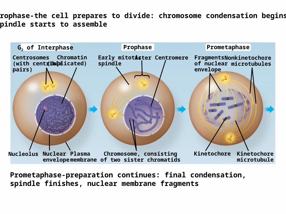
PrometaphaseProphaseG2 of Interphase
Nonkinetochoremicrotubules
Fragmentsof nuclearenvelope
Aster CentromereEarly mitoticspindle
Chromatin(duplicated)
Centrosomes(with centriolepairs)
Nucleolus Nuclearenvelope
Plasmamembrane
Chromosome, consistingof two sister chromatids
Kinetochore Kinetochoremicrotubule
Prophase-the cell prepares to divide: chromosome condensation begins, Spindle starts to assemble
Prometaphase-preparation continues: final condensation, spindle finishes, nuclear membrane fragments

Metaphase Anaphase Telophase and Cytokinesis
Cleavagefurrow
Nucleolusforming
Metaphaseplate
Centrosome atone spindle pole
SpindleDaughterchromosomes
Nuclearenvelopeforming
Metaphase-chromosomes line up at the middle of the cellAnaphase-sister chromatids separate to form new “daughter” chromosomes, pulled to opposite spindle poles
Telophase- daughter cells reorganize, reverse of prophase

Prophase
Fig. 12-6a
PrometaphaseG2 of Interphase

Fig. 12-6c
Metaphase Anaphase Telophase and Cytokinesis

The Mitotic Spindle
• The mitotic spindle is an apparatus of microtubules that controls chromosome movement during mitosis
• During prophase, assembly of spindle microtubules begins in the centrosome, the microtubule organizing center
• The centrosome replicates, forming two centrosomes that migrate to opposite ends of the cell, as spindle microtubules grow out from them

• During prometaphase, some spindle microtubules attach to the kinetochores of chromosomes and begin to move the chromosomes
• Different microtubules overlap with microtubules coming from the other centrosome (spindle pole).

Cytokinesis
• In animal cells, cytokinesis occurs by a process known as cleavage, forming a cleavage furrow
• In plant cells, a cell plate forms during cytokinesis

Cleavage furrow100 µm
Contractile ring ofmicrofilaments
Daughter cells
(a) Cleavage of an animal cell (SEM) (b) Cell plate formation in a plant cell (TEM)
Vesiclesformingcell plate
Wall ofparent cell
Cell plate
Daughter cells
New cell wall
1 µm

Binary Fission
• Prokaryotes (bacteria and archaea) reproduce by a simpler type of cell division called binary fission
• In binary fission, the chromosome replicates (beginning at the origin of replication), and the two daughter chromosomes actively move apart
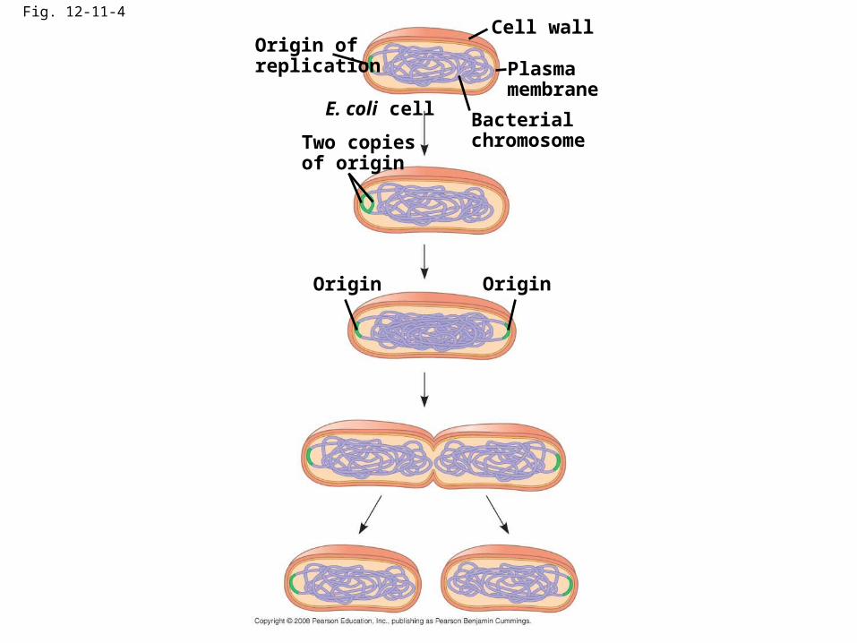
Fig. 12-11-4
Origin ofreplication
Two copiesof origin
E. coli cellBacterialchromosome
Plasmamembrane
Cell wall
Origin Origin

Concept 12.3: The eukaryotic cell cycle is regulated by a molecular control system
• The frequency of cell division varies with the type of cell
• These cell cycle differences result from regulation at the molecular level

Evidence for Cytoplasmic Signals
• The cell cycle appears to be driven by specific chemical signals present in the cytoplasm
• Some evidence for this hypothesis comes from experiments in which cultured mammalian cells at different phases of the cell cycle were fused to form a single cell with two nuclei

Experiment 1 Experiment 2
EXPERIMENT
RESULTS
S G1M G1
M MSS
When a cell in theS phase was fused with a cell in G1, the G1 nucleus immediatelyentered the Sphase—DNA was synthesized.
When a cell in theM phase was fused with a cell in G1, the G1 nucleus immediatelybegan mitosis—aspindle formed andchromatin condensed,even though thechromosome had notbeen duplicated.
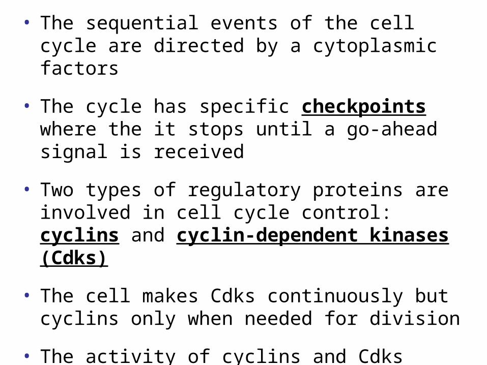
• The sequential events of the cell cycle are directed by a cytoplasmic factors
• The cycle has specific checkpoints where the it stops until a go-ahead signal is received
• Two types of regulatory proteins are involved in cell cycle control: cyclins and cyclin-dependent kinases (Cdks)
• The cell makes Cdks continuously but cyclins only when needed for division
• The activity of cyclins and Cdks fluctuates during the cell cycle-they serve as go-ahead signal at checkpoints

Fig. 12-14
SG1
M checkpoint
G2M
Controlsystem
G1 checkpoint
G2 checkpoint

Cyclin isdegraded
Cdk
MPF
Cdk
MS
G 1G2
checkpoint
Degradedcyclin
Cyclin
G2
Cyclin
accum
ulatio
n

• An example of an internal signal is that kinetochores not attached to spindle microtubules send a molecular signal that delays anaphase
• Some external signals are growth factors, proteins released by certain cells that stimulate other cells to divide

Loss of Cell Cycle Controls in Cancer Cells
• Cancer cells do not respond normally to the body’s control mechanisms
• Cancer cells may not need growth factors to grow and divide:
– They may make their own growth factor
– They may convey a growth factor’s signal without the presence of the growth factor
– They may have an abnormal cell cycle control system

• A normal cell is converted to a cancerous cell by a process called transformation
• Cancer cells form tumors, masses of abnormal cells within otherwise normal tissue
• If abnormal cells remain at the original site, the lump is called a benign tumor
• Malignant tumors invade surrounding tissues and can metastasize, exporting cancer cells to other parts of the body, where they may form secondary tumors

Multistep Model for Cancer
Tumor suppressors hold back cancer, oncogenes are genes that are capable of initiating cancerMutant oncogenes or tumor suppressor genes can be inherited

Environmental factors can trigger cancer
• Carcinogens are chemicals that lead to cancer
• Proto-oncogenes mutate to become fully active oncogenes
• Above shows several ways that proto-oncogenes can mutate

Viruses can trigger cancer
• Virus genes can remain in cells and act as oncogenes (v-oncogenes)
• Virus genes can remain in cell and activate cellular oncogenes (c-oncogenes)
• Cancer viruses, tumor viruses or oncoviruses

Example
• Mutations can lead to altered function at any step

• Environmental factors, genetic factors, viruses can trigger cancer
• They act through a multi-step model to de-regulate cell division
• They work through genes called oncogenes

NOTE CARD QUESTION
DISTINGUISH BETWEEN:
TUMOR SUPPRESSOR/ONCOGENE
V-ONCOGENE/C-ONCOGENE