Embed Size (px)
Citation preview

Overview of the Myelodysplastic Syndromes (MDS)
Brian A. Jonas, M.D., Ph.D. UC Davis School of Medicine
February 4, 2017
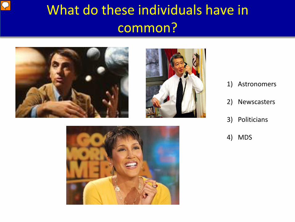
What do these individuals have in common?
1) Astronomers
2) Newscasters
3) Politicians
4) MDS

MDS Questions to be Answered
• What is MDS? • Why did I get MDS? • What does it mean for my life? • Is there treatment for it? • How should I be treated?
– When? – Why?
• What are some of the new advances in MDS?

Essentials for the Informed Pt with MDS
• Know your IPSS-R risk group • Know your treatment options
– Including transplant, clinical trials
• Know what your treatment goals are • Know the potential side effects of your
treatments • Know available MDS resources • Have a caregiver available/involved

The Myelodysplastic Syndromes
Blood, 2014.

The Myelodysplastic Syndromes (MDS)
• Heterogeneous group of clonal bone marrow failure syndromes
• 10-30,000 cases per year in US • Median age 76, > in males • Ineffective hematopoiesis
– Bleeding, infections, anemia • Transformation to AML • Variable clinical course
– Need for accurate prognostication
List et al. NEJM 2005. Ma et al. Cancer 2007.

Epidemiology of MDS

MDS Incidence Increases With Age
Aul et al. Leuk Res 1998. Radlund et al. Eur J Haematol 1995.

Predispositions and Risk Factors for MDS
• Acquired Risk Factors (common) – Age – Mutagen exposure (chemotherapy, radiation,
benzenes, tobacco) – Other hematologic disorders (e.g. AA, PNH)
• Heritable Predisposition (rare)

Pathogenesis of MDS

MDS is Like a Broken Down Assembly Line

Dysfunctional progression along the maturation pathway
Normal and Dysplastic Hematopoiesis

Pathogenesis of MDS
Ades et al. Lancet 2014.

Jaiswal et al, NEJM, 2014. Genovese et al, NEJM, 2014.
Aging is Associated with Clonal Hematopoiesis with Driver Mutations

MDS is a Cancer
Walter et al. NEJM 2012.

The MDS “Stem Cell” is the primitive Hematopoietic Stem Cell
Nilsson et al. Blood 2007. Tehranchi et al. NEJM 2010. Pang et al. PNAS 2013.

MDS is Associated with Chromosome Abnormalities…
40%
8% 22%
30%
Normal karyotype
Balanced abnormalities
Olney et al. Leuk Res 2006.
Other unbalanced abnormalities
Abnormal chromosome 5 and/or 7
Primary

Recurrent Mutations in MDS
Haferlach et al. Leukemia 2014; Bejar et al. Blood 2014.
Improved response to HMA Involved in methylation
Presentation and Diagnosis of MDS

MDS Presentation
• Symptoms – Some are asymptomatic – Fatigue – Weakness – Bruising – Infections
• Signs – Pallor – Ecchymoses
• CBC – Anemia (most common)
• Macrocytic – Neutropenia
• Pelger-Huet cells – Thrombocytopenia

Diagnosis of MDS
Bone Marrow Biopsy
and Aspirate
Morphology Cytogenetics FISH/Molecular Flow Cytometry

Differential Diagnosis of MDS
• Acute myeloid leukemia • Other MPNs • Aplastic anemia • Nutritional deficiencies • Medications

Prognostication of MDS

Prognostic Features in MDS
• Clinical – CBC, marrow blasts, cytogenetics – Age, PS, ferritin, LDH, β2M, marrow fibrosis – Treatment/Response
• Molecular – Specific mutations – Number of mutations

FAB and 2008 WHO Classification of MDS
Vardiman et al. Blood 2002. Vardiman et al. Blood 2009.
French-American-British (FAB) 2008 World Health Organization (WHO)
Refractory Anemia (RA) Refractory Cytopenia with Unilineage Dysplasia (RCUD)
“ RC with Multilineage Dysplasia (RCMD)
“ MDS associated with isolated del(5q)
RA with Ringed Sideroblasts (RARS) RARS with unilineage dysplasia
RCMD with ringed sideroblasts
RA with Excess Blasts (RAEB) RAEB-1 (5-9% blasts)
“ RAEB-2 (10-19% blasts)
RAEB in Transformation (RAEB-T) Acute Myeloid Leukemia (20+% blasts)
Chronic Myelomonocytic Leukemia (CMML) MDS/MPN Overlap
N/A MDS Unclassified (MDS-U)

2016 WHO Classification of MDS
NCCN Guidelines, MDS v1.2017.

International Prognostic Scoring System for MDS (IPSS)
Greenberg et al. Blood 1997.
Cytopenias: Hgb < 10 ANC < 1800 Plt < 100,000 Cytogenetics: Good – normal, -Y only, del(5q) only, del(20q) only Intermediate – +8, single misc, double abnormalities Poor – complex (≥3), abnormality of chromosome 7

IPSS
Greenberg et al. Blood 1997.
Years Years

Revised International Prognostic Scoring System
• IPSS-R built upon prior IPSS
• Multinational IWG-PM project
• 7,012 patients with median age 71
• MDS classified by FAB and WHO
• New MDS cytogenetic classification
• Considered depth of cytopenias, age, LDH, ferritin, b2M, fibrosis, and PS
Greenberg et al, Blood 2012. Schanz et al, JCO 2012.

IPSS-R: Determining the Score
Greenberg et al, Blood 2012.
Variable 0 0.5 1 1.5 2 3 4
Cytogenetics Very Good Good Intermediate Poor Very Poor
Marrow blasts (%) ≤2 >2-<5 5-10 >10
Hemoglobin ≥10 8-<10 <8
Platelets ≥100 50-<100 <50
ANC ≥0.8 <0.8
IPSS-R Score Values
Cytogenetic Risk Abnormalities
Very Good -Y, del(11q)
Good Normal, del(5q), del(12p), del(20q), double including del(5q)
Intermediate del(7q), +8, +19, i(17q), any other single or double
Poor -7, inv(3)/t(3q)/del(3q), double including -7/del(7q), complex = 3
Very Poor Complex >3

IPPS-R Risk IPSS-R Score Median OS (yr) 25% AML Progression (yr)
Very Low ≤1.5 8.8 NR
Low >1.5-3 5.3 10.8
Intermediate >3-4.5 3 3.2
High >4.5-6 1.6 1.4
Very High >6 0.8 0.7
Greenberg et al, Blood 2012.
IPSS-R: Calculating the Score

IPSS-R: Survival and AML Progression
Greenberg et al, Blood 2012. Schanz et al, JCO 2012.
• CBC values, bone marrow blasts, cytogenetics

IPSS-R Calculator from the MDS Foundation
Online tool and smartphone app available for free from the MDS Foundation: http://www.mds-foundation.org/interactive-tools/

• Rapid advances in understanding MDS pathogenesis
• Improvements in diagnostic and analytic tools • Future PSS likely to include:
– Flow cytometry – Gene mutations – Comorbidity assessments
Improving MDS Prognostication

Number of Driver Mutations Affects Prognosis
From Papaemmanuil et al, Blood 2013.

Recurrent and Prognostic Gene Mutations
Function Gene
Epigenetic/Chromatin Modifiers TET2, DNMT3A#, ASXL1, EZH2
Splicing SF3B1, SRSF2, U2AF1#, ZRSR2
Differentiation RUNX1
DNA Damage Response/Apoptosis TP53*, BCOR
Cohesin Complex STAG2
Signaling CBL
Recurrent in >5% of MDS patients across multiple studies Favorable prognostic impact Negative prognostic impact Neutral prognostic impact *Strong negative prognostic impact in therapy-related MDS #Strong negative prognostic impact in CMML Table adapted from: Haferlach et al, Leukemia 2014; Bejar et al, NEJM 2011; Papaemmanuil et al, Blood 2013; Walter et al, Leukemia 2013; and Thol et al; Blood 2012.

Combining Mutations with IPSS Can Improve Prognostication
IPSS and TP53, EZH2, ETV6, RUNX1 and ASXL1 mutations
From Bejar et al, NEJM 2011.

Revised IPSS-R: Incorporation of Mutations
Training Validation
Haferlach et al, Leukemia 2014.
Model: Gender, Age, IPSS-R Variables Mutations in: ASXL1, CBL, ETV6, EZH2, KRAS, LAMB4, NCOR2, NF1, NPM1, NRAS, PRPF8, RUNX1, TET2 and TP53

Treatment of MDS

Considerations for MDS Therapy
• Age, comorbidity, quality of life, and psychosocial assessments
• Treatment goals based on risk and mode of disease-related mortality
• All patients get “best supportive care” – Transfusions for anemia, thrombocytopenia – Antibiotics -/+ G-CSF for infections
• Iron chelation therapy may be required

FDA Drug Approvals
• Epo 1993; Darbepoetin 2002 – for chemotherapy-induced anemias
• GCSF 1996; Peg-GCSF 2002 – for infection (‘93 w/ Epo SUH)
• Azacitidine 2004 • Lenalidomide 2005 for (del)5q MDS • Decitabine 2006
– 2010: 5 day outpt regimen
• Deferasirox 2005; Deferiprone 2011 – for iron chelation

Treatment Approaches in MDS
Diagnosis of MDS
Higher Risk: IPSS-R Int*, HR, VHR
Lower Risk: IPSS-R VLR, LR, Int*
Treatment Goal Treatment Options
Alter disease natural history
Hematologic improvement
• Growth factors • Lenalidomide • Immune suppressive
therapy (IST) • HMA • Watch and Wait • Clinical Trial
• Hypomethylating agents (HMA)
• High-intensity chemotherapy (IC)
• Allogeneic HCT • Clinical Trial
* Differentiating features: age, performance status, ferritin, LDH

Treatment Options for Lower Risk MDS
• Supportive care (transfusions, antibiotics) • Anemia (EPO<500): Erythroid Stimulating Agents (ESAs)
– Erythropoietin (Procrit/Epogen) – Darbepoetin (Aranesp) +/- G-CSF (Neupogen)
• 5q-: Lenalidomide (Revlimid) • non-5q-: Lenalidomide (Revlimid) +/- Erythropoietin (Procrit/Epogen) • Int-1/’young’: ATG, cyclosporin • RBC transfusions >20-30u: Iron chelation
– Deferasirox (Exjade oral) or Deferoxamine (Desferal sc) – If ferritin >2500, goal is <1000
• Thrombocytopenia: – [Eltrombopag (Promacta), Romiplostim (Nplate)]
• Neutropenia: G-CSF (Neupogen/Neulasta) • Clinical trials

Therapeutically Targeted Subtypes of MDS
• RARS • 5q- • Hypoplastic/<60yo
HLA-DR15+ • CMML w/ t(5q31-33)/
PDGFRβ gene rearrang’t
• GCSF + Epo • Lenalidomide • Immunosuppression
(ATG, CSA) • Imatinib
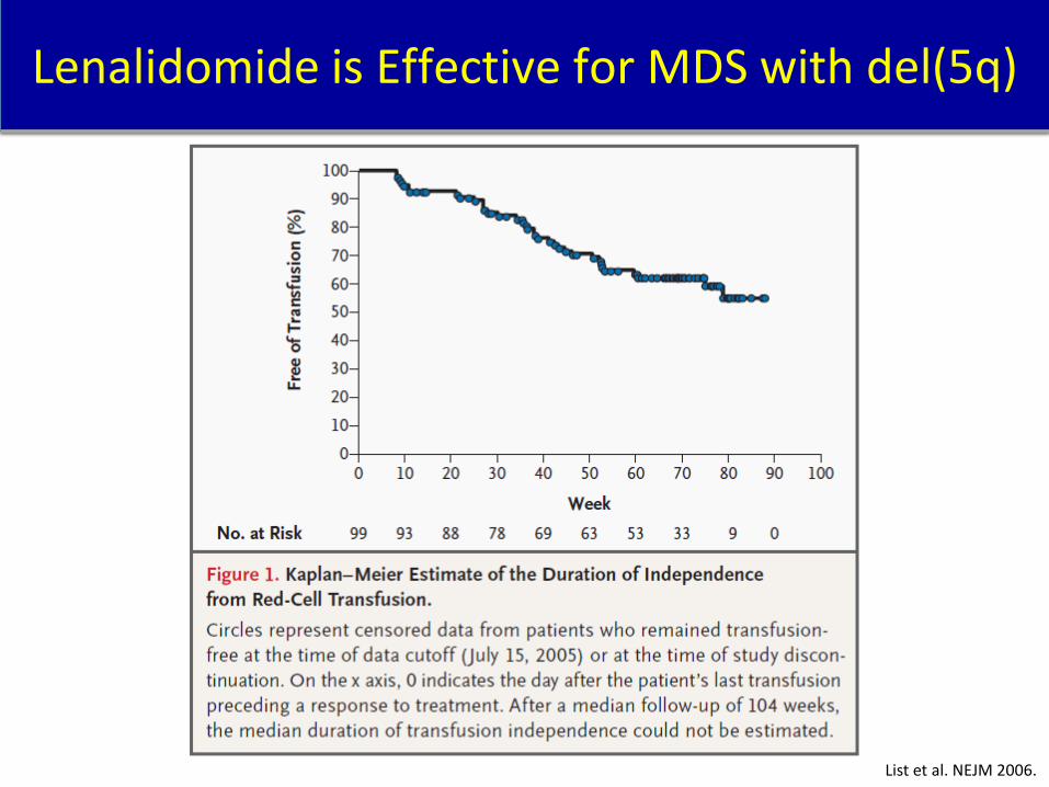
Lenalidomide is Effective for MDS with del(5q)
List et al. NEJM 2006.

List et al. NEJM 2006.
Lenalidomide is Effective for MDS with del(5q)

Lenalidomide is Also Effective for non-del(5q)
Raza et al. Blood 2008.

Treatment Options for Higher Risk MDS
• Supportive care (transfusions, antibiotics) • Low intensity therapy
– Azacitidine (Vidaza) – Decitabine (Dacogen) – Clinical trial
• High intensity therapy – Intensive chemotherapy (standard or clinical trial) – Stem Cell Transplant (standard or reduced intensity) --donor available, performance status, age

HMA inhibit DNA methyltransferases and induce DNA Hypomethylation and Gene Activation
• Hypomethylating agents: 5-Azacitidine (AZA) and 5-aza-2’-deoxycytidine (Decitabine/DAC) • Pyrimidine nucleoside analogs • AZA/DAC are incorporated into DNA in lieu of cytosine residue • Leads to inactivation of DMT • Leads to formation of newly synthesized DNA with unmethylated cytosine residues • Results in hypomethylation and transcription of previously quiescent genes
DAC DMT
A C G
mC G
T G C G Cm
: : : : : DMT
AZA
A Dc G
mC G
T G C G Cm
: : : : :
D M T
Silverman L. The Oncologist. 2001;6(S5):8-14.
(CpG Islands) DMT DMT

(N=358) Physician
Choice of 1 of 3 Conventional
Care Regimens (Best Supportive
Care (BSC) or LDAC or 7+3
Chemo)
AZA or BSC
AZA or LDAC
AZA or 7+3 Chemo
R A N D O M I Z E
AZA (n=117)
AZA (n=45)
AZA (n=17)
7+3 Chemo (n=25)
n=222
n=94
n=42
BSC (n=105)
LDAC (n=49)
Higher Risk MDS (IPSS Int-2 or HR) FAB-defined P3, international, multicenter, randomized
AZA-001 – AZA vs Conventional Care in Higher Risk MDS
AZA: 75mg/m2 SC days 1-7 every 28 days (at least 6 cycles) BSC: transfusions, G-CSF (for febrile neutropenia) LDAC: 20mg/m2 SC days 1-14 every 28 days (at least 4 cycles) IC (7+3 chemo): Cytarabine 100-200mg/m2/day CIV x7d Anthracycline IV daily x3d
Fenaux et al. Lancet Oncology 2009.

Patie
nts
(%)
0
10
30
40
50
60
Any HI HI-E Major
20
HI-P Major HI-N Major
49.2
28.7
39.5
10.6
32.6
14.0 19.1 18.0
Azacitidine CCR
Fenaux et al. Lancet Oncology 2009.
AZA-001: Hematologic Improvement (2000 IWG)

0 5 10 15 20 25 30 35 40 Time (months) From Randomization
0.0 0.1 0.2 0.3 0.4 0.5 0.6 0.7 0.8 0.9 1.0
Prop
ortio
n Su
rviv
ing
CCR AZA
Log-rank P=0.0001
HR=0.58 (95% CI: 0.43-0.77)
24.5 months 15 months
CI=confidence interval; HR=hazard ratio; ITT=intent-to-treat.
Azacitidine (HMA) Significantly Improves Survival in Higher Risk MDS
Fenaux et al. Lancet Oncology 2009.

Eligibility criteria n=223: •Intermediate- or high-risk MDS or CMML •Age > 60 years •Blast cell count 11%-30% or ≤ 10% with poor cytogenetics
R A N D O M I Z E
Decitabine n=119 15 mg/m2 IV 4h q8h, d 1-3 q6w ≤ 8 cycles
Best Supportive Care n=114
Stratification Cytogenics risk
group IPSS Primary vs
secondary Study center
EORTC-06011: Randomized Phase 3 Study of Low-Dose Decitabine vs BSC for Higher-Risk MDS
Lubbert et al, JCO 2011.

(months) 0 6 12 18 24 30 36 42
0
10
20
30
40
50
60
70
80
90
100
O N Number of patients at risk : 96 114 71 38 22 10 6 3 99 119 83 53 24 15 4 4
Median (months): 10.1 vs 8.5
HR = 0.88 , 95% CI (0.66, 1.17)
Logrank test: p=0.38
Supportive care Decitabine
Decitabine
Supportive care
Lubbert et al, JCO 2011.
EORTC-06011: Overall Survival

EORTC-06011: Response and Toxicity
Lubbert et al, JCO 2011.
Response BSC Decitabine
CR 0% 13%
PR 0% 6%
HI 2% 15%
SD 22% 14%
PD 68% 29%
Hypoplasia 0% 14%
Inevaluable 8% 8%
Decitabine arm: 26% went off protocol for treatment completion 16% for toxicity Why no survival advantage? Limited courses? More poor risk cytogenetics, older, lower PS? Dosing/schedule? Aza is better?

ADOPT Trial: Confirmation of 5-day Decitabine Dosing
Steensma et al. JCO 2009.
Initial decitabine study dosing is 15mg/m2 IV over 3h q8h x3d every 6 weeks Authors studied a convenient schedule for outpatients: 20mg/m2 IV over 1h days 1-5 every 4 weeks and confirmed Kantarjian et al. Blood 2007.
Median time to best response 1.7months

Meta-Analysis of HMA vs Conventional Care
Gurion et al. Haematologica 2010.
Overall Survival Time to AML or Death

HI
PR
CR
Patients (%)
0
100
Azacitidine (n=22)
Supportive care (n=23)
7
32
18 9
HI: 50% improvement in 1 or 2 peripheral blood counts or 50% decrease in transfusion requirements PR: 50% improvement in 3 peripheral blood counts and transfusion independent and ≤50% initial marrow blasts CR: Normalization of peripheral blood counts and ≤5% marrow blasts
Overall: 59%
Median time to response: 2 mos Median duration of response: 15 mos (all pts)
Silverman et al. JCO 2002
CALGB 9221: Responses in Lower-Risk MDS

Decitabine after AZA Failure can salvage some patients
Borthakur et al. Leuk Lymph 2008. Decitabine 20mg/m2 IV days 1-5 on a 28-day cycle

Prébet et al. JCO 2011.
435
HR MDS Post HMA Failure OS by Salvage Rx
Entire Cohort: Median OS 5.6 months 2yr OS 15%
2014 ASH Abstracts: 3275 (Nazha et al.): IPSS-R best predicts outcomes 3273 (Nazha et al.): SD after 6mo unlikely to improve -> clinical trials

n events mos 88 65 10 83 52 28 26 15 39 91 67 17
p=0.001
Jabbour et al. ASH 2013 abstract 388
LR MDS Post HMA Failure OS by Salvage Rx

Induction Chemotherapy for MDS
Beran et al. Cancer 2001. Kantarjian et al. Cancer 2006. Knipp et al. Cancer 2007. Malcovati et al, Blood, 2013.
Consider in: Younger fit patients <65-70 High blast percentage (>10%) Non-adverse cytogenetics Transplant candidate with donor Post-remission chemotherapy should be given
CR 40-60%, median duration CR <1yr Early mortality 17%, 5yr OS 8%

• 330 pts: 93 (28%) Rx with HMA and 237 (72%) with chemo Rx
• Multivariate analysis: worse OS with chemo Rx
Parameter HMA Intensive Chemo Rx
p value
-% CR + CRp 42 60 .01
-Median Rem. dur. (mos) 14.7 14.7
-%8-wk mortality 10 13
-median OS (mos) 18.8 14.6 .32
Nazha et al. Blood 2013: Abstract 2788.
HMA vs Induction chemotherapy in MDS with 10-30% blasts

Alessandrino et al. JCO 2013.
N=248 N=209
N=248 N=99 N=110
Retrospective analysis GITMO Adjusted for age, IPSS, donor type and conditioning intensity
HR=1.07 p=0.5
Allogeneic Transplant Can Cure MDS

Timing of Transplant in MDS
60-70yo: Koreth et al. JCO 2013.
Retrospective analysis of MDS patients <60 with MA MRD allo-HCT or 60-70yo with RIC MRD allo-HCT using a Markov decision model.
Low Int-1
Int-2 High
<60yo: Cutler et al. Blood 2004.
Della Porta et al, ASH 2014 Abstract#531: IPSS-R Int should be considered for Allo-HCT
Timing of Allogeneic Transplant in MDS

Pre-Transplant Therapy in MDS
Reviewed in: Mukherjee et al. BBMT 2014.
• Pretransplant blasts >5% and failure to achieve CR correlate with relapse with RIC HCT
• Retrospective analyses of IC before HCT show no convincing evidence
• No significant differences in outcomes between HMA and IC pre-transplant
• No significant benefit of HCT after HMA/IC compared to upfront HCT
• High-risk MDS patients should proceed directly to transplant without delay if possible – Fit younger patients may benefit from “rescue” IC – Older patients and those with poor-risk cytogenetics may
benefit from “bridging therapy” with HMA

Gerds et al. BBMT 2012.
*HR=0.72 (0.38-1.38) P=0.32
*HR=1.06 (0.45-2.54) P=0.88
*Adjusted for Cyto Risk, IPSS, Donor Source
Med age: aza 60; IC 47 High intensity: aza 40%; 100% IC RAEB-T: aza 6%; 33% IC Aza 2004-2010; IC 1992-2002
AZA vs IC Pre-Transplant

Iron Chelation Therapy
RBC transfusions: ≥20-30 Symptomatic anemia/Further RBC txn need
-- mainly Low, Intermediate-1 IPSS subtypes Evidence/pre-history of organ dysfunction
-- cardiac, hepatic, endocrine
Serum ferritin >2500→1000; ↑ Liver iron content
Rx: Deferrioxamine (Desferal) SQ or oral iron chelator Deferasirox (Exjade)

New Advances in MDS
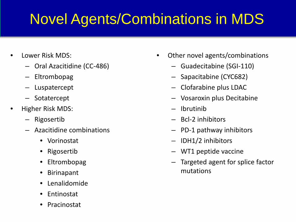
Novel Agents/Combinations in MDS
• Lower Risk MDS: – Oral Azacitidine (CC-486) – Eltrombopag – Luspatercept – Sotatercept
• Higher Risk MDS: – Rigosertib – Azacitidine combinations
• Vorinostat • Rigosertib • Eltrombopag • Birinapant • Lenalidomide • Entinostat • Pracinostat
• Other novel agents/combinations – Guadecitabine (SGI-110) – Sapacitabine (CYC682) – Clofarabine plus LDAC – Vosaroxin plus Decitabine – Ibrutinib – Bcl-2 inhibitors – PD-1 pathway inhibitors – IDH1/2 inhibitors – WT1 peptide vaccine – Targeted agent for splice factor
mutations

• None
• P1b Azacitidine + Ibrutinib • P1 Lenalidomide + Ibrutinib
• Lower risk (IPSS-R VL/L/I)
• Higher risk
(IPSS-R I/H/VH)
UC Davis Comprehensive Cancer Center MDS Trials
www.ucdmc.ucdavis.edu/CANCER/clinical_trials/

Stanford MDS Center: Biologically Focused Clinical Trials
• Luspatercept,III (TGFβ inhibitor for ring sideroblastic MDS)
• Spliceosome inhibitor, I/II (H3B-8800)
• Spliceosome inhibitor,I/II (H3B-8800) • AzaC & PD-L1 inhibitor,I/II (atezolizumab)
• RIC HSCT vs HMA,III
• Lower risk: (IPSS-R VL, Low, Int)
• Higher risk: (IPSS-R High, Very High) 1/2017

UCSF MDS Trials
• None
• MDM2 inhibitor • IDH1 inhibitor (AG-120)
• Lower risk (IPSS-R VL/L/I)
• Higher risk
(IPSS-R I/H/VH)

Summary and Concluding Thoughts

MDS Summary
• MDS is a heterogeneous group of BM failure syndromes
• Variable clinical presentation and course • Choice of therapy is primarily based on IPSS-R
score, symptoms, age and comorbidities • Understanding of pathogenesis,
prognostication and treatment is evolving • Novel biospecific therapies are being evaluated

MDS Resources
• Leukemia and Lymphoma Society • MDS Foundation • Aplastic Anemia and MDS Foundation • National Comprehensive Cancer Network
(NCCN) • UCD, UCSF and Stanford Cancer Centers
– Brian Jonas (UCD) – Peter Greenberg (Stanford) – Rebecca Olin (UCSF)

MDS Questions to be Answered
• What is MDS? • Why did I get MDS? • What does it mean for my life? • Is there treatment for it? • How should I be treated?
– When? – Why?
• What are some of the new advances in MDS?

Essentials for the Informed Pt with MDS
• Know your IPSS-R risk group • Know your treatment options
– Including transplant, clinical trials
• Know what your treatment goals are • Know the potential side effects of your
treatments • Know available MDS resources • Have a caregiver available/involved

Questions? Email: [email protected]