Embed Size (px)
Citation preview

Outer membrane permeability: Antimicrobialsand diverse nutrients bypass porins inPseudomonas aeruginosaJohanna Udea,1, Vishwachi Tripathia,1, Julien M. Buycka,1, Sandra Söderholma
, Olivier Cunratha,
Joseph Fanousa, Beatrice Claudia, Adrian Eglib,c, Christian Schlebergera, Sebastian Hillera,2, and Dirk Bumanna,2
aBiozentrum, University of Basel, CH-4056 Basel, Switzerland; bUniversity Hospital, University of Basel, CH-4056 Basel, Switzerland; and cDepartment ofBiomedicine, University of Basel, CH-4056 Basel, Switzerland
Edited by Staffan Normark, Karolinska Institutet, Stockholm, Sweden, and approved June 24, 2021 (received for review April 22, 2021)
Gram-negative bacterial pathogens have an outer membrane thatrestricts entry of molecules into the cell. Water-filled protein chan-nels in the outer membrane, so-called porins, facilitate nutrientuptake and are thought to enable antibiotic entry. Here, we de-termined the role of porins in a major pathogen, Pseudomonasaeruginosa, by constructing a strain lacking all 40 identifiable por-ins and 15 strains carrying only a single unique type of porin andcharacterizing these strains with NMR metabolomics and antimi-crobial susceptibility assays. In contrast to common assumptions,all porins were dispensable for Pseudomonas growth in rich me-dium and consumption of diverse hydrophilic nutrients. However,preferred nutrients with two or more carboxylate groups such assuccinate and citrate permeated poorly in the absence of porins.Porins provided efficient translocation pathways for these nutri-ents with broad and overlapping substrate selectivity while effi-ciently excluding all tested antibiotics except carbapenems, whichpartially entered through OprD. Porin-independent permeation ofantibiotics through the outer-membrane lipid bilayer was ham-pered by carboxylate groups, consistent with our nutrient data.Together, these results challenge common assumptions about therole of porins by demonstrating porin-independent permeation ofthe outer-membrane lipid bilayer as a major pathway for nutrientand drug entry into the bacterial cell.
membrane transport | bacterial outer membrane | lipid bilayer |diffusion | antimicrobial resistance
Antimicrobial resistance is a major worldwide threat to hu-man health. The World Health Organization has classified
Enterobacteriaceae, Pseudomonas aeruginosa, and Acinetobacterbaumannii as the most concerning pathogens (“critical priority”)(1). All three pathogens are Gram-negative bacteria with the char-acteristic inner and outer membranes. The outer membrane isa stringent permeability barrier that restricts the entry of mostmolecules and therefore presents a major challenge for the de-velopment of urgently needed novel antibiotics (2–5).The outer membrane consists of an asymmetric lipid bilayer
with lipopolysaccharide (LPS) in the outer leaflet and phos-pholipids in the inner leaflet and various outer-membrane pro-teins that are embedded in, or attached to, the lipid bilayer. LPScontains negatively charged phosphate and carboxylate groupsthat are cross-linked by divalent Mg2+ and Ca2+ cations, resultingin stable clusters of LPS molecules that reduce the permeationof small molecules by 10- to 100-fold compared to phospholipidbilayers (6). Some outer membrane proteins form water-filledchannels (so-called porins) that facilitate translocation of mole-cules through the outer membrane (4, 5). Enterobacteriaceaehave general “unspecific” porins that permit the entry of mole-cules with a size of up to 600 Da. By contrast, P. aeruginosa andA. baumannii have a large set of “specific” porins that permit theentry of only few molecules with sizes below 200 Da. In addition,all three pathogens have porins with mainly structural roles instabilizing the link between outer membrane and the underlying
peptidoglycan layer (OmpA and OprF). It has been proposedthat a small fraction of these structural porin molecules formlarge unspecific pores that permit entry of larger molecules atlow rates (7), but this model remains controversial.Antimicrobials and nutrients can penetrate the outer membrane
by two different pathways, through the lipid bilayer or throughporins. Hydrophobic molecules might predominantly use the lipidpathway, while hydrophilic molecules might prefer porins. However,the quantitative relevance of each pathway for outer-membranepermeability remains unknown (3, 8, 9). Even slow permeationpathways that mediate concentration-equilibration times in theorder of minutes (instead of seconds) can yield relevant intracellulardrug concentrations in bacteria with generation times of more than20 min, unless drug-efflux pumps and/or hydrolases diminish druglevels (2).Translocation pathways and their selectivity for specific physi-
cochemical properties of molecules are crucial for the rationalimprovement of drug entry into Gram-negative bacteria. Theimportant contribution of large cation-selective porins such asOmpF and OmpC for outer-membrane translocation into Enter-obacteriaceae enabled the establishment of rules for medicinal
Significance
Novel antibiotics are urgently needed to resolve the currentantimicrobial resistance crisis. For critical pathogens, drug entrythrough the cell envelope is one of the major challenges in thedevelopment of effective novel antibiotics. Envelope proteinsforming water-filled channels, so-called porins, are commonlythought to be essential for entry of hydrophilic molecules, butwe show here for the critical pathogen Pseudomonas aerugi-nosa that almost all antibiotics and diverse hydrophilic nutri-ents bypass porins and instead permeate directly through theouter membrane lipid bilayer. However, carboxylate groupshinder bilayer penetration, and Pseudomonas thus needs por-ins for efficient utilization of carboxylate-containing nutrientssuch as succinate. The major porin-independent entry route mightopen opportunities for facilitating drug delivery into bacteria.
Author contributions: J.U., V.T., J.M.B., S.H., and D.B. designed research; J.U., V.T., J.M.B.,S.S., O.C., J.F., B.C., A.E., and C.S. performed research; A.E. contributed new reagents/analytic tools; J.U., V.T., J.M.B., S.S., O.C., J.F., B.C., A.E., C.S., S.H., and D.B. analyzed data;and D.B. wrote the paper.
The authors declare no competing interest.
This article is a PNAS Direct Submission.
This open access article is distributed under Creative Commons Attribution-NonCommercial-NoDerivatives License 4.0 (CC BY-NC-ND).1J.U., V.T., and J.M.B. contributed equally to this work.2To whom correspondence may be addressed. Email: [email protected] or [email protected].
This article contains supporting information online at https://www.pnas.org/lookup/suppl/doi:10.1073/pnas.2107644118/-/DCSupplemental.
Published July 29, 2021.
PNAS 2021 Vol. 118 No. 31 e2107644118 https://doi.org/10.1073/pnas.2107644118 | 1 of 8
MICRO
BIOLO
GY
Dow
nloa
ded
by g
uest
on
Nov
embe
r 24
, 202
1

chemistry to improve whole-cell activities of antimicrobials againstthese bacteria (10–12). These porins have been extensively stud-ied, and in particular OmpF has a major impact on susceptibilityto various β-lactam antibiotics (13). However, an Escherichia coliΔompC ΔompF double mutant retains substantial susceptibility todiverse other antibiotics (9), suggesting alternative translocationpathways.For P. aeruginosa, physicochemical parameters favoring trans-
location have been more difficult to identify (10, 14, 15). BothP. aeruginosa and A. baumannii have lower outer-membranepermeability than Enterobacteriaceae for hydrophilic moleculesbecause they lack unspecific porins (16), making antimicrobialdevelopment particularly difficult for these critical pathogens.Specific porins might facilitate antibiotic entry into P. aeruginosa(17), but clear evidence for standard assay conditions is onlyavailable for penetration of carbapenems through OprD (18).Functional studies of individual porins in P. aeruginosa are hamperedby the large diversity of specific porins that are thought to each enableuptake of a few nutrients (19). Phenotypes of inactivating oneparticular porin might be masked by the numerous remainingother porins. To circumvent these issues, individual porins havebeen purified and reconstituted in artificial membranes, or expressedin E. coli, to determine their substrate specificity. However, theresults might not reflect porin functions in their native contextbecause their channel properties differ depending on the lipidenvironment (20, 21).In this study, we overcame these difficulties using extensive
mutagenesis. In contrast to previous assumptions, we show thatwild-type P. aeruginosa PA14 and a PA14 Δ40 mutant that lacksall identifiable 40 porin genes have indistinguishable susceptibilityto diverse antibiotics. Moreover, the Δ40 strain grew normally onrich media, and nutrient consumption assays revealed substantialporin-independent uptake of diverse hydrophilic nutrients. Bringingback individual porins accelerated uptake of some neutral/zwitterionicmolecules and was essential for efficient consumption of negativelycharged carboxylate-containing compounds. Instead of narrowsubstrate specificity, porins actually had broad overlapping substrateselectivity. These results demonstrate an unexpected but effi-cient porin-independent translocation pathway through the outer-membrane lipid bilayer for diverse hydrophilic compounds and allantipseudomonal antibiotics. A detailed understanding of this pathwaywill facilitate the development of novel antibiotics.
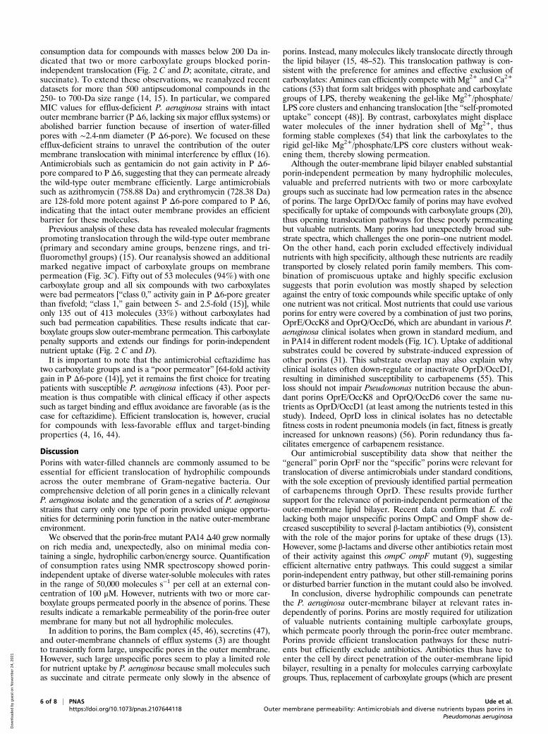
ResultsThe “General” Porin OprF Has Limited Relevance for Translocation ofAntimicrobials. OprF is one of the most abundant proteins in theP. aeruginosa outer membrane (4). Based on experiments withOprF reconstituted in liposomes, a minor open-channel con-former of OprF has been proposed to be the major entry path-way for various hydrophilic molecules including many antibiotics(7) (Fig. 1A). To test this idea in the native context of intact P.aeruginosa cells, we constructed mutants of the virulent clinicalisolate P. aeruginosa UCBPP-PA14 (22) that lacked oprF orexpressed chromosomal oprF variants with truncated C termini(oprF K188* and oprF V315*; the amino acid numbers includethe 24 amino acids of the signal peptide that are cleaved off duringmaturation) (23). These mutants would be partially (oprF V315*)or completely (oprF K188*) incapable of enlarging the N-terminaleight-strand β-barrel by incorporating further β-strands from theusually globular peptidoglycan-binding C-terminal domain (7).The ΔoprFmutant would also be unable to form dimers with fusedβ-barrels (24). All three P. aeruginosa mutants had wild-type fit-ness in rich culture media, as expected (23).If the large-channel conformer of OprF is indeed the major
entry route for antibiotics as proposed, all three mutants shouldbe less susceptible to antimicrobials. However, minimal inhibi-tory concentrations (MIC) and inhibition zones in disk diffusiontests (antibiograms) for diverse antibiotics (Fig. 1B) showed only
minor differences between wild type and mutants that weremostly within the accuracy of the respective assays (twofold forMIC values and 2 mm for inhibition zones). Piperacilin showedfourfold higher MIC against oprF mutants, but this was incon-sistent with unaltered inhibition zones. A minor impact of oprFmutations on susceptibility to β-lactam antibiotics has previouslybeen reported (13). Tetracycline had increased activity againstoprF mutants in both assays, suggesting potential indirect effectsof dysfunctional OprF increasing sensitivity to this translationalinhibitor, but not to aminoglycosides. The overall limited impactof OprF on antimicrobial susceptibility was not a result of in-creased outer-membrane permeability compensating for reducedentry through OprF in the mutants, because azithromycin andrifampin, which are sensitive indicators for outer-membrane bar-rier function in Pseudomonas (25, 26), remained poorly active inall three mutants. Together, these data indicate a limited role ofOprF in antimicrobial translocation across the outer membrane.
“Substrate-Specific” Porins Have Limited Impact on AntimicrobialTranslocation. In addition to OprF, P. aeruginosa encodes doz-ens of “substrate-specific” porins that might mediate antibioticuptake (19). One of these porins, OprD (also called OccD1),facilitates translocation of carbapenems (18), and OpdP/OccD3might also contribute to this under special circumstances (27). Inaddition, OpdH/OccK5 and OprE/OccK8 have been implicatedin translocation of the cephalosporin ceftazidime (28, 29), whereasOprO and OprP can transport fosmidomycin in vitro (30). Todetermine the relevance of these and other porins for the trans-location of diverse antimicrobials in intact bacteria, we constructeda series of porin deletion mutants. We generated clean gene de-letions to minimize potential polar effects on the expression ofdownstream genes. Initial characterization showed unaltered sus-ceptibilities (with the exception of the known OprD–carbapenemlink) in agreement with previous “resistome” data (27, 31–34). Totest the possibility that phenotypes of single porin mutants werebuffered by other porins, we mined the PA14 genome and iden-tified a total of 40 porin candidates (SI Appendix, Table S1).Several combinations of porin deletions still resulted in unalteredantibiotic susceptibilities.Eventually, we generated a strain, PA14 Δ40, that lacks all 40
porin genes as verified by whole-genome sequencing. During theconstruction this strain acquired nine secondary mutations in-cluding the loss of a duplicate transfer RNA-Asp gene and twononsynonymous mutations in protein-encoding genes. None of theaffected genes had a known association with outer-membrane per-meability or antimicrobial susceptibility (32–34) (SI Appendix, Sup-plementary Information Text and Table S2). PA14 Δ40 grew at ratescomparable to the wild type in rich culture media and showedwild-type susceptibility to diverse antimicrobials under standardassay conditions (Fig. 1B). The only clear change was a moder-ately reduced susceptibility (i.e., higher MIC values and smallerinhibition zones) to the carbapenems meropenem and imipe-nem, which could be explained almost entirely by the well-knownrole of OprD/OccD1.Together, these data demonstrate that the 40 porins are not
the major entry pathway for antibiotics under standard conditions.We cannot exclude that induction of certain porins with low ex-pression levels in standard Mueller–Hinton medium might permitantimicrobial entry under nonstandard conditions (27, 28). How-ever, mass spectrometry–based proteome analysis revealed thatP. aeruginosa porin abundance in Mueller–Hinton broth closelymimics porin patterns as observed in two different rodent infec-tion models (Fig. 1C), suggesting that standard assays comprise allclinically relevant porins.
Diverging Requirements of “Specific” Porins for Nutrient Uptake. ThePA14 Δ40 strain grew normally on rich media and on minimal mediacontaining 10 mM acetamide or arginine as the sole carbon/energy
2 of 8 | PNAS Ude et al.https://doi.org/10.1073/pnas.2107644118 Outer membrane permeability: Antimicrobials and diverse nutrients bypass porins in
Pseudomonas aeruginosa
Dow
nloa
ded
by g
uest
on
Nov
embe
r 24
, 202
1

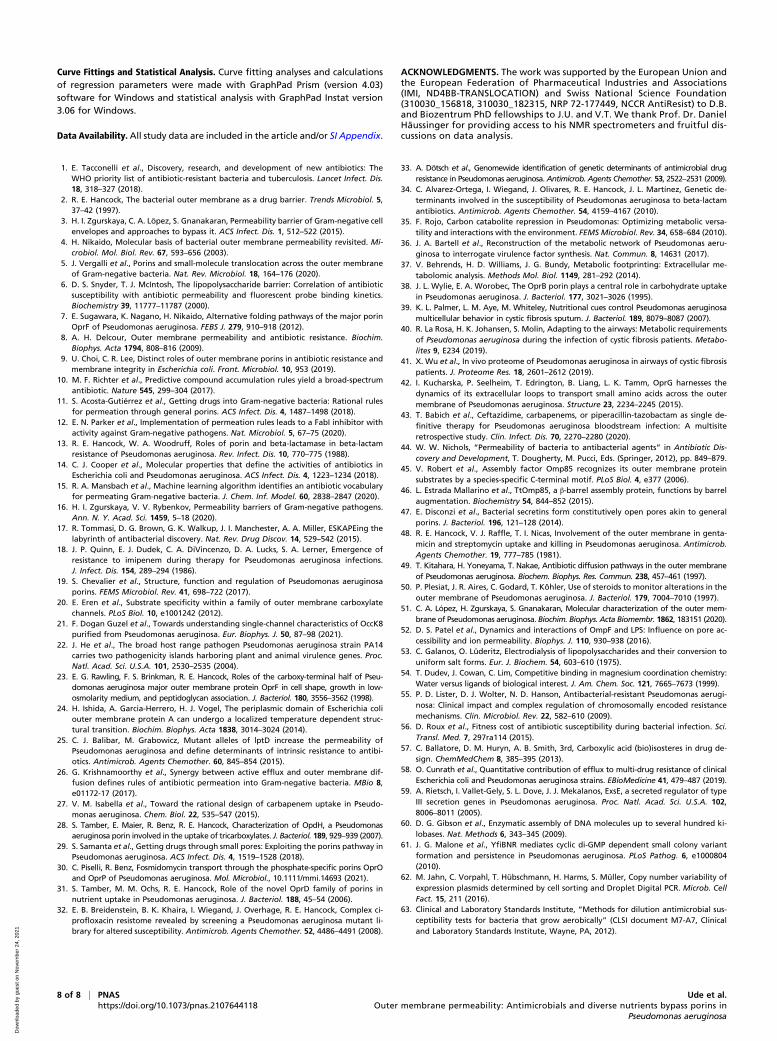
source but poorly on minimal media containing the otherwise pre-ferred carbon/energy sources (35) glutamate or succinate (Fig. 2Aand SI Appendix, Fig. S1A), indicating a key role of porins forefficient uptake of some but not all nutrients. To test this idea, wemixed 16 chemically diverse carbon/energy sources (organic acids,amino acids, and glucose) that are known to be utilized by PA14(36) at concentrations of 100 μM and quantified their consumptionby PA14 wild type and PA14 Δ40 using NMR spectroscopy (37)(Fig. 2B and SI Appendix, Figs. S1B and S2). We used comparativelylow nutrient concentrations to reduce interference by other po-tentially rate-limiting steps in nutrient utilization (i.e., transportacross the inner membrane and catabolism) (38) and to minimizethe contribution of inefficient unspecific translocation pathways(31). To enable consistent growth of PA14 Δ40 (and PA14), wealso included 10 mM acetamide, which was readily consumed byboth strains (Fig. 2 A, C, and D and SI Appendix, Fig. S1A). Wepreadapted the strains to this medium to ensure proper inductionof respective utilization pathways and to minimize lag phases thatoccurred after switching media with different nutrients.
P. aeruginosa wild type consumed all 16 components within afew hours with the typical pseudomonal preference for succinate,glutamine, proline, and asparagine (35) (Fig. 2 C and D). Theporin-free strain PA14 Δ40 consumed small (alanine) and posi-tively charged (histidine and arginine) nutrients at 80 to 100%of the wild-type rates (Fig. 2E). Although some porins such asOprD/OccD1 and closely related paralogs can permit translocationof arginine (20), they were obviously not required for wild-typearginine consumption rates (31). By contrast, all nutrients thathad two or more carboxylate groups (aconitate, aspartate, citrate,glutamate, and succinate) showed marked porin dependency (PA14Δ40 had <20% of wild-type consumption rates). Other compoundshad intermediate porin dependency (asparagine, glycine, glucose,glutamine, proline, pyruvate, and tyrosine), indicating that theporin-free outer membrane was partially permeable for thesecompounds, but porins facilitated translocation.Together, these data showed that the outer membrane of P.
aeruginosa permitted entry of hydrophilic/amphiphilic compoundswith substantial rates, even in the absence of all 40 identifiable
OprD OprG OprQ OprE FadL
Tsx OpdC OpdP OpdH OprB
OpdQ OprO
FadL3PA14_55320
OpdN OpdL OpdT OpdO OprP OpdI
OpdF OpdD OpdG AlgE
OprB3 OprB2 OpdJ
FadL2 SphA OpdK OpdR
PA14_39000 PA14_02890
OpdB
porin
cop
ies
perc
ell
100
101
102
103
104
105
Clinical isolates in-vitro
Mouse RatCation-adjusted MHB
UBCPP-PA14 in rodent pneumonia
PA14∆oprFoprF K188*oprF V315*∆oprD∆40
10242566416
41
0.250.0625
MIC
[μg/
mL]
Gen
tam
icin
Amik
acin
Tobr
amyc
inKa
nam
ycin
Col
istin
Cip
roflo
xaci
nN
orflo
xaci
n
Pipe
raci
llinTi
carc
illin
Ampi
cillin
Car
beni
cillin
Aztre
onam
Cef
epim
eC
efta
zidi
me
Mer
open
emIm
ipen
em
Tetra
cycl
ine
Azith
rom
ycin
Fosf
omyc
inR
ifam
pin
40353025201510
50
diam
eter
[mm
]
B
Peptidoglycan
N-terminal β-barrel
large pore encompassing both domains
V315
C
A
C-terminalpeptidoglycan-binding domain
Outer K188membrane
Fig. 1. Porin involvement in P. aeruginosa antimicrobial susceptibility. (A) Model of OprF with a large majority of two-domain conformer with a narrowouter-membrane β-barrel and a C-terminal domain linking the outer membrane with peptidoglycan and a minority of one-domain conformer with a largepore. (B) MIC (Upper) and antibiograms (Lower) of wild-type P. aeruginosa UCBPP-PA14 and various porin mutants. Means and SD of three experiments areshown. (C) Porin abundance in clinical P. aeruginosa strains in vitro and in UCBPP-PA14 in two rodent pneumonia models as determined by targetedproteomics.
Ude et al. PNAS | 3 of 8Outer membrane permeability: Antimicrobials and diverse nutrients bypass porins inPseudomonas aeruginosa
https://doi.org/10.1073/pnas.2107644118
MICRO
BIOLO
GY
Dow
nloa
ded
by g
uest
on
Nov
embe
r 24
, 202
1

porins. This includes the preferred carbon sources alanine andarginine but also other amino acids and glucose that are acces-sible for P. aeruginosa in millimolar concentrations in the lung ofcystic fibrosis (CF) and non-CF patients (39, 40). This compounduptake was not caused by general membrane leakage/permeabilizationin the absence of porins because small molecules with two or morecarboxylate groups (such as succinate) were effectively excluded.
Complex Substrate Selectivity of Individual Porins. To determine thecontribution of individual porins to nutrient uptake, we expressedsingle porin genes in the porin-free PA14 Δ40 background. For allconstructs we used the same PoprD promoter on low-copy plasmidsto minimize interference by specific porin-induction patterns (28).This strategy enabled determination of nutrient translocationthrough a single porin in the native membrane context withoutinterference by other porins. We focused on 13 porins that wedetected in various rodent infection models and in diverse clinicalisolates grown under standard conditions for clinical microbiology(Mueller–Hinton broth) (Fig. 1C). This set included all nine porins
detected in the lung of CF patients (41). For comparison, we in-cluded also the two porins OpdI/OccD5 and OpdL/OccK4 thatwere poorly expressed under all these conditions (“cryptic” porins).FadL and Tsx had no impact on consumption of any of the tested
nutrients (Fig. 3 A and B and SI Appendix, Fig. S3), consistent withtheir proposed selectivity for fatty acids and nucleosides, respec-tively, which were not included in our nutrient mix. OprG had nodetectable impact on nutrient consumption, arguing against animportant role for uptake of small amino acids including glycineand alanine (42) under native conditions, consistent with its nar-row channel consisting of eight β-strands (19). OprB enabled wild-type consumption rates specifically for glucose, as expected (38).The remaining porins enabled consumption of 3 to 11 differentnutrients at near wild-type levels. Importantly, no porin exceptfor OprB enabled consumption of glucose, indicating no generalmembrane leakage as a result of forced porin expression.Each of the other porins had a distinct substrate spectrum, while
at the same time each nutrient was able to translocate throughseveral different porins. The tricarboxylates, citrate and aconitate,
2
1
0
3
r c[m
olec
ules
s-1
cell-
1 ]
100500
acetamide
∆40alanine arginine histidine
100500
tyrosine glycine glucose asparagine
100500
pyruvate glutamine proline glutamate
100500
aspartate citrate aconitate succinate
cons
umpt
ion
[%]
100500
acetamide alanine arginine histidine
100500
tyrosine glycine glucose asparagine
100500
pyruvate glutamine proline glutamate
100500
aspartate citrate aconitate succinate
PA14
cons
umpt
ion
[%]
0.0
0.5
1.0
grow
thra
te[h
-1]
PA14∆ 40
4.0 3.5 3.0 2.5
δ(1H) [ppm]
2.0 1.5
BM2 mediumPA14 5h∆40 5h
cell density [108 CFU ml-1 01[ytisnedllec] 8 CFU ml-1]
0 h 3 h 5 h datapoints selected for linear regression linear regressionwith 95% CI
PA14∆40
x105
0 1 2 3 0 1 2 3 0 1 2 3 0 1 2 3 0 1 2 3 0 1 2 3 0 1 2 3 0 1 2 3
A
D
B
C
E
Fig. 2. Porin dependency of P. aeruginosa nutrient uptake. (A) Growth rates of P. aeruginosa PA14 wild-type and porin-free PA14 Δ40 in BM2 minimal mediacontaining a single energy/carbon source. (B) One-dimensional 1H-NMR spectrum of modified BM2 medium containing 16 different nutrients before and after5 h growth of P. aeruginosa PA14 wild type or porin-free PA14 Δ40. (C and D) PA14 nutrient consumption as measured by one-dimensional [1H] NMRspectroscopy. Each dot represents individual data for 1 of 21 independent cultures. (E) PA14 Δ40 nutrient consumption as measured by one-dimensional [1H]NMR spectroscopy. Each dot represents individual data for 1 of 21 independent cultures. Uptake rates for individual nutrients based on data shown in C andD. Means and SDs are shown.
4 of 8 | PNAS Ude et al.https://doi.org/10.1073/pnas.2107644118 Outer membrane permeability: Antimicrobials and diverse nutrients bypass porins in
Pseudomonas aeruginosa
Dow
nloa
ded
by g
uest
on
Nov
embe
r 24
, 202
1

entered almost exclusively through just two porins, OpdI/OccD5and OprQ/OccD6, but hardly through the previously postulatedOpdH/OccK5, consistent with negative electrophysiology data (28).The aromatic amino acid tyrosine translocated mainly through OpdP/OccD3 and OpdQ/OccK6, and partially through OpdI/OccD5.The previously implicated OpdT/OccD4 was not among our set ofexpressed porins. Glutamate translocated efficiently through severalporins of both the OpdK/OccK and OprD/OccD families (OprE/OccK8, OprQ/OccD6, OpdI/OccD5, OpdQ/OccK6, and OpdH/OccK5) and partially through OprD/OccD1 and OprO. Resultsfor OprE/OccK8 were consistent with recent liposome swellingassays (29). Succinate also translocated through members of bothporin subfamilies (OpdH/OccK5, OpdI/OccD5, OprE/OccK8, andOprQ/OccD6). The “cryptic” porin OpdI/OccD5 had transportcapabilities similar to the abundantly expressed and distantlyrelated subfamily member OprQ/OccD6, which might explainwhy OpdI/OccD5 was not expressed under standard conditions.The “cryptic” porin OpdL/OccK4 had an unusually narrow sub-strate spectrum with a preference for pyruvate, suggesting that itmight be induced when this nutrient is available. The broad rangeof substrates of some porins contrasted with efficient exclusion ofonly narrow sets of nutrients (e.g., glycine–OpdP, succinate–OpdQ,and proline–OprE).These data do not support the previously proposed distinct
substrate spectra of porin subfamilies (OprD/OccD1, positivelycharged amino acids; OpdK/OccK subfamily, net negative charge).This proposal was based on observations from porins expressed inE. coli (20) which might have affected their channel properties.
Recent electrophysiology data for OprE/OccK8 also question asimple dichotomy for substrates between the two subfamilies (29).Unsupervised clustering for transport capabilities observed in thisstudy yielded incomplete separation of the two subfamilies and nosignature substrates for either subfamily (Fig. 3B). Furthermore,the clustering did not conform with overall sequence similaritywithin each subfamily (19) (e.g., the sequence of OccD1 is closerto OccD3 and OccD6 than to OccD2, which is, however, moresimilar in terms of substrates).Finally, chemically similar substrates did cluster together (tri-
carboxylates citrate/aconitate; small negatively charged acids succi-nate/pyruvate; negatively charged amino acids glutamate/aspartate)although the zwitterionic amino acids formed two separate clusterstyrosine/glutamine/proline and glycine/asparagine, possibly drivenby molecular size. Together, these data show that certain porinsshare substrate preferences independently of their evolutionary re-latedness. This might facilitate identification of relevant structure–function relationships against commonly inherited channel proper-ties in future studies.Taken together, these data show 1) substantial porin-independent
translocation of certain nutrients and 2) broad porin substrate spectraand marked overlap, but also efficient porin-specific nutrientexclusion.
Carboxylate Groups Slow Translocation. Porin-independent translo-cation was crucial for antibiotic activity (Fig. 1 B and C). Com-pound properties that interfere with this pathway should thus beavoided in antimicrobial discovery and development. Our nutrient
B
0 1 42 3permeability class
0.0
0.2
0.4
0.6
0.8
fract
ion
2 x COO-
1 x COO-
no COO-
C
2
3
4
rc[m
olec
ules
/(s*c
ell)] PA14
∆40OprB OpdQ OprQOprD
1
0
x105
gluc
ose
acon
itate
ci
trate
su
ccin
ate
py
ruva
te
glut
amat
e
aspa
rtate
gl
ycin
e
aspa
ragi
ne
glut
amin
e
prol
ine
ty
rosi
ne
0
1
upta
keco
mpe
tenc
e
FadL Tsx
OprG OprB OpdL OprO OpdO OprE OpdH OpdI
OprQ OpdP OpdQ OprD OpdC
P< 0.00001
A
Fig. 3. Nutrient uptake through single porins in absence of other porins. (A) Nutrient consumption rates of PA14 Δ40 strains expressing a single porin from alow-copy-number plasmid. Means and averages for three independent cultures (single-porin strains) or 21 cultures (PA14, PA14 Δ40) are shown. (B) Substrateselectivity of 15 porins determined in single-porin strains. The capacity of each porin to boost nutrient consumption from baseline levels in PA14 Δ40 to wild-type levels is shown. Porins and nutrients are grouped based on unsupervised clustering. (C) Permeability of 472 antimicrobial compounds with detectableantipseudomonal activity (15). Following the published analysis, compounds are grouped in permeability classes according to the MIC values for efflux-deficient P. aeruginosa strains with intact (P Δ6) or abolished (P Δ6-pore) outer membrane permeability barrier (class 0, MIC of PA Δ6-pore is less than 20% ofPA Δ6; 1, 20 to 40%; 2, 40 to 60%, 3, 60 to 80%; 4 more than 80%). Statistical significance was analyzed with the χ2 test.
Ude et al. PNAS | 5 of 8Outer membrane permeability: Antimicrobials and diverse nutrients bypass porins inPseudomonas aeruginosa
https://doi.org/10.1073/pnas.2107644118
MICRO
BIOLO
GY
Dow
nloa
ded
by g
uest
on
Nov
embe
r 24
, 202
1
consumption data for compounds with masses below 200 Da in-dicated that two or more carboxylate groups blocked porin-independent translocation (Fig. 2 C and D; aconitate, citrate, andsuccinate). To extend these observations, we reanalyzed recentdatasets for more than 500 antipseudomonal compounds in the250- to 700-Da size range (14, 15). In particular, we comparedMIC values for efflux-deficient P. aeruginosa strains with intactouter membrane barrier (P Δ6, lacking six major efflux systems) orabolished barrier function because of insertion of water-filledpores with ∼2.4-nm diameter (P Δ6-pore). We focused on theseefflux-deficient strains to unravel the contribution of the outermembrane translocation with minimal interference by efflux (16).Antimicrobials such as gentamicin do not gain activity in P Δ6-pore compared to P Δ6, suggesting that they can permeate alreadythe wild-type outer membrane efficiently. Large antimicrobialssuch as azithromycin (758.88 Da) and erythromycin (728.38 Da)are 128-fold more potent against P Δ6-pore compared to P Δ6,indicating that the intact outer membrane provides an efficientbarrier for these molecules.Previous analysis of these data has revealed molecular fragments
promoting translocation through the wild-type outer membrane(primary and secondary amine groups, benzene rings, and tri-fluoromethyl groups) (15). Our reanalysis showed an additionalmarked negative impact of carboxylate groups on membranepermeation (Fig. 3C). Fifty out of 53 molecules (94%) with onecarboxylate group and all six compounds with two carboxylateswere bad permeators [“class 0,” activity gain in P Δ6-pore greaterthan fivefold; “class 1,” gain between 5- and 2.5-fold (15)], whileonly 135 out of 413 molecules (33%) without carboxylates hadsuch bad permeation capabilities. These results indicate that car-boxylate groups slow outer-membrane permeation. This carboxylatepenalty supports and extends our findings for porin-independentnutrient uptake (Fig. 2 C and D).It is important to note that the antimicrobial ceftazidime has
two carboxylate groups and is a “poor permeator” [64-fold activitygain in P Δ6-pore (14)], yet it remains the first choice for treatingpatients with susceptible P. aeruginosa infections (43). Poor per-meation is thus compatible with clinical efficacy if other aspectssuch as target binding and efflux avoidance are favorable (as is thecase for ceftazidime). Efficient translocation is, however, crucialfor compounds with less-favorable efflux and target-bindingproperties (4, 16, 44).
DiscussionPorins with water-filled channels are commonly assumed to beessential for efficient translocation of hydrophilic compoundsacross the outer membrane of Gram-negative bacteria. Ourcomprehensive deletion of all porin genes in a clinically relevantP. aeruginosa isolate and the generation of a series of P. aeruginosastrains that carry only one type of porin provided unique opportu-nities for determining porin function in the native outer-membraneenvironment.We observed that the porin-free mutant PA14 Δ40 grew normally
on rich media and, unexpectedly, also on minimal media con-taining a single, hydrophilic carbon/energy source. Quantificationof consumption rates using NMR spectroscopy showed porin-independent uptake of diverse water-soluble molecules with ratesin the range of 50,000 molecules s−1 per cell at an external con-centration of 100 μM. However, nutrients with two or more car-boxylate groups permeated poorly in the absence of porins. Theseresults indicate a remarkable permeability of the porin-free outermembrane for many but not all hydrophilic molecules.In addition to porins, the Bam complex (45, 46), secretins (47),
and outer-membrane channels of efflux systems (3) are thoughtto transiently form large, unspecific pores in the outer membrane.However, such large unspecific pores seem to play a limited rolefor nutrient uptake by P. aeruginosa because small molecules suchas succinate and citrate permeate only slowly in the absence of
porins. Instead, many molecules likely translocate directly throughthe lipid bilayer (15, 48–52). This translocation pathway is con-sistent with the preference for amines and effective exclusion ofcarboxylates: Amines can efficiently compete with Mg2+ and Ca2+
cations (53) that form salt bridges with phosphate and carboxylategroups of LPS, thereby weakening the gel-like Mg2+/phosphate/LPS core clusters and enhancing translocation [the “self-promoteduptake” concept (48)]. By contrast, carboxylates might displacewater molecules of the inner hydration shell of Mg2+, thusforming stable complexes (54) that link the carboxylates to therigid gel-like Mg2+/phosphate/LPS core clusters without weak-ening them, thereby slowing permeation.Although the outer-membrane lipid bilayer enabled substantial
porin-independent permeation by many hydrophilic molecules,valuable and preferred nutrients with two or more carboxylategroups such as succinate had low permeation rates in the absenceof porins. The large OprD/Occ family of porins may have evolvedspecifically for uptake of compounds with carboxylate groups (20),thus opening translocation pathways for these poorly permeatingbut valuable nutrients. Many porins had unexpectedly broad sub-strate spectra, which challenges the one porin–one nutrient model.On the other hand, each porin excluded effectively individualnutrients with high specificity, although these nutrients are readilytransported by closely related porin family members. This com-bination of promiscuous uptake and highly specific exclusionsuggests that porin evolution was mostly shaped by selectionagainst the entry of toxic compounds while specific uptake of onlyone nutrient was not critical. Most nutrients that could use variousporins for entry were covered by a combination of just two porins,OprE/OccK8 and OprQ/OccD6, which are abundant in various P.aeruginosa clinical isolates when grown in standard medium, andin PA14 in different rodent models (Fig. 1C). Uptake of additionalsubstrates could be covered by substrate-induced expression ofother porins (31). This substrate overlap may also explain whyclinical isolates often down-regulate or inactivate OprD/OccD1,resulting in diminished susceptibility to carbapenems (55). Thisloss should not impair Pseudomonas nutrition because the abun-dant porins OprE/OccK8 and OprQ/OccD6 cover the same nu-trients as OprD/OccD1 (at least among the nutrients tested in thisstudy). Indeed, OprD loss in clinical isolates has no detectablefitness costs in rodent pneumonia models (in fact, fitness is greatlyincreased for unknown reasons) (56). Porin redundancy thus fa-cilitates emergence of carbapenem resistance.Our antimicrobial susceptibility data show that neither the
“general” porin OprF nor the “specific” porins were relevant fortranslocation of diverse antimicrobials under standard conditions,with the sole exception of previously identified partial permeationof carbapenems through OprD. These results provide furthersupport for the relevance of porin-independent permeation of theouter-membrane lipid bilayer. Recent data confirm that E. colilacking both major unspecific porins OmpC and OmpF show de-creased susceptibility to several β-lactam antibiotics (9), consistentwith the role of the major porins for uptake of these drugs (13).However, some β-lactams and diverse other antibiotics retain mostof their activity against this ompC ompF mutant (9), suggestingefficient alternative entry pathways. This could suggest a similarporin-independent entry pathway, but other still-remaining porinsor disturbed barrier function in the mutant could also be involved.In conclusion, diverse hydrophilic compounds can penetrate
the P. aeruginosa outer-membrane bilayer at relevant rates in-dependently of porins. Porins are mostly required for utilizationof valuable nutrients containing multiple carboxylate groups,which permeate poorly through the porin-free outer membrane.Porins provide efficient translocation pathways for these nutri-ents but efficiently exclude antibiotics. Antibiotics thus have toenter the cell by direct penetration of the outer-membrane lipidbilayer, resulting in a penalty for molecules carrying carboxylategroups. Thus, replacement of carboxylate groups (which are present
6 of 8 | PNAS Ude et al.https://doi.org/10.1073/pnas.2107644118 Outer membrane permeability: Antimicrobials and diverse nutrients bypass porins in
Pseudomonas aeruginosa
Dow
nloa
ded
by g
uest
on
Nov
embe
r 24
, 202
1

in many current antibiotics) by isosteres (57) might be considered toaccelerate compound translocation across the P. aeruginosa outermembrane. Future studies should further characterize this largelyneglected translocation pathway, to identify additional molecularproperties that determine translocation rates of antimicrobials.
Materials and MethodsBacterial Strains and Growth Conditions. All P. aeruginosa mutants used inthis study are derived from the clinical isolate UCBBP-PA14 (22). In addition,we analyzed various P. aeruginosa clinical isolates from the University HospitalBasel strain collection. E. coli Sm10λpir was used for cloning and to conjugateplasmids into P. aeruginosa. All bacteria were cultured at 37 °C in lysogenybroth (LB) except for mating, for which we used P. aeruginosa grown over-night at 42 °C. For growth assays, bacteria were grown in overnight in LB andthen overnight in basal medium 2 (BM2; http://cmdr.ubc.ca/bobh/method/media-recipes/) minimal medium with 10 mM acetamide as carbon source.After washing, the bacterial were inoculated in BM2 containing the carbonsource of choice at an initial optical density at 600 nm (OD600) = 0.05.
Antibiotics and Reagents.Amikacin (disulfate salt; potency 77.60%), aztreonam(potency 92%), azithromycin (potency 92.70%), cefepime (hydrochloride,83.82%), ciprofloxacin (potency 78.60%), colistin (sulfate salt; potency,67.50%), gentamicin (sulfate salt, potency 67.70%), imipenem (monohydrate,potency 93.66%), kanamycin (sulfate salt, potency 83.16%), meropenem(trihydrate, potency 87.64%), piperacillin (sodium salt, potency 94.60%),tetracycline (potency 100%), ticarcillin (disodium salt, potency 90.62%),and tobramycin (potency 95.20%) were purchased from Sigma-Aldrich.Ampicillin (disulfate salt, potency 77.60%) and carbenicillin (disodium salt,potency 89.58%) were purchased from Roth. Ceftazidime was purchasedfrom European Pharmacopeia. Unless stated otherwise, all other reagentswere of analytical grade and were purchased from Sigma-Aldrich-Fluka.
Gene Deletion and Episomal Porin Expression. Strains and plasmids used in thisstudy are listed in SI Appendix, Table S3. Primers are listed in SI Appendix,Table S4.
Knockout vectors were constructed as described (58) with the followingmodifications. Seven hundred-base pair sequences of the flanking regions ofthe porin gene were PCR-amplified with primers designed with Snapgenesoftware (GSL Biotech LLC). The fragments were gel-purified and insertedinto pEXG2 plasmid (59) by Gibson assembly (60). The assembled plasmidwas transformed into competent SM10λpir prepared with Mix & Go (ZymoResearch Corporation) and plated on LB agar containing 50 μg/mL kanamycinand 15 μg/mL gentamicin. Sequenced-verified clones were mated for 4 h withPA14 strains at 37 °C. Single cross-over events were selected on plates containing15 μg/mL gentamicin and 20 μg/mL irgasan. Colonies were picked and grown inLB for 4 h and streaked on 5% sucrose plates overnight at 30 °C. P. aeruginosaclones were confirmed by sequencing and stored at −80 °C in LB containing 9%dimethyl sulfoxide. Whole-genome sequencing was done as described previously(58). Geneious Prime 2019.0.4 was used to map the reads to the P. aeruginosaUCBPP-PA14 reference sequence NC_008463.1 and to identify variations.
A plasmid backbone for expression of individual porins was constructedusing Gibson assembly of the TrfA-OriV origin of replication from pAD6 (61),a derivative of the low-copy-number plasmid RK2 (62), the gentamicin resis-tance cassette and origin of transfer (oriT) from pEXG2, rpsI and rrnB termi-nators, and the PoprD promoter amplified from PA14. A porin gene wasinserted downstream of PoprD. For electroporation of porin-expression plas-mids, 20-mL PA14 Δ40 overnight cultures in LB were washed thrice with ice-cold 0.3 M sucrose and resuspended in 100 μL cold 0.3 M sucrose. Electro-poration was done with 1 μL plasmid solution in 2-mm cuvettes at 25 μF/400Ohm/2.5 kV. After addition of 1 mL prewarmed LB and incubation for 1 h at37 °C, cells were plated on LB agar containing 15 μg/mL gentamicin.
Drug Susceptibility Tests. The MIC of drugs was determined by a twofolddilution assay in a 96-well plate according to Clinical and Laboratory Stan-dards Institute guidelines (inoculum of ∼106 colony-forming units [CFU]/mL;reading after 20- to 24-h incubation) in cation-adjusted Mueller–Hintonbroth (63). Growth of bacteria at 37 °C was examined by visual inspectionafter 20-h incubation. The MIC was defined as the lowest concentration ofan antibiotic that completely prevented visible cell growth. Drug susceptibilitywas also determined with antibiogram measurements with 20 different anti-biotics (Bio-Rad commercial disk). Overnight cultures of P. aeruginosa strainswere diluted at OD600 = 0.1 and were spread on 120- × 120-mm2 MHB II platesand air-dried in a laminar flow and then discs containing antibiotics wereplaced on the plates. Plates were incubated at 37 °C for 20 h. The diameter
of halos surrounding the discs were measured as an indication of growthinhibition.
Nutrient Consumption Assays. P. aeruginosa strains were grown overnight indefined nutrient medium (BM2 containing 10 mM acetamide and 100 μM of10 amino acids [alanine, arginine, asparagine, aspartate, glutamate, glutamine,glycine, histidine, proline, tyrosine], glucose, cis-aconitate, citrate, succinate, andpyruvate). Bacteria were washed and resuspended in prewarmed nutrient me-dium at OD600 = 0.005. Cultures were incubated at 37 °C and 180 rpm andsamples were taken after 2, 3, and 5 h of growth. OD600 and CFU were deter-mined at each time point. The remaining volumes were filtered through a0.2-μm pore filter and stored at −80 °C until NMR analysis.
NMR spectra were measured on a 600-MHz Bruker Avance III HD NMRspectrometer equipped with a cryogenic QCI-F probe. One-dimensional [1H]spectra were recorded with a free induction decay size of 32,000 points and256 transients at 298 K. Water was suppressed by excitation sculpting.Spectra were processed using TopSpin 3.6 by applying an exponential win-dow function with line broadening factor of 0.3 Hz and zero filling to 64,000points prior to Fourier transformation. For each substance, an isolated signalwas chosen for analysis (SI Appendix, Fig. S2). Peak intensities were deter-mined by comparison with nutrient medium as reference.
Analysis of Consumption Kinetics. We assumed that during exponentialgrowth, rC, the average consumption rate per bacterium of nutrient X fromthe medium is constant. The consumption rate of nutrient X by the bacterialpopulation is thus proportional to the cell density n(t):
d[X]dt
= −rC ·n(t) = −rC ·n0 · ekt ,
where n0 is the density of bacteria at t = 0 and k the growth rate constant.Integration results in the residual substance concentration at time T:
[X](T) = ∫ T0
d[X]dt
dt = [X]o − rCk·n(T )
and the total nutrient consumption MX, which depends linearly on the celldensity n(T):
MX (T ) = [X]o − [X](T ) = rCk·n(T).
The cell density relates to the OD600 via a proportionality factor z:
n(T) = NV0
= z ·OD600,
where N is the number of CFU determined in a volume V0 of cell culture. Inseparate calibration experiments, z was determined to be 9.8 × 10⁸ CFU·mL−1
and k was determined for each strain by exponential fitting of growth curves.The consumption rates rC was then obtained from linear fits of data pairs[Mx, n(T)].
To account for differential consumption preferences (delayed uptakecharacterized by 1) a lag-phase, 2) moderate uptake, and 3) fast uptake),different subsets of data points were used for linear regression modeling (asindicated in Fig. 1C):
1) Data points at t = 3 and 5 h2) Data points at t = 0, 2, 3, and 5 h3) Data points at t = 0, 2 and 3 h.
For calculating the uptake competence of each porin for each nutrient X,the consumption rate of the single-porin strain for X was normalized to arange defined by the value of the consumption rates of PA14 and Δ40:
uptake competence (porin)X = kX(porin) − kX(Δ40)kX(PA14) − kX(Δ40)
·100.
Values below 0 were set to 0 and values above 100 were set to 100.
Proteomics. P. aeruginosa porins were detected by targeted proteomics usingparallel reaction monitoring on a high-resolution and accurate mass instrumentwith absolute quantification using heavy-isotope-labeled reference peptides asdescribed previously (29). We analyzed PA14 and various clinical isolates grownto exponential phase in cation-adjusted Mueller–Hinton broth. We also rean-alyzed previously obtained blood or lung homogenates from mice and rats (29)that had been obtained at 24 h postinfection by intratracheal instillation of anagar bead containing 107 CFU of PA14.
Ude et al. PNAS | 7 of 8Outer membrane permeability: Antimicrobials and diverse nutrients bypass porins inPseudomonas aeruginosa
https://doi.org/10.1073/pnas.2107644118
MICRO
BIOLO
GY
Dow
nloa
ded
by g
uest
on
Nov
embe
r 24
, 202
1
Curve Fittings and Statistical Analysis. Curve fitting analyses and calculationsof regression parameters were made with GraphPad Prism (version 4.03)software for Windows and statistical analysis with GraphPad Instat version3.06 for Windows.
Data Availability. All study data are included in the article and/or SI Appendix.
ACKNOWLEDGMENTS. The work was supported by the European Union andthe European Federation of Pharmaceutical Industries and Associations(IMI, ND4BB-TRANSLOCATION) and Swiss National Science Foundation(310030_156818, 310030_182315, NRP 72-177449, NCCR AntiResist) to D.B.and Biozentrum PhD fellowships to J.U. and V.T. We thank Prof. Dr. DanielHäussinger for providing access to his NMR spectrometers and fruitful dis-cussions on data analysis.
1. E. Tacconelli et al., Discovery, research, and development of new antibiotics: TheWHO priority list of antibiotic-resistant bacteria and tuberculosis. Lancet Infect. Dis.18, 318–327 (2018).
2. R. E. Hancock, The bacterial outer membrane as a drug barrier. Trends Microbiol. 5,37–42 (1997).
3. H. I. Zgurskaya, C. A. Löpez, S. Gnanakaran, Permeability barrier of Gram-negative cellenvelopes and approaches to bypass it. ACS Infect. Dis. 1, 512–522 (2015).
4. H. Nikaido, Molecular basis of bacterial outer membrane permeability revisited. Mi-crobiol. Mol. Biol. Rev. 67, 593–656 (2003).
5. J. Vergalli et al., Porins and small-molecule translocation across the outer membraneof Gram-negative bacteria. Nat. Rev. Microbiol. 18, 164–176 (2020).
6. D. S. Snyder, T. J. McIntosh, The lipopolysaccharide barrier: Correlation of antibioticsusceptibility with antibiotic permeability and fluorescent probe binding kinetics.Biochemistry 39, 11777–11787 (2000).
7. E. Sugawara, K. Nagano, H. Nikaido, Alternative folding pathways of the major porinOprF of Pseudomonas aeruginosa. FEBS J. 279, 910–918 (2012).
8. A. H. Delcour, Outer membrane permeability and antibiotic resistance. Biochim.Biophys. Acta 1794, 808–816 (2009).
9. U. Choi, C. R. Lee, Distinct roles of outer membrane porins in antibiotic resistance andmembrane integrity in Escherichia coli. Front. Microbiol. 10, 953 (2019).
10. M. F. Richter et al., Predictive compound accumulation rules yield a broad-spectrumantibiotic. Nature 545, 299–304 (2017).
11. S. Acosta-Gutiérrez et al., Getting drugs into Gram-negative bacteria: Rational rulesfor permeation through general porins. ACS Infect. Dis. 4, 1487–1498 (2018).
12. E. N. Parker et al., Implementation of permeation rules leads to a FabI inhibitor withactivity against Gram-negative pathogens. Nat. Microbiol. 5, 67–75 (2020).
13. R. E. Hancock, W. A. Woodruff, Roles of porin and beta-lactamase in beta-lactamresistance of Pseudomonas aeruginosa. Rev. Infect. Dis. 10, 770–775 (1988).
14. C. J. Cooper et al., Molecular properties that define the activities of antibiotics inEscherichia coli and Pseudomonas aeruginosa. ACS Infect. Dis. 4, 1223–1234 (2018).
15. R. A. Mansbach et al., Machine learning algorithm identifies an antibiotic vocabularyfor permeating Gram-negative bacteria. J. Chem. Inf. Model. 60, 2838–2847 (2020).
16. H. I. Zgurskaya, V. V. Rybenkov, Permeability barriers of Gram-negative pathogens.Ann. N. Y. Acad. Sci. 1459, 5–18 (2020).
17. R. Tommasi, D. G. Brown, G. K. Walkup, J. I. Manchester, A. A. Miller, ESKAPEing thelabyrinth of antibacterial discovery. Nat. Rev. Drug Discov. 14, 529–542 (2015).
18. J. P. Quinn, E. J. Dudek, C. A. DiVincenzo, D. A. Lucks, S. A. Lerner, Emergence ofresistance to imipenem during therapy for Pseudomonas aeruginosa infections.J. Infect. Dis. 154, 289–294 (1986).
19. S. Chevalier et al., Structure, function and regulation of Pseudomonas aeruginosaporins. FEMS Microbiol. Rev. 41, 698–722 (2017).
20. E. Eren et al., Substrate specificity within a family of outer membrane carboxylatechannels. PLoS Biol. 10, e1001242 (2012).
21. F. Dogan Guzel et al., Towards understanding single-channel characteristics of OccK8purified from Pseudomonas aeruginosa. Eur. Biophys. J. 50, 87–98 (2021).
22. J. He et al., The broad host range pathogen Pseudomonas aeruginosa strain PA14carries two pathogenicity islands harboring plant and animal virulence genes. Proc.Natl. Acad. Sci. U.S.A. 101, 2530–2535 (2004).
23. E. G. Rawling, F. S. Brinkman, R. E. Hancock, Roles of the carboxy-terminal half of Pseu-domonas aeruginosa major outer membrane protein OprF in cell shape, growth in low-osmolarity medium, and peptidoglycan association. J. Bacteriol. 180, 3556–3562 (1998).
24. H. Ishida, A. Garcia-Herrero, H. J. Vogel, The periplasmic domain of Escherichia coliouter membrane protein A can undergo a localized temperature dependent struc-tural transition. Biochim. Biophys. Acta 1838, 3014–3024 (2014).
25. C. J. Balibar, M. Grabowicz, Mutant alleles of lptD increase the permeability ofPseudomonas aeruginosa and define determinants of intrinsic resistance to antibi-otics. Antimicrob. Agents Chemother. 60, 845–854 (2015).
26. G. Krishnamoorthy et al., Synergy between active efflux and outer membrane dif-fusion defines rules of antibiotic permeation into Gram-negative bacteria. MBio 8,e01172-17 (2017).
27. V. M. Isabella et al., Toward the rational design of carbapenem uptake in Pseudo-monas aeruginosa. Chem. Biol. 22, 535–547 (2015).
28. S. Tamber, E. Maier, R. Benz, R. E. Hancock, Characterization of OpdH, a Pseudomonasaeruginosa porin involved in the uptake of tricarboxylates. J. Bacteriol. 189, 929–939 (2007).
29. S. Samanta et al., Getting drugs through small pores: Exploiting the porins pathway inPseudomonas aeruginosa. ACS Infect. Dis. 4, 1519–1528 (2018).
30. C. Piselli, R. Benz, Fosmidomycin transport through the phosphate-specific porins OprOand OprP of Pseudomonas aeruginosa. Mol. Microbiol., 10.1111/mmi.14693 (2021).
31. S. Tamber, M. M. Ochs, R. E. Hancock, Role of the novel OprD family of porins innutrient uptake in Pseudomonas aeruginosa. J. Bacteriol. 188, 45–54 (2006).
32. E. B. Breidenstein, B. K. Khaira, I. Wiegand, J. Overhage, R. E. Hancock, Complex ci-profloxacin resistome revealed by screening a Pseudomonas aeruginosa mutant li-brary for altered susceptibility. Antimicrob. Agents Chemother. 52, 4486–4491 (2008).
33. A. Dötsch et al., Genomewide identification of genetic determinants of antimicrobial drugresistance in Pseudomonas aeruginosa. Antimicrob. Agents Chemother. 53, 2522–2531 (2009).
34. C. Alvarez-Ortega, I. Wiegand, J. Olivares, R. E. Hancock, J. L. Martínez, Genetic de-terminants involved in the susceptibility of Pseudomonas aeruginosa to beta-lactamantibiotics. Antimicrob. Agents Chemother. 54, 4159–4167 (2010).
35. F. Rojo, Carbon catabolite repression in Pseudomonas: Optimizing metabolic versa-tility and interactions with the environment. FEMS Microbiol. Rev. 34, 658–684 (2010).
36. J. A. Bartell et al., Reconstruction of the metabolic network of Pseudomonas aeru-ginosa to interrogate virulence factor synthesis. Nat. Commun. 8, 14631 (2017).
37. V. Behrends, H. D. Williams, J. G. Bundy, Metabolic footprinting: Extracellular me-tabolomic analysis. Methods Mol. Biol. 1149, 281–292 (2014).
38. J. L. Wylie, E. A. Worobec, The OprB porin plays a central role in carbohydrate uptakein Pseudomonas aeruginosa. J. Bacteriol. 177, 3021–3026 (1995).
39. K. L. Palmer, L. M. Aye, M. Whiteley, Nutritional cues control Pseudomonas aeruginosamulticellular behavior in cystic fibrosis sputum. J. Bacteriol. 189, 8079–8087 (2007).
40. R. La Rosa, H. K. Johansen, S. Molin, Adapting to the airways: Metabolic requirementsof Pseudomonas aeruginosa during the infection of cystic fibrosis patients. Metabo-lites 9, E234 (2019).
41. X. Wu et al., In vivo proteome of Pseudomonas aeruginosa in airways of cystic fibrosispatients. J. Proteome Res. 18, 2601–2612 (2019).
42. I. Kucharska, P. Seelheim, T. Edrington, B. Liang, L. K. Tamm, OprG harnesses thedynamics of its extracellular loops to transport small amino acids across the outermembrane of Pseudomonas aeruginosa. Structure 23, 2234–2245 (2015).
43. T. Babich et al., Ceftazidime, carbapenems, or piperacillin-tazobactam as single de-finitive therapy for Pseudomonas aeruginosa bloodstream infection: A multisiteretrospective study. Clin. Infect. Dis. 70, 2270–2280 (2020).
44. W. W. Nichols, “Permeability of bacteria to antibacterial agents” in Antibiotic Dis-covery and Development, T. Dougherty, M. Pucci, Eds. (Springer, 2012), pp. 849–879.
45. V. Robert et al., Assembly factor Omp85 recognizes its outer membrane proteinsubstrates by a species-specific C-terminal motif. PLoS Biol. 4, e377 (2006).
46. L. Estrada Mallarino et al., TtOmp85, a β-barrel assembly protein, functions by barrelaugmentation. Biochemistry 54, 844–852 (2015).
47. E. Disconzi et al., Bacterial secretins form constitutively open pores akin to generalporins. J. Bacteriol. 196, 121–128 (2014).
48. R. E. Hancock, V. J. Raffle, T. I. Nicas, Involvement of the outer membrane in genta-micin and streptomycin uptake and killing in Pseudomonas aeruginosa. Antimicrob.Agents Chemother. 19, 777–785 (1981).
49. T. Kitahara, H. Yoneyama, T. Nakae, Antibiotic diffusion pathways in the outer membraneof Pseudomonas aeruginosa. Biochem. Biophys. Res. Commun. 238, 457–461 (1997).
50. P. Plesiat, J. R. Aires, C. Godard, T. Köhler, Use of steroids to monitor alterations in theouter membrane of Pseudomonas aeruginosa. J. Bacteriol. 179, 7004–7010 (1997).
51. C. A. López, H. Zgurskaya, S. Gnanakaran, Molecular characterization of the outer mem-brane of Pseudomonas aeruginosa. Biochim. Biophys. Acta Biomembr. 1862, 183151 (2020).
52. D. S. Patel et al., Dynamics and interactions of OmpF and LPS: Influence on pore ac-cessibility and ion permeability. Biophys. J. 110, 930–938 (2016).
53. C. Galanos, O. Lüderitz, Electrodialysis of lipopolysaccharides and their conversion touniform salt forms. Eur. J. Biochem. 54, 603–610 (1975).
54. T. Dudev, J. Cowan, C. Lim, Competitive binding in magnesium coordination chemistry:Water versus ligands of biological interest. J. Am. Chem. Soc. 121, 7665–7673 (1999).
55. P. D. Lister, D. J. Wolter, N. D. Hanson, Antibacterial-resistant Pseudomonas aerugi-nosa: Clinical impact and complex regulation of chromosomally encoded resistancemechanisms. Clin. Microbiol. Rev. 22, 582–610 (2009).
56. D. Roux et al., Fitness cost of antibiotic susceptibility during bacterial infection. Sci.Transl. Med. 7, 297ra114 (2015).
57. C. Ballatore, D. M. Huryn, A. B. Smith, 3rd, Carboxylic acid (bio)isosteres in drug de-sign. ChemMedChem 8, 385–395 (2013).
58. O. Cunrath et al., Quantitative contribution of efflux to multi-drug resistance of clinicalEscherichia coli and Pseudomonas aeruginosa strains. EBioMedicine 41, 479–487 (2019).
59. A. Rietsch, I. Vallet-Gely, S. L. Dove, J. J. Mekalanos, ExsE, a secreted regulator of typeIII secretion genes in Pseudomonas aeruginosa. Proc. Natl. Acad. Sci. U.S.A. 102,8006–8011 (2005).
60. D. G. Gibson et al., Enzymatic assembly of DNA molecules up to several hundred ki-lobases. Nat. Methods 6, 343–345 (2009).
61. J. G. Malone et al., YfiBNR mediates cyclic di-GMP dependent small colony variantformation and persistence in Pseudomonas aeruginosa. PLoS Pathog. 6, e1000804(2010).
62. M. Jahn, C. Vorpahl, T. Hübschmann, H. Harms, S. Müller, Copy number variability ofexpression plasmids determined by cell sorting and Droplet Digital PCR. Microb. CellFact. 15, 211 (2016).
63. Clinical and Laboratory Standards Institute, “Methods for dilution antimicrobial sus-ceptibility tests for bacteria that grow aerobically” (CLSI document M7-A7, Clinicaland Laboratory Standards Institute, Wayne, PA, 2012).
8 of 8 | PNAS Ude et al.https://doi.org/10.1073/pnas.2107644118 Outer membrane permeability: Antimicrobials and diverse nutrients bypass porins in
Pseudomonas aeruginosa
Dow
nloa
ded
by g
uest
on
Nov
embe
r 24
, 202
1