Embed Size (px)
Citation preview
Osteomyelitis:Osteomyelitis:Pathophysiology &
Treatment Decisions
Clifford B. Jones, MD
Original Author: Clifford B. Jones, MD; March 2004 Revised February 2007 & February 2011

“One Should Especially Avoid Such Cases if One has a Respectable
Excuse, for the Favorable Chances are Few and the Risks are Many….

….Besides, if a Man does not Reduce the Fracture, He will be Thought Unskillful. If He does Reduce It, He will bring the Patient
Nearer to Death than Recovery.”
Hippocratic Writings, New York, Pelican Books, 1978

Fracture Management Goals
1. Osseous Union2. Restore Limb Function3. Avoid Complications
Osteomyelitis Results in:
1. Reduction in limb function2. Psychological & Social dysfunction3. Increased cost

Hansen’s 7 DsConcerning Prolonged Orthopaedic Problems
DespairDivorceDestitute
DepressionDelinquency
DefaultDeath
Sigvard Ted Hansen, 1997

Introduction• 350,000 long bone fxs/yr• Infection risk varies:
– Type I open – 10/1,000 infections– Type III open – up to 25%

Gustilo Open Fx ClassJBJS, 72A: 299-303, 1990
2%
7%
7%10-50%25-50%

Open Fractures
Type II Type IIIA
Type IIIB Type IIIB
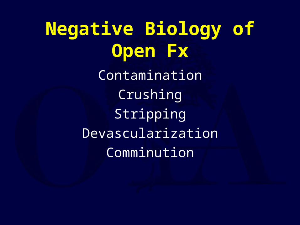
Negative Biology of Open Fx
ContaminationCrushingStripping
DevascularizationComminution

Blood SupplyRhinelander, CORR, 1974

Blood SupplyRhinelander, CORR, 1974
Normal - endosteal/medullary 2/3-3/4internal external
Fracture - periosteal/external majorityinternal external
Periosteal Blood Supply Important
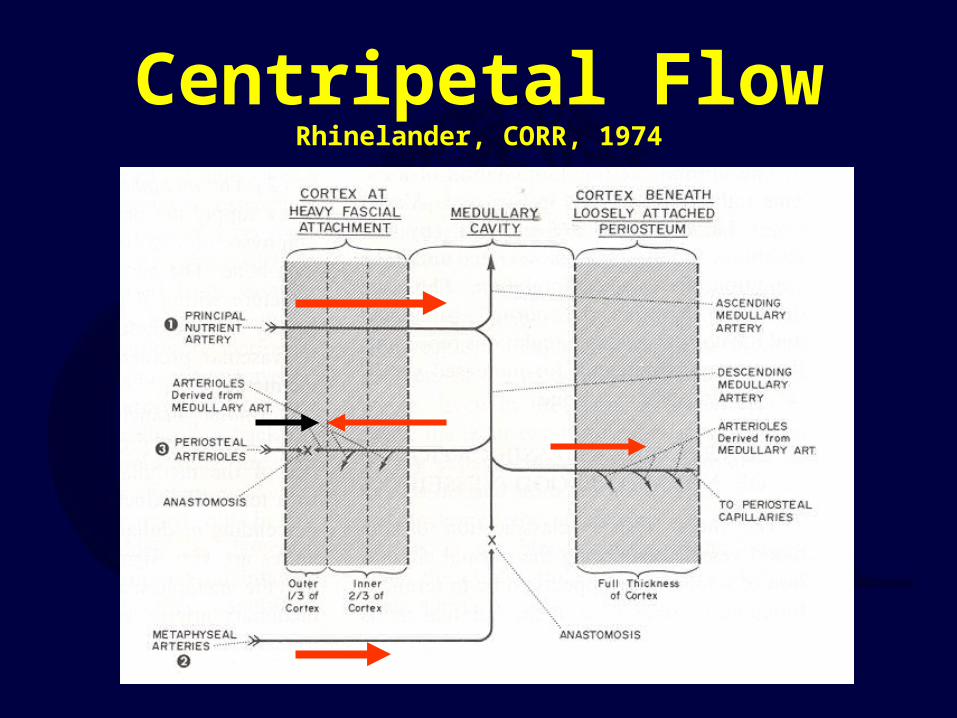
Centripetal FlowRhinelander, CORR, 1974

Initial Emergent Treatment
dTAntibiotics, IV
ReduceStabilize
Cover wound

Why infection risk high?Infection risk ≈ Fracture type (soft tissue)
Open fx = Contamination (70% cx +)
Open fx = Infected fx > 8 hours

Cost AnalysisInfection
– Increase cost 16-21%/pt– Increase hosp stay 36-50%/pt
Total Cost $ 271 million/yr

Definition• Group of conditions• “…presence of bacteria & an
inflammatory response causing progressive destruction of bone.”
– Fears, RL, et al, 1998
• “…suppurative process in bone caused by a pyogenic organism”
– Pelligrini, VD, et al, 1996

Why destruction of bone matrix?
Proteolytic enzymesHyperemiaOsteoclasts

Do Not Delay Tx & Dx

Classification• Waldvogel, 1971
– Classification based on pathogenesis
• May, 1989– 5 parts, post-traumatic tibial osteomyelitis
• Cierny & Mader, 1985– 4 factors affecting outcome– Host, site, extent of necrosis, degree of impairment

PathogenesisWaldvogel, 1971
1. Hematogenous2. Contiguous focus of infection3. Direct inoculation

AnatomicClassification(Cierny-Mader)
1985
I:I: II:II:
III:III: IV:IV:

Classification Break-DownI. Medullary
Endosteal nidus, min soft tissue involvement, ? Sinus tract
II. SuperficialSurface of bone, usu 2° to soft tissue defect
III. LocalizedLocalized sequestra, usu sinus tract, Usu stable s/p excision
IV. DiffusePermeative process, combination of I/II/III, Usu Unstable s/p excision

Physiologic Classification(Cierny-Mader, 1985)
A-Host: Good immune system & delivery
B-Host: Compromised hostBL: locally compromisedBS: systemically compromisedBC: combined
C-Host: Requires suppressive or no TxMinimal disabilityTx worse than dz, not a surgical candidate

Clinical Staging(Cierny-Mader, 1985)
Anatomic Type + Clinical StagePhysiologic ClassExample: IV BS tibial osteomyelitis = diffuse tibial lesion in a systemically compromised host

Types of Pathophysiology
Acute/Hematogenous
Chronic/Nonhematogenous

Acute/Hematogenous
• Anatomy (Hobo)– Sharp twist in metaphyseal capillaries
• Stasis (Trueta)– Decreased flow in capillaries & veins
• Combination (Morrissy)– Trauma & Bacteria

Acute/HematogenousProgression of Dz
• Cell death 2° to bacterial exotoxins bacterial culture medium worsens condition
Vascularity, leukocytosis, edema Pressure w/in rigid osseous container Pain, swelling, erythemaPotential for septic arthritis (knee, hip, shoulder)

Chronic/NonhematogenousS. aureus ↑
Pseudomonas aureginosa ↑Enterobacter
> 30% Polymicrobial> 30% Polymicrobial

Clinical Findings (varied)
ErythemaSwellingSinus TractDrainageLimpFluctuence
NoneNonePainPainTendernessTendernessFeverFeverHAHANausea/VomitingNausea/Vomiting
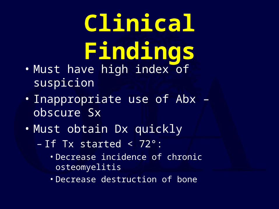
Clinical Findings• Must have high index of suspicion• Inappropriate use of Abx – obscure Sx• Must obtain Dx quickly
– If Tx started < 72°:• Decrease incidence of chronic osteomyelitis• Decrease destruction of bone

Laboratory DataAcute (Morrey, BF, OCNA, 1975)
WBC (25% of time)– Abnormal differential, Left Shift (65%)– Blood Cx – 50% positive
Chronic– Mild anemia, WESR, C-reactive protein– Possible leukocytosis with L shift– Blood Cx – usually negative
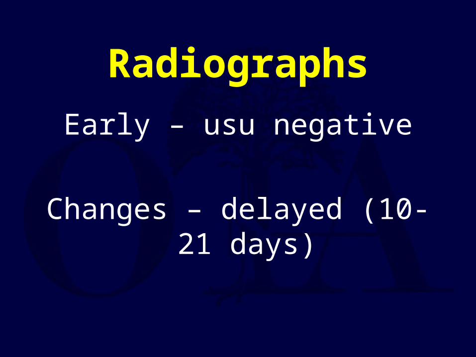
RadiographsEarly – usu negative
Changes – delayed (10-21 days)
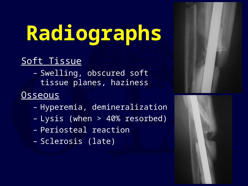
RadiographsSoft Tissue
– Swelling, obscured soft tissue planes, haziness
Osseous– Hyperemia, demineralization– Lysis (when > 40% resorbed)– Periosteal reaction– Sclerosis (late)

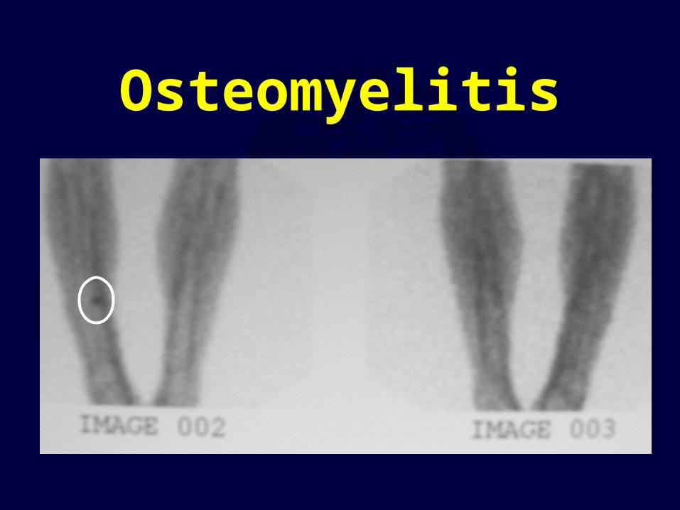
Radionucleotide Imaging
99M Tc
67Ga
111In WBC

99M Tc
• Action– binds to hydroxyapetite crystals
• Osteoblastic activity– Demineralized bone– Immature collagen

99M Tc• 3 Phase Bone Scan
1. Radionucleotide angiogram2. Immediate post injection blood pool3. Three hour: soft tissue, urinary excretion
• Diagnosis– Cellulitis: Phases 1 &2, no change 3– Osteomyelitis: Phases 1 & 2, focal 3
• Results: 94% sensitivity, 95% specificity– Rosenthal 1992, Schauwecker 1992

Cellulitis
Osteomyelitis

99M Tc: False Positive
DM foot d/oSeptic arthritis
Inflammatory bone dzAdjacent to pressure sores

99M Tc4 Phase Bone Scan
• New development• Action:
– Mature bone: uptake stops at 4 hr– Immature woven bone: cont’d uptake at 24 hr
• Problem: needs f/u imaging at 24 hr (compliance)• Gupta 1988, Israel 1987, Schauwecker 1992

67Ga
• Exudation of in vivo labeled serum protein– Transferrin, haptoglobin, albumin
• Results– 81% sensitivity, 69% specificity– Schauwecker, 1992
• Combination with Tc sensitivity, but specificity

111In WBC
• Used in combination (Seabold, 1989)– In/Tc: 88% accurate– Ga/Tc: 39% accurate
• Preparation problem rad dose to spleen, 18-24hr delay
• Spine (Whalen, Spine 1991)– 83% false negative use MRI

MRINo radiationGood soft tissue imagingImaging:
– T1 Dark– T2 Bright/Mixed

T1 bright T2 dark
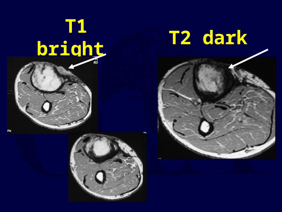
T1 bright T2 dark

MRI• Acute:
marrow fat granulation tissue H2O
• Chronic: thickened cortex– Low signal on all scans
• Cellulitis: no marrow changes

MRI ResultsSchauwecker, 1992
• Sensitivity 92-100%• Specificity 89-100%• Excellent for Spine (Modic, RCNA, 1986)
– Sens 96%, Spec 92%, Accuracy 94%• Soft tissue extension• Sinus tract formation
– Bright Tx from skin to bone

CT ImagingImage cortical and cancellous bone
Evaluate osseous adequacy of debridement
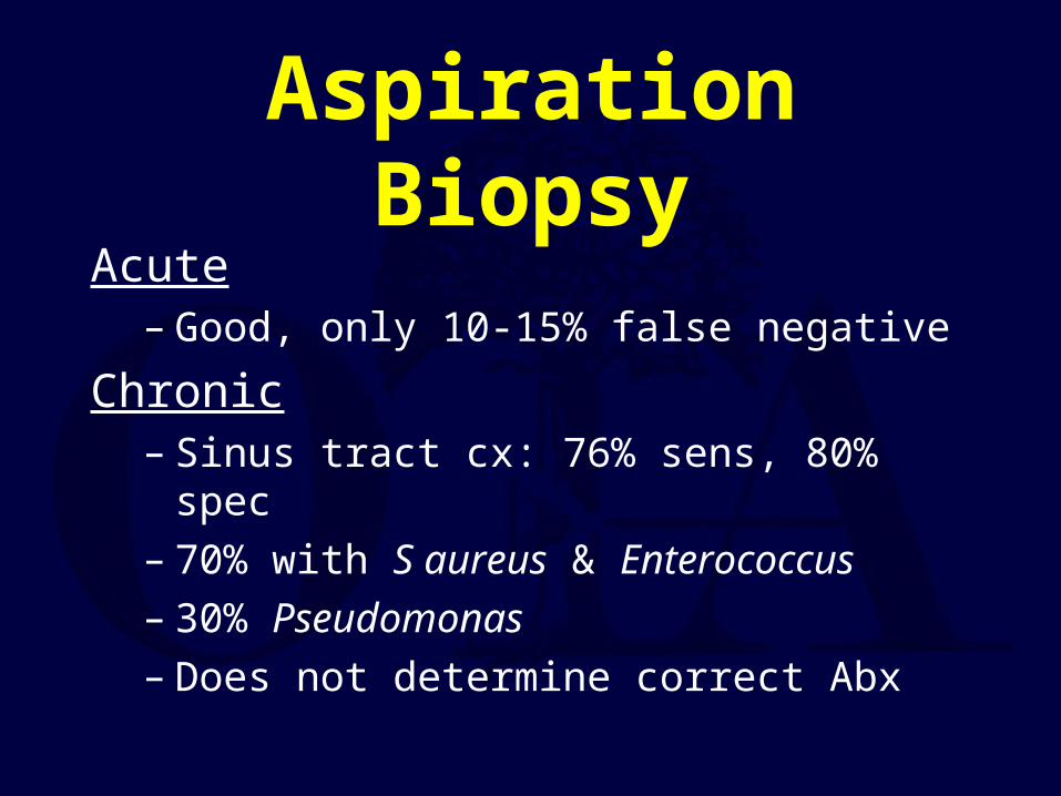
Aspiration BiopsyAcute
– Good, only 10-15% false negative
Chronic– Sinus tract cx: 76% sens, 80% spec– 70% with S aureus & Enterococcus– 30% Pseudomonas– Does not determine correct Abx

Acute/Hematogenous
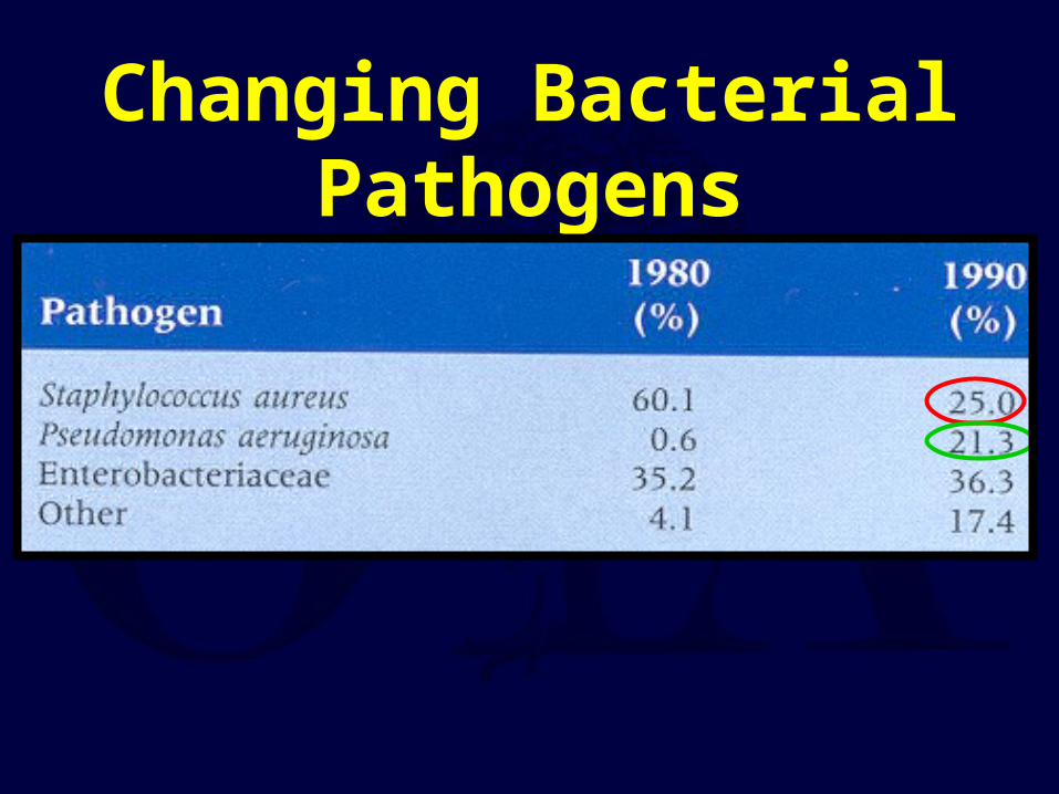
Changing Bacterial Pathogens

Resistant Bacterium - ESKAPE
E Enterococcus faecuimS Staphlococcus aureusK Klebsiella pneumoniaeA Acinobacter baumanniiP Pseudomonas aeruginosaE Enterobacter aerogenes

MSSA & MRSA
• MSSA Change to β lactam
• MRSA Treat ≤ MIC

Gram Negative Rods - SPICE
S SerratiaP PseudomonasI Indole positiveC CitrobacterE Enterobacter

Gram Negative
Rods

Proionibacterium acnes• Axillary bacteria (sebaceous glands)• Treated with:
– 1st: PCN or vanco– 2nd: Macrolides & Fluoroquinolones
• Long incubation time• Call lab – culture 2 wks, gram positive rods• Especially important for shoulder:
– Nonunions– Infections

Multilocus Polymerase Chain reaction & Electrospray Ionization/Mass Spectrometry
• Bacterial or fungal DNA is amplified by polymerase chain reaction and introduced into a mass spectroscopy by electrospray ionization
• The amplification procedure uses 16 S primers, and the primers can be varied to detect fungi and antibiotic resistance genes (eg, mec A).

Multilocus Polymerase Chain reaction & Electrospray Ionization/Mass Spectrometry
• Although culturing bacteria takes days, amplifying DNA takes hours
• Accurate, rapid point-of-care devices would be ideal for clinical use

Treatment Preventation
• Antibiotics – correct organism• Debridement – until viable tissue obtained• Irrigation• Wound care/coverage• Osseous & soft tissue stability
– Fx stability– Dead space management

New Oral Agents: MRSA
Zyvox/linazid po/iv ↓ plts
Synercid iv
Infectious Disease Consult

Stability Oxymoron
Hardware increased ↑ bacterial growth
&
Fracture stability (hardware) ↓ bacterial growth
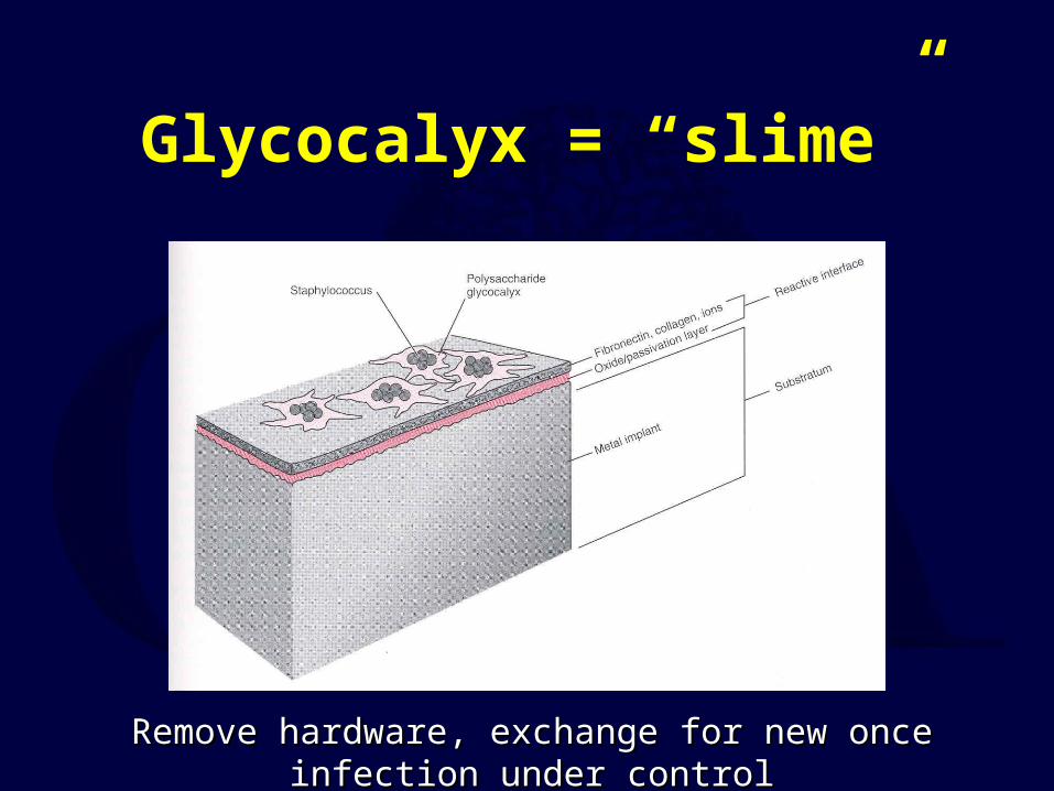
Glycocalyx = “slime”
Remove hardware, exchange for new once infection under controlRemove hardware, exchange for new once infection under control
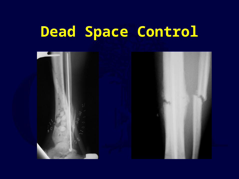
Dead Space Control

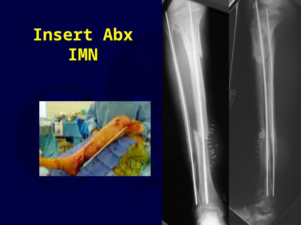
Abx IMN Materials & Methods
Research: Retrospective ReviewTime: 3 year period, 2 year F/U
Location: Level 1 Trauma Center

PatientsAge: 37 (range 18-67)
Femurs (n=4)Closed n=2Open n=2
Tibia (n=28)Closed n=2Open n=26
II: 4/26IIIA: 12/26IIIB: 10/28
10/28 open tibial fx with rotational or FTT for coverage

Antibiotic NailInserted Avg. 3 mo. (range 2 day – 23 mo.)
2 bags PMMA2.O g Vancomycin2.4 g Tobramycin32 Fr Chest Tube
3.2 mm Guide Wire

Incise & Debride WoundI&D Wound
I&D Canal
Reamers, Vent Hole

Presentation
44 M44 M4 bacterium4 bacterium
CoccidiomycosisCoccidiomycosis2 prior known “flare ups”2 prior known “flare ups”

Antibiotic IMN
32 Fr Chest Tube2 bags PMMA2.0 Vancomycin2.4 Tobramycin
Insert under pressure into chest tube while still “wet”
Insert 3.2 mm ball tip guide rod
Remove plastic before PMMA too hot and melting plastic chest tube
Insert Abx IMN

Wait until IMN Insertion
Wound HealedLabs ImprovedAnabolic Host
Usually 4-8 wks
(Average 4-8 wks)(Average 4-8 wks)

Example

Examples

Infected Tibial Nonunion
• 32 M• 2 ppd smoker• MCA 18 mo, 2 prior surgeries• Draining wound• “No one to take care of him”
– Translation No money

Presentation

Options
• Type IV BC
• Unstable with Osteo• Smoker, malnutrition• Local open wound
• Nothing• Revise with plate• Revise with nail• Revise with ex fix• Revise with Ilizarov• Amputation
Length +/-

Debridement of Skin & BoneDebridement of Skin & Bone
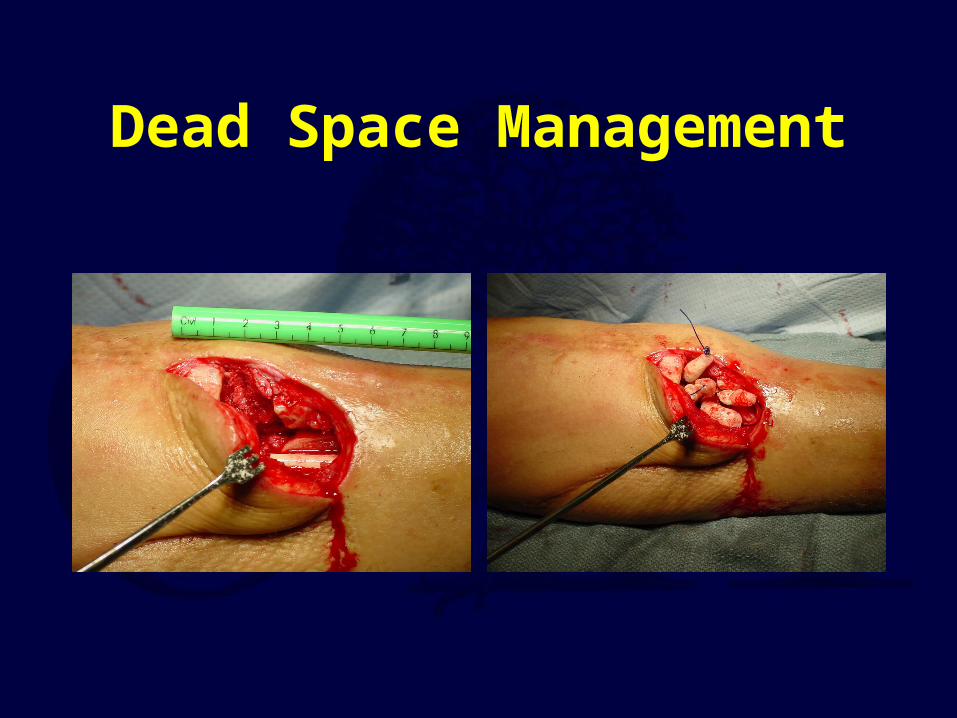
Dead Space Management

Stabilize NonunionStabilize Nonunion

Coverage of Wound

Lengthening Leg

Noncompliance - NonunionNoncompliance - Nonunion
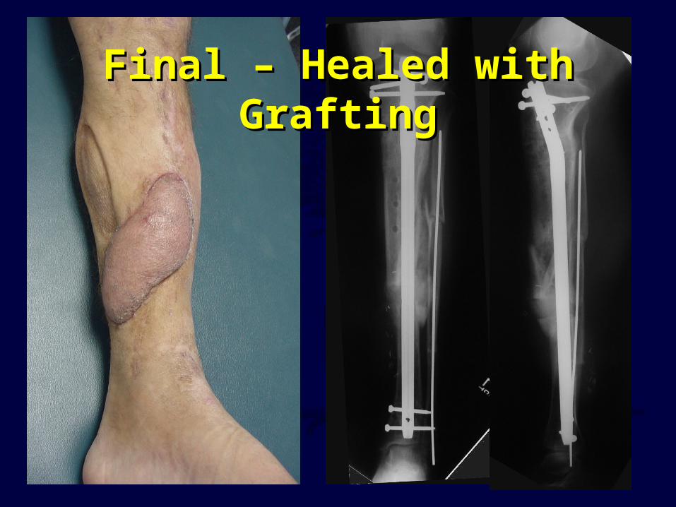
Final – Healed with GraftingFinal – Healed with Grafting

Infected Tibial Nonunion
• 38 yo M• Snuff tobacco• 1 pint vodka/day• 6 mo MCA with IIIB open tibia
Type I BS

Presentation

Initial Post opInitial Post op

3 mo

Exchange IMN at 4 ½ moExchange IMN at 4 ½ mo

Final at 18 moFinal at 18 mo
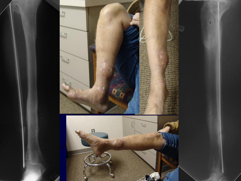
Example• 54 yo Male• Post-operative Pseudomonas osteomyelitis• Refractory to HW removal & Ancef• Healthy, non-smoking• Cierny III A Host
Photos from M Swiontkowski

Example 1
•Dead Space
•Calcaneal defect

Example 1• Debridement of all non-viable bone with
laser doppler• Defect filled with antibiotic PMMA• 6 wks antibiotics

Example 1, at 6 wks• Removal Abx beads• Bone grafting• Lateral arm flap• Infection eradication

Example• 47 yo Male, smoker• Presentation 2 months s/p ORIF closed proximal
tibia fx• Draining wound• Exposed HW• Cierny III BC Host
• Photos from M Swiontkowski

Example• Debridement• HW remains• Abx beads
Exposed plate

Example • Gastrocnemeus flap, STSG

Example • At 6 weeks• Remove Abx beads• Bone grafting• Healed wound and fracture
Example• At 5 yo, tibial osteomyelitis• Partially treated• At 62 yo, presentation to MD• Chronic draining tibial osteomyelitis• Cierny III BC Host
• Photos from M Swiontkowski

Example•Sinus tracts
•Chronic skin changes
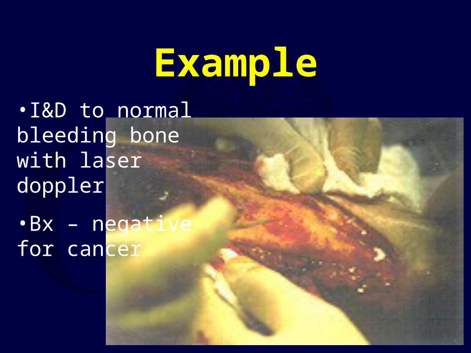
Example•I&D to normal bleeding bone with laser doppler
•Bx – negative for cancer

Example• Abx beads• Latissimus Flap• STSG

Example• Removal Abx beads at 6 wks• No bone graft – low demand
patient• Dz free at 8 years (70 yo)

Outcomes of Treatment

Antibiotic IMN


The Fate of Patients with a “Surprise” Positive Culture
After Nonunion Surgery
Olszewski D, Stucken C, Tornetta III P, Ricci W, Struebel P, Jones C, Sietsema D

Results• 460 patients
• Two cohort groups
– 98 cultures (21%) “surprise” positive
– 362 cultures (79%) negative

BacteriaType of Bacteria Number
Coagulase-negative Staphylococcus 45
Methicillin-resistant S. Aureus 12
Pseudomonas 8
Proprionibacterium 8
Methicillin-sensitive S. Aureus 7
Bacillus 4
Peptostreptococcus 3
Staph species unspecified 3
Enterococcus 2
Strep viridans 2
Clostridium 2
E. coli, Staph epidermidis, Beta hemolytic strep,
Serratia, Candida and Aspergillus 1

Positive Cultures• 98 with positive cultures
– 90 treated with antibiotics
• 6 – 8 week duration
• Culture specific
– 8 patients not treated
• “Presumed contaminant”

Union After Index
• Culture (+) = 66 / 90 (73%)• Culture (-) = 347 / 362 (96%)• P < 0.0001

Infection After Index
• Culture (+) = 11 / 90 (12%)• Culture (-) = 15 / 362 (4%)• P < 0.0001

Final Outcome• Culture (+) = 86 / 90 (95.5%)
– 24 Additional procedures – 9 / 13 Debridement only– 4 / 13 with 1 additional procedure– 4 / 90 (4.5%) infected nonunion– 2 BKA
• Culture (-) = 362 / 362 (100%)– 15 Additional procedures
• P < 0.0001
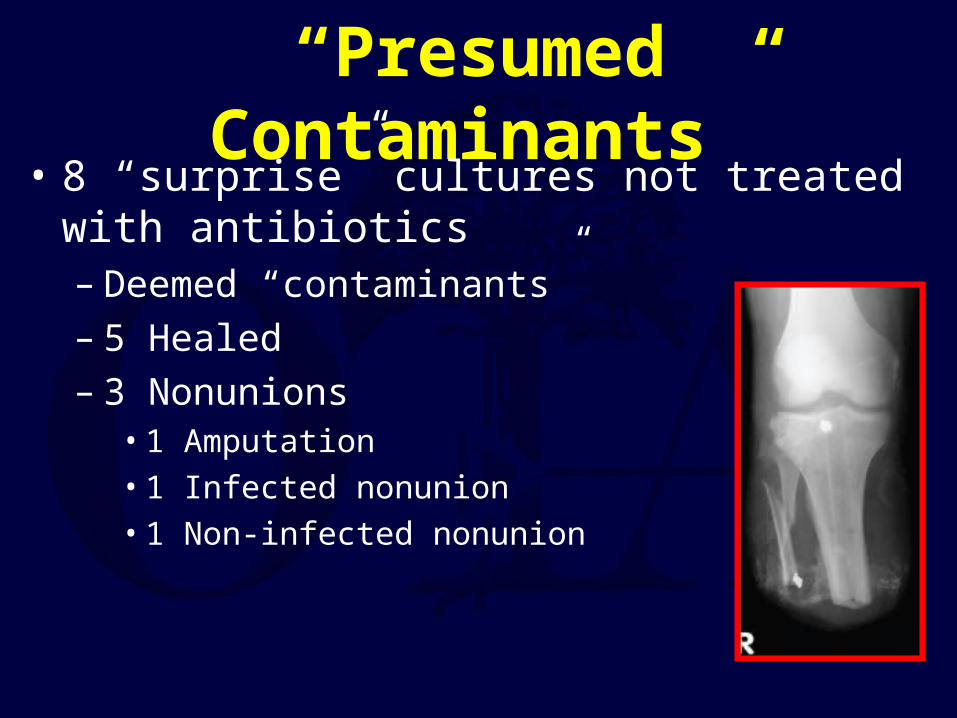
“Presumed Contaminants”• 8 “surprise” cultures not treated with antibiotics
– Deemed “contaminants”– 5 Healed– 3 Nonunions
• 1 Amputation• 1 Infected nonunion• 1 Non-infected nonunion

Culture Positive Culture Negative
Healed 73% 95.8%Infected Nonunion 13% 4%
Additional Procedures 27% 4%
Union at final follow-up
93% 100%
All Patients

Summary
• 21% of 460 “at risk” nonunions had surprise positive culture
• Staph species• 90 of 98 treated with antibiotics
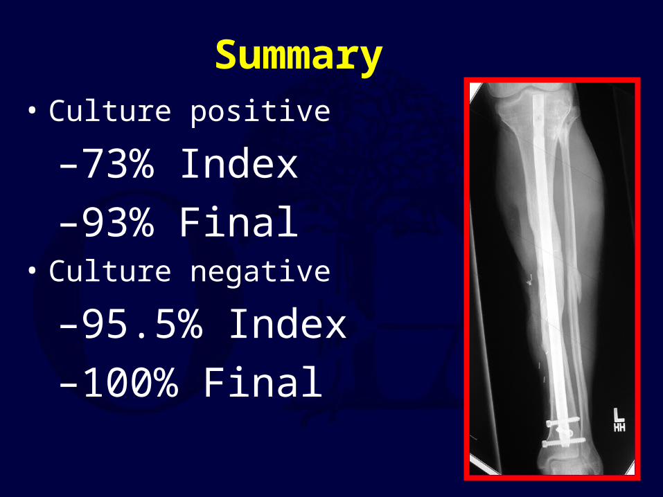
Summary• Culture positive
–73% Index–93% Final
• Culture negative
–95.5% Index–100% Final
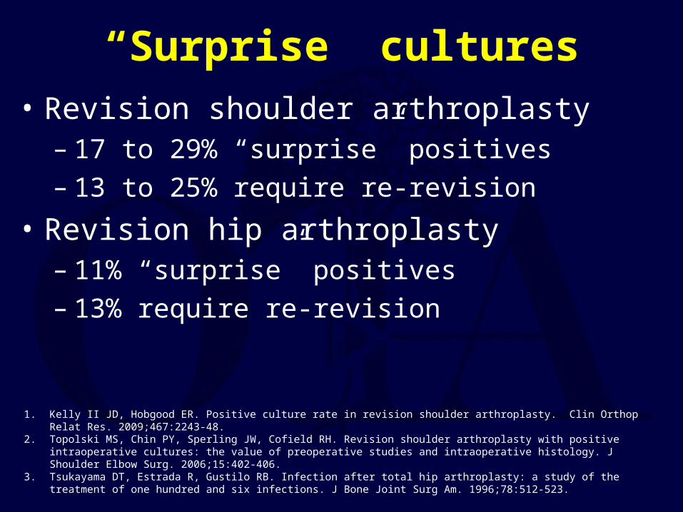
“Surprise” cultures• Revision shoulder arthroplasty
– 17 to 29% “surprise” positives– 13 to 25% require re-revision
• Revision hip arthroplasty – 11% “surprise” positives– 13% require re-revision
1. Kelly II JD, Hobgood ER. Positive culture rate in revision shoulder arthroplasty. Clin Orthop Relat Res. 2009;467:2243-48.2. Topolski MS, Chin PY, Sperling JW, Cofield RH. Revision shoulder arthroplasty with positive intraoperative cultures: the value of preoperative
studies and intraoperative histology. J Shoulder Elbow Surg. 2006;15:402-406.3. Tsukayama DT, Estrada R, Gustilo RB. Infection after total hip arthroplasty: a study of the treatment of one hundred and six infections. J Bone
Joint Surg Am. 1996;78:512-523.

Conclusions• 21% “surprise” positive cultures• 74% heal after initial index
procedure• 26% required additional procedures

Recommendations
• Counsel patients• Treat all positive cultures• Potentially offer two-stage procedures
– Unknown efficacy– 79% would be unnecessary

Conclusion
PreventionEarly DxEarly TxStabilize
Convert to Union ASAP
Return to General/Principles
Index
If you would like to volunteer as an author for the Resident Slide Project or recommend updates to any of the following slides, please send an e-mail to [email protected]