1
was sent a follow-up questionnaire to determine the outcome of receiving one of these sources of notification. 616 providers ordered DXA scans through our center over a four-year period. A survey was sent to the 308 providers who ordered at least 4 scans (median number). We received 96 surveys for a response rate of 31%. Results: 370 women were randomly enrolled in Groups 1 and 2. Baseline characteristics of the two groups were similar. At 3 to 6-month follow-up, 13 of 14 (93%) women in Group 1 with a T-score 2.5 or less (osteoporosis) were tak- ing prescription medication for osteoporosis, while only 8 of 14 (57%) women in Group 2 with a T-score of 2.5 or less were taking prescription medication (p 5 0.029). There was no difference in health-related behaviors such as consump- tion in dairy products or calcium supplements, smoking cessation, or level of ex- ercise. Of the 96 referring provider respondents, almost all responders (96%) agreed with sending DXA results directly to patients, and 85% thought they would more likely discuss DXA results if their patients received results directly from the testing center. Conclusion: Directly sending patients their DXA results increased the likelihood that prescription medication was started to treat osteoporosis. Al- most all responders to the provider survey agreed with sending results directly to patients, and most thought that this process increased the likelihood of discus- sing osteoporosis treatment with their patients at follow-up appointments. This model of direct reporting can be used for communicating other important test re- sults to patients to improve both patient safety and compliance with recommended therapy. Poster Number 163 Peripheral DXA UTILITY OF HEEL DXA IN DIAGNOSING OSTEOPOROSIS Tamara Vokes, MD, CCD, Associate Professor of Medicine, University of Chicago The current study examined how well heel DXA (PIXI, GE Medical Systems) di- agnosed osteoporosis in 861 subjects (769 female) referred for central BMD mea- surement as part of their medical care. Osteoporosis was defined as BMD T-score at or below 2.5 at central site or presence of prevalent vertebral fractures on VFA. There was a significant (p ! 0.0001) correlation between T-score of the heel and central sites with R2 of 0.38, 0.47, 0.52 and 0.48 for lumbar spine, fem- oral neck, total hip and lowest of hip or spine T-scores. However, the heel T-score was higher than the lowest of hip or spine T-score by almost 1 unit. Heel T-score of 1 or below had a sensitivity of 78.9% (95%CI 5 74.7e82.7) and specificity of 68.2% (63.7e72.6) in identifying patients with lowest central T-score of 2.5 or below. A heel T-score of 0 had a higher sensitivity of 95.0% (92.4e96.9) but lower specificity (39.9%; 35.3e44.6). The ability of heel BMD T-score to separate 138 women with and 631 without prevalent vertebral fractures was not as good as that of the lowest central T-score with the area under the receiver operating curve (ROC) of 0.631 vs. 0.677. The predictive ability of the models with heel and cen- tral BMD were closer if clinical risk factors such as age, history of peripheral frac- tures and use of glucocorticoids, were included (ROC of 0.782 vs. 0.792). We conclude that peripheral BMD of the calcaneus can be used for screening and sug- gest that T-score of 0 should prompt evaluation by central BMD. If central densi- tometry is not available, heel BMD can be used for assessment of fracture risk, particularly if combined with clinical risk factors. Poster Number 164 Prevention and Treatment of Osteoporosis OSTEOPOROSIS RISK FACTORS AND PREVENTION AMONG FEMALE PHYSICIANS IN NORTHERN CALIFORNIA Robert Bradford Mims, MD, Director, Endocrine Metabolic Clinic of Santa Rosa, CA While doing Bone Mineral Densities (BMDs) for Doctor’s Health Day, we en- countered one female physician (F-MD) with an unexpected low BMD who did not have any obvious Osteoporosis (OP) risk factors. We wondered if there were unique risk factors, and if F-MDs were practicing common OP prevention measures. We studied 36 F-MDs between the ages of 36e56, mean (M) age of 46 years, whose M Ht was 66 in (62e72), M Wt of 137.5 lbs (118e175), and 32 were White, 4 were Asian, and one Hispanic. Our OP risk factor assessment showed no one who smoked. 28/38 had 1e6 children (M 5 2), and 20/20 breastfed their children. Five F-MDs were menopausal, one PMP, 2 had declining menses, one took thyroid hormone, and 5 claimed Depression. There was only minimum use of alcohol, wine, coffee, colas, and only 3/36 took bone depleting meds. 95% of F-MDs continued from Hi School into medical practice, and worked for 5e23(M 5 13) years. For OP prevention 24/36 (67 %) were practicing OP preven- tion methods, most for the last five years. 56% took 1000e1200 mg of elemental calcium/d, and did adequate weight bearing exercise. Another 28% were physi- cally active described as ‘‘very busy.’’ Only three were immobile for up to six months, and only one consumed increased protein. The back BMDs for 3/36 F- MDs were below, and 9/36 BMDs were above population norms. No specific cause was found for the 3 low BMDs. Likewise, no hip BMDs were below population norms, and 5/36 were above norms. We conclude that no unique OP risk factors were identified in this group of Northern California F-MDs, and that most practice common OP prevention. Poster Number 166 Prevention and Treatment of Osteoporosis EFFICACY AND SAFETY OF MONTHLY ORAL IBANDRONATE IN POSTMENOPAUSAL WOMEN WITH OSTEOPENIA Michael R McClung, MD, Oregon Osteoporosis Center MA Bolognese, MD, Besthesda Health Research, Bethesda; F Sedarati, MD, Roche, Nutley, NJ; RR Recker, MD, MACP, FACE, Osteoporosis Research Center, Omaha, NE; PD Miller, MD, Colorado Center for Bone Research, Lakewood, CO Monthly ibandronate has been shown to increase bone mineral density (BMD) and reduce bone turnover in women with postmenopausal osteoporosis (PMO) but its effectiveness has not been investigated in women with osteopenia. This study ex- amined the efficacy and safety of monthly oral ibandronate (150 mg) treatment in postmenopausal women with osteopenia. This 1-year, double-blind, placebo-con- trolled, randomized study enrolled ambulatory postmenopausal women aged 45e60 years with baseline lumbar spine (LS) BMD T-score! 1.0 and O 2.5 and baseline proximal femur BMD T-score O 2.5. The primary endpoint was the relative change from baseline (%) in mean LS BMD at 1 year (intent-to-treat population). Adverse events and safety laboratory parameters were monitored con- tinuously. All patient received calcium and vitamin D supplements. A total of 77 women received monthly ibandronate and 83 women received placebo. The sub- jects who received ibandronate achieved larger increases in LS BMD after 1 year compared with placebo. At 1 year, the relative mean change in LS BMD from baseline was 3.6% for women receiving ibandronate and -0.4% for women receiving placebo (see Figure). The mean relative change in total hip BMD was 1.5% for women receiving ibandronate and -0.9% in women receiving placebo. Monthly ibandronate was well tolerated and the incidence of adverse events was similar between treatment groups. The most common treatment-related adverse events in patients receiving ibandronate were dyspepsia (5.2%), myalgia (5.2%), and influenza-like illness (5.2%). Treatment of appropriate patients with monthly ibandronate therapy may prevent PMO. Abstracts 461 Journal of Clinical Densitometry: Assessment of Skeletal Health Volume 11, 2008

Osteoporosis Risk Factors and Prevention Among Female Physicians In Northern California

Embed Size (px)

Citation preview

was sent a follow-up questionnaire to determine the outcome of receiving one ofthese sources of notification. 616 providers ordered DXA scans through our centerover a four-year period. A survey was sent to the 308 providers who ordered atleast 4 scans (median number). We received 96 surveys for a response rate of31%. Results: 370 women were randomly enrolled in Groups 1 and 2. Baselinecharacteristics of the two groups were similar. At 3 to 6-month follow-up, 13 of14 (93%) women in Group 1 with a T-score �2.5 or less (osteoporosis) were tak-ing prescription medication for osteoporosis, while only 8 of 14 (57%) women inGroup 2 with a T-score of �2.5 or less were taking prescription medication(p 5 0.029). There was no difference in health-related behaviors such as consump-tion in dairy products or calcium supplements, smoking cessation, or level of ex-ercise. Of the 96 referring provider respondents, almost all responders (96%)agreed with sending DXA results directly to patients, and 85% thought they wouldmore likely discuss DXA results if their patients received results directly from thetesting center. Conclusion: Directly sending patients their DXA results increasedthe likelihood that prescription medication was started to treat osteoporosis. Al-most all responders to the provider survey agreed with sending results directlyto patients, and most thought that this process increased the likelihood of discus-sing osteoporosis treatment with their patients at follow-up appointments. Thismodel of direct reporting can be used for communicating other important test re-sults to patients to improve both patient safety and compliance with recommendedtherapy.

Poster Number 163 Peripheral DXA

UTILITY OF HEEL DXA IN DIAGNOSING OSTEOPOROSIS

Tamara Vokes, MD, CCD, Associate Professor of Medicine, University ofChicago

The current study examined how well heel DXA (PIXI, GE Medical Systems) di-agnosed osteoporosis in 861 subjects (769 female) referred for central BMD mea-surement as part of their medical care. Osteoporosis was defined as BMD T-scoreat or below �2.5 at central site or presence of prevalent vertebral fractures onVFA. There was a significant (p ! 0.0001) correlation between T-score of theheel and central sites with R2 of 0.38, 0.47, 0.52 and 0.48 for lumbar spine, fem-oral neck, total hip and lowest of hip or spine T-scores. However, the heel T-scorewas higher than the lowest of hip or spine T-score by almost 1 unit. Heel T-score of�1 or below had a sensitivity of 78.9% (95%CI 5 74.7e82.7) and specificity of68.2% (63.7e72.6) in identifying patients with lowest central T-score of �2.5or below. A heel T-score of 0 had a higher sensitivity of 95.0% (92.4e96.9) butlower specificity (39.9%; 35.3e44.6). The ability of heel BMD T-score to separate138 women with and 631 without prevalent vertebral fractures was not as good asthat of the lowest central T-score with the area under the receiver operating curve(ROC) of 0.631 vs. 0.677. The predictive ability of the models with heel and cen-tral BMD were closer if clinical risk factors such as age, history of peripheral frac-tures and use of glucocorticoids, were included (ROC of 0.782 vs. 0.792). Weconclude that peripheral BMD of the calcaneus can be used for screening and sug-gest that T-score of 0 should prompt evaluation by central BMD. If central densi-tometry is not available, heel BMD can be used for assessment of fracture risk,particularly if combined with clinical risk factors.

Poster Number 164 Prevention and Treatmentof Osteoporosis

OSTEOPOROSIS RISK FACTORS AND PREVENTIONAMONG FEMALE PHYSICIANS IN NORTHERNCALIFORNIA

Robert Bradford Mims, MD, Director, Endocrine Metabolic Clinic of SantaRosa, CA

While doing Bone Mineral Densities (BMDs) for Doctor’s Health Day, we en-countered one female physician (F-MD) with an unexpected low BMD who didnot have any obvious Osteoporosis (OP) risk factors. We wondered if therewere unique risk factors, and if F-MDs were practicing common OP preventionmeasures. We studied 36 F-MDs between the ages of 36e56, mean (M) age of46 years, whose M Ht was 66 in (62e72), M Wt of 137.5 lbs (118e175), and32 were White, 4 were Asian, and one Hispanic. Our OP risk factor assessmentshowed no one who smoked. 28/38 had 1e6 children (M 5 2), and 20/20 breastfedtheir children. Five F-MDs were menopausal, one PMP, 2 had declining menses,one took thyroid hormone, and 5 claimed Depression. There was only minimum

use of alcohol, wine, coffee, colas, and only 3/36 took bone depleting meds.95% of F-MDs continued from Hi School into medical practice, and worked for5e23(M 5 13) years. For OP prevention 24/36 (67 %) were practicing OP preven-tion methods, most for the last five years. 56% took 1000e1200 mg of elementalcalcium/d, and did adequate weight bearing exercise. Another 28% were physi-cally active described as ‘‘very busy.’’ Only three were immobile for up to sixmonths, and only one consumed increased protein. The back BMDs for 3/36 F-MDs were below, and 9/36 BMDs were above population norms. No specific causewas found for the 3 low BMDs. Likewise, no hip BMDs were below populationnorms, and 5/36 were above norms. We conclude that no unique OP risk factorswere identified in this group of Northern California F-MDs, and that most practicecommon OP prevention.

Poster Number 166 Prevention and Treatmentof Osteoporosis

EFFICACY AND SAFETY OF MONTHLY ORALIBANDRONATE IN POSTMENOPAUSAL WOMEN WITHOSTEOPENIA

Michael R McClung, MD, Oregon Osteoporosis Center

MA Bolognese, MD, Besthesda Health Research, Bethesda; F Sedarati, MD,Roche, Nutley, NJ; RR Recker, MD, MACP, FACE, Osteoporosis Research Center,Omaha, NE; PD Miller, MD, Colorado Center for Bone Research, Lakewood, CO

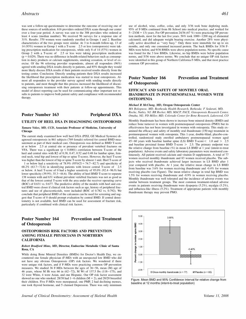
Monthly ibandronate has been shown to increase bone mineral density (BMD) andreduce bone turnover in women with postmenopausal osteoporosis (PMO) but itseffectiveness has not been investigated in women with osteopenia. This study ex-amined the efficacy and safety of monthly oral ibandronate (150 mg) treatment inpostmenopausal women with osteopenia. This 1-year, double-blind, placebo-con-trolled, randomized study enrolled ambulatory postmenopausal women aged45e60 years with baseline lumbar spine (LS) BMD T-score!�1.0 and O�2.5and baseline proximal femur BMD T-score O�2.5. The primary endpoint wasthe relative change from baseline (%) in mean LS BMD at 1 year (intent-to-treatpopulation). Adverse events and safety laboratory parameters were monitored con-tinuously. All patient received calcium and vitamin D supplements. A total of 77women received monthly ibandronate and 83 women received placebo. The sub-jects who received ibandronate achieved larger increases in LS BMD after 1year compared with placebo. At 1 year, the relative mean change in LS BMDfrom baseline was 3.6% for women receiving ibandronate and -0.4% for womenreceiving placebo (see Figure). The mean relative change in total hip BMD was1.5% for women receiving ibandronate and -0.9% in women receiving placebo.Monthly ibandronate was well tolerated and the incidence of adverse events wassimilar between treatment groups. The most common treatment-related adverseevents in patients receiving ibandronate were dyspepsia (5.2%), myalgia (5.2%),and influenza-like illness (5.2%). Treatment of appropriate patients with monthlyibandronate therapy may prevent PMO.

Abstracts 461

Journal of Clinical Densitometry: Assessment of Skeletal Health Volume 11, 2008