Embed Size (px)
Citation preview


OSCEs for Medical Finals

Companion website
This book is accompanied by a companion website:
www.wiley.com/go/khan/osces
featuring:• Downloadable checklists from the book• Survey showing which OSCE stations have a high chance of appearing in finals
This book is also available as an e-book.For more details, please seewww.wiley.com/buy/9780470659410or scan this QR code:
OSCEs for Medical FinalsHamed KhanMBBS DGM MRCGP MRCP (London)
GP Principal and Undergraduate Tutor
Oxted, Surrey
Iqbal KhanBSc MBBS
FY2 Doctor
Homerton University Hospital NHS Foundation Trust
Akhil GuptaBSc MBBS
Specialist Registrar in Anaesthetics
London Deanery
Nazmul HussainMBBS MRPharmS
GP ST3
Newham GP Vocational Training Scheme
Sathiji NageshwaranBSc MBBS
FY2 Doctor
Royal Free London NHS Foundation Trust
A John Wiley & Sons, Ltd., Publication

This edition first published 2013 © 2013 by John Wiley & Sons, Ltd.
Wiley-Blackwell is an imprint of John Wiley & Sons, formed by the merger of Wiley’s
global Scientific, Technical and Medical business with Blackwell Publishing.
Registered office: John Wiley & Sons, Ltd, The Atrium, Southern Gate, Chichester, West
Sussex, PO19 8SQ, UK
Editorial offices: 9600 Garsington Road, Oxford, OX4 2DQ, UK
The Atrium, Southern Gate, Chichester, West Sussex, PO19 8SQ, UK
111 River Street, Hoboken, NJ 07030-5774, USA
For details of our global editorial offices, for customer services and for information about
how to apply for permission to reuse the copyright material in this book please see our
website at www.wiley.com/wiley-blackwell.
The right of the author to be identified as the author of this work has been asserted in
accordance with the UK Copyright, Designs and Patents Act 1988.
All rights reserved. No part of this publication may be reproduced, stored in a retrieval
system, or transmitted, in any form or by any means, electronic, mechanical,
photocopying, recording or otherwise, except as permitted by the UK Copyright, Designs
and Patents Act 1988, without the prior permission of the publisher.
Designations used by companies to distinguish their products are often claimed as
trademarks. All brand names and product names used in this book are trade names,
service marks, trademarks or registered trademarks of their respective owners. The
publisher is not associated with any product or vendor mentioned in this book. This
publication is designed to provide accurate and authoritative information in regard to the
subject matter covered. It is sold on the understanding that the publisher is not engaged in
rendering professional services. If professional advice or other expert assistance is required,
the services of a competent professional should be sought.
Library of Congress Cataloging-in-Publication Data
OSCEs for medical finals / Hamed Khan ... [et al.].
p. ; cm.
Objective structured clinical examinations for medical finals
Includes bibliographical references and index.
ISBN 978-0-470-65941-0 (pbk. : alk. paper) – ISBN 978-1-118-44190-9 (eMobi) –
ISBN 978-1-118-44191-6 (ePDF/ebook) – ISBN 978-1-118-44192-3 (ePub)
I. Khan, Hamed. II. Title: Objective structured clinical examinations for medical finals.
[DNLM: 1. Clinical Medicine–Examination Questions. 2. Clinical Competence–
Examination Questions. 3. Communication–Examination Questions. 4. Medical
History Taking–Examination Questions. 5. Physical Examination–Examination
Questions. WB 18.2]
616.0076–dc23
2012024677
A catalogue record for this book is available from the British Library.
Wiley also publishes its books in a variety of electronic formats. Some content that appears
in print may not be available in electronic books.
Cover design by Sarah Dickinson
Set in 8.75/11 Minion pt by Toppan Best-set Premedia Limited
1 2013

Contributors, vii
Acknowledgements, viii
Preface, ix
Part 1: ExaminationsTop Tips, 1
1. Cardiovascular, 2
2. Respiratory, 7
3. Abdominal, 10
4. Peripheral nervous system, 20
5. Central nervous system, 28
6. Ophthalmoscopy, 37
7. Cerebellar, 40
8. Speech, 44
9. Thyroid, 48
10. Breast, 53
11. Rectal, 56
12. Hernia, 60
13. Testicular, 64
14. Vascular (arterial), 68
15. Vascular (venous), 73
16. Ulcer, 76
17. Shoulder, 80
18. Hand, 87
19. Hip, 93
20. Knee, 98
21. Confirming death, 105
Part 2: HistoriesTop Tips, 107
22. General lethargy and tiredness, 109
23. Weight loss, 112
24. Chest pain, 115
25. Palpitations, 118
26. Cough, 122
Contents
27. Shortness of breath, 125
28. Haemoptysis, 128
29. Diarrhoea, 132
30. Abdominal pain, 137
31. Abdominal distension, 143
32. Haematemesis, 148
33. Rectal bleeding, 152
34. Jaundice, 155
35. Dysphagia, 158
36. Headache, 161
37. Loss of consciousness, 165
38. Tremor, 168
39. Dizziness, 172
40. Joint pain, 177
41. Back pain, 183
42. Pyrexia of unknown origin, 191
43. Ankle swelling, 195
44. Needlestick injury, 199
45. Preoperative assessment, 201
Part 3: Communication skillsTop Tips, 205
46. Breaking bad news, 208
47. Explaining medication, 211
48. Explaining a procedure, 215
49. Inhaler technique and asthma medication, 220
50. Exploring reasons for non-compliance, 222
51. Counselling for an HIV test, 225
52. Post mortem consent, 228
53. Explaining a DNAR (Do Not Attempt Resuscitation) decision, 230
54. Explaining post-myocardial infarction medication, 233
55. Dealing with an angry patient, 236
56. Carrying out a handover, 239
v

vi Contents
Part 4: ProceduresTop Tips, 243
57. Urinary catheterisation, 245
58. Insertion of nasogastric tube, 248
59. Venepuncture/phlebotomy, 252
60. Intramuscular injection, 254
61. Intravenous cannulation, 257
62. Intravenous drug administration, 260
63. Arterial blood gas analysis, 262
64. Measuring peak expiratory flow rate, 267
65. Performing and interpreting ECGs, 271
66. Scrubbing up in theatre, 276
67. Suturing, 278
68. Basic life support, 282
69. Advanced life support, 286
70. Completing a death certificate, 291
Index, 293
Companion website
This book is accompanied by a companion website:
www.wiley.com/go/khan/osces
featuring:• Downloadable checklists from the book• Survey showing which OSCE stations have a high chance of appearing in finals

vii
Contributors
We are grateful to the following doctors and medical students for their contributions to this book.
Contributors to the chaptersShifa RahmanManpreet Sahamey Ruth-Mary deSouzaGillian LandymoreRavi Naik
Contributors to the medical school tablesSaba AliAli AlidinaNina ArnesenSvitlana AustinJames BestKerry BosworthLisa BurtonSangeetha ChandragopalEmily ClarkLaura ClarkeRebecca CritchleyNicola DavisRuth-Mary deSouzaPippa DwanMatthew EversonMartin FawcettClare Fernandes
Lyndsey ForbesRachel FrielUshma GadhviHarminder GillCatherine HatzantonisElizabeth HockleyLaura HopkinsTowhid ImamZara JaulimMichelle KamedaJennifer KellyPamini LedchumykanthanAlmas MalikSathiji NageshwaranRavi NaikSania Naqvi
Siva NathanAllan NghiemGary NicholsonClarissa PerksAnna RebowskaElissa ScotlandCharly SengheiserNadir SohailCharlotte SpilsburySarah ThompsonElizabeth Khadija TissinghChristine WahbaJohn WahbaSiobhan WildAnna WillcockAhila Yogendra

viii
Acknowledgements
We are immensely grateful to the multitude of friends and colleagues who helped us with various aspects of this book. They include the following:• All of the patients who kindly permitted us to use their photos in this book• All the staff at Eversley Medical Centre who assisted us with finding patients with signs that could be pho-tographed – specifically Dr John Chan, Dr Colette Boateng and practice nurses Pauline Kearney and Cheryl Mirador• Dr Vivek Chayya and Dr Alison Barbour for their advice on gastroenterology• Dr Sara Khan, Dr Kartik Modha, Dr Nazia Khan and Dr Siva Nathan for their help in recruiting contributors
• Saiji Nageshwaran and Vaitehi Nageshwaran for reviewing several of the chapters• Mr Ian Skipper for his unparalleled IT expertise and assistance• Dr Khalid Khan for helping us develop the idea from which this book was derived, and for reviewing, proof-reading and critiquing the final manuscript• All of our parents and families, without whose patience and support this project would never have succeeded
We are also grateful to the Medical Womens Federa-tion, Tiko’s GP Group and the Muslim Doctors Asso-ciation for helping us recruit contributors through their organisations.

ix
Preface
The student begins with the patient, continues with the patient, and ends his studies with the patient, using books and lectures as tools, as means to an end.
Sir William Osler
Few will disagree that the recent overhauls in medical training, together with higher numbers of medical students being trained, has made medicine far more competitive than before. Medical students today have to make definitive career choices much earlier on than they would have had in years gone by, and to start building a portfolio of achievements such as audits and publications very early on at medical school. Time has become even more precious than it was before, and it is understandable that medical students today will opt for concise focused textbooks rather than sprawl-ing prosaic texts, some of which have been used over many generations and gained an almost legendary status.
This book is perhaps unique in that it has been written by a group of doctors who range from those in career-grade posts who have completed postgraduate training and have been OSCE examiners themselves, to those who have very recently sat their finals. We have collated our experiences to create a textbook that we have made as focused, easy to read and, above all, as exam-orientated as possible. While doing this, we have worked hard to ensure that we include everything nec-essary not only to pass finals, but also to achieve excel-lent marks and hopefully merits and distinctions.
The structure is based on four sections – clinical examinations, histories, communication skills and pro-cedures. At the beginning of each of these sections, there is a ‘Top Tips’ page that has generic advice for any OSCE station of that section which would help you
boost your marks and performance regardless of what the station is.
Each section is divided into chapters based on the stations we feel are most likely to appear in OSCEs at medical schools. Practice makes perfect – and more so in OSCEs than in any other form of assessment. That is why we have started each chapter with a checklist of items reflecting the areas you are likely to be marked on. You should use these to perfect and consolidate your routines, and also when practising OSCEs with friends and on patients. You should ideally do this in a pair or a group of three, with one student doing the station as a candidate and one allocating mock ‘marks’ using the checklists to assess the candidate’s performance.
Following this in each section, we have included tables that summarise the most common conditions that are likely to present in finals OSCEs. We have ensured that the information on the conditions in these tables is as focused and exam-oriented as possible. There is also a ‘Hints and tips for the exam’ section in which we have summarised key advice and common pitfalls that finalists tend to make.
We hope that this book will make your revision not only thorough and focused, but also enjoyable. We have spent a lot of time working with our publishers to make the text as vibrant, colourful and easy to read as pos-sible, with a plethora of tables, illustrations and photos that will not only make it easy to remember key ideas and principles, but also make the topic more interesting.
We wish you the very best of luck with your finals OSCEs, and hope that you find this book both enjoy-able and useful.
Hamed Khan


OSCEs for Medical Finals, First Edition. Hamed Khan, Iqbal Khan, Akhil Gupta, Nazmul Hussain, and Sathiji Nageshwaran.© 2013 John Wiley & Sons, Ltd. Published 2013 by John Wiley & Sons, Ltd.
1
Part 1: Examinations
Top tips
Do:• Memorise the steps: The most important thing that OSCE examiners are looking for is an ability to carry out a full examination with reasonable technique and speed. At finals level, you will be forgiven for missing a few signs, and the vast majority of the marks on the mark schemes are allocated for going through the motions and doing all the ‘steps’. In contrast, at post-graduate level, for example for the MRCP exam, you would be expected to pick up all the major signs, and be penalised heavily for missing them.• Always suggest a number of possible differential diagnoses: Very few doctors will be able conclusively to put their finger on a diagnosis after examining a patient for 10 minutes without a history. Offering a number of differentials means that you have a higher chance of at least mentioning the correct one, even if it is not at the top of your list. It will also show a healthy awareness of your own limitations.• Practise, practise and practise: The best way to do this is by seeing patients, having a friend to assess you using our checklists and then getting critical (but con-structive) feedback from them. Swapping roles and watching colleagues examine is more useful than most students think, as it will reinforce the steps of the exam-ination, and you may see them use techniques and skills that you would not otherwise have thought of. Doing all the major examinations should become such a normal routine for you that you can do it without thinking about what the next step will be – just like riding a bicycle or driving a car.
Don’t:• Don’t be nervous: Most people have problems in OSCEs not because of poor technique or knowledge, but because of anxiety and nervousness. Don’t be over-whelmed by the occasion, and don’t be intimated by an examiner’s grilling. You will find it much easier to focus on your technique and findings if you are relaxed, and most examiners only grill students who are doing well,
as they do not waste their breath on those whom they have decided are a lost cause!• Don’t worry about minutiae: Medicine is not an exact science, and different doctors have different ways of examining patients, most of which yield the right conclusions. At undergraduate level, all the examiners are looking for is a decent, fluent technique that appears to be well practised. Don’t spend ages trying to figure out exactly how much the chest should expand, or whether the cricoid–sternal notch distance is three finger breadths or four. • Don’t hurt the patient: This is the only unforgivable sin in the OSCE. Its always a good idea to start your examination by asking if the patient is in pain anywhere, and reassuring them that if you unintentionally cause pain during the examination, you will be happy to stop. Students often have a tendency to ignore patients saying ‘Ouch!’ and pretending that they have not heard it, but this is definitely the worst thing you can do. If you do cause pain, acknowledge it immediately, apologise unre-servedly and offer to stop – both examiners and patients will appreciate your honesty and professionalism.
Generic points for all examination stationsHELP:H: ‘Hello’ (introduction and gains consent)E: Exposure (nipples to knees/down to groins)L: LightingP: Positions correctly (supine), asks if patient is in any
painWashes handsInspects from end of bed for paraphernaliaInspects patient (scars, etc.)Thanks patientOffers to help patient get dressedWashes handsPresents findingsOffers appropriate differential diagnosisSuggests appropriate further investigations and
managementFor joints only: Look → Feel → Move → Active/
passive/resisted

2
1 Cardiovascular
Checklist P MP F
HELP:
H: ‘Hello’ (introduction and gains consent)
E: Exposure
L: Lighting
P: Positions at 45 degrees, asks if patient is in any pain
Washes hands
Inspection:
• From end of bed: ECG, GTN spray
• Scars: thoracotomy, mitral valvotomy
• Pacemaker
Hands:
• Clubbing (infective endocarditis, cyanotic heart disease, atrial myxoma)
• Signs of infective endocarditis (splinter haemorrhages, Janeway lesions, Osler’s nodes)
Radial pulse:
• Rate
• Rhythm (regular or irregular)
• Character (collapsing, slow rising)
• Radial–radial delay
Requests blood pressure
Eyes:
• Xanthelasma
• Corneal arcus
• Anaemia
Face:
• Malar flush
Mouth
• ‘CDD’ (central cyanosis, dental hygiene, dehydration)
Checklist P MP F
Neck:
• Jugular venous pressure (raised >4 cm)
• Palpates carotid pulse (character)
Palpates apex beat
Checks if apex beat is displaced in axilla
Palpates sternal edges and subclavicular areas for thrills
Auscultates chest:
• Mitral area/apex beat (5th intercostal space [ICS], midclavicular line)
• Tricuspid area (4th ICS, right sternal edge)• Pulmonary area (2nd ICS, right sternal edge)• Aortic area (2nd ICS, left sternal edge)• Palpate carotid or brachial pulse
simultaneously to time murmur
Cardiac manoeuvres:
• Auscultates mitral area with patient lying on left side and in expiration for murmur of mitral stenosis
• Auscultates aortic area with patient sitting forward and in expiration for murmur of aortic regurgitation
Auscultates lung bases for pulmonary oedema
Palpates shins or ankles for peripheral oedema
Thanks patient
Offers to help patient get dressed
Washes hands
Presents findings
Offers appropriate differential diagnosis
Suggests appropriate further investigations and management
OVERALL IMPRESSION:
Examinations: 1 Cardiovascular 3
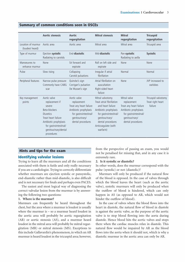
Summary of common conditions seen in OSCEs
Aortic stenosis Aortic regurgitation
Mitral stenosis Mitral regurgitation
Tricuspid regurgitation
Location of murmur (loudest heard)
Aortic area Aortic area Mitral area Mitral area Tricuspid area
Type of murmur Ejection systolicRadiating to carotids
End-diastolic Mid-diastolic Pan-systolicRadiating to axilla
Systolic
Manoeuvres to enhance murmur
None Sit forward and expirate
Roll on left side and expirate
None None
Pulse Slow rising CollapsingCarotid pulsations
Irregular if atrial fibrillation
Normal Normal
Peripheral features Narrow pulse pressureCommonly have CABG
scar
Quincke’s signCorrigan’s pulsationDe Musset’s sign
Atrial fibrillation on auscultation
Right-sided heart failure
None JVP increased to earlobes
Key management points
Aortic valve replacement if severe
Beta-blockersDiureticsTreat heart failureAntibiotic prophylaxis
for gastrointestinal/genitourinary/dental procedures
Aortic valve replacement
Treat any heart failureAntibiotic prophylaxis
for gastrointestinal/genitourinary/dental procedures
Mitral valvotomyTreat atrial fibrillation
and heart failureAntibiotic prophylaxis
for gastrointestinal/genitourinary/dental procedures
Anticoagulate (with warfarin)
Mitral valve replacement
Treat any heart failureAntibiotic prophylaxis
for gastrointestinal/genitourinary/dental procedures
Tricuspid valvotomyTreat right heart
failure
Hints and tips for the exam
Identifying valvular lesionsTrying to learn all the murmurs and all the conditions associated with them is futile and only really necessary if you are a cardiologist. Trying to correctly differentiate whether murmurs are ejection systolic or pansystolic, end-diastolic rather than mid-diastolic, is also difficult and is not necessary for finals and perhaps even PACES.
The easiest and most logical way of diagnosing the correct valvular lesion from the murmur is by answer-ing the following two questions:1. Where is the murmur?Murmurs can frequently be heard throughout the chest, but the area where a murmur is loudest is usually where the murmur is – so a murmur heard loudest in the aortic area will probably be aortic regurgitation (AR) or aortic stenosis (AS), and a murmur heard loudest in the mitral area will probably be mitral regur-gitation (MR) or mitral stenosis (MS). Exceptions to this include Gallavardin’s phenomenon, in which an AR murmur is heard loudest in the tricuspid area; however,
from the perspective of passing an exam, you would not be penalised for missing that, and in any case it is extremely rare.2. Is it systolic or diastolic?In other words, does the murmur correspond with the pulse (systolic) or not (diastolic)?
Murmurs will only be produced if the natural flow of the blood is opposed. In the case of valves through which the blood leaves the heart (such as the aortic valve), systolic murmurs will only be produced when the outflow of blood is hindered, which can only happen in AS (as opposed to AR, which would not hinder the outflow of blood).
In the case of valves where the blood flows into the heart in diastole, the natural flow of blood in diastole is against the aortic valve, as the purpose of the aortic valve is to stop blood flowing into the aorta during diastole. Hence blood hits the aortic valves and stops there when the cardiac muscles relax in diastole. This natural flow would be impaired by AR as the blood flows into the aorta when it should not, which is why a diastolic murmur in the aortic area can only be AR.

4 Examinations: 1 Cardiovascular
Also remember that you should not hear a murmur with a replaced valve unless it is leaking.
Identifying which valve has been replacedRemember that the pulse correlates with the first heart sound, which is the mitral valve closing. (The second heart sound is the aortic valve closing.)• If the loudest sound of the valve closing correlates with the pulse, it is the first heart sound, indicating that the mitral valve has been replaced.• If the loudest sound of the valve does not correspond with the pulse, it is the second heart sound, indicating that the aortic valve has been replaced.• The location of the loudest sounds may also be helpful. Bioprosthetic valves sound the same as normal heart valves, so it would be unfair for examiners to expect you to identify them.
Apex beatThe apex beat is palpable in the 5th intercostal space, and is displaced to the apex in MR. Various char-acters of the apex beat have been described, such as ‘heaving’ and ‘thrusting’; differentiating between them is extremely difficult and probably beyond the scope of a 10-minute OSCE. Other than this, it is more likely to cause confusion than add anything substantive.
The best course of action is to describe where the apex beat it, and whether it is palpable or not. An impalpable apex beat is often caused by obesity, hyperinflation of the lungs, dextrocardia or poor technique.
ScarsFigures 1.1–1.5 show scars and other signs that you will need to note on your examination of the patient.
Questions you could be asked
Q. Which organism causing infective endocarditis is associated with underlying bowel cancer?A. Streptococcus bovis – a colonoscopy should be con-sidered in all patients presenting who are found to have Streptococcus bovis.Q. What is the most common cause of tricuspid regurgitation?A. Most cases of tricuspid regurgitation are ‘func-tional’, due to dilatation of the right ventricle (so that the tricuspid valves flop downwards). This could arise for a number of reasons, such as right heart failure, congestive heart failure and pulmonary hypertension.
If this seems too complex, remember that diastolic murmurs are usually ‘ARMS’ (AR or MS), and the area where it is loudest is probably where the murmur is.
Right versus left• LEFT-sided murmurs are louder in EXPIRATION.• RIGHT-sided murmurs are louder in INSPIRATION.This is because more blood flows into the intrathoracic cavity and lungs on inspiration, and hence more blood flows through the right-sided heart valves as these supply the lungs. The converse is true for left-sided murmurs.
It is vital to ask patients to hold their breath when using this test, but you must not ask them to do this for too long as this can cause the patient pain and you will fail the exam. Its often a good idea to hold your own breath at the same time so that you will know when it is getting too long to allow your patient to breath normally.
Timing the murmurTiming murmurs is something that both students and experienced doctors have difficulty with. Just remem-ber to palpate the pulse when listening to the heart sound, and see if you hear the murmur at the same time as you feel the pulse.• If the murmur is WITH the pulse, it is a SYSTOLIC murmur.• If the murmur if NOT WITH the pulse, it is a DIASTOLIC murmur.Use a central pulse such as the carotid or brachial to do this, otherwise it will not be accurate.
Diastolic murmursA number of conditions can cause diastolic murmurs, but the most common ones are AR and MS – this can be easily memorised using the mnemonic ‘ARMS’.
Diastolic murmurs are very difficult to elicit for even the most experienced doctors, and if you can hear a murmur easily, it is most likely to be systolic. However, if you do manage to identify a diastolic murmur, it is handy to remember that MS murmurs are much quieter than AR murmurs, and if you can auscultate a diastolic murmur throughout the chest, it is much more likely to be AR than MS.
Valve replacementsIf you see a midline sternotomy scar, you should imme-diately bring your ear close to the patient’s chest and listen carefully for the clicking noise that is indicative of the closing of a metallic valve replacement – this can easily be heard without a stethoscope.

Examinations: 1 Cardiovascular 5
Figure 1.1 Graft scar from leg vein removal in coronary artery bypass grafting
Figure 1.2 Chest scar in coronary artery bypass grafting
Figure 1.3 Xanthelasma
Figure 1.4 Corneal arcus
Figure 1.5 Indication of pacemaker insertion

6 Examinations: 1 Cardiovascular
Q. How should a patient with suspected heart failure be investigated in primary care?A. According to the NICE guidelines (NICE, 2010), the primary investigation of choice is the blood level of brain natriuretic peptide (BNP)– patients with normal results are unlikely to have heart failure, and those with a BNP level >400 pg/mL should be investigated urgently (within 2 weeks).
Reference
National Institute for Health and Clinical Excellence (2010) Chronic heart failure: Management of chronic heart failure in adults in primary and secondary care. Available from http://www.nice.org.uk/nicemedia/live/13099/50526/50526.pdf (accessed June 2012).
7
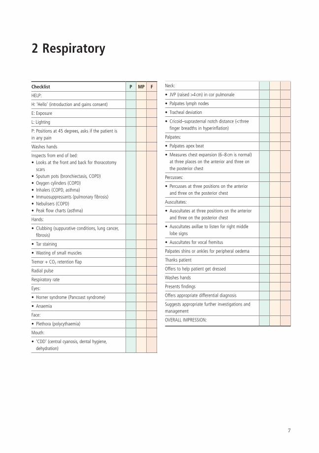
2 Respiratory
Checklist P MP F
HELP:
H: ‘Hello’ (introduction and gains consent)
E: Exposure
L: Lighting
P: Positions at 45 degrees, asks if the patient is in any pain
Washes hands
Inspects from end of bed:• Looks at the front and back for thoracotomy
scars• Sputum pots (bronchiectasis, COPD)• Oxygen cylinders (COPD)• Inhalers (COPD, asthma)• Immuosuppressants (pulmonary fibrosis)• Nebulisers (COPD)• Peak flow charts (asthma)
Hands:
• Clubbing (suppurative conditions, lung cancer, fibrosis)
• Tar staining
• Wasting of small muscles
Tremor + CO2 retention flap
Radial pulse
Respiratory rate
Eyes:
• Horner syndrome (Pancoast syndrome)
• Anaemia
Face:
• Plethora (polycythaemia)
Mouth:
• ‘CDD’ (central cyanosis, dental hygiene, dehydration)
Checklist P MP F
Neck:
• JVP (raised >4 cm) in cor pulmonale
• Palpates lymph nodes
• Tracheal deviation
• Cricoid–suprasternal notch distance (< three finger breadths in hyperinflation)
Palpates:
• Palpates apex beat
• Measures chest expansion (6–8 cm is normal) at three places on the anterior and three on the posterior chest
Percusses:
• Percusses at three positions on the anterior and three on the posterior chest
Auscultates:
• Auscultates at three positions on the anterior and three on the posterior chest
• Auscultates axillae to listen for right middle lobe signs
• Auscultates for vocal fremitus
Palpates shins or ankles for peripheral oedema
Thanks patient
Offers to help patient get dressed
Washes hands
Presents findings
Offers appropriate differential diagnosis
Suggests appropriate further investigations and management
OVERALL IMPRESSION:
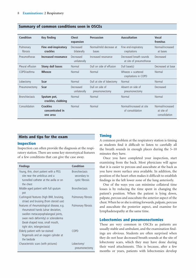
8 Examinations: 2 Respiratory
Summary of common conditions seen in OSCEs
Condition Key finding Chest expansion
Percussion Auscultation Vocalfremitus
Pulmonary fibrosis
Fine end-inspiratory crackles
Decreased bilaterally
Normal/mild decrease at bases
Fine end-inspiratory crepitations
Normal/increased at bases
Pneumothorax Increased resonance Decreased unilaterally
Increased resonance Decreased breath sounds at site of pneumothorax
Decreased
Pleural effusion Stony dull bases Normal Dull on side of effusion Dull base(s) Decreased at base
COPD/asthma Wheeze Normal Normal Wheeze + scattered crepitations in COPD
Normal
Lobectomy Scar Normal Dull at site of lobectomy Normal Normal
Pneumonectomy Scar Decreased unilaterally
Dull on side of pneumonectomy
Absent on side of pneumonectomy
Decreased
Bronchiectasis Sputum pot, crackles, clubbing
Normal Normal Normal Normal
Consolidation Crackles concentrated in one area
Normal Normal Normal/increased at site of consolidation
Normal/increased at site of consolidation
Findings Condition
Young, thin, short patient with a PEG site near the umbilicus and a tunnelled catheter at the axilla or on the chest
Bronchiectasis secondary to cystic fibrosis
Middle-aged patient with full sputum pot
Bronchiectasis
Cushingoid features (high BMI, bruising, striae) and bruising (from steroid use)
Pulmonary fibrosis
Features of rheumatological disease, e.g. rheumatoid hands (ulnar deviation, swollen metacarpophalangeal joints, swan neck deformity) or scleroderma (beak-shaped nose, small mouth, tight skin, telangiectasia)
Pulmonary fibrosis
Elderly patient with tar-stained fingernails and an oxygen cylinder at the bedside
COPD
Characteristic scars (with pictures) Lobectomy/pneumonectomy
Hints and tips for the exam
InspectionInspection can often provide the diagnosis at the respi-ratory station. There are some key stereotypical features of a few conditions that can give the case away.
TimingA common problem at the respiratory station is timing as students find it difficult to listen to carefully all the breath sounds in enough places during the 5–10 minutes they have.
Once you have completed your inspection, start examining from the back. Most physicians will agree that it is easier to percuss and auscultate at the back as you have more surface area available. In addition, the position of the heart often makes it difficult to establish findings in the left lower zone of the lung anteriorly.
One of the ways you can minimise collateral time losses is by reducing the time spent in changing the patient’s position. When the patient is lying down, palpate, percuss and auscultate the anterior aspect of the chest. When he or she is sitting forwards, palpate, percuss and auscultate the posterior aspect, and examine for lymphadenopathy at the same time.
Lobectomies and pneumonectomiesThese are very common in OSCEs as patients are usually stable and ambulant, and the examination find-ings are obvious. Students are often surprised when they do not hear decreased breath sounds at the site of lobectomy scars, which they may have done during their ward attachments. This is because, after a few months or years, patients with lobectomies develop
Examinations: 2 Respiratory 9
Questions you could be asked
Q. Why are spontaneous pneumothoraces more common in tall men?A. There are a number of theories for this. One is that the difference between the intrapleural pressure of the apex and the base is greater in taller people, making it easier for a pneumothorax to form spontaneously. Another is that any anatomical defects or blebs will become more stretched if the length if the lung is longer, as is the case in taller individuals.Q. Why might you hear breath sounds over an area of the lung that has been excised in a lobectomy?A. See ‘Lobectomies and pneumonectomies’ above.Q. Name three causes of bibasal crepitations with club-bing in a patient.A. See ‘Creps and clubbing’ above.
compensatory hyperinflation, and lung tissue fills up areas it was removed from. This will not be the case immediately after lobectomy surgery as sufficient time has not surpassed for compensatory hyperinflation to occur.
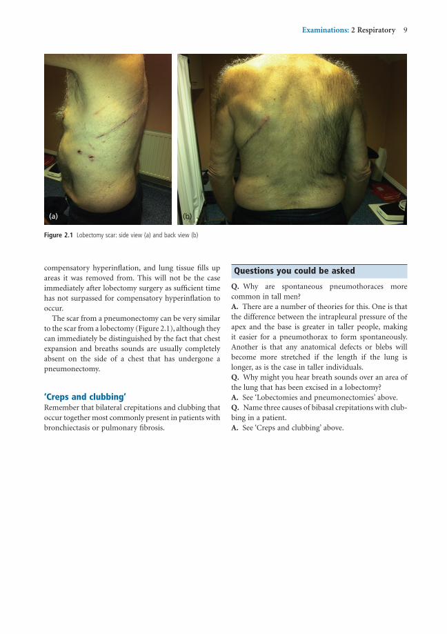
The scar from a pneumonectomy can be very similar to the scar from a lobectomy (Figure 2.1), although they can immediately be distinguished by the fact that chest expansion and breaths sounds are usually completely absent on the side of a chest that has undergone a pneumonectomy.
‘Creps and clubbing’Remember that bilateral crepitations and clubbing that occur together most commonly present in patients with bronchiectasis or pulmonary fibrosis.
Figure 2.1 Lobectomy scar: side view (a) and back view (b)
(a) (b)
10
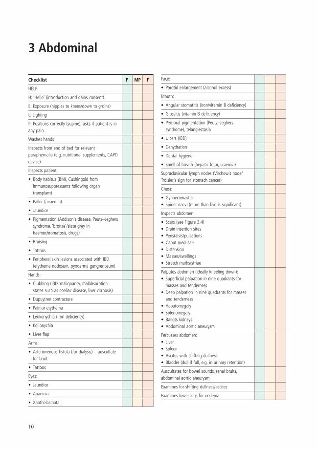
3 Abdominal
Checklist P MP F
HELP:
H: ‘Hello’ (introduction and gains consent)
E: Exposure (nipples to knees/down to groins)
L: Lighting
P: Positions correctly (supine), asks if patient is in any pain
Washes hands
Inspects from end of bed for relevant paraphernalia (e.g. nutritional supplements, CAPD device)
Inspects patient:
• Body habitus (BMI, Cushingoid from immunosuppressants following organ transplant)
• Pallor (anaemia)
• Jaundice
• Pigmentation (Addison’s disease, Peutz–Jeghers syndrome, ‘bronze’/slate grey in haemochromatosis, drugs)
• Bruising
• Tattoos
• Peripheral skin lesions associated with IBD (erythema nodosum, pyoderma gangrenosum)
Hands:
• Clubbing (IBD, malignancy, malabsorption states such as coeliac disease, liver cirrhosis)
• Dupuytren contracture
• Palmar erythema
• Leukonychia (iron deficiency)
• Koilonychia
• Liver flap
Arms:
• Arteriovenous fistula (for dialysis) – auscultate for bruit
• Tattoos
Eyes:
• Jaundice
• Anaemia
• Xanthelasmata
Checklist P MP F
Face:
• Parotid enlargement (alcohol excess)
Mouth:
• Angular stomatitis (iron/vitamin B deficiency)
• Glossitis (vitamin B deficiency)
• Peri-oral pigmentation (Peutz–Jeghers syndrome), telangiectasia
• Ulcers (IBD)
• Dehydration
• Dental hygiene
• Smell of breath (hepatic fetor, uraemia)
Supraclavicular lymph nodes (Virchow’s node/Troisier’s sign for stomach cancer)
Chest:
• Gynaecomastia• Spider naevi (more than five is significant)
Inspects abdomen:
• Scars (see Figure 3.4)• Drain insertion sites• Peristalsis/pulsations• Caput medusae• Distension• Masses/swellings• Stretch marks/striae
Palpates abdomen (ideally kneeling down):• Superficial palpation in nine quadrants for
masses and tenderness• Deep palpation in nine quadrants for masses
and tenderness• Hepatomegaly• Splenomegaly• Ballots kidneys• Abdominal aortic aneurysm
Percusses abdomen:• Liver• Spleen• Ascites with shifting dullness• Bladder (dull if full, e.g. in urinary retention)
Auscultates for bowel sounds, renal bruits, abdominal aortic aneurysm
Examines for shifting dullness/ascites
Examines lower legs for oedema
Examinations: 3 Abdominal 11
Checklist P MP F
Tells examiner he would like to complete the examination by examining the following:• Hernial orifices (with cough/sitting up)• Genitalia• Rectum• Lymph nodes• Urine dipstick
Thanks patient
Offers to help patient get dressed
Checklist P MP F
Washes hands
Presents findings
Offers appropriate differential diagnosis
Suggests appropriate further investigations and management
OVERALL IMPRESSION:
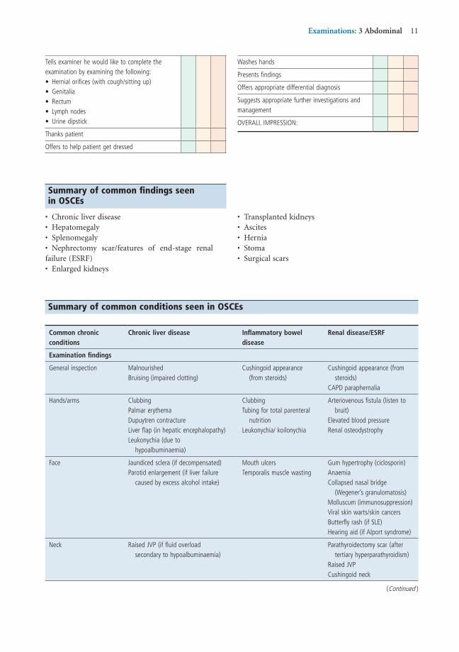
Summary of common findings seen in OSCEs
• Chronic liver disease• Hepatomegaly• Splenomegaly• Nephrectomy scar/features of end-stage renal failure (ESRF)• Enlarged kidneys
• Transplanted kidneys• Ascites• Hernia• Stoma• Surgical scars
Summary of common conditions seen in OSCEs
Common chronicconditions
Chronic liver disease Inflammatory bowel disease
Renal disease/ESRF
Examination findings
General inspection MalnourishedBruising (impaired clotting)
Cushingoid appearance (from steroids)
Cushingoid appearance (from steroids)
CAPD paraphernalia
Hands/arms ClubbingPalmar erythemaDupuytren contractureLiver flap (in hepatic encephalopathy)Leukonychia (due to
hypoalbuminaemia)
ClubbingTubing for total parenteral
nutritionLeukonychia/ koilonychia
Arteriovenous fistula (listen to bruit)
Elevated blood pressureRenal osteodystrophy
Face Jaundiced sclera (if decompensated)Parotid enlargement (if liver failure
caused by excess alcohol intake)
Mouth ulcersTemporalis muscle wasting
Gum hypertrophy (ciclosporin)AnaemiaCollapsed nasal bridge
(Wegener’s granulomatosis)Molluscum (immunosuppression)Viral skin warts/skin cancersButterfly rash (if SLE)Hearing aid (if Alport syndrome)
Neck Raised JVP (if fluid overload secondary to hypoalbuminaemia)
Parathyroidectomy scar (after tertiary hyperparathyroidism)
Raised JVPCushingoid neck
(Continued )
12 Examinations: 3 Abdominal
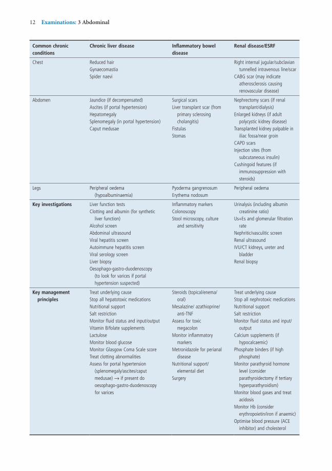
Common chronicconditions
Chronic liver disease Inflammatory bowel disease
Renal disease/ESRF
Chest Reduced hairGynaecomastiaSpider naevi
Right internal jugular/subclavian tunnelled intravenous line/scar
CABG scar (may indicate atherosclerosis causing renovascular disease)
Abdomen Jaundice (if decompensated)Ascites (if portal hypertension)HepatomegalySplenomegaly (in portal hypertension)Caput medusae
Surgical scarsLiver transplant scar (from
primary sclerosing cholangitis)
FistulasStomas
Nephrectomy scars (if renal transplant/dialysis)
Enlarged kidneys (if adult polycystic kidney disease)
Transplanted kidney palpable in iliac fossa/near groin
CAPD scarsInjection sites (from
subcutaneous insulin)Cushingoid features (if
immunosuppression with steroids)
Legs Peripheral oedema (hypoalbuminaemia)
Pyoderma gangrenosumErythema nodosum
Peripheral oedema
Key investigations Liver function testsClotting and albumin (for synthetic
liver function)Alcohol screenAbdominal ultrasoundViral hepatitis screenAutoimmune hepatitis screenViral serology screenLiver biopsyOesophago-gastro-duodenoscopy
(to look for varices if portal hypertension suspected)
Inflammatory markersColonoscopyStool microscopy, culture
and sensitivity
Urinalysis (including albumin creatinine ratio)
Us+Es and glomerular filtration rate
Nephritic/vasculitic screenRenal ultrasoundIVU/CT kidneys, ureter and
bladderRenal biopsy
Key management principles
Treat underlying causeStop all hepatotoxic medicationsNutritional supportSalt restrictionMonitor fluid status and input/outputVitamin B/folate supplementsLactuloseMonitor blood glucoseMonitor Glasgow Coma Scale scoreTreat clotting abnormalitiesAssess for portal hypertension
(splenomegaly/ascites/caput medusae) → if present do oesophago-gastro-duodenoscopy for varices
Steroids (topical/enema/ oral)
Mesalazine/ azathioprine/ anti-TNF
Assess for toxic megacolon
Monitor inflammatory markers
Metronidazole for perianal disease
Nutritional support/elemental diet
Surgery
Treat underlying causeStop all nephrotoxic medicationsNutritional supportSalt restrictionMonitor fluid status and input/
outputCalcium supplements (if
hypocalcaemic)Phosphate binders (if high
phosphate)Monitor parathyroid hormone
level (consider parathyroidectomy if tertiary hyperparathyroidism)
Monitor blood gases and treat acidosis
Monitor Hb (consider erythropoietin/iron if anaemic)
Optimise blood pressure (ACE inhibitor) and cholesterol
Examinations: 3 Abdominal 13
Hints and tips for the exam
Hepatomegaly and splenomegalyHepatomegaly and splenomegaly are also very common findings at this station in finals. We have discussed various key tips below to help you in both the diagnosis and the discussion.
Examining large livers and spleens• Start low in the right iliac fossa, so that you do not miss giant organomegaly.
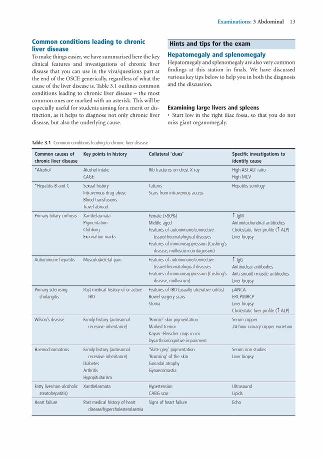
Common conditions leading to chronic liver diseaseTo make things easier, we have summarised here the key clinical features and investigations of chronic liver disease that you can use in the viva/questions part at the end of the OSCE generically, regardless of what the cause of the liver disease is. Table 3.1 outlines common conditions leading to chronic liver disease – the most common ones are marked with an asterisk. This will be especially useful for students aiming for a merit or dis-tinction, as it helps to diagnose not only chronic liver disease, but also the underlying cause.
Table 3.1 Common conditions leading to chronic liver disease
Common causes of chronic liver disease
Key points in history Collateral ‘clues’ Specific investigations to identify cause
*Alcohol Alcohol intakeCAGE
Rib fractures on chest X-ray High AST:ALT ratioHigh MCV
*Hepatitis B and C Sexual historyIntravenous drug abuseBlood transfusionsTravel abroad
TattoosScars from intravenous access
Hepatitis serology
Primary biliary cirrhosis XanthelasmataPigmentationClubbingExcoriation marks
Female (>90%)Middle-agedFeatures of autoimmune/connective
tissue/rheumatological diseasesFeatures of immunosuppression (Cushing’s
disease, molluscum contagiosum)
↑ IgMAntimitochondrial antibodiesCholestatic liver profile (↑ ALP)Liver biopsy
Autoimmune hepatitis Musculoskeletal pain Features of autoimmune/connective tissue/rheumatological diseases
Features of immunosuppression (Cushing’s disease, molluscum)
↑ IgGAntinuclear antibodiesAnti-smooth muscle antibodiesLiver biopsy
Primary sclerosing cholangitis
Past medical history of or active IBD
Features of IBD (usually ulcerative colitis)Bowel surgery scarsStoma
pANCAERCP/MRCPLiver biopsyCholestatic liver profile (↑ ALP)
Wilson’s disease Family history (autosomal recessive inheritance)
‘Bronze’ skin pigmentationMarked tremorKayser–Fleischer rings in irisDysarthria/cognitive impairment
Serum copper24-hour urinary copper excretion
Haemochromatosis Family history (autosomal recessive inheritance)
DiabetesArthritisHypopituitarism
‘Slate grey’ pigmentation‘Bronzing’ of the skinGonadal atrophyGynaecomastia
Serum iron studiesLiver biopsy
Fatty liver/non-alcoholic steatohepatitis)
Xanthelasmata HypertensionCABG scar
UltrasoundLipids
Heart failure Past medical history of heart disease/hypercholesterolaemia
Signs of heart failure Echo

14 Examinations: 3 Abdominal
• Use the radial aspect of your index finger – but if that doesn’t work, use your finger with your hands pointing up towards the patient’s head.• Keep your fingers absolutely still as the patient breathes in and out.• Make sure that you move your hand upwards supe-riorly by no more than 2 cm as the patient breathes in and out. If you leave too large a distance as you move up, there is a risk that you may miss the edge of the liver or spleen.• For the liver, percussion is almost as discriminatory as palpation. It is also useful to differentiate between lung hyperinflation pushing the liver down, and true hepatomegaly. The superior aspect of the liver usually lies between the 4th and 6th ribs, and continues down to the last rib at the inferior border of the rib cage; hence, there should be dullness in all of this area. Hyperinflation pushing down the liver is confirmed if percussion is resonant significantly below the 6th rib.• For the spleen, use your left hand to stabilise the left ribs in order to prevent them from being pushed towards the left as you palpate the spleen with your right hand. If you still have difficulty, roll the patient on to the right side and repeat this.• When you do find an enlarged liver or spleen, esti-mate the size of hepatomegaly in centimeters rather than ‘finger breadths’, which vary from person to person (depending on how big their fingers are!).• Avoid the business of trying to identify the liver char-acteristics (e.g. whether it ‘firm’, ‘hard’ or ‘soft’, or pul-satile, or nodular or smooth). Doing this in an exam will make the patient uncomfortable and use up your valuable time without achieving very much. Once a large liver or spleen has been identified, the most logical way of defining its characteristics would be to carry out some sort of imaging – usually an ultrasound of the abdomen.
Systematic differentiation of the underlying causes of hepatomegaly and splenomegaly• A large liver and/or spleen is a very common finding at finals OSCE stations. Make sure that you have a generic system for categorising the causes, so that you can reel off a list of differential diagnoses quickly, con-fidently and systematically.• Always try to use all the signs to help you devise a differential diagnosis. However, if you find an enlarged spleen or liver and have no clue what the cause is, go for conditions that can cause hepatomegaly and splenomegaly either individually or together – the first column of Table 3.2 summarises these.
• Don’t be too pedantic when distinguishing between gigantic, moderate and mild splenomegaly. Identifying splenomegaly and giving a reasonable list of differential diagnoses and investigations will usually be enough to score a decent pass. Distinguishing between mild/moderate and gigantic splenomegaly will help to get you into the merit/distinction range. Remember that the spleen has to be at least double or triple its normal size to be palpable.• Remember to piece the other parts of your examina-tion together to complete the diagnostic jigsaw. All the conditions that cause hepatomegaly or splenomegaly have several peripheral signs so look out for these and use them to support your differential diagnosis.
Renal casesAlthough students often worry about getting a ‘renal case’ in finals, it can often be a blessing in disguise. The differential diagnosis is relatively straightforward, and the signs are easy to elicit.
Fundamentally, there are only two findings in renal cases – those of ESRF, and ballotable enlarged kidneys.
End-stage renal failureThere are potentially three findings that are all attribut-able to ESRF:• Nephrectomy scar (Figure 3.2): Inspect carefully for this, making sure that you look all the way around the lumbar/flank regions through to the back. Finding a nephrectomy scar is alone sufficient to devise a full list of differential diagnoses and a management plan.• Palpable transplanted kidney: This is usually near the groin/iliac fossa with a small scar at the site.• Signs of dialysis use (arteriovenous fistula, right internal jugular vein line, CAPD scars; Figure 3.3): A slicker way of describing this is ‘renal replacement therapy’, which covers them all – and also sounds more impressive!
Whichever of these signs the patient has, the under-lying condition is always ESRF.
The four most common causes of ESRF are as follows:1) Diabetes2) Hypertension3) Adult polycystic kidney disease (APKD)4) GlomerulonephritisOnce you have got to this stage, your investigations and management should be guided by your differential diagnosis. However, if you are still struggling, merely discuss the generic investigations and management strategies for patients with ESRF, as discussed in the summary table above.
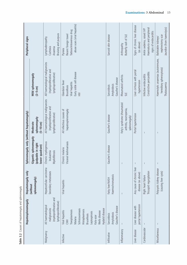
Examinations: 3 Abdominal 15
Tabl
e 3.
2 Ca
uses
of h
epat
omeg
aly
and
sple
nom
egal
y
Hep
atos
plen
omeg
aly
Hep
atom
egal
y on
ly(w
itho
ut
sple
nom
egal
y)
Sple
nom
egal
y on
ly (
wit
hout
hep
atom
egal
y)Pe
riph
eral
sig
ns
Gig
anti
c sp
leno
meg
aly
(pal
pabl
e in
rig
ht
low
er q
uadr
ant)
Mod
erat
esp
leno
meg
aly
(5–1
0 cm
)
Mild
spl
enom
egal
y(2
–5 cm
)
Mal
igna
ncy
All h
aem
atol
ogica
l m
alig
nanc
ies
(mye
lopr
olife
rativ
e an
d lym
phop
rolif
erat
ive)
Hepa
toce
llula
r car
cinom
aSe
cond
ary
met
asta
ses
Chro
nic
mye
loge
nous
le
ukae
mia
Mye
lofib
rosis
All h
aem
atol
ogica
l mal
igna
ncie
s (m
yelo
prol
ifera
tive
and
lymph
opro
lifer
ativ
e)
All h
aem
atol
ogica
l mal
igna
ncie
s (m
yelo
prol
ifera
tive
and
lymph
opro
lifer
ativ
e)
Lym
phad
enop
athy
Cach
exia
Anae
mia
Brui
sing
and
purp
ura
Infe
ctiv
eVi
ral h
epat
itis
CMV
Toxo
plas
mos
isM
alar
iaSc
hist
osom
iasis
Hist
opla
smos
isBr
ucel
losis
Lept
ospi
rosis
Kala
-aza
rW
eils
dise
ase
Hyda
tid d
iseas
e
Vira
l hep
atiti
sCh
roni
c m
alar
iaVi
scer
al le
ishm
ania
sisAl
l inf
ectio
us c
ause
s of
he
pato
sple
nom
egal
yG
land
ular
feve
rBr
ucel
losis
Vira
l hep
atiti
sEa
rly s
ickle
cel
l dise
ase
HIV
Pyre
xia
Rece
nt fo
reig
n tra
vel
Tatto
os/in
trave
nous
dru
g ab
use
scar
s (v
iral h
epat
itis)
Infil
trativ
eSa
rcoi
dosis
Amylo
idos
isG
auch
er’s
dise
ase
Fatty
live
r/NAS
HHa
emoc
hrom
atos
isG
auch
er’s
dise
ase
Gau
cher
’s di
seas
eSa
rcoi
dosis
Amylo
idos
isG
auch
er’s
dise
ase
Sarc
oid
skin
dise
ase
Infla
mm
ator
y–
––
Felty
’s sy
ndro
me
(rheu
mat
oid
arth
ritis,
neu
trope
nia,
sp
leno
meg
aly)
Rheu
mat
oid
arth
ritis
SLE
Arth
ropa
thy
Butte
rfly
rash
of S
LE
Live
r dise
ase
Live
r dise
ase
with
porta
l hyp
erte
nsio
nAn
y ca
use
of c
hron
ic liv
er
dise
ase
(as
abov
e)–
Porta
l hyp
erte
nsio
nLi
ver c
irrho
sis w
ith p
orta
l hy
perte
nsio
nSi
gns
of c
hron
ic liv
er d
iseas
e (a
s ab
ove)
Card
iova
scul
ar–
Righ
t hea
rt fa
ilure
Tricu
spid
regu
rgita
tion
––
Infe
ctiv
e en
doca
rditi
sCo
nstri
ctiv
e pe
ricar
ditis
Ankl
e oe
dem
a, ra
ised
JVP
Haem
atur
ia a
nd p
erip
hera
l sig
ns o
f end
ocar
ditis
Misc
ella
neou
s–
Polyc
ystic
kid
ney
dise
ase
(cau
sing
liver
cys
ts)
––
Haem
olyt
ic an
aem
ias
(aut
oim
mun
e,
here
dita
ry s
pher
ocyt
osis)
Thal
assa
emia
Ballo
tabl
e ki
dney
s/
neph
rect
omy
scar
Jaun
dice
(fro
m h
aem
olys
is)

16 Examinations: 3 Abdominal
occurs because of tertiary hyperparathyroidism and although it looks like clubbing, with prominence of the distal phalanges, what actually happens is that the prox-imal phalanges become narrow, and this makes the distal phalanges look prominent despite being normal.
Pseudoclubbing is common after renal replacement therapy – patients with long-standing secondary hyperparathyroidism (due to low calcium levels) develop parathyroid hyperplasia, leading to increased parathyroid hormone production that becomes auton-omous of the negative feedback system. Once a patient is undergoing renal replacement therapy and their calcium levels normalise, the parathyroid continues producing excess parathyroid hormone, which results in hypercalcaemia and resorption of bone from the proximal phalanges, causing them to narrow.• Chronic liver disease and features of ESRF in the same patient: This is rare, but don’t let it put you off. The most likely cause is hepatitis C (leading to chronic liver disease), which also causes membranous glomeru-lonephritis (leading to ESRF).• Spleen versus kidney: When palpating the left side of the abdomen, it can sometimes be difficult to distin-guish a ballotable kidney from a spleen. Table 3.3 below summarises the key differences.
Figure 3.2 Nephrectomy scar
Figure 3.3 Right internal jugular tunnelled catheter (for dialysis)
Figure 3.1 Scar from splenectomy after a road traffic accident, also showing the drain insertion site
Ballotable/ enlarged kidneysBallotable enlarged kidneys can be palpated in the lateral lumbar regions. As with ESRF, you only need to remember a short list of differential diagnosis:• APKD• Renal cell carcinoma• Bilateral hydronephrosis (secondary to obstruction, e.g. by an external mass, prostate enlargement, etc.)• Amyloidosis (primary or secondary)The key investigations with all of these are imaging (CT of the kidney, ureter and bladder/IVU) and renal biopsy, with the management depending on the under-lying cause.
Rare findings• Clubbing versus pseudoclubbing: Although these conditions look similar on examination, the underlying causes are fundamentally different. Pseudoclubbing

Examinations: 3 Abdominal 17
5) Nephrectomy scar (rarely adrenalectomy scar)1. Classic caesarean section scar/hysterectomy scar2. Appendicectomy scar: at McBurney’s point3. Caesarean section scar (suprapubic)4. Inguinal hernia scar5. Femoral hernia scar
Abdominal massesIf you find a mass, try to answer two questions in your mind.1. Where is the mass?First identify the quadrant where the mass is located, and then think of the organs in that quadrant from which the mass might originate (Figure 3.5).
Table 3.3 Spleen or kidney?
Spleen Kidney
Cannot get above the spleen Should be able to get above the kidney
Moves downwards and medially with inspiration
No movement with breathing
Not ballotable BallotablePalpable notch (medial aspect) No notch
Figure 3.4 Common abdominal scars
1
23
45
67
8 9
10
Figure 3.5 Location of organs in the abdomen
LungLiver
Gallbladder
StomachPancreas
Abdominal aorta
LungSpleen
Pancreas (rarely)
LiverKidneyUreter
StomachSmall intestine/transverse colon
Abdominal aorta
SpleenKidneyUreter
UreterOvary
Fallopian tubeCaecum
(appendix)
BladderUterusCervix
(referred pain from testicles)
UreterOvary
Fallopian tubeSigmoid colon
Theoretically, the spleen should be dull while the kidney has traditionally been documented in most texts to be ‘resonant’. This is, however, more theoretical than realistic as in practice both kidneys and spleens feel dull on percussion.
Abdominal scarsAs with all OSCEs, the key findings in abdominal exam-ination are often established on inspection (Figure 3.4):1) Rooftop scar
• Partial hepatectomy• Pancreatic surgery• Accessing aorta
2) Kocher incision• Cholecystectomy
3) ‘Mercedes-Benz’ scar• Liver transplant• Gastric surgery• Oesophageal surgery
4) Midline laparotomy• Colon surgery• Aortic abdominal aneurysm surgery

18 Examinations: 3 Abdominal
Remember to describe the mass accurately and logi-cally – see Chapter 10 on breast examination for a table of characteristics that you should aim to describe.2. What is the lesion?As with everything in OSCEs, the key is to have a generic method of categorising potential differential diagnoses. The categories below can be used to devise a differential diagnosis for a mass in almost any of the nine quadrants:• Tumour
• Benigni. Cyst (liver, renal)
ii. Fibroids (in the pelvic area in women)iii. Vascular (abdominal aortic aneurysm)
• Malignanti. Primary
ii. Secondaryiii. Lymphoma
• Infection• Abscess• Tuberculosis (usually ileocaecal)
• Inflammatory bowel disease• Crohn’s disease (in right iliac fossa)• Diverticular disease (left iliac fossa)
Key investigationsThe crux of investigating a mass is to visualise it and to get a tissue sample from it. Hence the following inves-tigations are most important:• Imaging: CT/MRI scan• Endoscopy: colonoscopy for colon, oesophago-gastro-duodenoscopy for oesophagus/stomach, cystos-copy for bladder• Biopsy: for any non-vascular mass
StomasStomas (Figures 3.6 and 3.7) feature more commonly in finals than most students think, and they are actually quite easy to examine and talk about. The most common stomas are ileostomies and colostomies, and the key feature that distinguishes them is their location. Table 3.4 summarises the key features.
Questions you could be asked
Q. What is the one investigation you would do in a patient with known portal hypertension in order to reduce mortality?A. The key features of portal hypertension are:
• Splenomegaly• Ascites• Caput medusae• Oesophageal varices
Figure 3.6 Stoma
Figure 3.7 Percutaneous endoscopic gastrostomy (PEG)
Although the answer to this is debatable, the most important investigation would be an oesophago-gastro-duodenoscopy to identify varices, and more importantly to band them and prevent torrential acute severe gastrointestinal bleeding.Q. How big does the spleen have to be before it is palpable?A. About twice its normal size.