Embed Size (px)
Citation preview


1
OSCE Dossier
2018
Authors (Year 4 Medical Students) Ayat Al-Zghoul
Bushra Arafa
Hashim Ahmad Mohammad
Haya Yanes
Linda Al-bsould
Raghad Bataineh
Reem Akiely
Majd Rawashdeh
Mohammad Qussay Al-Sabbagh

1
Cardiovascular System

2
3
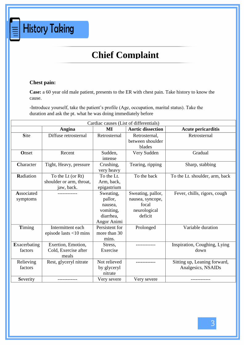
Chest pain:
Case: a 60 year old male patient, presents to the ER with chest pain. Take history to know the
cause.
-Introduce yourself, take the patient’s profile (Age, occupation, marital status). Take the
duration and ask the pt. what he was doing immediately before
Cardiac causes (List of differentials)
Angina MI Aortic dissection Acute pericarditis
Site Diffuse retrosternal Retrosternal Retrosternal,
between shoulder
blades
Retrosternal
Onset Recent Sudden,
intense
Very Sudden Gradual
Character Tight, Heavy, pressure Crushing,
very heavy
Tearing, ripping Sharp, stabbing
Radiation To the Lt (or Rt)
shoulder or arm, throat,
jaw, back.
To the Lt.
Arm, back,
epigastrium
To the back To the Lt. shoulder, arm, back
Associated
symptoms
------------ Sweating,
pallor,
nausea,
vomiting,
diarrhea,
Angor Animi
Sweating, pallor,
nausea, syncope,
focal
neurological
deficit
Fever, chills, rigors, cough
Timing Intermittent each
episode lasts <10 mins
Persistent for
more than 30
mins.
Prolonged Variable duration
Exacerbating
factors
Exertion, Emotion,
Cold, Exercise after
meals
Stress,
Exercise
------------ Inspiration, Coughing, Lying
down
Relieving
factors
Rest, glyceryl nitrate Not relieved
by glyceryl
nitrate
------------ Sitting up, Leaning forward,
Analgesics, NSAIDs
Severity ------------ Very severe Very severe ------------
Chief Complaint

4
Notes If unstable angina the
pain is more severe,
occurs at rest,
increasing in frequency
and duration
May be silent
in elderly,
diabetics and
females
------------ Viral etiologies may be preceded
by flu-like respiratory or GI
symptoms
Risk factors Age (♂>45 ♀>55), smoking, HTN,
Hyperlipidemia, DM, previous attacks,
Family history of premature CAD
(♂<55 ♀<65)
HTN, Marfan
syndrome,
Ehlers-Danlos
syndrome,
Weight lifting,
Pregnancy in 3rd
trimester
------------
Non-cardiac causes
Take the duration and ask the pt. what he was doing immediately before
Pleural pain Chest wall pain Mediastinal
pain
Esophageal pain
Site Well localized Wide area Retrosternal.
Central
Retrosternal or epigastric
Onset Sudden Sudden Variable Sudden
Character Sharp, Stabbing Generalized
tightness
Dull, aching,
gnawing
Dull, burning
Radiation To the neck and shoulder
tip (if diaphragmatic area is
involved)
To the Epigastrium (if the
pleura overlying the lower
six ribs is involved)
------------ To the medial
side of the arm
(in Pancoast’s
tumor)
To the arms and back
Associated
symptoms
------------ Cough ------------ Heartburn and acid reflux
Timing ------------ Wakes the pt
from sleep
Wakes the pt.
from sleep
Wakes the pt. from sleep
Exacerbating
factors
Inspiration and coughing Coughing Coughing (in
tracheobronchial
infections)
Lying flat
Relieving
factors
------------ ------------ ------------ Nitrites sometimes relieve
Severity ------------ ------------ ------------ Usually mild
DDx Pneumonia, PE,
pneumothorax, fractured
ribs
Rib fractures,
intercostal
muscle injury,
HZV,
malignancy
Lymphoma,
thymoma,
infection of
tracheobronchial
tree)
Esophageal spasm, GERD,
hiatus hernia

5
Review of
systems Ask about SOB, Palpitation, Syncope, Claudication, Ankle swelling
Cough, Wheezes, Hemoptysis
Fever, Chills, Wt. loss, Anorexia, Nausea, Vomiting
Past medical and
surgical
HTN, hyperlipidemia, DM, previous caths and stents, recent infections, previous heart
surgeries
Drug Hx NSAIDs, B-blockers, Thyroxine, Cocaine
Family Hx Family Hx of heart disease or premature CAD
Social Hx Smoking history (# of pack years), alcohol, travel history

6
Shortness of breath (SOB)/ Dyspnea:
Case: a 53 year-old man presents to your clinic complaining of SOB, take detailed history to
know the cause.
-Introduce yourself, take the patient’s profile (age, occupation, marital status).
Lis
t of
com
mon
dif
feren
tials
Cardiac Heart Failure:
-Take the duration
-Timing (At night, With exertion, At rest)
-if at night: does it occur immediately when lying flat (Orthopnea), or does it occur later
during sleep waking the patient gasping for air (PND)?
-ask about the # of pillows used to sleep
-If with exertion: ask about baseline, how it progressed? how does it affect daily life?
(Check box 7.7 in the next page to assess severity)
-Is it relieved with diuretics? (Ask about compliance to drugs and diet)
-Associated symptoms: Fatigue, coughing with frothy blood sputum, ankle swelling,
abdominal distention, oliguria, chest pain, palpitations.
IHD:
-Take the duration
-Is it exacerbated with exercise? Is t relieved by glyceryl nitrate?
-Associated symptoms: chest pain, palpitation, sweating, nausea, pallor.
Others:
-Ask about Fever, chills, rigors, cough
Respiratory
Ask about the duration (Check box 7.6 in the next page for DDx).
Asthma:
-Typically wakes the pt. from sleep around 3-5 am.
-Associated with wheezes and cough (either dry or productive with white viscid mucoid
sputum).
-Relieved by inhalers.
-Ask about exposure to allergens (shaking bedding, hoovering, and mowing the lawn.
Exposure to cats, dogs, horses and tree pollens), smoke, perfumes, fumes, cold air or
drugs ( Aspirin, NSAIDs).
-Ask if related to exercise and if it continues to worsen 5-10 minutes after stopping
activity (Exercise-induced asthma).
-Ask if it improves on weekends or holidays (occupational asthma).
-Causes of exacerbation:
Stress (emotion and exercise)
Infection (runny nose, fever, Chills, sore throat, chest pain, yellow-green sputum).
Exposure to irritants (allergens, smoking, cold air, drugs).
COPD:
-Typically is worse upon waking in the morning. Improved after coughing clear, grey
mucoid sputum.
-Associated with wheezes and productive cough.
PE:
-Very sudden in onset.
-Exacerbated with upright posture, relieved by lying flat, Associated with pleuritic chest
pain, , cough with hemoptysis, tachycardia, pallor, sweating.

7
Pneumonia:
-Onset within hours to days.
-Associated with: Acute productive cough (purulent green sputum), Fever, Chills,
Pleuritic chest pain, Wt. loss and history of URTI.
-Ask about abdominal pain, nausea and vomiting to consider lower lobe pneumonia.
Hematology Anemia:
-SOB without chest pain.
-Associated with: headache, fatigue, loss of concentration, dizziness upon standing,
palpitation, bone pain, Wt. loss, hx of bleeding or easy bruising.
Psychogenic -Occurs suddenly at rest or while talking.
-Associated with: Lightheadedness, Dizziness, Tingling in the fingers and around the
mouth, Chest tightness.
Past Medical hx: DM, HTN, Hyperlipidemia, Malignancy, hx of recent surgery or bed rest, Alpha-1-
antitrypsin deficiency.
Drug hx: Aspirin, NSAIDs, B-blockers, Ca+2
Channel blockers, Inhalers.
Family hx: Family Hx of atopy (Allergic Rhinits, sinusitis, Eczema, Conjunctivitis, Asthma), Hx of
Lung Ca. or IHD.
Social hx: Smoking Hx (calculate pack years), travel Hx, Hx. of vaccination

8
Palpitations:
Case: a 40 year-old lady presents to ER complaining of palpitations, ask relevant questions to
know the cause of her complaint.
-Introduce yourself, take the patient’s profile (age, occupation, marital status). Take the
duration and ask what she was doing immediately before.
Sinus tachycardia SV Tach Extra-systoles A Fib V tach
Onset Gradual Sudden with
jump
Sudden Sudden Sudden
Character Regular, fast Regular, fast Jump or missed
beat followed
by a strong beat
Irregular, fast
(slow in elderly)
Regular, fast
Associated
symptoms
------------ Polyuria, chest
tightness,
lightheadedness
------------ Polyuria, SOB Presyncope,
syncope, chest
tightness
Timing Lasts for a few
mins
Lasts for mins-
hours
Brief Variable Variable
Exacerbating
factors
Anxiety, stress,
exercise, caffeine,
alcohol, smoking
Bending
Usually occur
at rest
Fatigue,
caffeine,
alcohol,
Exercise,
alcohol
Exercise
Relieving
factors
------------ -Valsalva
maneuver
-Carotid sinus
pressure
Walking or
exercise
------------ ------------
Severity Mild to moderate Moderate to
severe
Mild Very variable Severe
Causes -Drugs (B2-
agonists,Thyroxine,
Antihistamines,
Decongestants,
Amphetamine,
Cocaine, Ecstasy)
-Infection and
sepsis (Fever)
-Thyrotoxicosis
(Wt. Loss, Heat
intolerance,
Sweating)
-Active bleeding
(hematemesis,
melena, hematuria)
-Anemia (fatigue,
weakness, SOB)
-Pregnancy
-
Pheochromocytoma
(headache,
sweating)
No underlying
heart disease
-Excessive
alcohol,
caffeine, or
tobacco
consumption
-Drugs
(Tricyclic
antidepressants,
digoxin)
-IHD
-Valvular heart
disease (Mitral
stenosis)
-HTN
-
Cardiomyopathy
-Sick sinus
syndrome
-Alcohol excess
-Congenital
heart disease
-Constrictive
pericarditis
-
Hyperthyroidism
-OSA
-Asthma/COPD
-Idiopathic
-People with
cardiomyopathy
or previous MI
-People with
pacemakers or
Intra-cardiac
devices have
increased risk of
V. Tach

9
Review of systems SOB, Chest pain, Syncope, Claudication, Ankle swelling, Fever, Headache, Dizziness.
Past medical and
surgical Hx
Rheumatic fever, previous heart attacks, anemia, hyperthyroidism, HTN, DM, HF,
endocarditis, previous caths or stents, previous heart surgeries.
Drug Hx Thyroxine, B-agonists, Digoxin, Anti-depressants, Diuretics
Family Hx Family hx of heart disease or sudden death
Social Hx Smoking (#of pack years), alcohol, caffeine intake

10
Syncope:
Case: a 35 year-old man presents to ER after a brief LOC, take history to know the cause.
-Introduce yourself, take the patient’s profile (age, occupation, marital status). Take the
duration and ask what he was doing immediately before.
Postural Hypotension Neuro-cardiogenic
syncope (Vasovagal
attack)
Arrhythmias Mechanical
Obstruction to
CO
Onset Suddenly upon standing Sudden ------------ ------------
Prodrome ------------ Light headedness, tinnitus,
dark vision, sweating,
pallor.
When he wakes up he is
flushed and vomiting.
Palpitations, chest
pain, SOB
------------
Duration 1-2 mins <60 seconds ------------
Exacerbating
factors
Standing Standing for long periods
in warm weather,
emotions, anxiety, cough
------------ Exertion, during
exercise
Relieving
factors
------------ Elevation of legs ------------ ------------
DDx Hypovolemia,
septicemia, autonomic
neuropathy,
Drugs (diuretics, anti-
hypertensives,
vasodilators)
Vasovagal attack
If frequent: hypersensitive
carotid sinus syndrome
Tachyarrhythmia:
V. Tach
Brady-arrhythmia:
Complete heart
block
Severe aortic
stenosis, HOCM,
PE, Aortic
dissection
Review of systems Chest pain, SOB, palpitation, claudication, ankle swelling, cough
Nausea, vomiting, diarrhea, melena, hematemesis
Fever, fatigue, pallor, anxiety
Weakness, numbness, jerky moves, tongue biting, amnesia after attack
Past medical and
surgical Hx
Anemia, Bleeding, prolonged vomiting or diarrhea, prolonged fasting, seizures, HTN,
DM
Drug hx Vasodilators, Nitrates, ACEIs
Family Hx Bleeding, seizures

11
Lower Limb swelling:
Case: a 60 year-old lady presents with LL swelling. Take focused history to know the cause.
-Introduce yourself, take the patient’s profile (age, occupation, marital status)
-Ask general questions about the complaint:
Take the duration.
Site: Unilateral or Bilateral?
Extent: Up to what level?
Associated symptoms: Are there color changes? Are the limbs tender? Do they feel hot? Is there itching?
Progression: Do they progress with activity or throughout the day? Or with lying down?
Other sites: Periorbital? Abdomen? Genitalia? Back? Hands?
Bilateral lower limb swelling list of DDx:
1- Heart Failure: Ask about PND, orthopnea, chest pain, palpitation, claudication, abdominal swelling, oliguria, nocturia. Ask about compliance to diet and
medications
2- Chronic venous insufficiency: Hx of varicose veins, DVT.
3- Nephrotic syndrome: Frothy urine, oliguria, Wt. gain
4- Liver disease: Jaundice, Pruritis, Abdominal distention, Anorexia, GI bleeding, Vomiting,
Diarrhea, Easy Bruising, Alcohol consumption
5- Lymphatic obstruction: Hx. Of malignancy, If ♀ ask about pregnancy
6- Dietary: Protein deficiency, Thiamine deficiency
7- Drugs: NSAIDs, steroids, Ca+2 Ch. Blockers (Nifedipine, Amlodipine)
8- Immobility: Recent surgeries and hospital stay, Hx of stroke or Parkinson’s disease
9- Hypothyroidism (Myxedema): Cold intolerance, Wt. gain, Decreased Appetite
10- Increased capillary permeability: Septicemia (Fever, recent infections), allergy (is he allergic to
something? Was there an exposure to the allergen?)
Unilateral lower limb swelling list of DDx:
1- DVT: Ask about all risk factors in box 6.43 (Check next page)
2- Soft tissue infection (ex: Cellulitis): Fever, chills, redness, hotness, tenderness,
site of entry
3- Trauma
4- Immobility: Recent hospital stay, hx of stroke or Parkinson’s disease
5- Lymphedema: Hx of previous lymph node resection and radiotherapy
6- Venous Obstruction: Hx of pelvic tumor, AV fistula
7- Joint disease: Pain, hotness, redness, skin rash, decreased range of movement

12
Past Medical Hx Previous hx of lower limb swelling, HTN, DM, Hyperlipidemia, IHD
Drug Hx (Box 21.1) NSAIDs, steroids, Ca+2 Ch. Blockers (Nifedipine, Amlodipine)
Social Hx Smoking, Alcohol intake

13

14
Greet your patient, introduce yourself then ask for permission to examine him.
Wash your hands and ensure adequate privacy, warmth and illumination.
Stand to the Rt. Side of the patient.
Expose the anterior chest to the umbilicus, position the patient in semi recombinant position
(45o).
General examination:
First impression: The pt is conscious, alert, oriented to time, place and person.
Not in distress, no hoarseness of voice, not cyanosed, no IV lines.
Vitals: RR, Temp, BMI, BP, Pulses
Pulses:
-Radial Comment on rate, rhythm, compressibility, radio-radial asymmetry (as in
subclavian or aortic disease), ask if he has pain in his shoulder then examine for collapsing
pulse (occurs in Aortic Regurgitation And Patent Ductus Arteriosus), radio-femoral delay (as
in Coarctation of the aorta), pulse deficit (Difference between radial pulse and HR by
auscultation more than 10, occurs in A. Fib).
-Brachial Comment on rate, rhythm, volume (Small, Large, Normal),
character (Collapsing as in Aortic Regurgitation and Patent Ductus Arteriosus, Slow-Rising
as in Aortic Stenosis, Bisferens as in Concomitant Aortic Regurg. and Aortic Stenosis or
HOCM, Alternans as in Advanced Heart failure), compressibility, brachio-brachial delay.
Take BP then look for pulsus paradoxus and postural hypotension.
-Carotid NEVER assess both simultaneously, use the thumb that’s contralateral to the side
examined, comment on rate, rhythm, volume, character, compressibility.
-Lower limb pulses (Femoral, Popliteal, Posterior tibial, Dorsalis pedis) comment if
palpable or not.
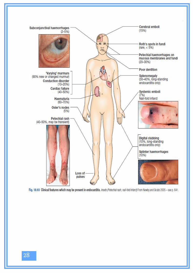
Hands: inspect for Clubbing, Splinter hemorrhages, Peripheral cyanosis, Tar stains, skin
and tendon Xanthomata, Janeway’s lesions, Osler’s nodes.
Palpate for temperature (using the dorsum of your hand) and comment if sweaty or dry.
Then examine for Fine tremor and Flapping tremor (Asterexis).
Examination of CVS

15
Face: comment on Conjunctival Pallor, Jaundice, Conjunctival Hemorrhage, Corneal Urcus,
Xanthelasma, Malar flush, Central cyanosis, Dental carries, Angular stomatitis, Glossitis.
Ask to perform fundoscopy looking for Roth’s spots or hypertensive retinopathy.
Neck: Examine for scars, visible masses, distended veins.
16
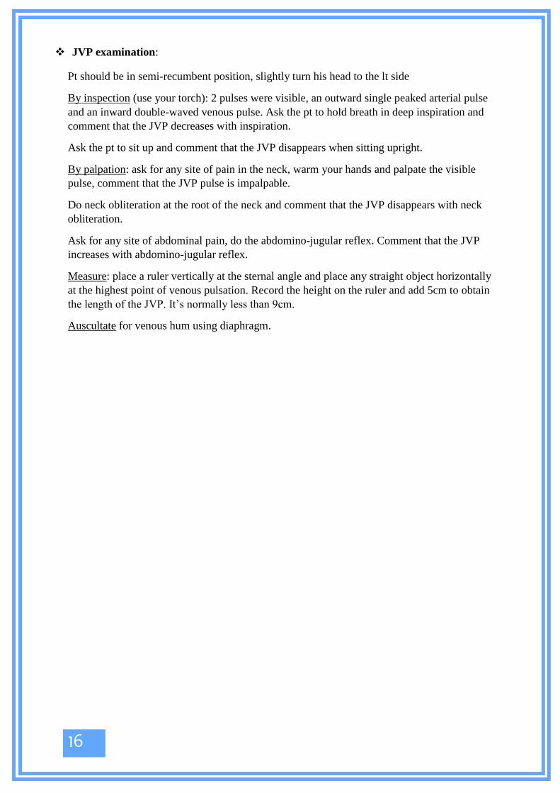
JVP examination:
Pt should be in semi-recumbent position, slightly turn his head to the lt side
By inspection (use your torch): 2 pulses were visible, an outward single peaked arterial pulse
and an inward double-waved venous pulse. Ask the pt to hold breath in deep inspiration and
comment that the JVP decreases with inspiration.
Ask the pt to sit up and comment that the JVP disappears when sitting upright.
By palpation: ask for any site of pain in the neck, warm your hands and palpate the visible
pulse, comment that the JVP pulse is impalpable.
Do neck obliteration at the root of the neck and comment that the JVP disappears with neck
obliteration.
Ask for any site of abdominal pain, do the abdomino-jugular reflex. Comment that the JVP
increases with abdomino-jugular reflex.
Measure: place a ruler vertically at the sternal angle and place any straight object horizontally
at the highest point of venous pulsation. Record the height on the ruler and add 5cm to obtain
the length of the JVP. It’s normally less than 9cm.
Auscultate for venous hum using diaphragm.

17
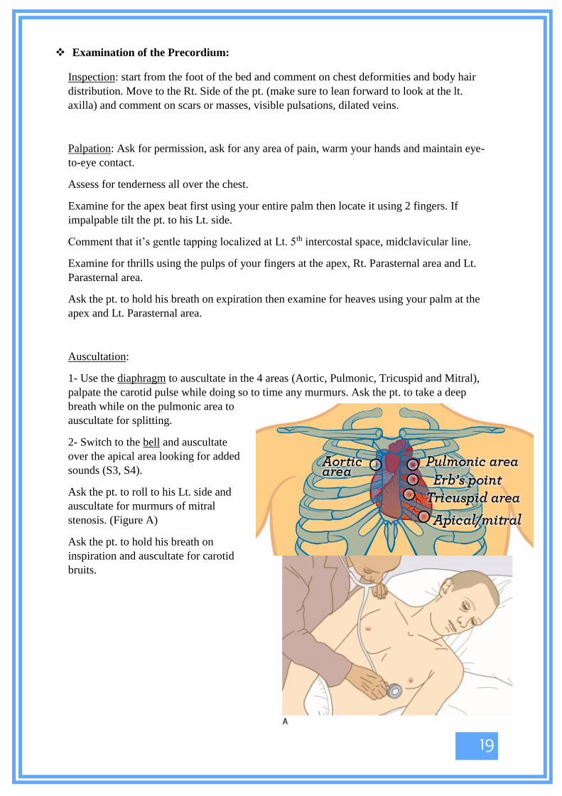
Examination of the Precordium:
Inspection: start from the foot of the bed and comment on chest deformities and body hair
distribution. Move to the Rt. Side of the pt. (make sure to lean forward to look at the lt.
axilla) and comment on scars or masses, visible pulsations, dilated veins.
Palpation: Ask for permission, ask for any area of pain, warm your hands and maintain eye-
to-eye contact.
Assess for tenderness all over the chest.
Examine for the apex beat first using your entire palm then locate it using 2 fingers. If
impalpable tilt the pt. to his Lt. side.
Comment that it’s gentle tapping localized at Lt. 5th
intercostal space, midclavicular line.
Examine for thrills using the pulps of your fingers at the apex, Rt. Parasternal area and Lt.
Parasternal area.
Ask the pt. to hold his breath on expiration then examine for heaves using your palm at the
apex and Lt. Parasternal area.
Auscultation:
1- Use the diaphragm to auscultate in the 4 areas (Aortic, Pulmonic, Tricuspid and Mitral),
palpate the carotid pulse while doing so to time any murmurs. Ask the pt. to take a deep
breath while on the pulmonic area to
auscultate for splitting.
2- Switch to the bell and auscultate
over the apical area looking for added
sounds (S3, S4).
Ask the pt. to roll to his Lt. side and
auscultate for murmurs of mitral
stenosis. (Figure A)
Ask the pt. to hold his breath on
inspiration and auscultate for carotid
bruits.

18
3- Switch to the diaphragm again and ask the pt. to sit up, lean forward and hold his breath in
full expiration. Place the stethoscope’s diaphragm on “Erb’s area” to auscultate for murmur
of aortic regurgitation. (Figure B)
While the pt. is sitting upright auscultate for
basal lung crackles.
Test for sacral edema.
Lower Limbs
Exposure should be all the way up to the umbilicus (for cultural concerns expose to the mid-
thigh).
Inspection: Look at all leg aspects for signs of ischemia:
Amputated Limb or toe, Thick hypertrophied nails, Peripheral cyanosis, Pallor, Hair loss,
Skin discoloration, Skin changes (Dry skin or Shiny skin), decreased muscle bulk, Charcot’s
joint deformity, Obvious swelling. Look at the heel and between the toes for ulcers.
Look at all leg aspects for signs of chronic venous insufficiency:
Visible distended / tortuous veins, Obvious scars, Obvious swelling, Redness or
Hyperpigmentation, Thick skin, Ulcers.
Palpation: -Feel for Temperature difference.
-Feel for palpable masses, Coarse skin, Tenderness on squeezing the muscle.
-Test for capillary refill by compressing a toenail (normally it’s restored in 2 seconds
maximum).
-Feel the pulses:
Dorsalis Pedis: Just lateral to the extensor halluces longus muscle, in the middle of the foot.
Posterior Tibial: 2cm below and 2cm behind the medial malleolus or midway between the
medial malleolus and the heel.
Popliteal: Press firmly behind the 30o flexed knee.
Femoral: Midway between the ASIS and the symphysis pubis; just below the inguinal
ligament.
-Do neurological exam for power, sensation and vibration sense.
-Check for edema by pressing using your thumb on the shaft of tibia then above the medial
malleolus.
-Measure the circumference of both legs at the same point (10cm below the tibial tuberosity).
Special tests: -When examining for PAD: Perform Buerger’s test (Next page).
-When examining for Venous disease : Perform Trendelenburg test (Next page).
Auscultation: -Auscultate over femoral arteries for bruits.

19

20
ECG interpretation:
1-STEMI
ST-elevation in leads II, III, aVF Inferior wall MI Occlusion of Rt. Coronary artery.
ST- elevation in leads I, aVL, V5, V6 Lateral wall MI Occlusion of circumflex
artery.
ST-elevation in leads V1,V2,V3,V4 Anterior wall MI Occlusion of LAD.
ST-depression in leads V1, V2 Posterior wall MI.
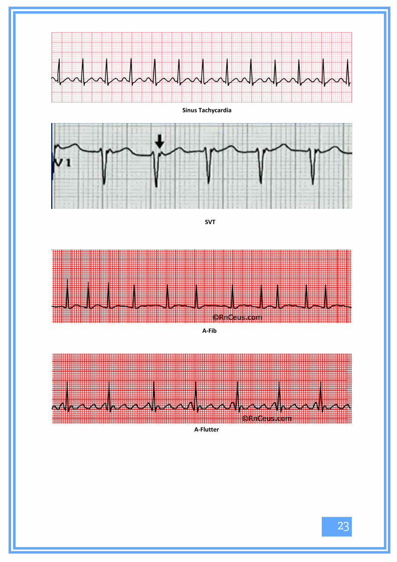
2- Tachycardia (HR >100)
Is it fast? Is it regular? QRS
(normally <3
small squares)
P wave
present?
------ Diagnosis
✔ ✔ Narrow ✔ Sinus
tachycardia
✔ ✔ Narrow X (Instead
there’s a
retrograde P
wave in V1)
SVT
✔ X Narrow X A. Fib
✔ ✔ Narrow ✔✔✔ موجودة
وبقوة
)more than one
P wave for each
QRS complex)
A. Flutter
✔ X Narrow ✔ and each P
wave has a
distinct
morphology
MAT
✔ ✔ Wide X Looks good V. Tach
✔ X Wide X Looks like a
total mess
V. Fib

21
Sinus Tachycardia
SVT
A-Fib
A-Flutter

22
MAT
V-Tach
V-Fib

23
3- Bradycardia (HR<60)
Is it
slow?
Look at the PR intervals Are there
dropped
beats?
Dx
✔ They’re of normal length (< 5 small squares) and they’re fixed X Sinus
bradycardia
✔ They’re elongated (>5 small squares) but fixed X 1st degree AV
block
✔ There’s progressive prolongation of PR intervals then a
dropped beat ✔ 2
nd degree AV
block type 1
(Wenckebach
block)
✔ PR intervals are of normal length and fixed then a dropped
beat ✔ 2
nd degree AV
block type 2
(Mobitz type)
✔ No relation between PR intervals ✔✔✔
(A LOT)
3rd
degree AV
block
(complete
heart block)

24
4- Acute Pericarditis
Diffuse ST-elevation in all leads except leads V1 and aVR (these have ST depression), with
PR depression in all leads except aVR (it has PR elevation).
5- Axis deviation
Always look at Leads I and aVF Dx
Lead I Lead aVF
+ve +ve Normal axis
-ve +ve Rt. Axis deviation
+ve -ve Lt. Axis deviation
-ve -ve Severe axis deviation mostly
to the right
Acute Pericarditis

25
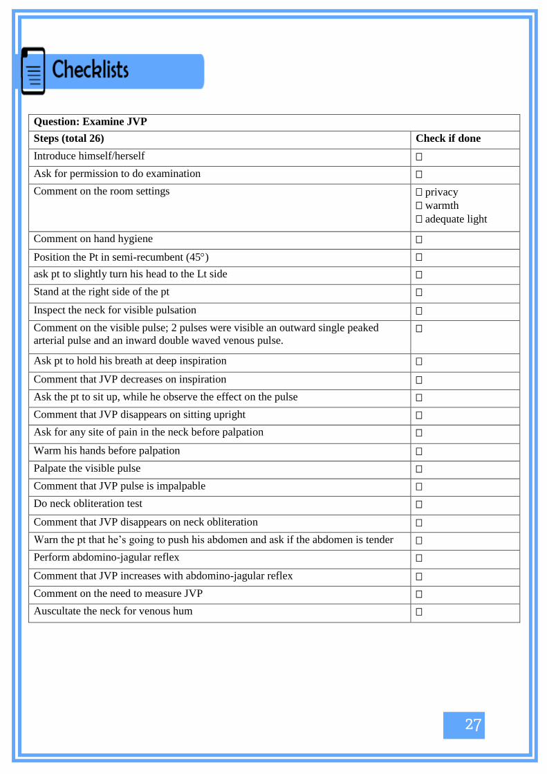
Question: Examine JVP
Steps (total 26) Check if done
Introduce himself/herself
Ask for permission to do examination
Comment on the room settings privacy
warmth
adequate light
Comment on hand hygiene
Position the Pt in semi-recumbent (45)
ask pt to slightly turn his head to the Lt side
Stand at the right side of the pt
Inspect the neck for visible pulsation
Comment on the visible pulse; 2 pulses were visible an outward single peaked
arterial pulse and an inward double waved venous pulse.
Ask pt to hold his breath at deep inspiration
Comment that JVP decreases on inspiration
Ask the pt to sit up, while he observe the effect on the pulse
Comment that JVP disappears on sitting upright
Ask for any site of pain in the neck before palpation
Warm his hands before palpation
Palpate the visible pulse
Comment that JVP pulse is impalpable
Do neck obliteration test
Comment that JVP disappears on neck obliteration
Warn the pt that he’s going to push his abdomen and ask if the abdomen is tender
Perform abdomino-jagular reflex
Comment that JVP increases with abdomino-jagular reflex
Comment on the need to measure JVP
Auscultate the neck for venous hum

26
Question: Do Inspection and Palpation of the Precordium:
Steps (total 28) Check if done
Introduce himself/herself
Ask for permission to do examination
Comment on the room settings privacy
warmth
adequate light
Comment on hand hygiene
Position the Pt in semi-recumbent (45)
Exposure of anterior chest to the umbilicus
Stand at the foot of the bed
At the foot of the bed comment on chest deformities and body hair distribution chest deformities
body hair distribution
Stand at the right side of the pt
At the right side of the pt inspect for scars
Look at the axilla at the left of the pt for scars
At the right side comment on visible pulsation (location )
Warm his hands
Ask for any tender area on the chest
Maintain eye-to-eye contact throughout palpation
Comment on chest tenderness
Locate the apex beat
Comment on the apex beat; gently tapping
location
Ask the pt to hold his breath at expiration after a deep inspiration for palpation
of heaves
Palpate for left ventricular heave at the apex using palm
Palpate for right ventricular heaves at the left sternal margin
Palpate for thrills using the pulps of your fingers at apex and both sides of
sternum at apex
at right side of sternum
at left side of sternum

27
28

29

30
List of Resources:
1- Davidson’s Principles and Practice of Medicine, 22nd
edition.
2- Macleod’s Clinical Examination, 13th
edition.
3- Macleod’s Clinical Diagnosis.
4- The Only EKG Book You’ll Ever Need, 7th
edition.
5- Hanan Mansour’s History Taking and OSCE Examination Dossier.
6- Aiman Juma’a’s OSCE Dossier.

15

16
❖ Greet your patient, introduce yourself then ask for permission to examine him.
Wash your hands and ensure adequate privacy, warmth and illumination.
Stand to the Rt. Side of the patient.
Expose the anterior chest to the umbilicus, position the patient in semi recombinant position
(45o).
❖ General examination:
First impression: The pt is conscious, alert, oriented to time, place and person.
Not in distress, no hoarseness of voice, not cyanosed, no IV lines.
Vitals: RR, Temp, BMI, BP, Pulses
Pulses:
-Radial Comment on rate, rhythm, compressibility, radio-radial asymmetry (as in
subclavian or aortic disease), ask if he has pain in his shoulder then examine for collapsing
pulse (occurs in Aortic Regurgitation And Patent Ductus Arteriosus), radio-femoral delay (as
in Coarctation of the aorta), pulse deficit (Difference between radial pulse and HR by
auscultation more than 10, occurs in A. Fib).
-Brachial Comment on rate, rhythm, volume (Small, Large, Normal),
character (Collapsing as in Aortic Regurgitation and Patent Ductus Arteriosus, Slow-Rising
as in Aortic Stenosis, Bisferens as in Concomitant Aortic Regurg. and Aortic Stenosis or
HOCM, Alternans as in Advanced Heart failure), compressibility, brachio-brachial delay.
Take BP then look for pulsus paradoxus and postural hypotension.
-Carotid NEVER assess both simultaneously, use the thumb that’s contralateral to the side
examined, comment on rate, rhythm, volume, character, compressibility.
-Lower limb pulses (Femoral, Popliteal, Posterior tibial, Dorsalis pedis) comment if
palpable or not.
❖ Hands: inspect for Clubbing, Splinter hemorrhages, Peripheral cyanosis, Tar stains, skin
and tendon Xanthomata, Janeway’s lesions, Osler’s nodes.
Palpate for temperature (using the dorsum of your hand) and comment if sweaty or dry.
Then examine for Fine tremor and Flapping tremor (Asterexis).
Examination of CVS

17
❖ Face: comment on Conjunctival Pallor, Jaundice, Conjunctival Hemorrhage, Corneal Urcus,
Xanthelasma, Malar flush, Central cyanosis, Dental carries, Angular stomatitis, Glossitis.
Ask to perform fundoscopy looking for Roth’s spots or hypertensive retinopathy.
❖ Neck: Examine for scars, visible masses, distended veins.

18
❖ JVP examination:
Pt should be in semi-recumbent position, slightly turn his head to the lt side
By inspection (use your torch): 2 pulses were visible, an outward single peaked arterial pulse
and an inward double-waved venous pulse. Ask the pt to hold breath in deep inspiration and
comment that the JVP decreases with inspiration.
Ask the pt to sit up and comment that the JVP disappears when sitting upright.
By palpation: ask for any site of pain in the neck, warm your hands and palpate the visible
pulse, comment that the JVP pulse is impalpable.
Do neck obliteration at the root of the neck and comment that the JVP disappears with neck
obliteration.
Ask for any site of abdominal pain, do the abdomino-jugular reflex. Comment that the JVP
increases with abdomino-jugular reflex.
Measure: place a ruler vertically at the sternal angle and place any straight object horizontally
at the highest point of venous pulsation. Record the height on the ruler and add 5cm to obtain
the length of the JVP. It’s normally less than 9cm.
Auscultate for venous hum using diaphragm.
19
❖ Examination of the Precordium:
Inspection: start from the foot of the bed and comment on chest deformities and body hair
distribution. Move to the Rt. Side of the pt. (make sure to lean forward to look at the lt.
axilla) and comment on scars or masses, visible pulsations, dilated veins.
Palpation: Ask for permission, ask for any area of pain, warm your hands and maintain eye-
to-eye contact.
Assess for tenderness all over the chest.
Examine for the apex beat first using your entire palm then locate it using 2 fingers. If
impalpable tilt the pt. to his Lt. side.
Comment that it’s gentle tapping localized at Lt. 5th intercostal space, midclavicular line.
Examine for thrills using the pulps of your fingers at the apex, Rt. Parasternal area and Lt.
Parasternal area.
Ask the pt. to hold his breath on expiration then examine for heaves using your palm at the
apex and Lt. Parasternal area.
Auscultation:
1- Use the diaphragm to auscultate in the 4 areas (Aortic, Pulmonic, Tricuspid and Mitral),
palpate the carotid pulse while doing so to time any murmurs. Ask the pt. to take a deep
breath while on the pulmonic area to
auscultate for splitting.
2- Switch to the bell and auscultate
over the apical area looking for added
sounds (S3, S4).
Ask the pt. to roll to his Lt. side and
auscultate for murmurs of mitral
stenosis. (Figure A)
Ask the pt. to hold his breath on
inspiration and auscultate for carotid
bruits.

20
3- Switch to the diaphragm again and ask the pt. to sit up, lean forward and hold his breath in
full expiration. Place the stethoscope’s diaphragm on “Erb’s area” to auscultate for murmur
of aortic regurgitation. (Figure B)
While the pt. is sitting upright auscultate for
basal lung crackles.
Test for sacral edema.
❖ Lower Limbs
Exposure should be all the way up to the umbilicus (for cultural concerns expose to the mid-
thigh).
Inspection: Look at all leg aspects for signs of ischemia:
Amputated Limb or toe, Thick hypertrophied nails, Peripheral cyanosis, Pallor, Hair loss,
Skin discoloration, Skin changes (Dry skin or Shiny skin), decreased muscle bulk, Charcot’s
joint deformity, Obvious swelling. Look at the heel and between the toes for ulcers.
Look at all leg aspects for signs of chronic venous insufficiency:
Visible distended / tortuous veins, Obvious scars, Obvious swelling, Redness or
Hyperpigmentation, Thick skin, Ulcers.
Palpation: -Feel for Temperature difference.
-Feel for palpable masses, Coarse skin, Tenderness on squeezing the muscle.
-Test for capillary refill by compressing a toenail (normally it’s restored in 2 seconds
maximum).
-Feel the pulses:
Dorsalis Pedis: Just lateral to the extensor halluces longus muscle, in the middle of the foot.
Posterior Tibial: 2cm below and 2cm behind the medial malleolus or midway between the
medial malleolus and the heel.
Popliteal: Press firmly behind the 30o flexed knee.
Femoral: Midway between the ASIS and the symphysis pubis; just below the inguinal
ligament.
-Do neurological exam for power, sensation and vibration sense.
-Check for edema by pressing using your thumb on the shaft of tibia then above the medial
malleolus.
-Measure the circumference of both legs at the same point (10cm below the tibial tuberosity).
Special tests: -When examining for PAD: Perform Buerger’s test (Next page).
-When examining for Venous disease : Perform Trendelenburg test (Next page).
Auscultation: -Auscultate over femoral arteries for bruits.

21

22
❖ ECG interpretation:
1-STEMI
▪ ST-elevation in leads II, III, aVF Inferior wall MI Occlusion of Rt. Coronary artery.
▪ ST- elevation in leads I, aVL, V5, V6 Lateral wall MI Occlusion of circumflex
artery.
▪ ST-elevation in leads V1,V2,V3,V4 Anterior wall MI Occlusion of LAD.
▪ ST-depression in leads V1, V2 Posterior wall MI.
2- Tachycardia (HR >100)
Is it fast? Is it regular? QRS
(normally <3
small squares)
P wave
present?
------ Diagnosis
✔ ✔ Narrow ✔ Sinus
tachycardia
✔ ✔ Narrow X (Instead
there’s a
retrograde P
wave in V1)
SVT
✔ X Narrow X A. Fib
✔ ✔ Narrow ✔✔✔ موجودة
وبقوة
)more than one
P wave for each
QRS complex)
A. Flutter
✔ X Narrow ✔ and each P
wave has a
distinct
morphology
MAT
✔ ✔ Wide X Looks good V. Tach
✔ X Wide X Looks like a
total mess
V. Fib
23
Sinus Tachycardia
SVT
A-Fib
A-Flutter

24
MAT
V-Tach
V-Fib

25
3- Bradycardia (HR<60)
Is it
slow?
Look at the PR intervals Are there
dropped
beats?
Dx
✔ They’re of normal length (< 5 small squares) and they’re fixed X Sinus
bradycardia
✔ They’re elongated (>5 small squares) but fixed X 1st degree AV
block
✔ There’s progressive prolongation of PR intervals then a
dropped beat ✔ 2nd degree AV
block type 1
(Wenckebach
block)
✔ PR intervals are of normal length and fixed then a dropped
beat ✔ 2nd degree AV
block type 2
(Mobitz type)
✔ No relation between PR intervals ✔✔✔
(A LOT)
3rd degree AV
block
(complete
heart block)

26
4- Acute Pericarditis
Diffuse ST-elevation in all leads except leads V1 and aVR (these have ST depression), with
PR depression in all leads except aVR (it has PR elevation).
5- Axis deviation
Always look at Leads I and aVF Dx
Lead I Lead aVF
+ve +ve Normal axis
-ve +ve Rt. Axis deviation
+ve -ve Lt. Axis deviation
-ve -ve Severe axis deviation mostly
to the right
Acute Pericarditis
27
Question: Examine JVP
Steps (total 26) Check if done
Introduce himself/herself
Ask for permission to do examination
Comment on the room settings privacy
warmth
adequate light
Comment on hand hygiene
Position the Pt in semi-recumbent (45)
ask pt to slightly turn his head to the Lt side
Stand at the right side of the pt
Inspect the neck for visible pulsation
Comment on the visible pulse; 2 pulses were visible an outward single peaked
arterial pulse and an inward double waved venous pulse.
Ask pt to hold his breath at deep inspiration
Comment that JVP decreases on inspiration
Ask the pt to sit up, while he observe the effect on the pulse
Comment that JVP disappears on sitting upright
Ask for any site of pain in the neck before palpation
Warm his hands before palpation
Palpate the visible pulse
Comment that JVP pulse is impalpable
Do neck obliteration test
Comment that JVP disappears on neck obliteration
Warn the pt that he’s going to push his abdomen and ask if the abdomen is tender
Perform abdomino-jagular reflex
Comment that JVP increases with abdomino-jagular reflex
Comment on the need to measure JVP
Auscultate the neck for venous hum

28
Question: Do Inspection and Palpation of the Precordium:
Steps (total 28) Check if done
Introduce himself/herself
Ask for permission to do examination
Comment on the room settings privacy
warmth
adequate light
Comment on hand hygiene
Position the Pt in semi-recumbent (45)
Exposure of anterior chest to the umbilicus
Stand at the foot of the bed
At the foot of the bed comment on chest deformities and body hair distribution chest deformities
body hair distribution
Stand at the right side of the pt
At the right side of the pt inspect for scars
Look at the axilla at the left of the pt for scars
At the right side comment on visible pulsation (location )
Warm his hands
Ask for any tender area on the chest
Maintain eye-to-eye contact throughout palpation
Comment on chest tenderness
Locate the apex beat
Comment on the apex beat; gently tapping
location
Ask the pt to hold his breath at expiration after a deep inspiration for palpation
of heaves
Palpate for left ventricular heave at the apex using palm
Palpate for right ventricular heaves at the left sternal margin
Palpate for thrills using the pulps of your fingers at apex and both sides of
sternum at apex
at right side of sternum
at left side of sternum

29

30

31

32
List of Resources:
1- Davidson’s Principles and Practice of Medicine, 22nd edition.
2- Macleod’s Clinical Examination, 13th edition.
3- Macleod’s Clinical Diagnosis.
4- The Only EKG Book You’ll Ever Need, 7th edition.
5- Hanan Mansour’s History Taking and OSCE Examination Dossier.
6- Aiman Juma’a’s OSCE Dossier.

33
Chapter 2: Respiratory System
-X year old male pt presented with a history of cough of X days duration, take a good
history and list the differential diagnosis.
Differential diagnosis:
• Acute cough (<3 weeks): foreign body aspiration, upper respiratory tract
infection (yellow sputum), Lower respiratory tract infections (pneumonia,
TB) (purulent sputum, can be blood stained).
• Chronic cough (>8 weeks):
Productive: COPD (in adult only, clear mucoid sputum, purulent if active
infection is present), TB (blood stained, where TB is endemic), bronchiectasis
(purulent sputum), pulmonary edema - HF (pink watery sputum), lung CA
(blood stained sputum), pulmonary embolism (frothy pink sputum), cystic
fibrosis (purulent sputum, in children), lung abscess (purulent sputum).
Non productive: asthma, post nasal drip, GERD, drugs (ACEI), sarcoidosis,
and neuromuscular disease.
• 3-8 weeks is called subacute or acute persistent
History
» Knock the door, introduce yourself, take permission and start taking patient profile
(age, occupation, admission details). Listen to the case carefully.
» Chief compliant: cough and Duration: to know if acute or chronic
❖ Clarify the chief complaint :
» Productive or non productive (sputum): if productive ask about :
✓ The consistency of the sputum, is it easy to evacuate or hard mass?
✓ Amount:
large volume (bronchiectasis), large volume on single occasion (lung abscess
rupture), large volume of watery sputum (pulmonary edema, if pink:
bronchioalveolar ca), mucous plug (asthma).
Cough; productive, non-productive
& hemoptysis

34
✓ Taste or smell: foul (infection, bronchiectasis, abscess).
✓ Solids: foreign body, aspergellosis
✓ color:
» Special sounds with cough (does it have a special sound?) :
bovine cough (left recurrent laryngeal paralysis or invasion),
violent repetitive cough in children (foreign body),
painful parking cough (laryngeal infection, inflammation),
» Timing: nocturnal cough (asthma), less in holidays (occupational asthma), during a
drug course (dry cough due to ACEI), cough during swallowing (neuromuscular
abnormality).
» Any preceding event: runny nose and fever/chills (infection), sore throat 2 weeks
ago then resolved (reactivation), exercise ( exercise induced asthma )
» Does it change or increase with specific conditions: recumbent posture
(COPD,GERD), temperature (bronchiectasis) , after exercise or exposure to
allergens , irritants (asthma).
» moist cough (bronchiectasis), smoker's moist cough at morning (chronic bronchitis),
paroxysmal dry cough (bronchial hyperactivity after viral infection with asthma).
» How severe is it ?! Ask if the patient experience vomiting, fatigue or dizziness after
cough?!
» Any similar pervious attack ?
» Noisy breathing:
Stridor ( infection/inflammation, e.g. acute epiglottitis, tumors of the trachea and
main bronchi, extrinsic compression by lymph nodes, anaphylaxis and foreign
body),
wheezy breathing (mainly asthma and COPD).

35
» Associated symptoms: feeling of hotness, chills ,rigor, fatigue, dyspnea, chest pain,
hemoptysis, anorexia , weight loss
Red flag symptoms: (important)
• Hemoptysis • Breathlessness • Fever • Chest pain • Weight loss
pleuritic chest pain: sharp, stabbing, non-central chest pain, increased by
inspiration and coughing, feature of PE, pneumonia, pneumothorax.
*** since it’s a respiratory case then the smoking is very important , ask about
active and passive smoking ? pack yeas ? make sure that the patient understand
that water pipes also involved in smoking?
*** if you are suspecting asthma , it’s very important to ask about family
history , atopy, allergy ?
*** if you suspect malignancy it is good to ask about metz symptoms to the
liver, brain , bone and adrenal .also try to ask about paraneoplastic syndrome
symptoms such as hypercalcemia symptoms ( bones, stones, groans and mental
overtone ) , ask about hyponatremia symptoms due to SIADH , Cushing
syndrome ,,,etc.
❖ Review of system
» Review the systems focusing on GI
symptoms : abdominal pain (can be seen in
lower lobe pneumonia), nausea, vomiting,
diarrhea, anorexia..
❖ Past medical and surgical history
» Previous similar attack (asthmatic attacks) ,
other lung disease, heart diseases or heart
failure (pulmonary HTN), allergy, GERD,
chronic sinusitis (bronchiectasis), DVT with
PE.
❖ Drug history
» ACEI, NSAIDs, B-blockers…
❖ Family history
» Asthma, eczema and hay fever (atopy),
Cystic fibrosis is (AR), α1- antitrypsin
deficiency (AR), other lung diseases in the
family.

36
❖ Social history
» Smoking with pack years (COPD), alcohol (neuromuscular), occupation (occupational
asthma), recent travel (TB), pets (allergy, asthma).
❖ If the pt has hemoptysis as chief compliant (not as a color of sputum only),ask
about the following:
✓ Make sure that it is hemoptysis not hematemesis?!
✓ Is the blood fresh or mixed with sputum ?!
✓ Amount and appearance: large (lung CA, bronchiectasis, TB), blood clots (lung ca)
✓ Frequency: intermittent (bronchiectasis), daily for a week (CA,TB, lung, abscess),
sudden single episode (PE and infarction) associated with hematuria (Goodpasture’s syndrome, Wegener's granulomatosis
✓ Ask the patient if there’s bleeding elsewhere from other body orifices (bleeding
tendency), if he’s taking any antiplatelet or anticoagulant, or if there’s family history
of recurrent bleeding.
Introduce yourself, take permission, insure privacy, warmth and light, make sure of
good exposure and position >>>>The patient must be semi sitting and exposure from
the neck to the umbilicus.
➢ General look :
• The patient is conscious, alert and oriented to time, place and person.
• The patient is lying/sitting/semi sitting.
• Oxygen mask or ventilator.
• Comfortable or distressed.
• Using accessory muscles for respiration (sternocleidomastoid, platysma and
trapezius Muscles) .
• Audible sounds (wheezes, stridor…) Ask the patient to cough and then
breathe deeply in and out with the mouth wide open.
• Cyanosis of the lips and underside of the tongue.
• Obese, thin, catechetic ?
➢ Vital signs (HR, BP, respiratory rate, temperature, O2 sat and BMI)
• Diastolic pressure of <60 mmHg is associated with increased mortality in
community-acquired pneumonia
• In pneumothorax, hypotension may indicate the development of ‘tension’ with
reduction in venous return to the heart and risk of cardiac arrest.

37
• Pulsus paradoxus: a fall in diastolic blood pressure of more than 10mmHg can
occur in cardiac tamponade and asthma .
➢ Hands
• Hot/cold.
• Sweaty/dry. • Palmar erythema (indicates CO2 retention as in COPD,
cirrhosis, hyperthyroidism and polycythemia).
• Cyanosis.
• Tar stains on fingers and nails, Nicotine stains on dominant
index and middle finger. • Fine tremor (seen in patients taking 𝛽-agonist or
theophylline bronchodilator inhalers).
• Asterixis: Ask the patient to hyperextend his wrists
and abduct his fingers for 30 seconds and observe for
flapping tremor. Seen in cases of CO2 retention and
severe ventilator failure in end organ damage.
Unilateral asterixis is due to a structural abnormality in
the contralateral cerebral hemisphere. Patients with a
flapping tremor cannot maintain their grip.
• Clubbing:
It rarely develops quickly over several weeks with
empyema.
• If tenderness of the wrist is also present, suspect
hypertrophic pulmonary osteoarthropathy (squamous cell
cancer).
• Yellow-nail syndrome is associated with lymphedema
and an exudative pleural effusion.
•
➢ Face
• Sclera and conjunctiva for pallor and jaundice.
• Jaundice is first seen in the mucosa beneath the tongue;
it results from a
congested liver failure due to a chronic pulmonary
disease.
• Pallor is seen from anemia.
• Ptosis ,Horner’s syndrome in Pancoast lung cancer.

38
• Tongue, mouth ulcers and dental hygiene.
➢ Neck
• Scars.
• Vein engorgements.
• Lymph nodes (a palpable supraclavicular node strongly
suggests metastatic spread of lung cancer; localized cervical
lymphadenopathy is a common presenting feature of
lymphoma).
• Pamperton’s sign >>> ask the patient to raise both hands and
observe for Facial flushing, distension of neck veins and
stridor ; positive in SVC obstruction
• JVP.
➢ Chest
➢ Inspection
• When you inspect the posterior thorax, make sure to ask the patient to sit up
and cross his hands.
• Symmetrical, bilateral, abdominothoracic/thoracoabdominal breathing.
• Check for paradoxical breathing (abdomen moves inward; seen in bilateral
phrenic nerve damage or severe COPD).
• Scars for surgeries (make sure you ask the patient to lift his arms to check the
sides).
• Swellings, dilated superficial veins and subcutaneous nodules.
• Antero-posterior: transverse diameter ratio (normally 5:8).
• Check for deformities:
✓ Barrel-shaped hyperinflated chest with
intercostal indrawing in COPD patients.
✓ Kyphoscoliosis (can be due to childhood
poliomyelitis or spinal TB) causes CO2
retention and cor pulmonale.
✓ Pectus carinatum (pigeon chest) with
prominent Harrison’s sulci can be caused
by uncontrolled childhood asthma,
osteomalacia or rickets.
✓ Pectus excavatum (funnel chest) is
usually asymptomatic but in severe cases,
it may cause left heart displacement and
reduced ventilator capacity.
➢ Palpation
• Make sure you warm your hands and palpate with your hands horizontally in a
continuous pattern .
• Ask the patient if he is feeling any pain; start from the area furthest from the
pain and gently palpate it at the end.
• Superficial palpation for tenderness (maintain eye contact to observe any
discomfort or pain), subcutaneous emphysema and superficial masses
(lipomas).
39
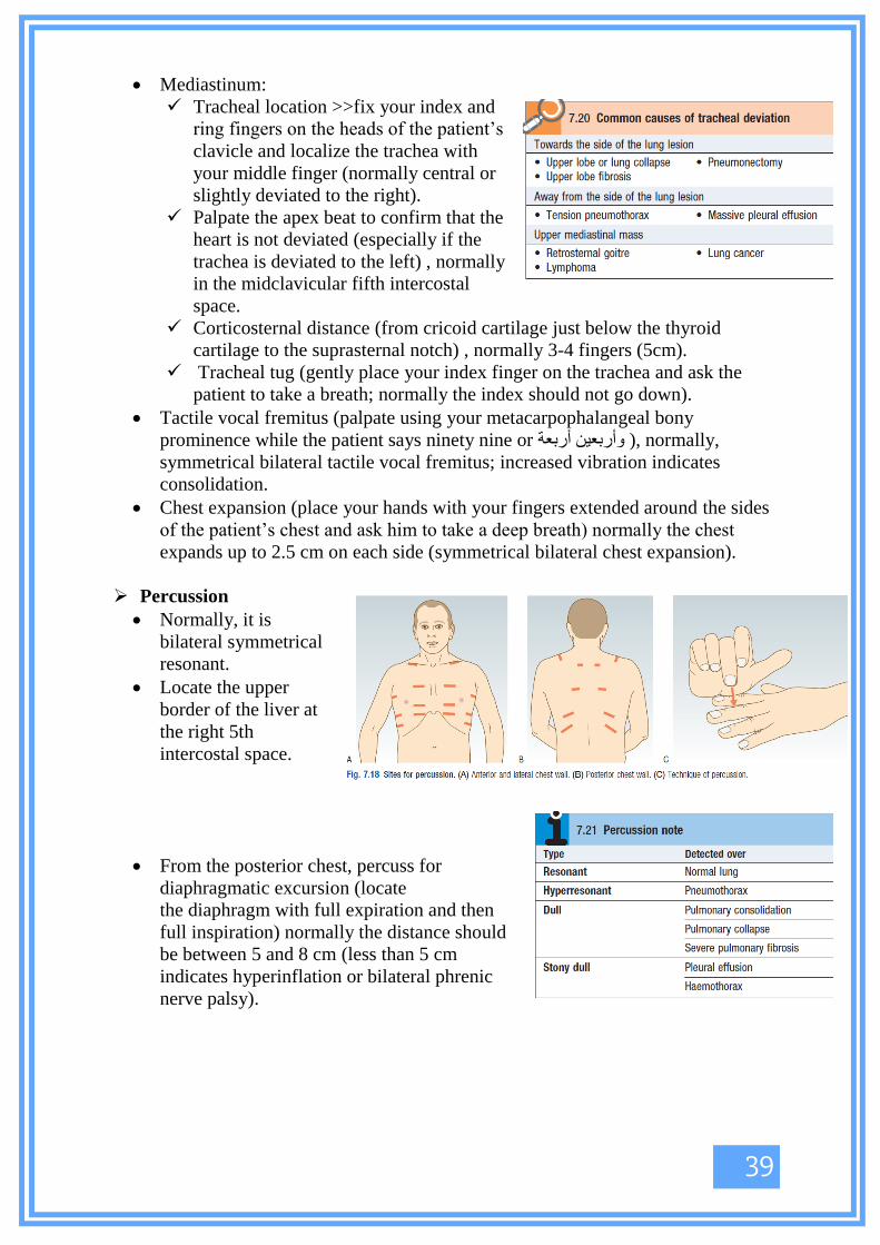
• Mediastinum:
✓ Tracheal location >>fix your index and
ring fingers on the heads of the patient’s
clavicle and localize the trachea with
your middle finger (normally central or
slightly deviated to the right).
✓ Palpate the apex beat to confirm that the
heart is not deviated (especially if the
trachea is deviated to the left) , normally
in the midclavicular fifth intercostal
space.
✓ Corticosternal distance (from cricoid cartilage just below the thyroid
cartilage to the suprasternal notch) , normally 3-4 fingers (5cm).
✓ Tracheal tug (gently place your index finger on the trachea and ask the
patient to take a breath; normally the index should not go down).
• Tactile vocal fremitus (palpate using your metacarpophalangeal bony
prominence while the patient says ninety nine or وأربعين أربعة ), normally,
symmetrical bilateral tactile vocal fremitus; increased vibration indicates
consolidation.
• Chest expansion (place your hands with your fingers extended around the sides
of the patient’s chest and ask him to take a deep breath) normally the chest
expands up to 2.5 cm on each side (symmetrical bilateral chest expansion).
➢ Percussion
• Normally, it is
bilateral symmetrical
resonant.
• Locate the upper
border of the liver at
the right 5th
intercostal space.
• From the posterior chest, percuss for
diaphragmatic excursion (locate
the diaphragm with full expiration and then
full inspiration) normally the distance should
be between 5 and 8 cm (less than 5 cm
indicates hyperinflation or bilateral phrenic
nerve palsy).

40
➢ Auscultation
• Good or diminished air entry.
• Symmetrical bilateral vesicular breathing (abnormally can be bronchial in cases
of lung consolidation, localized pulmonary fibrosis, non-occluding collapsed
lung and at the top of a pleural effusion).
• Do not auscultate the clavicle.
• Added sounds (wheeze, crepitation, pleural rub, crackling and pneumothorax
click) ask the patient to clear his throat by cough as crackling decreases after
that in bronchiectasis
• Vocal resonance (ask the patient to say ninety nine or وأربعين أربعة ) normally, it
is symmetrical bilateral vocal resonance; the numbers are only clearly audible
in cases of consolidation.
• Whispering pectoriloquy (higher
sounds indicate consolidation).
• Egophony (ask the patient to say the
letter ‘e’, normally it is heard as an
‘e’; in pneumonia, the letter ‘a’ is
heard).
➢ Other
• Liver.
• Ascites.
• Pitting edema (can result from cor
pulmonale).
• DVT.
• Erythema nodosum (present in
sarcoidosis, SLE and TB).
• Non-tender subcutaneous nodules
may occur in patients with
disseminated cancer.
41
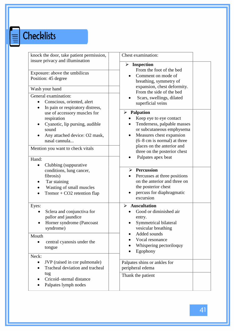
knock the door, take patient permission,
insure privacy and illumination
Chest examination:
➢ Inspection
From the foot of the bed
• Comment on mode of
breathing, symmetry of
expansion, chest deformity.
From the side of the bed
• Scars, swellings, dilated
superficial veins
Exposure: above the umbilicus
Position: 45 degree
Wash your hand
General examination:
• Conscious, oriented, alert
• In pain or respiratory distress,
use of accessory muscles for
respiration
• Cyanotic, lip pursing, audible
sound
• Any attached device: O2 mask,
nasal cannula...
➢ Palpation
• Keep eye to eye contact
• Tenderness, palpable masses
or subcutaneous emphysema
• Measures chest expansion
(6–8 cm is normal) at three
places on the anterior and
three on the posterior chest
• Palpates apex beat
Mention you want to check vitals
Hand: • Clubbing (suppurative
conditions, lung cancer,
fibrosis)
• Tar staining
• Wasting of small muscles
• Tremor + CO2 retention flap
➢ Percussion
• Percusses at three positions
on the anterior and three on
the posterior chest
• percuss for diaphragmatic
excursion
Eyes:
• Sclera and conjunctiva for
pallor and jaundice
• Horner syndrome (Pancoast
syndrome)
➢ Auscultation
• Good or diminished air
entry.
• Symmetrical bilateral
vesicular breathing
• Added sounds
• Vocal resonance
• Whispering pectoriloquy
• Egophony
Mouth
• central cyanosis under the
tongue
Neck:
• JVP (raised in cor pulmonale)
• Tracheal deviation and tracheal
tug
• Cricoid–sternal distance
• Palpates lymph nodes
Palpates shins or ankles for
peripheral edema
Thank the patient

42
A 50-year-old gentle man presented with SOB m productive cough with audible
breathing sounds he is smoker (45 pack year). Give differential diagnosis. Examine
him looking for signs of COPD.
➢ General look :
• Conscious alert oriented ( if the patient came with co2 retention he may be
drossy )
• Posture of the patient (If the
patient sits forward with the hands/arms
on the thighs or knees to ‘fix’ the
shoulder girdle, he raises the clavicles
and upper chest, increasing lung volume
and negative intrathoracic pressure then
think of emphysema )
• Thin or obese ( thin
>>emphysema , obese >>
chronic bronchitis)
• Cyanosis ( central under the
tongue and peripheral lips ,
conjunctiva ) >> ( blue
colored patients >> chronic bronchitis , pink colored patient >>> emphysema )
• Level of consciousness as mentioned before .
• Tachypnea and Use of accessory muscles mainly the sternocleidomastoid .
• Pursed lips .
• Use of extra equipments : o2 support, inhalers .
• Note any unusual noisy breathing sounds
➢ Vital signs :
• RR ( > 25 tachypnea , > 35 respiratory distress )
• HR ( could be tachycardia )
• temperature .
• o2 sat ( patient hypoxic )
• blood pressure .
COPD

43
➢ hands :
• tarry stain , NO clubbing if it’s found then think of lung malignancy , familial
or another cause ( COPD by itself DONOT cause Clubbing !)
• palmar erythema in co2 retention in COPD
• flapping tremor ( Co2 toxicity ) .
➢ neck :
• use of accessory muscles ,distended vessels , JVP ( elevated then think for Core
Pulmonale ) , look for tracheal tug ( because of hyperinflation ) ,
➢ chest :
✓ inspection : breathing pattern , barrel chest ( anteroposterior : transverse
>5:7) with intercostal indrawing in COPD patients.
• palpation: the chest expansion may be reduced .
• percussion : hyperresonance chest .decrease the dullness of the liver and heart
.
• Auscultation : Diminished air entry , vesicular breathing , prolonged
expiratory time , added sounds ( end _expiratory wheeze ,coarse crackles with
inspiration due to sputum accumulation if found ) .
➢ Then mention that you are going to look for sign of cor pulmonale : Symptoms
of fluid overload : ankle swelling , ascites , increase the JVP . hepatomegaly,
Parasternal heave , loud P2 +/- tricuspid incompetency murmurs .

44
A 20-year-old gentle man presented with SOB ,chest tightness, cough with audible
breathing sounds , nonsmoker , the patient mentioned that the symptoms become
worse at night specially 4:00 am and with exercise . give differential diagnosis .
Examine him looking for signs of Asthma.
➢ General look:
• Conscious alert oriented to place, time and people (severe cases patient >> agitation /
drowsiness).
• Cyanosis (central under the tongue and peripheral lips, conjunctiva) >> central
cyanosis in severe cases .
• Tachypnea and use of accessory muscles mainly the sternocleidomastoid .
• Use of extra equipments : o2 support, inhalers.
• Note any unusual noisy breathing sounds
• If there is any skin rash and eczema .
• Look for any sign of acute attack :
diaphoresis , tachypnea, wheezing, speaking in incomplete sentences , use accessory
muscles . paradoxical movement of the chest and abdomen during the inspiration .
hemodynamically instability .
➢ Vital signs :
• RR ( > 25 tachypnea , > 30 respiratory distress )
• HR ( could be tachycardia )
• temperature .
• o2 sat ( patient may be hypoxic )
• blood pressure ( may be hypotensive and pulsus paradoxus ) .
➢ hands :
• no specific findings .but you have to inspect looking for any sign of allergy such as
eczema
➢ neck :
• tracheal tug >> hyperinflation , may have elevated JVP , no other specific findings .
➢ chest :
➢ inspection :
✓ breathing pattern ,
✓ Pectus carinatum (pigeon chest) with prominent Harrison’s sulci can be caused by
uncontrolled childhood asthma, osteomalacia or rickets.
➢ palpation: the chest expansion may be reduced .
➢ percussion : hyperresonance chest .decrease the dullness of the lover and heart
➢ Auscultation : Diminished air entry , vesicular breathing , prolonged expiratory time
, added sounds ( end _expiratory wheeze) .
• Mention that you are going for signs of atopy : eczema, rhinitis and nasal polyps.
Asthma

45
Pneumonia pt will present with acute cough as a chief complain.
Pneumonia syndrome could be typical vs Atypical.
Typical (S. pneumonia , H-
influenza , G-ve )
Atypical ( mycoplasma , chlamydia
m viruses )
Sudden onset
High grade fever >39 wit chills
Productive cough with green /
yellow sputum - purulent (could
be dry at first) .
Toxic looking pt
Loss of appetite
Pleuritic chest pain
SOB
( plural effusion )
Gradual ( headache , sore throat ,
fatigue, myalgia )
Low grade fever
Dry cough
Non- toxic look
Good appetite
On physical exam:
tachycardia , tachypnea , late
inspiratory crackles , bronchial
breathing , increased TVF ,
plural friction rub , dullness
Pulse-temperature dissociation (
normal pulse despite fever ) ,
wheeze , crackles
On X-RAY :
Lobar consolidation
Diffuse bilateral infiltrates with
minimal consolidation
Pneumonia

46
IN history ask about :
Risk factor : ( for both types )
* weight loss
* DM , chronic liver disease, lung diseases
* current smoker ( more than 1 pack –year )
* previous Respiratory infections
* hospital stay
*increased risk of aspiration (stroke , epilepsy, surgeries )
* malignancy > chemotherapy > neutropenia > infection .
Social history : travel history , vaccination
For investigation :
CBC
CXR
Gram stain and sputum culture
Blood culture
serology not useful in acute case .
Note :
pt could have upper abdominal pain m if the pneumonia in the lower lobe .
47
A 40-year-old gentle man presented with excessive day time sleepiness and recurrent
airway obstruction, take full history.
❖ Patient profile: M>F, middle aged - usually overweight.
❖ Diagnostic approach to OSA
history
✓ Symptoms while sleeping (seek for witness)
Snoring [partial airway obstruction], apnea [complete airway obstruction],
arousal or chocking [arousal is seen on EEG or the pt move his limbs, while in
choking there is full awakening], choking episode is described as snoring
pausing grunting noise snoring again, all in addition to Nocturia.
✓ Symptoms in the morning
Headache at morning , Unrefreshing sleep, restless sleep.
✓ Symptoms during the day
Impaired cognitive function ( concentration and memory ) ,
irritability/personality change, decreased libido, excessive daytime sleepiness
[due to sleep fragmentation].
Increased risk for RTA
Past medical and surgical history
» Can be associated with nasal polyps, acromegaly and hypothyroidism.
» Increased risk for DM, systemic and pulmonary HTN, IHD, cardiac arrhythmia and
strokes.
Drugs, alcohol
Physical exam
Perform full physical examination focusing on the following:
✓ Neck : JVP examination, thyroid exam (pt with hypothyroidism can develop
OSA), neck circumference, cricosternal distance
✓ Mouth: mallampati classification
Obstructive sleep apnea (OSA)

48
✓ Chest:
Sings of hyperinflation:
* On inspection: barrel chest, intercostal retraction and paradoxical breathing
(if sever hyperinflation).
* On palpation: increased AP: transverse diameter (more than 5:7),
symmetrical decreased chest movement
* Hyper resonant percussion note
* On auscultation: Decreased air entry, prolonged expiratory phase, wheezes,
loud A2 (if associated with HTN).
Sings of pulmonary HTN: (don’t forget the increased risk of pulmonary HTN in
these pts)
* Left parasternal heave (due to RT ventricular hypertrophy)
* Wide S2 splitting, pansystolic murmur in case of tricuspid regurgitation.
Diagnosis
Polysomnography (overnight sleep study) confirms the diagnosis.

49

50

51

52
❖ Always make sure of the patient's profile, date and time of x-ray.
❖ Check the technique by assessing the following
» Positioning: AP vs PA (scapula is out of the field in PA)
» Adequacy : both shoulders , diagram and upper abdomen should be
included in the CXR.
» Inspiration: pt should take deep breath, if there is adequate
inspiratory effort at least 5-6 Ant. Ribs above the diaphragm AND 8-
10 Post. Ribs should be viewed.
» Penetration: intervertebral spaces above and below the heart should
be radiolucent, and the outline of vertebra behind the heart should be
identified.
» Rotation:
check if the CXR is centralized equal distance between medial end
of clavicles and central line (midline).
» Orientation:
verify right and left sides (gastric bubble should be on the left side).
❖ Start Interpretation of the CXR, use any systemic approach you like, here we
will discuss ABCDEF approach:
» Airway (trachea): Midline vs. deviated or rotated, foreign body in
trachea, endotracheal tube position.
» Bones (clavicles, ribs, humeri, etc): move with the external border of
the bone, any steepness indicate fracture.
» Cardiomediastinal silhouette: check cardiac size (more than 50% of
the transverse diameter of the thorax is cardiomegaly)
» Diaphragms and the costophrenic angles: put in your mind that RT
diaphragm is slightly higher than left diaphragm.
» Expanded lungs (lung fields, soft tissues)
» Foreign bodies(tubes, lines, wires, devices, etc)
❖ Lung is the most important part to comment on, compare Rt & Lt, look for:
» Pulmonary infiltrates or consolidation: pneumonia
» Cavitations: TB, lung CA especially squamous cell carcinoma
» Dilated airways: bronchiectasis
» Hyperinflation and emphysema: COPD
How to read chest x-ray

53
» Line demarcating a hyperlucent space: pnemothorax
» Prominent pulmonary vessels >>> pulmonary HTN
» Blunted costophrenic angel: plural effusion
» If you found cardiomegaly with prominent pulm. vessels: Pulm. HTN
& CHF

54
Chapter 3: Gastroenterology and
Hepatology
Whenever you take a history, try to divide it into the following parts:
1- History of Present Illness:
a) General questions that should be asked to all patients with a certain symptom.
b) Asking relevant questions that will either support or refute a certain differential
diagnosis.
2- Past medical history and medications:
Ask about previous diseases, drugs or risk factors that might be associated with the
differential diagnosis.
3- Social history and family history.
So, in the history station you have to bear these things in mind:
- Be wide! Don’t focus your questions on one differential diagnosis. Even uncommon
entities causing the symptom in the question are listed in the checklist of the examiner.
- Know the system involved in the station. If you get lost, ask about all symptoms, risk
factors, or any associations related to that system.
- Don’t memorize checklists or questions. Try to understand the differential diagnoses
and formulate your own way of approaching symptoms.
Don’t study history taking disease by disease, study it chief complaint by chief complaint.
General Considerations

55
OSCE Vignette: A 56-year-old man presented with difficulty swallowing. He is losing
weight because he is unable to eat. Obtain a detailed history trying to know the cause of his
swallowing problem.
----------------------------------------------------------------------------------------------------------------
Dysphagia is difficulty swallowing. The most important thing in the history is to know whether
it is due to oropharyngeal or esophageal problem, and to know whether it comes with solids,
liquids or both.
General Questions:
- Always ask for clarification (Do not confuse dysphagia with early satiety, the inability
to complete a full meal because of premature fullness, or globus, the feeling of a lump
in the throat. Globus does not interfere with swallowing and is not related to eating).
- Dysphagia is an alarming GI symptom, so it always indicates a serious pathology.
Always investigate it.
- Dysphagia is either (1) Oropharyngeal: bulbar palsy, pseudobulbar palsy, myasthenia
gravis, and pharyngeal pouch) or (2) Esophageal: esophageal dysphagia can be (a)
structural (i.e. dysphagia due to complete or partial occlusion of the esophageal lumen)
or (b) dysmotility (i.e. impairment of peristalsis).
- How to differentiate between these types of dysphagia based on the history?
• Was there difficulty swallowing solids, liquids or both?
Only liquids: Neurological If it’s neurological, ask about diplopia, tremor,
and weakness.
Both: Motility disorder (e.g achalasia, CNS, or pharyngeal causes).
Solids then liquids: suspect a stricture (benign or malignant).
• Is it difficult to initiate a swallowing movement?
Yes: Suspect bulbar palsy, especially if patient coughs on swallowing.
• Is swallowing painful (odynophagia)?
Yes: Suspect ulceration (malignancy, esophagitis, viral infection or Candida in
immunocompromised, or poor steroid inhaler technique) or spasm.
• Is the dysphagia intermittent or is it constant and getting worse?
Dysphagia

56
Intermittent: suspect esophageal spasm.
Constant and worsening: suspect malignant stricture.
• Does the neck bulge or gurgle on drinking?
Yes: Suspect a pharyngeal pouch.
• Ask about red flag symptoms:
1- Weight loss 2- Loss of appetite
3- Hematemesis, melaena
4- Progressive and persistent
• Past medical history:
GERD, PUD, scleroderma, iron deficiency.
• Medications:
Taking pills without water (erosive
esophagitis), use of antacids (related to GERD
and PUD).
• Social history:
Smoking, alcohol intake (risk factors of
esophageal cancer and GERD).
• Family history:
Family history of esophageal cancer,
neuromuscular diseases.
Solids and liquids to the same degree
Oropharyngeal
Dysmotility
Liquids more than
solids
• Systemic Sclerosis
Solids and Liquids
Structural

57
Introduces himself, gains consent
Ask for clarification
Onset (how it started):
• Sudden • Gradual
• Character: • Fluids • Solids or both
Time:
Duration
• Intermittent, continuous, progressive
Level: where does food/liquid feel like it is getting ‘stuck’
• Exacerbating or relieving factors
Define at which stage the dysphagia occurs:
When initiating swallowing
• After swallowing has been initiated
• Pain (esophageal/abdominal), odynophagia
• Trauma, foreign body
• Feeling of a lump in the throat
• Previous episodes of dysphagia
• Family members/contacts with similar
‘Red flags’:
Weight loss
Loss of appetite
Hematemesis, melaena
Progressive and persistent
Past medical history: • Stroke • Thyroid problems • PUD and GERD
Family history:
• Upper gastrointestinal tract cancer
Drug history: • NSAIDs • Bisphosphonates
Social history: • Alcohol (peptic ulcer disease, gastritis) • Smoking
• Illicit drug use • Diet: spicy foods ( peptic ulcer disease )

58
Differential diagnosis of abdominal pain:
Acute abdominal pain (this is a pure surgical topic):
• History of Present Illness:
- Ask about SOCRATES of pain.
- Associated symptoms: (this depends on the type of abdominal pain described by
SOCRATES).
❖ Gastrointestinal/colorectal symptoms:
• Nausea/vomiting • Bowel habit, diarrhoea/constipation • Flatulence
• Fevers • Dysphagia • Dyspepsia
• Bloating/abdominal swelling (generalized/localized)
• IBD symptoms: arthralgia, eye symptoms, skin features, oral ulcers, bloody diarrhea.
❖ Liver/hepatic symptoms:
• Right upper quadrant pain • Jaundice • Ankle swelling
Abdominal Pain
Differential diagnosis of acute abdominal pain based on the site

59
❖ Gallstone symptoms:
• Jaundice • Right upper quadrant pain radiating
to shoulders
• Dark stools • Pale urine
❖ Renal symptoms:
• Location and character:
• Loin to groin + flank + colicky: renal stones
• Flank + burning dysuria: pyelonephritis
• Generalized lethargy
• Pruritus • Ankle swelling
If the patient is female:
❖ Gynecological symptoms
• Correlation with menstrual periods
• Menorrhagia • Irregular periods
• Vaginal discharge
❖ Obstetric symptoms
• Possibility of patient being pregnant
• Last menstrual period • Contraception •
Vaginal bleeding (with severe abdominal pain = ectopic
pregnancy until proven otherwise).

60
Introduces himself, gains consent
Ask for clarification
History of Present Illness:
• Site
• Onset (how it started): - Sudden - Gradual
• Character: - Colicky (renal stones) - Sharp/sudden (rupture of viscus) - Burning
(peptic ulcer disease) - Dull
• Radiation: - To back (abdominal aortic aneurysm, ruptured duodenal ulcer)
- To testicles/groin (hernia) - To shoulders (gallbladder) - Loin to groin
(renal stone)
• Time: Duration/ Intermittent, continuous, progressive.
• Alleviating factors: Dietary factors, relieved by bowel motion?, relieved by a
certain position (lying on one side, or leaning forward), relieved by abstinence
from food?.
• Exacerbating factors: Dietary factors, increased by swallowing?
(esophagus/stomach), fatty foods (gallstones), acidic/spicy foods, hot drinks
(peptic ulcer disease).
- Does it increase by movement or breathing?
• Severity (from 0-10)
Associated symptoms: As described above.
Red Flags of acute abdominal pain: Weight loss, bleeding (upper GI bleed or lower
GI bleed), loss of appetite.
Past medical and surgical history:
• Previous surgeries
• Hepatitis, or history of blood transfusions
• IBD, IBS.
Social history: Smoking, alcohol intake
Medications: NSAIDs, antacids, use of laxatives.
Family history: Ask about relevant conditions related to the history (IBD, colon
cancer … etc.).
61
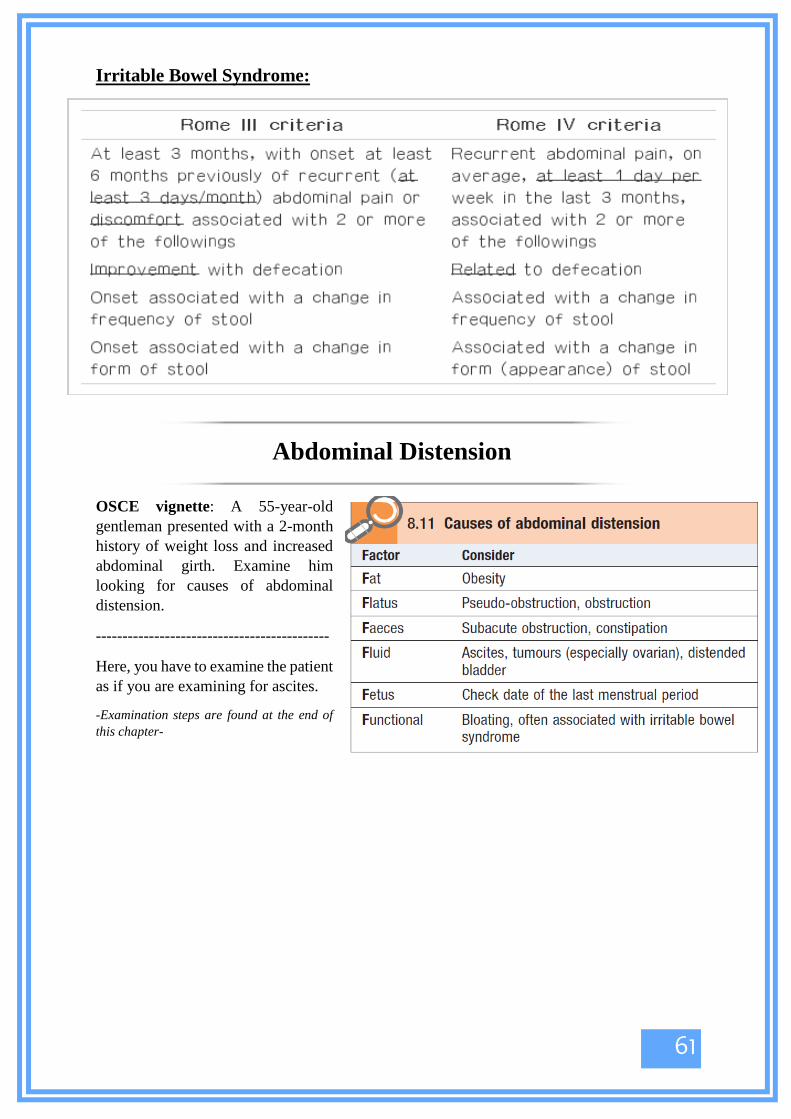
Irritable Bowel Syndrome:
OSCE vignette: A 55-year-old
gentleman presented with a 2-month
history of weight loss and increased
abdominal girth. Examine him
looking for causes of abdominal
distension.
--------------------------------------------
Here, you have to examine the patient
as if you are examining for ascites.
-Examination steps are found at the end of
this chapter-
Abdominal Distension
62
- Hematemesis is vomiting of blood from the GI tract. It indicates upper GI bleed.
- Upper GI bleed could be:
a) Active bleeding: presents with bright red blood or clots.
b) Modest bleeding or bleeding that has ceased: presents with coffee-ground blood
(blood gets degraded by gastric pepsin and becomes dark and granular just like
coffee).
Ask about the character of bleeding to know whether it’s active or old.
- Upper GI bleed is any bleeding that occurs above the ligament of Treitz (above
the duodenojejunal junction). So, blood may come from:
1- Esophagus: esophageal varices, Mallory-Weiss tear, esophagitis.
2- Stomach: PUD, gastritis, gastric cancer.
3- Duodenum: duodenitis.
- Each of these differentials has a characteristic history that you should be familiar
with;
• PUD: Usually presents with a history of epigastric pain and hematemesis. There is an
association with H. pylori, NSAIDs, steroids and alcohol.
• Mallory-Weiss tear: Presents with hematemesis that was preceded by forceful retching
with non-bloody vomit.
• Cancer: Associated with weight loss and generalized weakness and fatigue.
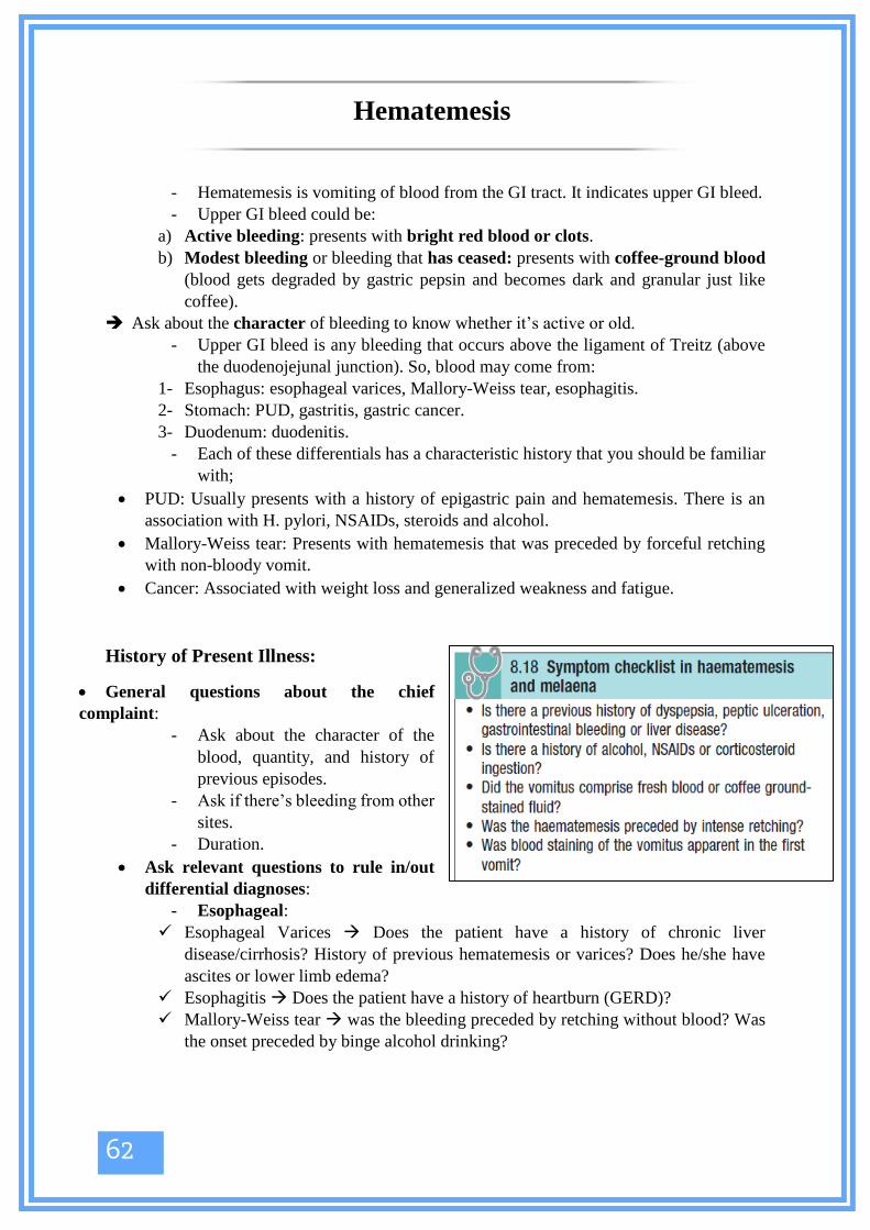
History of Present Illness:
• General questions about the chief
complaint:
- Ask about the character of the
blood, quantity, and history of
previous episodes.
- Ask if there’s bleeding from other
sites.
- Duration.
• Ask relevant questions to rule in/out
differential diagnoses:
- Esophageal:
✓ Esophageal Varices Does the patient have a history of chronic liver
disease/cirrhosis? History of previous hematemesis or varices? Does he/she have
ascites or lower limb edema?
✓ Esophagitis Does the patient have a history of heartburn (GERD)?
✓ Mallory-Weiss tear was the bleeding preceded by retching without blood? Was
the onset preceded by binge alcohol drinking?
Hematemesis
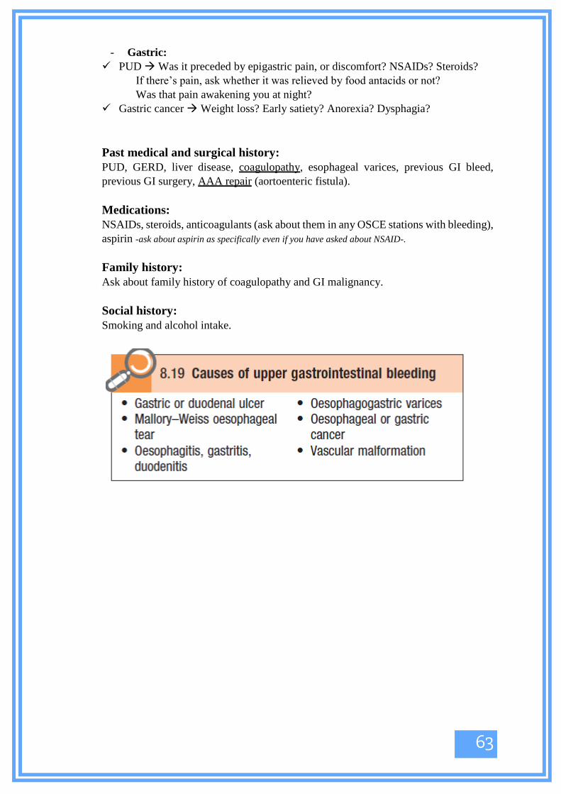
63
- Gastric:
✓ PUD Was it preceded by epigastric pain, or discomfort? NSAIDs? Steroids?
If there’s pain, ask whether it was relieved by food antacids or not?
Was that pain awakening you at night?
✓ Gastric cancer Weight loss? Early satiety? Anorexia? Dysphagia?
Past medical and surgical history:
PUD, GERD, liver disease, coagulopathy, esophageal varices, previous GI bleed,
previous GI surgery, AAA repair (aortoenteric fistula).
Medications:
NSAIDs, steroids, anticoagulants (ask about them in any OSCE stations with bleeding),
aspirin -ask about aspirin as specifically even if you have asked about NSAID-.
Family history:
Ask about family history of coagulopathy and GI malignancy.
Social history:
Smoking and alcohol intake.

64
Introduces himself, gains consent
Asks for clarification (distinguishes hematemesis from hemoptysis)
• Details about the chief complaint:
1- Volume: If large volume, patient needs to be assessed and
resuscitated immediately.
2- Number of episodes
3- Character/color
- Coffee grounds
- Dark clots
- Fresh, bright red
- Mixed with vomitus
4- Onset (what brought it on):
- Medications, alcohol
- Vomiting/retching (Mallory–Weiss tear)
- Sudden -esophageal varices-
5- Precipitating factors:
- Alcohol → Mallory-Weiss tear
- NSAIDs
- Trauma to abdomen
6- Previous episodes of hematemesis
• Associated symptoms:
- Melena
- Abdominal/chest pain
- Symptoms of shock (faintness, shortness of breath)
- Bleeding, bruising elsewhere, epistaxis
- Anemia (fatigue, dyspnea on exertion)
- Dysphagia, odynophagia
- Vomiting
- Liver/hepatic symptoms: • Right upper quadrant pain • Jaundice
• Ankle swelling and ascites
- Weight loss.
• PMH: GERD, PUD, liver problems, coagulopathy.
• Family history: coagulopathies, cancer
• Medications: NSAIDs, steroid, aspirin, warfarin.
• Social history: Smoking, alcohol

65
Jaundice can be:
a. Pre-hepatic: caused by hemolysis (so we should ask about sickle cell anemia
and thalassemia in the history).
b. Hepatic: caused by any hepatic disease (so we should ask about symptoms
related to cirrhosis, hepatitis, and autoimmune liver diseases like autoimmune
hepatitis, PBC and PSC).
c. Post-hepatic: caused by obstruction to bile flow (so we should ask about
symptoms of gallstones and bile duct stones).
See the checklist below (next page),
and go through it while looking at the
following tables.
Jaundice
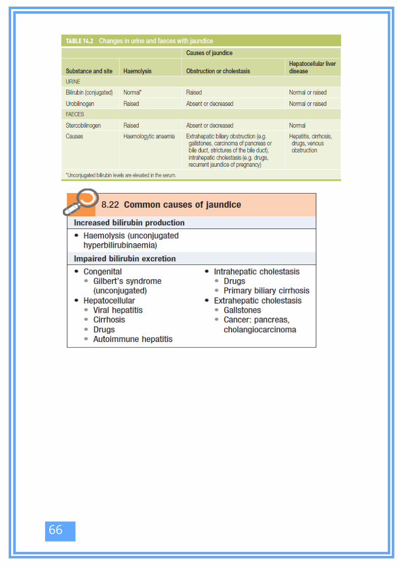
66

67
• Introduces himself, gains consent
• History of present illness:
- How was the jaundice discovered – did the patient notice it, or was
it someone else?
- Onset (what brought it on, how it started)
- Time:
• Duration
• Intermittent (e.g. Gilbert’s syndrome), continuous, progressive
• Fevers
• Previous episodes of jaundice
• Family members/contacts with similar symptoms
• Associated symptoms:
- Gallstones, biliary duct obstruction: Abdominal pain, pale stools,
itching, steatorrhea, dark urine
- Liver symptoms: Abdominal swelling, ankle swelling, bleeding,
bruising (liver failure and impaired synthetic function)
- Autoimmune conditions: Arthralgia, vitiligo, skin rashes (systemic
lupus erythematosus)
- Risk factors for viral hepatitis
• Hepatitis B and C: Contaminated needles, intravenous drug abuse,
blood transfusions, tattoos.
• Foreign travel/contacts
• PMH: previous jaundice, hemolytic anemias (sickle cell anemia and
thalassemia), IBD (linked to PSC)
• Family history: any family member with similar disease (may
indicate acute viral hepatitis)
• Medications: Hepatitis B immunization, antibiotics, statins,
acetaminophen, anti-TB medications.
• Social history: alcohol intake, IV drug abuse.

68
Details about Important GI Diseases

69
OSCE Vignette: A 62-year-old gentleman presented with a change in mental status.
He was an alcoholic and an IV-drug abuser in his twenties, and was diagnosed with
hepatitis B after that. Examine this patient looking for signs of chronic liver disease.
--------------------------------------------------------------------------------------------------------
- All types of chronic liver disease will ultimately lead to liver cirrhosis.
- Liver cirrhosis causes four types of signs:
1. Signs related to impaired liver function (decreased synthesis of albumin> edema, clotting
factors> easy bruisability... etc.).
2. Signs related to hyperestrinism (impaired breakdown of estrogen causes gynecomastia, spider
angiomata, loss of sexual hair, testicular atrophy, and palmar erythema).
3. Signs related to portal hypertension.
4. Signs related to hepatic encephalopathy and liver failure (asterixis and fetor hepaticus)
Chronic Liver disease

70
Ascites

71
How to differentiate between the spleen and
the kidney?

72
- Inspection:
Comment on abdominal distention.
See whether the flanks are full or not.
- Shifting dullness:
• With the patient supine, percuss from the midline out to the flanks. Note
any change from resonant to dull, along with areas of dullness and
resonance.
• Keep your finger on the site of dullness in the flank and ask the patient to
turn on to his opposite side.
• Wait for 10 seconds to allow any ascites to gravitate, then percuss again.
If the area of dullness is now resonant, shifting dullness is present,
indicating ascites.
- Fluid thrill:
• If the abdomen is tensely distended and you are not certain whether
ascites is present, feel for a fluid thrill.
• Place the palm of your left hand flat against the left side of the patient’s
abdomen and flick a finger of your right hand against the right side of
the abdomen.
Ascites Examination

73
• If you feel a ripple against your
left hand, ask an assistant or the
patient to place the edge of his
hand on the midline of the
abdomen. This prevents
transmission of the impulse via
the skin rather than through the
ascites. If you still feel a ripple
against your left hand, a fluid
thrill is present (only detected in
gross ascites).
-

74
Abdominal Examination

75

76

77

78

79
Chapter 4: Endocrine System

80
This part includes the most probable topics that show up in 4th year OSCE, these are:
Hyper/ hypothyroidism history and physical exam.
Cushing’s syndrome history and physical exam.
Lower limb examination in diabetic patient and DM follow up.
History of hirsutism.
Acromegaly physical exam.
Calcium disorders history.
Before we start, few notes related to more than one topic you should remember:
❖ Testing for proximal myopathy: ask the
patient to stand from a sitting position with arms
crossed; an inability to do this suggests proximal
muscle wasting.
❖ Diplopia: test this by asking the patient to follow
your finger by his eyes, without moving the head,
while you are drawing H shape with your finger in
the air.
❖ Testing for carpal tunnel syndrome (CTS):
1- Tinel’s sign: by percussion with tip of your index over median nerve close to wrist. Ask the
patient for any shooting tingling sensation elicited upon percussion
2- Phalen’s sign: ask patient to do reverse prayer’s sign, ask whether he felt any pain elicited
3- Look for thenar muscle wasting
❖ Cervical lymph nodes:
❖ visual fields examination:

81
❖ General endocrine findings on physical exam are summarized in the following
picture:

82
OSCE stations may be as the following:
Take a proper history differentiating between hyper and hypothyroidism.
Ask relevant questions to know the cause of hyper OR hypothyroidism.
➢ Quick review: The thyroid axis is involved in the regulation of cellular differentiation and metabolism in virtually all
nucleated cells, so that disorders of thyroid function have diverse manifestations. Follicular epithelial
cells synthesize thyroid hormones by incorporating iodine into the amino acid tyrosine. The thyroid
secretes predominantly thyroxine (T4) and only a small amount of triiodothyronine (T3), the more
active hormone; ~85% of T3 in blood is produced from peripheral conversion of T4. They both
circulate in plasma almost entirely (> 99%) bound to transport proteins, mainly thyroxine-binding
globulin (TBG).
➢ Plan: I. Ask about symptoms of hyper/hypothyroidism regardless the cause.
II. Symptoms caused by anatomical location of goiter -if present-.
III. Past thyroid history and family history.
IV. Ask by differentials.
I. Symptoms of hyper or hypothyroidism: When asking to differentiate, you need to ask many questions related to almost all systems of the body; thus, you
need to be systematic in order not to miss any:
System Symptoms in
hypothyroidism
Symptoms in
hyperthyroidism
CNS Depressed mood, apathy Nervousness, anxiety, irritability
and tremor
CVS Palpitation, SOB.
GI Constipation, decreased appetite. Diarrhea, increased appetite.
GUS menorrhagia Oligomenorrhea or amenorrhea *In males: there may be
gynecomastia, decreased libido and
erectile dysfunction.
MSS Dry hands, symptoms of carpel
tunnel syndrome, brittle nails and
coarse hair or alopecia.
Sweaty hands, oncholysis,
alopecia and muscle weakness
Thyroid gland

83
Others -very important- Cold intolerance, husky voice
and weight gain.
Heat intolerance and weight loss.
Ask about eye problems:
exophthalmos, diplopia, lid lag. Seen mostly in Grave’s.
II. Symptoms caused by anatomical location of goiter -if present-:
Dysphagia -compression on the esophagus-.
Dyspnea and SOB -compression on the trachea-.
Dysphonia.
III. Past thyroid history and family history:
Previous thyroid diseases in general.
Family history of any thyroid diseases.
IV. Ask by differentials:
Causes of hypothyroidism Causes of hyperthyroidism
Hashimoto thyroiditis Toxic MNG/adenoma Post total thyroidectomy.
Grave’s disease
Post I131 treatment HCG-induced hyperthyroidism
-Associated with pregnancy or
Trophoblastic Tumors
Congenital, i.e. Thyroid agenesis or
dysplasia.
Hyperthyroidism due to thyrotropin
secretion (TSH-oma).
Medications, i.e. Lithium and Amiodarone Inherited non-immune hyperthyroidism
Iodine deficiency Thyroxine induced or amiodarone induced.
Central hypothyroidism Subacute thyroiditis “daQuevarian”:
Thyroid infiltration, i.e. Riedel’s struma,
amyloidosis, and hemochromatosis
How to ask:
- Causes of hypothyroidism: I. Hashimoto thyroiditis:
It an autoimmune disorder so you have to ask:
If the patient has any other autoimmune diseases especially: vitiligo,
DM type 1, primary adrenal insufficiency and pernicious anemia.
Family history of any autoimmune diseases.
II. Postpartum thyroiditis:
Ask about last pregnancy and whether the symptoms are related to the
postpartum period or not -symptoms arise within 1-6 months after
childbirth-.
III. Post total thyroidectomy: did the patient undergo thyroid surgery?
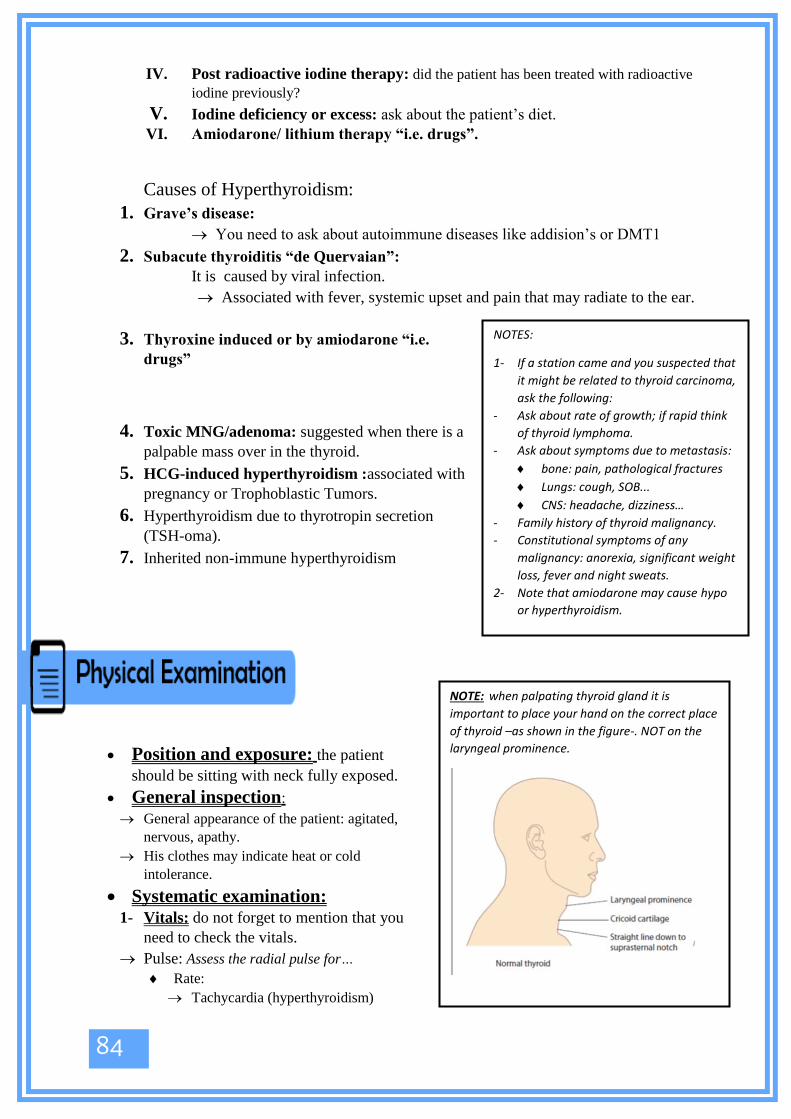
84
IV. Post radioactive iodine therapy: did the patient has been treated with radioactive
iodine previously?
V. Iodine deficiency or excess: ask about the patient’s diet. VI. Amiodarone/ lithium therapy “i.e. drugs”.
Causes of Hyperthyroidism:
1. Grave’s disease:
You need to ask about autoimmune diseases like addision’s or DMT1
2. Subacute thyroiditis “de Quervaian”:
It is caused by viral infection.
Associated with fever, systemic upset and pain that may radiate to the ear.
3. Thyroxine induced or by amiodarone “i.e.
drugs”
4. Toxic MNG/adenoma: suggested when there is a
palpable mass over in the thyroid.
5. HCG-induced hyperthyroidism :associated with
pregnancy or Trophoblastic Tumors.
6. Hyperthyroidism due to thyrotropin secretion
(TSH-oma).
7. Inherited non-immune hyperthyroidism
• Position and exposure: the patient
should be sitting with neck fully exposed.
• General inspection:
General appearance of the patient: agitated,
nervous, apathy.
His clothes may indicate heat or cold
intolerance.
• Systematic examination: 1- Vitals: do not forget to mention that you
need to check the vitals.
Pulse: Assess the radial pulse for…
Rate:
Tachycardia (hyperthyroidism)
NOTES:
1- If a station came and you suspected that
it might be related to thyroid carcinoma,
ask the following:
- Ask about rate of growth; if rapid think
of thyroid lymphoma.
- Ask about symptoms due to metastasis:
bone: pain, pathological fractures
Lungs: cough, SOB...
CNS: headache, dizziness…
- Family history of thyroid malignancy.
- Constitutional symptoms of any
malignancy: anorexia, significant weight
loss, fever and night sweats.
2- Note that amiodarone may cause hypo
or hyperthyroidism.
NOTE: when palpating thyroid gland it is
important to place your hand on the correct place
of thyroid –as shown in the figure-. NOT on the
laryngeal prominence.

85
Bradycardia (hypothyroidism)
Rhythm – irregular (atrial fibrillation) – thyrotoxicosis
2- Hands:
Inspect the patients’ hands for…
Dry skin (hypothyroid) /Increased sweating (hyperthyroid)
Temperature: Cold -in hypo-/ -hyper-warm
Thyroid acropachy “in Graves’ disease”.
Palmar erythema in hyperthyroidism.
Peripheral tremor in hyperthyroidism.
Test for carpel tunnel syndrome -seen in hypothyroidism-
3- Face:
Inspect the face for…
• Dry skin – hypothyroidism / Sweating – hyperthyroidism
• Eyebrows: loss of the outer third in hypothyroidism (rare)
• Eyes:
I. Exophthalmos (anterior displacement of the eye out of the orbit) in Graves’: inspect from the
front, side and above.
II. Lid retraction: note if the sclera is visible above the iris -seen in Graves’ disease-
III. Inspect for any redness / inflammation of the conjunctiva -seen in hyperthyroidism-.
IV. Diplopia: Eye movement can be restricted in Graves’ disease due to abnormal connective
tissue deposition in the orbit and extra-ocular muscles and this may result in diplopia.
V. Lid lag.
4- Thyroid gland examination:
• Inspection:
Inspect the neck for any:
Skin changes.
Scars -may indicate thyroid surgery.
Mass -if found describe its site, size, shape and skin over it “the 4 Ss’”
Ask the patient to:
Sallow -thyroid normally moves with swallowing-
Protrude the tongue –thyroid do NOT move while protruding the tongue, thyroglossal
cyst does-.
Open his mouth and inspect if there is a lingual thyroid.
To raise his arms above the head notice any facial congestion and cyanosis -
Pemberton's sign-.
• Palpation: do NOT forget to ask the patient if there is any pain.
From Anterior: note any tenderness.
From posterior with slight flexion of the neck:
Palpate each thyroid lobe by itself, and comment on the symmetry of the lobes, if there
is any masses then describe them ( 4 Ss, tenderness, temperature, consistency,
attachment, pulsation, surface, edges, mobility), any thrills.
Ask the patient once again to sallow and protrude the tongue while palpating this time.

86
Cervical Lymph nodes.
Trachea for deviation.
Carotid pulse: to check if there is berry sign which is the absence of carotid pulsation
on palpation.
• Percussion:
Percuss the clavicle bilaterally to see if there is any dullness -retrosternal goiter-.
• Auscultate:
For bruits -found in Grave’s’ disease-.
• finish your examination with:
Testing the deep tendon reflexes –delayed relaxation in hypothyroidism/ hyperreflexia in
hyperthyroidism-.
Inspect the lower limbs for pretibial myxedema -in Graves’-
Test for Proximal myopathy; proximal myopathy is associated with hyperthyroidism.

87

88
*Here is a simple picture; you may revise the examination by writing the steps in your own way*

89
NOTE: this part will be brief as it is unlikely to show up in the OSCE as a history-.
➢ Quick review: Cushing’s syndrome is due to a chronic excess of glucocorticoids. Glucocorticoids
(e.g., cortisol) stimulate lipolysis, the release of amino acids from the muscles, and
gluconeogenesis by the liver. Cortisol inhibits all stages of the inflammatory process and also
affects the bones by decreasing the protein matrix. Its immunosuppressive effect is on T cells
and their associated cell-mediated immunity and delayed hypersensitivity. Excess cortisol can
additionally stimulate mineralocorticoid and androgen receptors with the clinical appearance
of aldosterone excess (hypertension, hypokalemia, and alkalosis). Cortisol does not bind
androgen receptors. So glucocorticoids have multiple effects on the human body, so when
taking history or examining a patient with Cushing’s we are supposed to look for all effects of
steroids excess.
➢ Plan: I. You need to ask about the symptoms of excess steroids regardless the cause.
II. Then, ask by differentials.
I. Symptoms of excess steroids regardless the cause:
1- Ask about recent weight gain and how much?
2- Ask the patient if he/she bruises easily?
3- Has skin become thin recently?
4- Ask if the patient has recently been more susceptible to infections?
5- Ask about visual problems as steroids could cause cataract.
6- Ask about problems of excess androgens: acne and excess hair in females.
7- In females, ask about menstrual irregularity or amenorrhea.
8- Ask about proximal myopathy: you may ask for example “if the patient is facing difficulty
getting up from the chair?”
9- Ask whether the patient has been diagnosed with diabetes or not, or symptoms of
hyperglycemia.
10- Ask about pathological fractures.
Cushing’s Syndrome

90
II. Ask by differential:
1- Exogenous administration of steroids: so ask the patient if he is taking steroids for any
reason or not. It is extremely important to ask about this, as the most common cause of
Cushing’s is iatrogenic.
2- Cushing disease: i.e. excess ACTH production from the pituitary. Ask the patient if he has
visiual problems suggesting a pituitary mass and ask whether the patient has noted any
hyperpigmentation over his body.
3- ACTH secreting tumors: ask whether the patient has been diagnosed with malignancy and
ask about general constitutional symptoms of malignancy.
4- Adrenal neoplasia: suggested when the symptoms develops rapidly and ask about flank pain
or heaviness, however these two are not common symptoms to be found.
5- Finally, it is important to exclude things that causes pseudo-Cushing’s and these include:
depression, alcoholism and primary obesity.
Examining a patient for signs of Cushing's syndrome involves a general inspection of the patient,
along with a systematic examination of the main organs involved in the disease.
• Position and exposure: inspect the patient generally while standing then ask him/ her
kindly to lie down in supine position.
Exposure: hands, face, chest, abdomen and lower limbs.
• General Inspection: Look at the patient as a whole; remember that patients with Cushing’s have a characteristic
appearance that may be noted easily:
central obesity
stooped posture (due to osteoporotic damage)
Generalized changes over the body, including:
bruising
Striae -purple and wider than 1 cm are the characteristic striae of Cushing’s-
Thin skin
• Systemic Examination: Examine each of the following body parts separately:
1- Vitals: blood pressure may be raised.
2- Hands and arms:
Bruising
Thin arms.
Hyperpigmentation in skin creases -REM: seen when Cushing’s is caused by excess ACTH-.
Note whether there is changes suggesting rheumatoid diseases indicating an iatrogenic cause
of the Cushingoid features.
Test for proximal myopathy.

91
3- Face:
Rounded face (moon face).
Visible vessels on the cheeks suggesting thin skin or telangiectasia.
Greasy skin.
Acne.
Hirsutism.
Plethora: “important as this is only seen in true Cushing’s not in pseudocushing’s”
At this point you may tell the examiner that you want to test Visual fields (looking for a
bitemporal hemianopia that may be the result of an ACTH producing pituitary tumor pressing
on the optic chiasm)
4- Shoulders:
Supra-clavicular fat pads.
Buffalo hump.
5- Chest: gynecomastia in males.
6- Abdomen:
Central obesity
Purple Striae.
Bruising.
Renal transplant scars (patient may be on long term steroids causing the Cushing's
syndrome).
Palpate the abdomen for any adrenal masses: rarely found.
7- Legs:
Skin ulceration “poor wound healing”.
Palpate for edema -due to hypertension secondary to Cushing’s-
test for proximal myopathy
8- Finally, you may examine the Spine: for spinal tenderness (may occur with osteoporosis if
vertebral fractures are found).

92

93
*Here is a simple picture; you may revise the examination by writing steps in your own way*
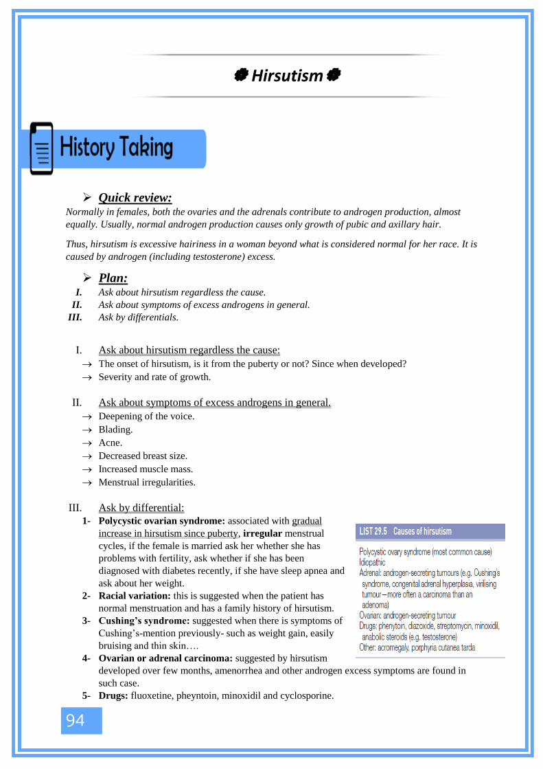
94
➢ Quick review: Normally in females, both the ovaries and the adrenals contribute to androgen production, almost
equally. Usually, normal androgen production causes only growth of pubic and axillary hair.
Thus, hirsutism is excessive hairiness in a woman beyond what is considered normal for her race. It is
caused by androgen (including testosterone) excess.
➢ Plan: I. Ask about hirsutism regardless the cause.
II. Ask about symptoms of excess androgens in general.
III. Ask by differentials.
I. Ask about hirsutism regardless the cause:
The onset of hirsutism, is it from the puberty or not? Since when developed?
Severity and rate of growth.
II. Ask about symptoms of excess androgens in general.
Deepening of the voice.
Blading.
Acne.
Decreased breast size.
Increased muscle mass.
Menstrual irregularities.
III. Ask by differential:
1- Polycystic ovarian syndrome: associated with gradual
increase in hirsutism since puberty, irregular menstrual
cycles, if the female is married ask her whether she has
problems with fertility, ask whether if she has been
diagnosed with diabetes recently, if she have sleep apnea and
ask about her weight.
2- Racial variation: this is suggested when the patient has
normal menstruation and has a family history of hirsutism.
3- Cushing’s syndrome: suggested when there is symptoms of
Cushing’s-mention previously- such as weight gain, easily
bruising and thin skin….
4- Ovarian or adrenal carcinoma: suggested by hirsutism
developed over few months, amenorrhea and other androgen excess symptoms are found in
such case.
5- Drugs: fluoxetine, pheyntoin, minoxidil and cyclosporine.
Hirsutism

95
➢ Note that menstrual cycle:
Irregular since puberty in PCOS
Amenorrhea in ovarian or adrenal carcinoma.
Normal and regular in racial variation

96
❖ History taking: -in brief-
➢ Quick review:
Diabetes mellitus is a clinical syndrome characterized by hyperglycemia due to absolute or relative
deficiency of insulin. Long-standing metabolic derangement can lead to the development of
complications of diabetes, which characteristically affect the eye, kidney and nervous system.
➢ Symptoms of hyperglycemia: Diabetes mellitus may present with a classical triad of symptoms:
• Polyuria (and nocturia): due to osmotic diuresis caused by glycosuria.
• Thirst: due to the resulting loss of fluid and electrolytes.
• Weight loss: due to fluid depletion and breakdown of fat and muscle secondary to insulin
deficiency.
Other common symptoms are:
• Tiredness, mood changes, poor concentration and blurred vision (due to glucose-induced
changes in lens refraction).
• Bacterial and fungal skin infection are common because of the combination of
hyperglycemia impaired immune resistance and tissue ischemia. Itching of the genitalia
(pruritus vulvae in women, balanitis in men) is due to Candida yeast infection (thrush).
• Headache.
Diabetes mellitus
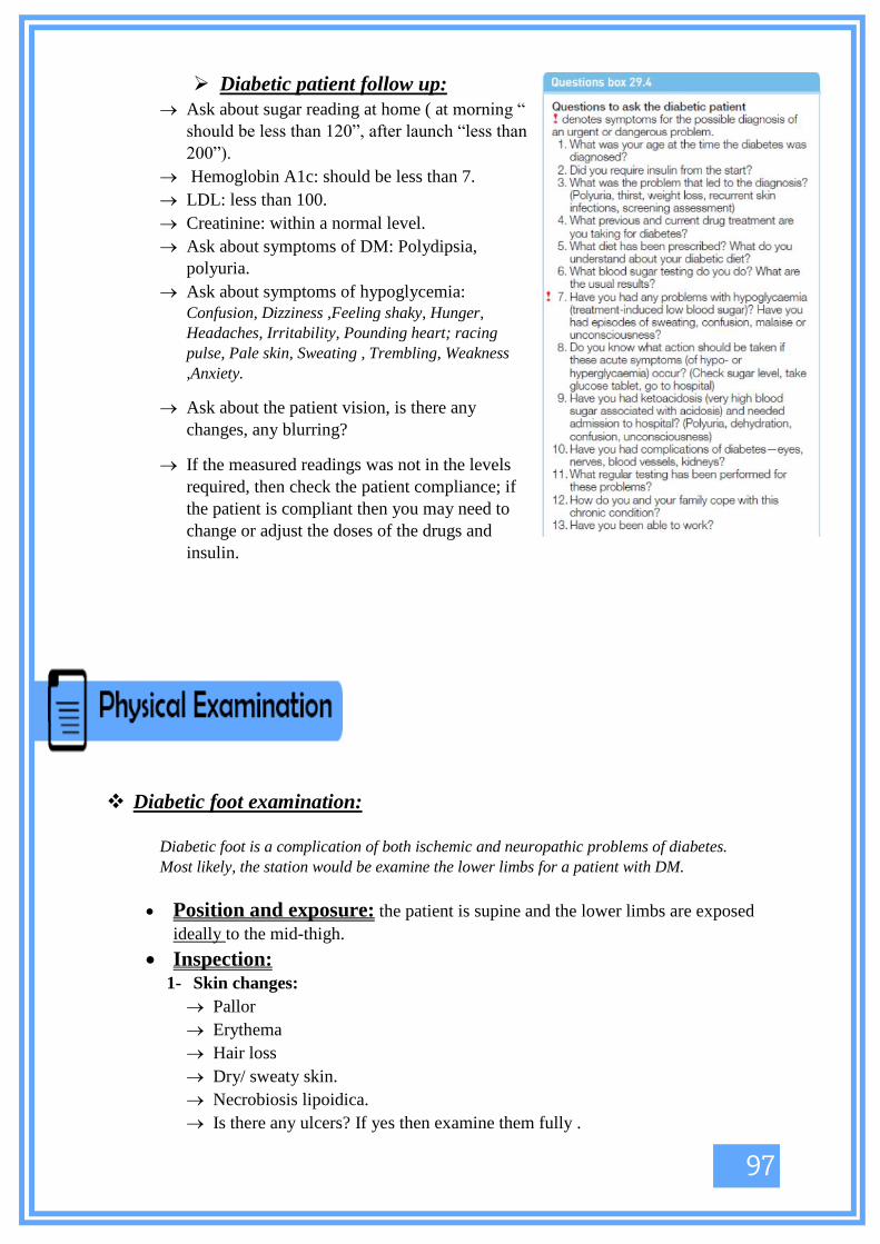
97
➢ Diabetic patient follow up:
Ask about sugar reading at home ( at morning “
should be less than 120”, after launch “less than
200”).
Hemoglobin A1c: should be less than 7.
LDL: less than 100.
Creatinine: within a normal level.
Ask about symptoms of DM: Polydipsia,
polyuria.
Ask about symptoms of hypoglycemia:
Confusion, Dizziness ,Feeling shaky, Hunger,
Headaches, Irritability, Pounding heart; racing
pulse, Pale skin, Sweating , Trembling, Weakness
,Anxiety.
Ask about the patient vision, is there any
changes, any blurring?
If the measured readings was not in the levels
required, then check the patient compliance; if
the patient is compliant then you may need to
change or adjust the doses of the drugs and
insulin.
❖ Diabetic foot examination:
Diabetic foot is a complication of both ischemic and neuropathic problems of diabetes.
Most likely, the station would be examine the lower limbs for a patient with DM.
• Position and exposure: the patient is supine and the lower limbs are exposed
ideally to the mid-thigh.
• Inspection: 1- Skin changes:
Pallor
Erythema
Hair loss
Dry/ sweaty skin.
Necrobiosis lipoidica.
Is there any ulcers? If yes then examine them fully .

98
Is there any gangrene? If yes then examine them fully.
Inspect between the toes checking for any infection.
Inspect the heel; you may notice hyperkeratosis.
2- Musculoskeletal changes:
Joint deformities –Charcot joint, claw toe, hammer toe or hallux valgus.
Notice any muscle wasting.
3- Finally, the nails: check for thickening of the nail or onchogryphosis.
• Palpation:
Temperature -always compare to the other side-
Capillary refill. Normally capillary refill takes 2-3 seconds.
Pulses of the lower limb –femoral, popliteal, dorsalis pedis and posterior
tibial- .
Edema if found.
• Neural exam:
Sensation (do not forget to ask the patient to close his eyes then test for it).
Vibration (tested with tuning fork over a bony prominence).
Proprioception (do not forget to ask the patient to close his eyes then test for
it).
Test the reflexes.
All of these may be absent or reduced.
• Finishing your exam:
Check the patient’s gait: as Charcot joint may affect the gait-.
Mentioned that it is indicated to measure Ankle Brachial index “ABI”.

99

100
➢ Quick review: Acromegaly is caused by excess GH secretion, usually from pituitary macroadenoma. If GH
hypersecretion occurs before epiphyseal fusion, then gigantism will result. More commonly, GH
excess occurs after epiphyseal closure and acromegaly ensues. Acromegaly is associated with
cardiomyopathy, increased incidence of diabetes mellitus, hypertension and colonic cancer.
• Position and exposure: at 45° and expose chest fully and abdomen.
• General Inspection: Patient Well or unwell?
Voice (deep and slow): deepening of the voice is a sign of acromegaly.
• Systemic examination:
1- Vitals:
BP (acromegaly is associated with increased incidence of hypertension).
Palpate the Pulse.
2- Hands:
Inspection:
Large hands; coarse, sweaty skin.
Diabetic finger prick marks. -indicating DM-
Clubbing.
Changes of osteoarthritis are common due to skeletal overgrowth.
Examine for carpel tunnel syndrome.
3- Arms :
Palpate behind the medial epicondyle looking for ulnar nerve thickening, which is found in
acromegaly.
Test for proximal myopathy
4- Face:
Prominent supraorbital ridges.
Large nose and lips.
Prognathism (prominent mandible) and malocclusion.
Wide spaces between the teeth.
Macroglossia.
Scars from previous surgery.
5- Eyes:
Test the Visual fields (looking for bitemporal hemianopia). This is due to mass effect from
the pituitary gland.
Acromegaly
101
Fundoscopy (papilloedema secondary to raised ICP, diabetic and hypertensive changes): just
mention.
6- Axilla:
Acanthosis nigricans.
Skin tags (called molluscum fibrosum, which are non-tender skin colored protrusions)
7- Neck:
Goiter (increased thyroid size and vascularity)
Acanthosis nigricans: in the back of the neck.
8- Chest:
Coarse body hair.
Gynecomastia. –if associated with hyperprolactinemia-
Feel for displaced apex.
Listen for any murmurs.
Listen to lung bases if any suspicion of heart failure.
9- Abdomen:
Organomegaly (examine for hepatomegaly and splenomegaly).
10- Legs:
Proximal myopathy.
Peripheral edema (if associated heart failure).
Foot drop may be present due to common peroneal nerve entrapment.
11- Finally mention that you should look for testicular atrophy, as acromegaly may be
associated with mixed pituitary tumors, with hyperprolactinemia or due to gonadotropin
deficiency due to enlarging pituitary.

102
103

104
❖ Hypercalcemia: Remember that symptoms of hypercalcemia are:
*Stones, bones, abdominal moans and psychic groans*
➢ Plan: I. Ask about symptoms of hypercalcemia regardless the cause.
II. Ask by differentials.
I. Symptoms of hypercalcemia:
1. Renal symptoms:
Polyuria
Nocturia
Dehydration
Stones
2. Skeletal symptoms:
Ask about bone pain and pathological fractures.
3. Neurological manifestations:
Lethargy
Weakness
Loss of consciousness
Confusion
4. GI:
Nausea
Anorexia
Constipation
History of peptic ulcer
II. Ask by differentials:
Most common causes you need to ask about are:
1- Primary hyperparathyroidism: has no specific symptoms other than the mentioned above.
2- Malignancy related hypercalcemia: most commonly associated with lung cancer so ask about
general respiratory symptoms, general symptoms of any malignancy ( like weight loss) or history
of malignancy.
3- Multiple myeloma: associated with weight loss, back pain and symptoms of decreased immunity
- recurrent infections-.
4- Drugs: like thiazides.
5- Familial hypercalcemia hypocalciuria.
Calcium Disorders
105
❖ Hypocalcemia:
➢ Plan: I. Ask about symptoms of hypocalcemia regardless the cause.
II. Ask by differentials.
I. Symptoms of hypercalcemia:
Tingling around mouth and the fingers
Muscle cramps
Fits or seizures
II. Ask by differential :
1- Recent surgery of thyroid or parathyroid.
2- Diseases of malabsorption.
3- Renal diseases.
4- History of recent acute pancreatitis.
5- Hypocalcemia of malignancy.
6- Hypomagnesemia.
7- Pseudo-hypoparathyroidism: this disease is associated with skeletal deformity in general and
short 5th or 4th finger or toe specially.
8- Vitamin D deficiency.

106
Chapter 5: Hematology
** You will notice that this system is not presented in the usual way as the CVS, RS, or
GI (simply because it’s very different). However, we hope you find it simple and easy.
** Contents
1) Anemia (very common in OSCE)
- Revision of some basics and some diseases
- History
- Physical exam
2) Thrombocytopenia, as part of the topic ‘bleeding tendency’
- Revision of some basics and some diseases
- History
- Physical exam
3) Multiple Myeloma
- Revision of some basics
- History
- Physical exam
4) Some Useful Images
5) Past paper

107
Anemia
❖ Revision of some basics I. Anemia is defined as “reduction in circulating RBC mass”. Since hemoglobin (Hb),
hematocrit (Hct) and RBC count are used as surrogates for RBC mass (which is
difficult to measure), we can say that anemia is defined as “reduction in Hct or Hb
concentration”.
Anemia is defined as Hb <13.5 g/dL in males, and < 12.5 g/dL in females (normal
Hb is 13.5-17.5 g/dL in males and 12.5-16.0 g/dL in females). These values are
according to Pathoma.
II. Anemia could present as isolated anemia or as part of pancytopenia (Anemia +
Thrombocytopenia + neutropenia).
III. Classification
• Based on mean corpuscular volume (MCV), anemia can be classified as
Microcytic (MCV < 80 μm3), Normocytic (MCV = 80 -100 μm3), or Macrocytic
(MCV > 100 μm3).
• If Microcytic anemia (MCV < 80), the differential diagnosis includes the
following:
- Iron deficiency anemia,
- Anemia of chronic disease,
- Thalassemia,
- Sideroblastic anemia (includes lead poisoning, pyridoxine deficiency, toxic
effects of alcohol).
• If Macrocytic anemia (MCV > 100), the differential diagnosis includes the
following:
- Vitamin B12 deficiency and/or folate deficiency,
- Liver disease.
- Others; alcoholism, drugs (e.g. Fluorouracil (5-FU))
• If Normocytic anemia, the differential diagnosis includes the following:
{“TARAB”+H}
- Tumor
- Aplastic anemia
- Renal failure (decreased erythropoietin production)
- Anemia of chronic disease (chronic inflammation, malignancy)
- Bone marrow fibrosis
- HEMOLYSIS (Hereditary spherocytosis, Sickle cell anemia, Paroxysmal
nocturnal hemoglobinuria (PNH), G6PD deficiency, Immune hemolytic anemia
(IHA), Microangiopathic hemolytic anemia (MAHA), Malaria).

108
• For the classification to be more accurate and more clinically relevant, we should
look at the reticulocyte index.
- The reticulocyte count is an important initial test in evaluation of anemia
because it indicates whether effective erythropoiesis is occurring in the bone
marrow. (in fact, you should look at the reticulocyte index before the MCV as
you’ll see in a minute).
- A reticulocyte index > 2% implies excessive RBC destruction or blood loss.
The bone marrow is responding to increased RBC requirements (the bone
marrow is trying to compensate what has been lost/destroyed).
- A reticulocyte count < 2%, in the presence of anemia, implies inadequate RBC
production by the bone marrow.
IV. Clinical features of anemia:
• A variety of non-specific complaints: fatigue/weakness, shortness of breath,
palpitations, headache, dizziness/lightheadedness, poor concentration, diarrhea,
nausea, vague abdominal discomfort.
• Pallor of conjunctiva or skin (“Have you -or someone you know- noticed that you
look pale?”)
• Angina, especially with preexisting coronary artery disease.
• Hypotension (also ask about postural lightheadedness) and tachycardia.
• Signs of the underlying cause- jaundice if hemolytic anemia, blood in stool if GI
bleeding,…
V. Important investigations when it comes to anemia include:
- CBC (we’re mostly concerned with Hb/Hct, reticulocyte index -if present-, and
RBC indices especially MCV)
- Blood smear
- Specific tests according to your suspicions; like Iron studies, Vitamin B12 and
folate levels,..

109
VI. Evaluation of anemia (general approach) - from Step-Up:
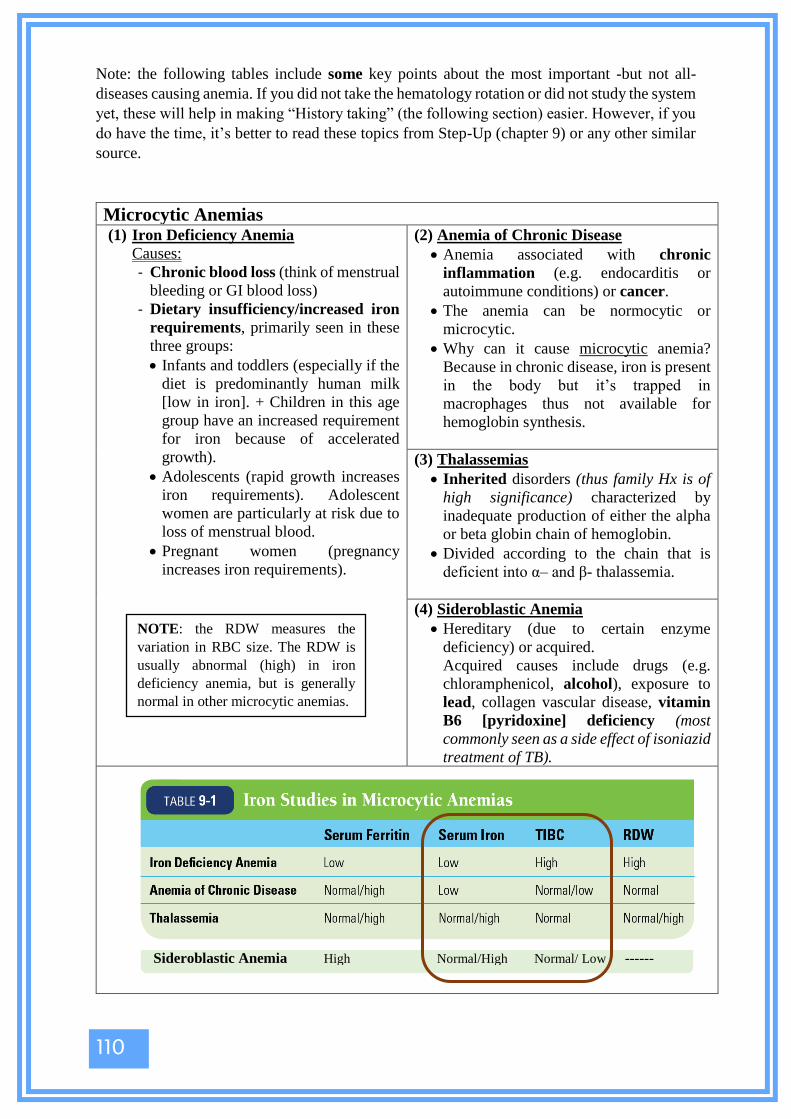
110
Note: the following tables include some key points about the most important -but not all-
diseases causing anemia. If you did not take the hematology rotation or did not study the system
yet, these will help in making “History taking” (the following section) easier. However, if you
do have the time, it’s better to read these topics from Step-Up (chapter 9) or any other similar
source.
Microcytic Anemias (1) Iron Deficiency Anemia
Causes:
- Chronic blood loss (think of menstrual
bleeding or GI blood loss)
- Dietary insufficiency/increased iron
requirements, primarily seen in these
three groups:
• Infants and toddlers (especially if the
diet is predominantly human milk
[low in iron]. + Children in this age
group have an increased requirement
for iron because of accelerated
growth).
• Adolescents (rapid growth increases
iron requirements). Adolescent
women are particularly at risk due to
loss of menstrual blood.
• Pregnant women (pregnancy
increases iron requirements).
(2) Anemia of Chronic Disease
• Anemia associated with chronic
inflammation (e.g. endocarditis or
autoimmune conditions) or cancer.
• The anemia can be normocytic or
microcytic.
• Why can it cause microcytic anemia?
Because in chronic disease, iron is present
in the body but it’s trapped in
macrophages thus not available for
hemoglobin synthesis.
(3) Thalassemias
• Inherited disorders (thus family Hx is of
high significance) characterized by
inadequate production of either the alpha
or beta globin chain of hemoglobin.
• Divided according to the chain that is
deficient into α– and β- thalassemia.
(4) Sideroblastic Anemia
• Hereditary (due to certain enzyme
deficiency) or acquired.
Acquired causes include drugs (e.g.
chloramphenicol, alcohol), exposure to
lead, collagen vascular disease, vitamin
B6 [pyridoxine] deficiency (most
commonly seen as a side effect of isoniazid
treatment of TB).
NOTE: the RDW measures the
variation in RBC size. The RDW is
usually abnormal (high) in iron
deficiency anemia, but is generally
normal in other microcytic anemias.
Sideroblastic Anemia High Normal/High Normal/ Low ------
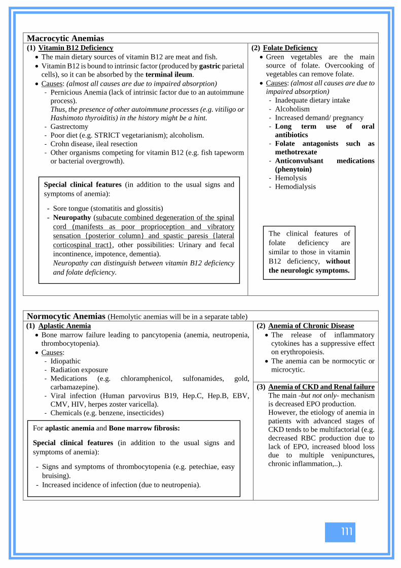
111
Macrocytic Anemias (1) Vitamin B12 Deficiency
• The main dietary sources of vitamin B12 are meat and fish.
• Vitamin B12 is bound to intrinsic factor (produced by gastric parietal
cells), so it can be absorbed by the terminal ileum.
• Causes: (almost all causes are due to impaired absorption)
- Pernicious Anemia (lack of intrinsic factor due to an autoimmune
process).
Thus, the presence of other autoimmune processes (e.g. vitiligo or
Hashimoto thyroiditis) in the history might be a hint.
- Gastrectomy
- Poor diet (e.g. STRICT vegetarianism); alcoholism.
- Crohn disease, ileal resection
- Other organisms competing for vitamin B12 (e.g. fish tapeworm
or bacterial overgrowth).
(2) Folate Deficiency
• Green vegetables are the main
source of folate. Overcooking of
vegetables can remove folate.
• Causes: (almost all causes are due to
impaired absorption)
- Inadequate dietary intake
- Alcoholism
- Increased demand/ pregnancy
- Long term use of oral
antibiotics
- Folate antagonists such as
methotrexate
- Anticonvulsant medications
(phenytoin)
- Hemolysis
- Hemodialysis
Normocytic Anemias (Hemolytic anemias will be in a separate table)
(1) Aplastic Anemia
• Bone marrow failure leading to pancytopenia (anemia, neutropenia,
thrombocytopenia).
• Causes:
- Idiopathic
- Radiation exposure
- Medications (e.g. chloramphenicol, sulfonamides, gold,
carbamazepine).
- Viral infection (Human parvovirus B19, Hep.C, Hep.B, EBV,
CMV, HIV, herpes zoster varicella).
- Chemicals (e.g. benzene, insecticides)
(2) Anemia of Chronic Disease
• The release of inflammatory
cytokines has a suppressive effect
on erythropoiesis.
• The anemia can be normocytic or
microcytic.
(3) Anemia of CKD and Renal failure
The main -but not only- mechanism
is decreased EPO production.
However, the etiology of anemia in
patients with advanced stages of
CKD tends to be multifactorial (e.g.
decreased RBC production due to
lack of EPO, increased blood loss
due to multiple venipunctures,
chronic inflammation,..).
Special clinical features (in addition to the usual signs and
symptoms of anemia):
- Sore tongue (stomatitis and glossitis)
- Neuropathy (subacute combined degeneration of the spinal
cord (manifests as poor proprioception and vibratory
sensation {posterior column} and spastic paresis {lateral
corticospinal tract}, other possibilities: Urinary and fecal
incontinence, impotence, dementia).
Neuropathy can distinguish between vitamin B12 deficiency
and folate deficiency.
The clinical features of
folate deficiency are
similar to those in vitamin
B12 deficiency, without
the neurologic symptoms.
For aplastic anemia and Bone marrow fibrosis:
Special clinical features (in addition to the usual signs and
symptoms of anemia):
- Signs and symptoms of thrombocytopenia (e.g. petechiae, easy
bruising).
- Increased incidence of infection (due to neutropenia).

112
Hemolytic Anemias
(1) Sickle Cell Anemia
• Autosomal recessive disorder that results when the normal Hb A is
replaced by the mutant Hb S. Sickle cell disease is caused by
inheritance of two Hb S genes (homozygous).
• Hb S may be distinguished from Hb A by electrophoresis because of
the substitution of an uncharged valine for a negatively charged
glutamic acid at the sixth position of the β-chain.
• Under reduced oxygen conditions (e.g., acidosis, hypoxia, changes
in temperature, dehydration, and infection) the Hb molecules
polymerize, causing the RBCs to sickle. Sickled RBCs obstruct
small vessels, leading to ischemia (see Clinical Pearl 9-3).
• Hemolysis is predominantly extravascular (so keep jaundice and
gallstones in your mind).
Note: I recommend reading “clinical features of sickle cell anemia” from
page 336 in Step-Up.
(2) Hereditary Spherocytosis
• Autosomal dominant
inheritance of a defect in the
gene coding for spectrin and
other RBC proteins.
• There is a loss of RBC
membrane surface area without
a reduction in RBC volume,
necessitating a spherical shape.
The spherical RBCs become
trapped and destroyed in the
spleen (by macrophages)—
hence the term extravascular
hemolysis. We expect jaundice, gallstones,
splenomegaly.
(3) G6PD Deficiency
• An X-linked recessive
disorder that primarily affects
men.
• Known precipitants include;
fava beans, sulfonamides,
nitrofurantoin, primaquine,
dimercaprol, and infection.
• Presents as episodic hemolytic
anemia that is usually drug-
induced.
• Hemolysis is predominantly
(but not exclusively)
intravascular.
• Dark urine and jaundice are
usually present.
Clinical Features
1. Signs and symptoms of anemia
2. Signs and symptoms of underlying disease (e.g. bone crisis in sickle cell disease)
3. Jaundice (due to unconjugated bilirubin), bilirubin gallstones (cholelithiasis), hepatosplenomegaly.
These are more seen with extravascular hemolysis (mainly in spleen and liver).
4. Dark urine color (due to hemoglobinuria, not bilirubin) may be present. This indicates intravascular
hemolysis.
5. Lymphadenopathy (in chronic cases).

113
(4) Autoimmune Hemolytic Anemia (AIHA)
• Production of auto-antibodies toward RBC membrane antigen(s)
which leads to destruction of these RBCs.
• Two types;
1) Warm AIHA (autoantibody is IgG which binds optimally to
RBC membranes at 37O).
Produces usually extravascular hemolysis – the primary site
of RBC sequestration is the spleen. Splenomegaly is a
common feature.
Causes: primary (idiopathic), secondary to lymphomas,
leukemias (CLL), other malignancies, SLE (most common
cause), drugs (penicillin, cephalosporins, α -methyldopa)
2) Cold AIHA (autoantibody is IgM which binds optimally to
the RBC membrane at cold temperatures (usually 0OC TO
5OC).
Produces mainly intravascular hemolysis. (but extravascular
hemolysis is also possible).
Jaundice is expected if significant hemolysis is present.
Causes: can be idiopathic or due to infection (such as
Mycoplasma pneumoniae or infectious mononucleosis).
(5) Paroxysmal Nocturnal
Hemoglobinuria (PNH)
• An acquired disorder that affects
hematopoietic stem cells and cells of
all blood linages.
• This is caused by deficiency of anchor
proteins that link complement-
inactivating proteins to blood cell
membranes the deficiency of this
anchoring mechanism results in an
unusual susceptibility to complement
mediated lysis of RBCs, WBCs, and
platelets.
• Results in intravascular hemolysis that
usually occurs episodically, often at
night during sleep.
** Other causes of hemolysis include:
• Microangiopathic Hemolytic Anemia (MAHA)
key points about this entity will be mentioned in the “Bleeding
Tendency” section).
• Malaria
Special clinical features (in addition to
the usual signs and symptoms of
anemia):
- Pancytopenia
- Thrombosis of venous systems can
occur (e.g. of the hepatic veins
[Budd-Chiari syndrome]).
**destroyed platelets release their
cytoplasmic contents into
circulation, inducing thrombosis).

114
Note: the question will NOT simply be “Take brief history from
a patient with anemia to know the cause of the anemia”.
Instead, it is usually given in a way that is similar to the
following:
[“Mr. ~~~ presented to you complaining of fatigue, headache,
and dizziness (or SOB, or poor concentration, or diarrhea, …
and so on). Take brief/ focused history to reach a diagnosis.”
+/- a clinical aid (CBC results for example)].
The question/examiner might want you to reach a specific
diagnosis in the end (and in this case, his answers would be
directed towards one specific entity), or he might just want you
to ask the questions that would cover the possible differential
diagnoses (in this case, he’ll be mostly “listening” to your
questions rather than giving you precise answers).
Either way, I recommend using the following three steps while
taking history:
The first step: Knowing that the case is anemia
The second step: knowing if the anemia is isolated or is part of
pancytopenia (to narrow down the possibilities of the diagnosis)
The third step: Proceed with history taking (according to the
general outline) to know the exact cause of anemia.
(1) Knowing that the case is anemia
How:
1. By asking about symptoms of anemia. For example, if Mr.~~~ came complaining of
fatigue and dizziness, you should ask about other symptoms like SOB (+ its relation to
exertion), pallor, poor concentration,…
If many – not necessarily all – are positive, this is probably anemia.
2. If there is a clinical aid in the question (for example, the CBC results), it will usually
provide a hint that this is anemia (for example, Hb level = 10 g/dL).
(2) Knowing if the anemia is isolated or is part of pancytopenia
1. By asking about the following
• Recurrent infections (a result of neutropenia)
• Symptoms of skin bleeding Petechiae (ask about small red spots on the skin) and
easy bruising.
Symptoms of mucosal bleeding epistaxis, hemoptysis, GI bleeding, hematuria,
and menorrhagia.
(results of thrombocytopenia)
Please keep the general outline for
history taking always in your mind.
- Patient profile
- Chief complaint and its details.
- Associated symptoms
• Constitutional symptoms
(fever, malaise, weight loss,
chills and rigors, night
sweats).
• Other symptoms of the same
system
- Review of systems
- Past medical (including blood
transfusion), past surgical,
OBG.
- Drug history
- Family history
- Social history
• Life style; including diet
• Smoking
• Alcohol
• Occupation
• Address
• Travel

115
2. Look at the WBC and platelet counts in the CBC,
if present.
Normal WBC count: 5,000-10,000 /μL
(5-10 K/μL)
Normal platelet count: 150,000-450,000 /μL
(150-450 K/μL).
Note: usually -not always- the question will be more
towards isolated anemia to get closer to a specific
diagnosis. However even if it is isolated anemia, you
usually get points for asking about the previous two
points. [let the examiner see that you understand what
you’re asking about ;)].
Note 2: multiple myeloma (discussed in a later
section) causes anemia due to BM infiltration and
renal failure. It also causes recurrent infections due to
deprivation of normal immunoglobulins.
(3) Reach the exact cause of anemia (the diagnosis) According to the general outline:
• Patient profile, if you didn’t ask about it in the
beginning
• Chief complaint (whatever it was, ask about its
onset, duration, course, and progression, and any
other details that come to your mind)
• Associated symptoms
- Constitutional symptoms (fever, malaise, weight
loss, …)
- Other symptoms of the same system [The presence
of other symptoms of anemia, Recurrent
infections, petechiae and easily bruising, epistaxis,
… (if you still haven’t asked about them)].
• Review of systems
Be fast when asking, and if certain diagnoses are in
your mind, focus on them.
For example, if you’re suspecting:
- Iron deficiency anemia ask about dyspnea on
exertion, postural (orthostatic) lightheadedness,
melena, hematemesis, epistaxis, pica (cravings for non-nutritive substances like sand, ice,
or paint or others), injury, menstruation in females.
- Pernicious anemia ask about the symptoms of other autoimmune conditions like vitiligo
or Hashimoto thyroiditis.
- Hemolytic anemia ask about jaundice, gall stones, dark urine, …
- Sickle cell anemia ask about severe bone pain (pain crisis), acute severe chest pain and
SOB (acute chest syndrome), painful swelling at the dorsa of the hand and feet (dactylitis),
Causes of pancytopenia include:
• Damage to bone marrow precursors e.g.
Aplastic Anemia.
• Myelophthesic process (pathologic
process (e.g. metastatic cancer) that
replaces the bone marrow resulting in
pancytopenia.
• Myelodysplastic syndromes (increased
blasts -but less than 20%- in the bone
marrow)
& Acute leukemias (>20% blasts in the
bone marrow)
increased blasts “crowd out” normal
hematopoiesis resulting in
pancytopenia.
• Myelofibrosis (bone marrow fibrosis)
• Drugs (e.g. certain chemotherapy drugs
cause BM suppression)
Important Note: it might look like there’s a lot
to ask about (especially in the review of
systems, past medical, and drug history),
however, the hints in the case (like the CBC or
the answers of the examiner usually reduce the
possibilities, so pay good attention to the hints
to save time and make your questions directed
towards a specific group of diagnoses.
For example: if the question asks you find out
the exact diagnosis, and the CBC reveals the
presence of microcytic anemia (this scenario
came in the year 2017/2018- first semester),
your questions should focus on: Iron deficiency
anemia, thalassemia, sideroblastic anemia, and
anemia of chronic disease, in an attempt to
know which one of them is the right diagnosis.
(This is why studying the causes/risk factors of
each disease -which were mentioned in the
previous tables- is of high significance).

116
joint pain, priapism, ophthalmologic complications, abdominal pain, … (There are many
others. This is why reading the clinical features of sickle cell anemia from page 336 in Step-Up
was recommended ;p)
- The previous are only some examples, but apply the same principle whatever the disease
you’re thinking of, and remember that for the purpose of history taking in the OSCE, it’s
better to know the key points about many diseases, than to know every single detail about
few diseases.
• Past medical history
- Chronic diseases (SLE, RA, chronic renal disease, chronic infections like TB,
Malignancies, …)
Note: if you ask “Do you have SLE?” or “Do you have chronic renal disease?” .., the
examiner will sometimes NOT give you a direct yes/no, so you might need to elaborate
and ask about SYMPTOMS of SLE or SYMPTOMS of chronic renal disease, and so on.
- Don’t forget to ask about history of blood transfusion as this might provide a clue to the
diagnosis (for example, a patient with thalassemia or sickle cell anemia might be
dependent on blood transfusions).
• Past surgical history
- Ask about any surgery done, and focus on GI surgeries (e.g. colon resection due to
malignancy, ileal resection, gastric sleeve,…)
• OBG
- Asking about the severity of menses in female patients is super important when it comes
to anemia.
o Regularity of period and the frequency (ask about the number of days between the
beginning of each menses and the next; i.e. length of the cycle).
o Heaviness (ask about the numbers of days of menses, the presence of clots in blood, the
frequency of changing pads)
- Ask about current pregnancy
• Drug history
- Aspirin, NSAIDs, anti-platelets, anti-coagulants (e.g. warfarin).
- Chemotherapy
- Carbamazepine, phenytoin, chloramphenicol can cause aplastic anemia
- Penicillin, cephalosporins can cause drug-induced immune hemolytic anemia
There are many other drugs. The previous are only some important examples
• Family history (very important)
- Thalassemia
- Sickle cell anemia
- G6PD deficiency [X-linked recessive so if the patient is a male (which is most probably
the case) don’t forget to ask about the maternal uncles and maternal grandfather of the
patient].
- Hereditary spherocytosis
• Social history
- Life style including diet ask about meat consumption, vegetables consumption, fava
beans sensitivity, raw fish consumption [might cause fish tapeworm (diphyllobothrium
latum) infestation vit.B12 deficiency].
- Smoking and Alcohol
- Occupation and address (example: radiation exposure aplastic anemia)
- Travel (significant if you’re suspecting malaria for example).

117
(Signs of anemia on examination)
Please follow the numbers in order to be systematic and not forget important
points.
The following figure is mostly about “anemia" in general with some points about specific diseases.
However, keep in your mind that the specific cause of anemia might have its own signs.
IDA: Iron deficiency anemia HA: Hemolytic anemia
General
- Fatigue and in distress
- Pallor
- Jaundice (HA)
1
Hands
- Koilonychia (IDA)
- Changes consistent with
certain chronic diseases
(example: Palmar
erythema, leukonychia,
and half and half nails in
CLD,, clubbing in lung
cancer or chronic IBD,,
etc.)
- Vitiligo (pernicious
anemia)
3
Chest
- S3
- Palpitations
- Tachycardia
5
Head and Neck
- Eyes: look for pallor, or
jaundice (HA)
- Mouth: look for angular
stomatitis, and atrophic
glossitis (IDA),
hypertrophied
tongue/beefy raw
appearance (folate or B12
def.)
- Lymphadenopathy
(chronic disease)
- Raised JVP
4
Specific points for β-thalassemia
- Distortion of bones due to expansion of
bone marrow space short limbs, rib
deformities, chipmunk face.
- Growth retardation and failure to thrive
- Skull has a crew cut appearance on
skull X-ray.
*
Lower Limbs
- Look for bruising (also in upper
limbs) – pancytopenia
- Neuropathy: loss of position and
vibratory sensation in the lower
extremities, ataxia, upper motor
neuron signs [spasticity, weakness,
increased deep tendon reflexes,
Babinski sign] – B12 deficiency.
7
Vital Signs
- Heart Rate: tachycardia
- Blood pressure: assess for
hypotension
- Respiratory rate
- Temperature: assess for
fever
2
Always check for
orthostatic changes in
anemia.
Abdomen
- Hepatomegaly/
splenomegaly (in certain
hemolytic anemias).
6
If the patient is likely getting blood transfusions (e.g. sickle cell
anemia or β-thalassemia , you should mention that you need to
examine for signs of iron over load; bronzed skin, liver disease signs,
diabetes signs, hypothyroidism signs,… (clinical features of
hemochromatosis).

118
Bleeding Tendency (the following text focuses on non-traumatic causes
of bleeding)
❖ Revision of some basics ❖ Hemostasis occurs in two stages: primary and secondary
• Primary hemostasis forms a weak platelet plug and is mediated by interaction
between platelets and the vessel wall.
• Secondary hemostasis stabilizes the platelet plug and is mediated by the
coagulation cascade.
Disorders of primary hemostasis
❖ Are usually due to abnormalities of platelets; they’re divided into quantitative disorders
or qualitative disorders.
❖ Platelets abnormalities usually cause mucocutaneous bleeding (skin and mucous
membranes)
• Symptoms of mucosal bleeding include epistaxis, hemoptysis, GI bleeding
(including gum bleeding), hematuria, and menorrhagia. Intracranial bleeding
occurs with severe thrombocytopenia.
• Symptoms of skin bleeding include petechiae (1-2 mm), purpura (>3mm),
ecchymosis (>1cm) and easy bruising. Petechiae are a sign of thrombocytopenia
and are not usually seen with qualitative disorders.
❖ Useful laboratory studies include:
- Platelet count – normal: 150,000-450,000 /μL (150-
450 K/μL).
- Bleeding time – normal: 2-7 minutes; prolonged with
quantitative or qualitative platelet disorders.
- Blood smear – used to assess the number, size, and
shape of platelets.
- Bone marrow biopsy (often not done) – used to assess
megakaryocytes, which produce platelets.
Disorders of secondary hemostasis
❖ Are usually due to factor abnormalities.
❖ Coagulation factors abnormalities usually cause deep
tissue bleeding into muscles and joints (hemarthrosis)
and re-bleeding after surgical procedures (e.g.
circumcision and wisdom tooth extraction). Deep
tissue bleeding is more severe than mucocutaneous
bleeding.
❖ Useful laboratory studies include:
- Prothrombin time (PT), measures extrinsic
(factor VII) and common (factor II, V, X, and
fibrinogen) pathways of the coagulation
cascade.
INR is derived of PT
- Partial thromboplastin time (PTT) measures
intrinsic (factors XII, XI, IX, VIII) and
common (factor II, V, X, and fibrinogen)
pathways of the coagulation cascade.
Thrombocytopenia is defined as
platelet count less than 150
K/μL (normal : 150-450 K/μL).
As in anemia, thrombocytopenia
could present as isolated
thrombocytopenia or as part of
pancytopenia (Anemia +
thrombocytopenia +
neutropenia).

119
The following tables contain key points about important -but not all- diseases related to this
topic.
Quantitative Disorders of primary hemostasis Idiopathic/Immune Thrombocytopenic Purpura (ITP)
• Autoimmune production of IgG directed against platelet antigens (e.g. GPIIb/IIIa).
• Usually a diagnosis of exclusion.
Heparin Induced Thrombocytopenia (HIT)
• Platelet destruction that arises secondary to heparin therapy.
• Fragments of destroyed platelets may activate remaining platelets leading to thrombosis. (so the
main feared complication of Heparin-Induced Thrombocytopenia is actually THROMBOSIS ;p)
HUS and TTP discussed in the following page (under the title Microangiopathic Hemolytic Anemia)
Qualitative Disorders of primary hemostasis (normal platelet count, abnormal platelet function)
Von Willebrand Disease
• Considered a qualitative platelet
disorder and a coagulation disorder
at the same time.
• Due to genetic vWF deficiency.
Platelet adhesion is impaired.
• Autosomal dominant
Bernard-Soulier Syndrome
• Qualitative platelet disorder.
• Due to genetic GPIb deficiency.
Platelet adhesion is impaired.
• Autosomal recessive.
• Mild thrombocytopenia.
• Blood film shows enlarged platelets.
Glanzmann Thrombasthenia
• Qualitative platelet disorder.
• Due to genetic GPIIb/IIIa
deficiency.
Platelet aggregation is impaired.
• Autosomal recessive
Aspirin
Aspirin irreversibly inactivates
cyclooxygenase lack of TXA2
impairs aggregation.
Uremia
• Disrupts platelet function; both adhesion and aggregation
are impaired.
• Look for signs of uremia (nausea, vomiting, chest pain,
itching, uremic smell, decreased level of
consciousness,…)
Remember:
Platelet adhesion is dependent on
vWF and GPIb.
Platlet aggregation is dependent on
GPIIb/IIIa, and fibrinogen as a
linking molecule.
See the figure to the right.

120
Disorders of secondary hemostasis (disorders of coagulation)
Hemophilia A
• Genetic deficiency of factor 8.
• X-linked recessive disorder.
Affects male patients primarily.
Hemophilia B
• Genetic deficiency of factor 9.
• X-linked recessive disorder.
Affects male patients primarily.
Vitamin K Deficiency
• Vitamin K is activated in the liver by epoxide reductase.
• Clotting factors 10, 9, 7, 2, and proteins C &S depend on
activated vitamin K for their activation in the liver.
• Sources of vitamin K include diet (e.g. leafy green
vegetables) and synthesis by gut flora.
• Causes of vitamin K deficiency:
- Long-term antibiotic therapy (suppression of gut flora)
- Malabsorption of fat soluble vitamins (e.g. small bowel
disease, obstructive jaundice)
- Newborns (due to lack of GI colonization by bacteria)
- Warfarin (blocks epoxide reductase, thus, vitamin K will
not be activated).
Von Willebrand Disease Books put this disease with disorders of
coagulation since it resembles a genetic
deficiency of the Von Willebrand
factor. However, remember what we
said in the previous page (It also causes
qualitative platelet problem since its
deficiency impairs platelet adhesion). In fact, it’s worth mentioning that
patients with Von Willebrand
disease present with skin and
mucosal bleeding. Deep bleeding is
usually not seen.
Liver disease
1. Decreased production of most coagulation factors (most
clotting factors are produced in the liver).
2. Decreased activation of vitamin K by epoxide reductase.
DIC discussed in the following text (under the title
Microangiopathic Hemolytic Anemia)
❖ Microangiopathic Hemolytic Anemia (MAHA)
• Pathologic formation of platelet microthrombi in small vessels.
• Results:
- Platelets are consumed in the formation of microthrombi thrombocytopenia mucocutaneous
bleeding.
- In certain cases (like DIC), coagulation factors are also consumed deep bleeding.
- RBCs are ‘sheared’ as they cross microthrombi, resulting in hemolytic anemia with
schistocytes (sheared RBCs) seen on blood film.
• MAHA is not a single disease, it’s in fact seen in several disorders such as:
(1). HUS (Hemolytic Uremic Syndrome)
- Classically seen in children with E coli O157:H7 infection which
results from exposure to undercooked beef.
- Most important clinical features are the triad:
Hemolytic anemia + thrombocytopenia + uremia/renal failure
Some say that HUS is the most common cause of acute renal failure
in pediatric age group.
(2). TTP (Thrombotic Thrombocytopenic Purpura)
- Patients with TTP lack functional ‘ADAM-TS-13 enzyme’.
- Clinical features:
The triad of HUS + fever + neurological symptoms

121
(3). HELLP (Hemolysis, Elevated Liver enzymes & Low Platelet count)
- A life-threatening condition that can occur in pregnancy, where microthrombi
mainly occur in the liver vasculature.
(4). DIC (Disseminated Intravascular Coagulation)
- Pathologic activation of the coagulation cascade leading to widespread
microthrombi that result ischemia and infarction.
- The consumption of platelets and factors can result in either type of
bleeding.
- DIC is almost always secondary to another disease process (e.g.
Obstetric complications, Sepsis, Malignancy, Major tissue injury,
Shock/circulatory collapse, Snake venom).
Note: As in anemia, The question will NOT simply be “Take brief history from a patient with
[thrombocytopenia or hemophilia, or …] to know the cause”.
The question/examiner might want you to reach a specific diagnosis in the end (and in this
case, his answers would be directed towards one specific entity), or he might just want you to
ask the questions that would cover the possible differential diagnoses (in this case, he’ll be
mostly “listening” to your questions rather than giving you precise answers).
Either way, I recommend using the following steps while taking history:
The first step: Knowing that the case is “bleeding tendency”, and what type (mucocutaneous
Vs. deep).
The second step: Proceed with history taking to know the exact cause of the bleeding.
(1) Knowing that the case is ‘bleeding tendency’
You should know that from the symptoms that the patient comes with.
- Epistaxis, purpura (often described as “small red spots on the skin”), hemoptysis, GI
bleeding, menorrhagia, hematuria mucocutaneous bleeding.
- Bleeding after surgeries or bleeding into joints deep bleeding.
Important note (regarding hemoptysis, hematuria, hematemesis, and
hematochezia/melena): You should be very careful when it comes to these
symptoms, because their presence might signify platelet disorders (e.g.
thrombocytopenia), or a problem in the system they belong to (e.g. TB in the
case of hemoptysis, or GN in the case of hematuria).
- How to tell? see next page
Note: another
cause of MAHA
is prosthetic
heart valves.
Note:
Anemia, followed by
thrombocytopenia,
are high yield topics
when it comes to
hematology history
stations in OSCE.

122
- Always ASK about bleeding from other sites. For example, if a patient comes complaining
of coughing blood, you should ask him about nose bleed, gum bleeding, small red spots on
the skin, vomiting blood, blood in urine, heavy menses in females, and so on. If the answer
is negative regarding the other sites, most probably the answer is towards diseases of the
respiratory system rather than a bleeding tendency.
Of course, other parts of the history like associated symptoms will also help you, but asking
first about bleeding from other sites will be a good start to know which system to focus on.
Please keep in your mind that the patient does not have to bleed from all mentioned sites to
consider the case a bleeding tendency. For example, the patient might come with purpura
only, or purpura with epistaxis and so on, but the note was added to remind you to be careful
when it comes to the ‘H’ symptoms (hemoptysis, hematuria, hematemesis, and
hematochezia/melena).
(2) Knowing the cause of bleeding
The following two schemes are according to Dr. Hussam Al-Qaisi’s seminar. They were added
because they are very useful in narrowing the possibilities when it comes to bleeding (both;
clinically and in exams). You can use them as an aid in addition to the general outline for
history taking, and the few points you know about each disease.
How to ask about personal or family history of bleeding (bleeding in general):
Easy bruising, menorrhagia, prolonged bleeding after tooth extraction, required blood
transfusions in previous surgery,… in the patient himself and his relatives; most importantly,
first degree relatives (and maternal uncles/maternal grandfather in X-linked recessive disorders).
No Yes
Usually normal platelet count in the CBC
Deep Bleeding
DIC
Personal or family
history of bleeding?
Congenital Acquired
Trauma
Drugs
Vitamin K
deficiency
Hemophilia A
Hemophilia B Liver
disease

123
Most
pro
bab
ly, th
is i
s IT
P
(a d
iagnosi
s of
excl
usi
on)
No
Yes
If
all
ar
e
neg
ativ
e, a
nd t
he
blo
od fi
lm
does
n’t
rev
eal
a
pro
ble
m
Look a
t th
e pla
tele
t
count
in t
he
CB
C
Mu
cocu
tan
eou
s B
leed
ing
Lo
w p
late
let
cou
nt
(Th
rom
bo
cyto
pen
ia)
No
rmal
pla
tele
t co
unt
(Qu
ali
tati
ve
pro
ble
m)
Ap
last
ic a
nem
ia
Acu
te l
eukem
ia
MD
S
Myel
ofi
bro
sis
Dru
gs
…
Per
sonal
or
fam
ily
his
tory
of
ble
edin
g?
Con
gen
ita
l
Von W
ille
bra
nd
Dis
ease
Gla
nzm
ann
Thro
mb
asth
enia
Ber
nar
d S
oli
er S
yn
dro
me
Acq
uir
ed
Ure
mia
Dru
gs
(e.g
. as
pir
in,
clo
pid
ogri
l)
Look a
t th
e H
b a
nd W
BC
co
un
t in
th
e C
BC
+ l
ook a
t th
e blo
od
fil
m i
f av
aila
ble
Only
thro
mbocy
topen
ia
Thro
mb
ocy
top
enia
and
anem
ia
Pan
cyto
pen
ia
MA
HA
(sch
isto
cyte
s [s
hea
red
RB
Cs]
o
n b
lood
fil
m)
HU
S
TT
P
DIC
HE
LL
P
Val
ve
hem
oly
sis
Dru
gs?
(hep
arin
,
cert
ain
anti
bio
tics
,
oth
ers)
Sig
ns/
sym
pto
ms
of
au
toim
mu
ne
pro
cess
? (l
ike
SL
E f
or
exam
ple
)
Sig
ns
/sym
pto
ms
of
Infe
ctio
n?
(fev
er,
leukocy
tosi
s..)

124
(Signs of thrombocytopenia on examination)
The following figure is mostly about “thrombocytopenia" in general with some points about specific
diseases. However, keep in your mind that the specific cause of thrombocytopenia might have its own
signs.
MAHA: Microangiopathic Hemolytic anemia
General
Look for:
- Pallor (MAHA)
- Jaundice (MAHA)
1
Hands and
Upper Limbs
- Petechia/ bruising
- Splinter hemorrhages
- (remember that
thrombocytopenia is
present in several
rheumatological diseases
like SLE, RA, and
vasculitis)
3
Head and Neck
- Eyes: look for
conjunctival pallor, or
jaundice (MAHA)
- Gum bleed, epistaxis
4
Lower Limbs
- Bruising and petechiae.
6
Vital Signs
- Heart Rate
- Blood pressure
- Respiratory rate
- Temperature
2
Abdomen
Look for splenomegaly
5

125
Multiple Myeloma
❖ Revision of some basics
I. Multiple myeloma is a neoplastic proliferation of a single plasma line that produces
monoclonal immunoglobulin. This leads to enormous copies of one specific
immunoglobulin (usually of the IgG or IgA type).
II. Incidence is increased after the age 50.
III. As the disease process advances, bone marrow elements are replaced by malignant
plasma cells. Therefore, anemia, leukopenia, and thrombocytopenia may be present in
advanced disease.
IV. Clinical features (the following are the most important, but there are others)
• Skeletal manifestations
- Bone pain with hypercalcemia – due to osteolytic lesions,
fractures, and vertebral collapse. Occurs especially in the
low back or chest (ribs) and jaw (mandible).
- Pathologic fractures
- Loss of height secondary to collapse of vertebrae.
• Anemia (normocytic normochromic) – due to BM infiltration
and renal failure.
• Renal failure (myeloma kidney)
• Recurrent infections
• Cord compression (rare but may occur secondary to a
plasmacytoma or fractured bone fragment).
V. Investigations
BLOOD
• Serum protein electrophoresis (SPEP)
and immunofixation.
(to look for monoclonal spike and identify
the type of the monoclonal Ig).
• CBC (leukopenia, thrombocytopenia, and
anemia may be present especially in
advanced disease) and peripheral blood
smear (RBCs are in Rouleaux formation)
• Serum Calcium (hypercalcemia)
• Serum albumin, serum total protein.
• Serum Creatinine
• Serum free light chain
• ESR
URINE
• Urine protein electrophoresis
and immunofixation.
• Assess for the presence Bence
Jones protein.
BONE
• Bone marrow biopsy
• Bone imaging (X-ray to
detect lytic lesions, MRI for
spinal cord compression)

126
Again, IF multiple myeloma comes as a history station, the question will probably NOT be
direct, so the first step would be to know that the case is multiple myeloma (you would know
from the presenting symptoms or the lab results in the question, and so on).
Follow the general outline for history taking but focus on the clinical features of multiple
myeloma (ask about bone pain with its SOCRATES, symptoms of anemia and fatigue,
symptoms of chronic renal failure [see nephrology part], recurrent infections, neurological
symptoms).
Three important final notes:
• Please revise lymph nodes examination (from chapter 3 of Macleod’s) and examination of
hepatomegaly and splenomegaly (from Macleod’s or the GI section of the dossier), as these two
topics are very very very important in hematology and other systems. They’re also considered high
yield topics in the OSCE.
• The following 4 figures are useful so please study them : ).
• I really hope you found this section enjoyable, and I apologize for any mistake I may have made.
Wish you all best of luck : ).
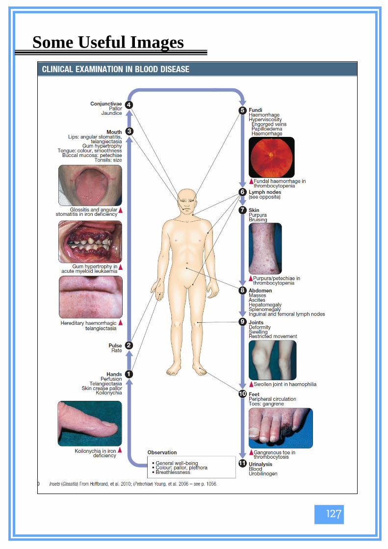
127
Some Useful Images

128

129

130

131
Past Paper (These are just the available ones (from the past two or three years [2016-2018] only))
* Take history form a patient “with symptoms of anemia” (+ hints like CBC) to know the
diagnosis.
* Take history from a patient “with symptoms of anemia” (+ hints like CBC) to know the
diagnosis. What is the diagnosis?
The answer was one of the hemolytic anemias.
* “A female patient presented to you complaining of fatigue, lightheadedness, and shortness
of breath since ---. Her Hemoglobin level was 8, MCV = 70…..”
1. Take brief history to know the cause of the symptoms
2. What is the diagnosis?
The answer was “Iron deficiency anemia, due to heavy menses” (after excluding other
causes while taking the history).
* “A man presented to you with fatigue, dyspnea, and disorientation. His hemoglobin level
= 5.4 and MCV =119, and probably some other hints.”
1. What is the diagnosis? It was vitamin B12 deficiency
2. Examine him specifically for the type of anemia he has. Don’t forget neurological
exam.
* Examine for hepatosplenomegaly.

1
Chapter 6: Nephrology
Case scenario: A 40-year old lady presented with red colored urine. Take a focused history of her
complaint.
Physical Examination
Start by introducing yourself and asking about patient profile (age,
gender,..)
HPI:
Characterize the hematuria
o Take the duration and frequency, was this the first time?
o Establish the color of the urine and character of blood (fresh
vs clots), clots are more typical of glomerular disease while
fresh of trauma or infection
o At what point during urination is the blood seen? (initial-
urethra, end-bladder neck or prostate , throughout – bladder
or kidney.
o Associated with pain? If yes, ask SOCRATES. Site: Flank
(renal stones), Loin (Loin pain-hematuria), Suprapubic
(cystitis)
o Apperance of the urine? Frothy? Indicates GN
o Exacerbated by cold or exercise?
o Constitutional symptoms (fever, chills, weight
loss,fatigue,nightsweats) TB, Malignancy
o Other urinary symptoms (dysuria, frequency, urgency)
o Specific symptoms: CVS (heart failure), rheumatological
(joint pain) (SLE),hearing loss (Alport), bleeding tendecy and
bruising
o Recent trauma(rhabdomyolysis), infection (URTI, skin,
pharyingitis), or menustration.
o Strenous exercise or ingestion of food causing
pseudohematuria (blackberries, beetroot).
Meds: NSAIDs, Warfarin, Heparin, Aspirin, Penicillin,Rifampicin,
Rifabutin, Clofazamine, Entacapone
PMHx: HTN, kidney stones, heart disease, SLE, bleeding disorders
FHx: alport disease, HTN, malignancy, adult polycystic kidney
disease
SHx: smoking, occupational exposure to rubber, dyes
Hematuria
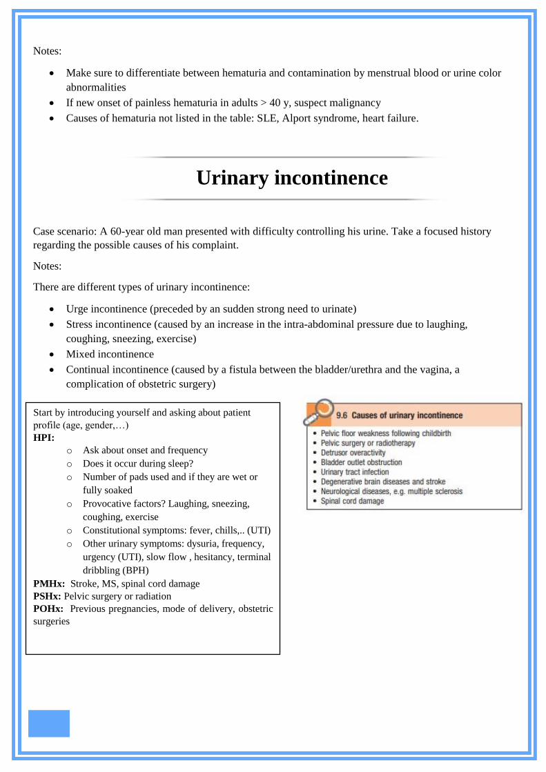
Notes:
• Make sure to differentiate between hematuria and contamination by menstrual blood or urine color
abnormalities
• If new onset of painless hematuria in adults > 40 y, suspect malignancy
• Causes of hematuria not listed in the table: SLE, Alport syndrome, heart failure.
Case scenario: A 60-year old man presented with difficulty controlling his urine. Take a focused history
regarding the possible causes of his complaint.
Notes:
There are different types of urinary incontinence:
• Urge incontinence (preceded by an sudden strong need to urinate)
• Stress incontinence (caused by an increase in the intra-abdominal pressure due to laughing,
coughing, sneezing, exercise)
• Mixed incontinence
• Continual incontinence (caused by a fistula between the bladder/urethra and the vagina, a
complication of obstetric surgery)
Start by introducing yourself and asking about patient
profile (age, gender,…)
HPI:
o Ask about onset and frequency
o Does it occur during sleep?
o Number of pads used and if they are wet or
fully soaked
o Provocative factors? Laughing, sneezing,
coughing, exercise
o Constitutional symptoms: fever, chills,.. (UTI)
o Other urinary symptoms: dysuria, frequency,
urgency (UTI), slow flow , hesitancy, terminal
dribbling (BPH)
PMHx: Stroke, MS, spinal cord damage
PSHx: Pelvic surgery or radiation
POHx: Previous pregnancies, mode of delivery, obstetric
surgeries
Urinary incontinence

3
Case scenario: A 6-year old boy presented with facial puffiness. Urine dipstick showed +3 protein, take a
focused history on the possible causes of his condition. What is your diagnosis?
• Classical features:
1. Nephrotic range proteinuria >3.5 g/day
2. Hypoalbuminemia
3. Hyperlipidemia
4. Edema
Other features: hypertension, recurrent infections and hypercoagulable state
• UA:
Inspection: frothy urine
Dipstick: +3 protein
Microscopy: fatty/waxy casts
Primary
1. Minimal Change Disease
-Commonly seen in children
-Either idiopathic or secondary to
Hodgkin's lymphoma or drugs (NSAIDs,
Lithium)
-Not associated with HTN
2. Focal Segmental Glomerulosclerosis
-Either idiopathic or secondary to
diseases (sickle cell disease, lymphoma,
HIV, or other GN) or drugs (heroin,
lithium)
3. Membranous Glomerulonephritis
-Most commonly seen in adults
-Can be idiopathic OR
-Secondary to:
1. Drugs (gold, penicillamine)
2. Solid Malignancies (lung, breast,
colon, prostate)
3. Infections (hepatitis B/C, malaria,
streptococcal, schistosomiasis)
4. Autoimmune diseases (SLE, RA,
sjogren, sarcoidosis)
Secondary
1. Diabetic nephropathy
-Occurs in both type 1 and 2
2. Amyloidosis
-Causes:
1. Familial Mediterranean Fever (FMF)
Symptoms:
Paroxysmal attacks of fever, abdominal
pain, chest pain.
*Ask if the patient takes colchicine to
prevent amyloidosis.
2. Multiple Myeloma
Symptoms:
CRAB
C: Hypercalcemia (nausea, vomiting,
abdominal pain, constipation…. Etc)
R: Renal failure
A: Anemia (dizziness, SOB on exertion,
fatigue)
B: Bone lesions (bone pain)
Nephrotic Syndrome
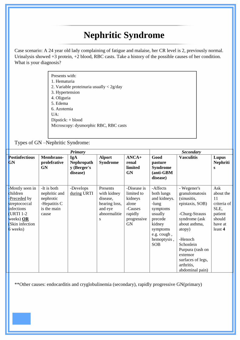
Case scenario: A 24 year old lady complaining of fatigue and malaise, her CR level is 2, previously normal.
Urinalysis showed +3 protein, +2 blood, RBC casts. Take a history of the possible causes of her condition.
What is your diagnosis?
Types of GN –Nephritic Syndrome:
Secondary Primary
Lupus
Nephriti
s
Vasculitis Good
pasture
Syndrome
(anti-GBM
disease)
ANCA+
renal
limited
GN
Alport
Syndrome
IgA
Nephropath
y (Berger's
disease)
Membrano-
prolefrative
GN
Postinfectious
GN
Ask
about the
11
criteria of
SLE,
patient
should
have at
least 4
- Wegener's
granulomatosis
(sinusitis,
epistaxis, SOB)
-Churg-Strauss
syndrome (ask
about asthma,
atopy)
-Henoch
Schonlein
Purpura (rash on
extensor
surfaces of legs,
arthritis,
abdominal pain)
-Affects
both lungs
and kidneys.
-lung
symptoms
usually
precede
kidney
symptoms
e.g. cough ,
hemoptysis ,
SOB
-Disease is
limited to
kidneys
alone
-Causes
rapidly
progressive
GN
Presents
with kidney
disease,
hearing loss,
and eye
abnormalitie
s
-Develops
during URTI
-It is both
nephritic and
nephrotic
-Hepatitis C
is the main
cause
-Mostly seen in
children
-Preceded by
streptococcal
infections
(URTI 1-2
weeks) OR
(Skin infection
6 weeks)
**Other causes: endocarditis and cryglobulinemia (secondary), rapidly progressive GN(primary)
Presents with:
1. Hematuria
2. Variable proteinuria usually < 2g/day
3. Hypertension
4. Oliguria
5. Edema
6. Azotemia
UA:
Dipstick: + blood
Microscopy: dysmorphic RBC, RBC casts
Nephritic Syndrome

5
Case scenario: A 37 year old lady presented to the ER with 48-hour diarrhea and vomiting. She can't
remember passing urine today. Her labs showed CR level of 1.9, it was previously normal. What is your
diagnosis? What are the diagnostic criteria? Take a detailed history of other possible causes of her condition.
Kidney Disease: Improving Global Outcomes (KDIGO) defines AKI as any one of the following
1- Increase in serum creatinine by 0.3 mg/dL or more from baseline within 48 hours OR
2- Increase in serum creatinine to 1.5 times baseline within one week OR
3- Urine output less than 0.5 mL/kg/h for 6 hours.
Causes of AKI
Pre-renal (caused by
underperfusion of the kidneys,
either from true loss of volume or
decreased effective arterial blood
flow)
Renal(caused by a dysfunction in
any part of the kidney, either the
glomeruli, tubules, interstitium or
vasculature)
Post-renal( caused by obstruction
to the urine flow)
1.Hypovolemia
Causes:
a. Dehydration which could be due
to poor fluid intake, working in
hot conditions, vomiting*,
diarrhea*, polyuria, burns, third
spacing (e.g. ascites) or excessive
diuretic use
*Symptoms of gastroenteritis
b. Blood loss
1.Acute tubular necrosis
Caused by either ischemia or a
nephrotoxin
a. Ischemic ATN can be due to
shock or any condition that causes
sudden hypoperfusion
b. Nephrotoxic ATN causes
include: free myoglobin
(rhabdomyolysis) , free
hemoglobin (intravascular
hemolysis), contrast, antibiotics
(e.g.
aminoglycosides,vancomycin)
Antifungal (e.g. amphotericin B)
chemotherapeutic agents (e.g.
cisplastin) , NSAIDs
1.Urethral obstruction
It commonly occurs due to benign
prostatic hyperplasia, symptoms
are: hesitancy, slow flow,
incomplete emptying ,frequency, ,
terminal dribbling, and nocturia
Other causes: kidney stones,
tumors, vaginal or uterine prolapse
2.Hypotension
Causes:
a. Sepsis
b. Hypovolemia
d. Excessive use of
antihypertensive medications
2. Glomerulonephritis
Symptoms include frothy urine,
red colored urine, swelling
2. Bilateral ureteral obstruction or
unilateral obstruction of a solitary
functioning kidney
Ask about symptoms of kidney
stones or family history of renal
tubular acidosis type 1 which
predisposes to formation of
calcium kidney stones
Other causes: retroperitoneal
fibrosis
Acute Kidney Injury
(a(AKI

***Exclude pre-renal and post-renal causes before moving to renal causes of AKI
3.Decreased cardiac output
Occurs in CHF.
3. Acute interstitial disease
It is usually a result of an allergic
rxn to a drug e.g. antibiotics,
NSAIDs, or PPI
Symptoms include: fever, skin
rash, swelling
Less commonly it can be due to an
infection e.g. pyelonephritis
3. Medications which crystalize in
the urine:
1.Methotroxate
2.Ganciclovir
3.Acyclovir
4.Vascular disease limiting renal
blood flow (embolus)
Does not present with any specific
symptom. It usually causes
elevated blood pressure that is
poorly controlled with drugs
4. Vascular disease
a. Renal artery/vein thrombosis
b. Thrombotic thrombocytopenic
purpura (TTP). Symptoms
include: neurological (e.g.
confusion, seizures) , bruising,
pallor and fatigue
c. Hemolytic-uremic syndrome
(HUS), it is the most common
cause of AKI in children
5.Medications which impair renal
autoregulation
a.NSAIDs
b.ACE-I/ARB
c.Cyclosporin
6.Hepatorenal syndrome
Ask about causes of cirrhosis and
symptoms of portal hypertension
e.g. abdominal distention,
vomiting blood, black tarry stool,
bleeding tendency, infections,
dizziness, SOB on exertion

7
Case scenario: A 72 year old man referred to you after routine monitoring of his renal function demonstrated
steadily increasing CR levels. Renal ultrasound showed small kidneys and his BP is 150/92. Take a focused
history regarding the possible causes of his condition.
CKD is defined as any of the following:
1. Kidney damage for > 3 months, with or without decrease in GFR, with either pathological abnormalities or
markers of kidney damage.
2. GFR < 60 mL/min/1.73m2 for >3 months, with or without kidney damage.
Stages of CKD:
GFR Description Stage
=>90 Kidney damage
with normal or
high GFR
CKD1
60-89 Kidney damage
with mild
decrease in GFR
CKD2
30-59 Moderate
decrease in GFR
CKD3
15-30 Severe decrease
in GFR
CKD4
<15 End-stage kidney
disease (dialysis
requiring)
CKD5
Most common causes of CKD:
1. Diabetes
2. Hypertension
3. Chronic GN
4. Vascular diseases e.g. renal artery stenosis, renal vein thrombosis
4. Unrecovered AKI
5. Polycystic kidney disease (consanguineous family)
6. Congenital defects in the kidney or bladder
7. Recurrent kidney stone formation
6. Long-term use of drugs that affect the kidneys e.g. NSAIDs
Chronic Kidney Injury
InjuryI(a(AKI
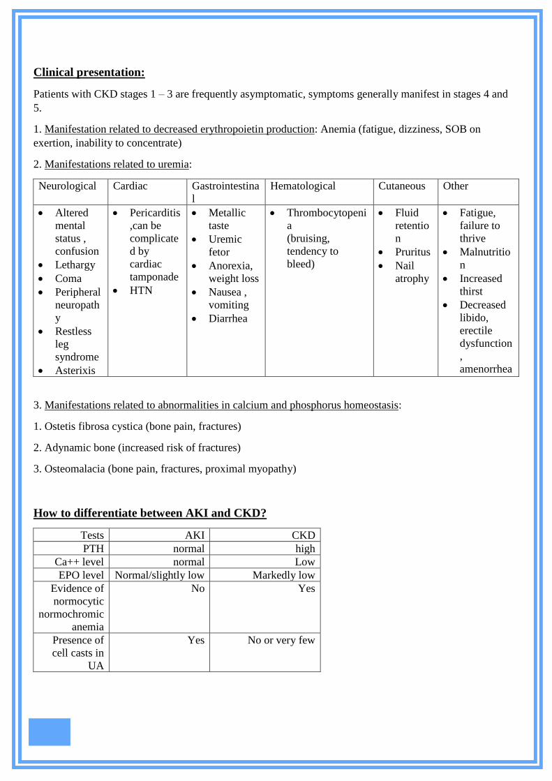
Clinical presentation:
Patients with CKD stages 1 – 3 are frequently asymptomatic, symptoms generally manifest in stages 4 and
5.
1. Manifestation related to decreased erythropoietin production: Anemia (fatigue, dizziness, SOB on
exertion, inability to concentrate)
2. Manifestations related to uremia:
Other Cutaneous Hematological Gastrointestina
l
Cardiac Neurological
• Fatigue,
failure to
thrive
• Malnutritio
n
• Increased
thirst
• Decreased
libido,
erectile
dysfunction
,
amenorrhea
• Fluid
retentio
n
• Pruritus
• Nail
atrophy
• Thrombocytopeni
a
(bruising,
tendency to
bleed)
• Metallic
taste
• Uremic
fetor
• Anorexia,
weight loss
• Nausea ,
vomiting
• Diarrhea
• Pericarditis
,can be
complicate
d by
cardiac
tamponade
• HTN
• Altered
mental
status ,
confusion
• Lethargy
• Coma
• Peripheral
neuropath
y
• Restless
leg
syndrome
• Asterixis
3. Manifestations related to abnormalities in calcium and phosphorus homeostasis:
1. Ostetis fibrosa cystica (bone pain, fractures)
2. Adynamic bone (increased risk of fractures)
3. Osteomalacia (bone pain, fractures, proximal myopathy)
How to differentiate between AKI and CKD?
CKD AKI Tests
high normal PTH
Low normal Ca++ level
Markedly low Normal/slightly low EPO level
Yes No Evidence of
normocytic
normochromic
anemia
No or very few Yes Presence of
cell casts in
UA

9
Case scenario: A 26 year old lady, diagnosed with HTN 8 months ago, is complaining of poorly controlled
hypertension despite her compliance to treatment. Take a focused history on the possible causes of her
condition.
Primary HTN
Risk factors:
1. Age : >55 y for males, >65 y
for females
2. Gender: males>females
3. Race: African Americans
4. Obesity: BMI =>30
6. Alcohol
7. Smoking
8. Dyslipidemia
9. Salt intake
10. Diabetes
11. Heart disease
12. CKD
13. Family history
14. Stress
15. Sedentary lifestyle
Secondary HTN
When to suspect secondary
HTN?
1. If new onset of HTN at a
young age <30 y or at an old
age >50 y
2. If poorly controlled with
drugs (>3-4 drugs)
3. If there are clinical or lab
features specific for a disease
e.g. episodic
HTN/flushing/palpitations,
low TSH, hypokalemia
Causes:
1. Renal diseases: adult
polycystic kidney disease,
renal artery stenosis,
glomerular disease.
2. Endocrine diseases: cushing
syndrome, conn's syndrome,
pheochromocytoma, hyper/
hypothyroisidm,
hyperparathyroidism
3. Coarctation of the aorta
4. Obstructive sleep apnea
5. Pregnancy
6. Medications: OCP,
NSAIDS, TCA, cyclosporine,
steroids.
Few hints to help you ask about
these possible causes are found in
the next page.
Classification of blood pressure ranges (2017):
• Normal =< 120/<80
• Prehypertension 120-139/80-89
• HTN, Stage 1: 140-159/90-99
• HTN, Stage 2: >160/>100
Hypertension

Physical signs related to secondary causes and complications of HTN
Causes of secondary HTN that you can ask about it:
Cushing’s syndrome: ask about weight gain, striae ( بالجلد تششققات ), proximal myopathy, having a
moon face.
Pheochromocytoma: palpitations, headaches, dizziness, flushing.
Hyperthyroidism: heat intolerance, weight loss, tremor, excessive sweating.
Hypothyroidism: cold intolerance, weight gain, hair loss.
OSA: snoring, restless sleep, increased daytime sleepiness.
Ask about anxiety or recurrent panic attacks ( ذعر من غير سبب حاالت)

11
General principles:
• Clean catch (midstream urine sample)
• < 4 hours old
Sequence of urinalysis
Inspection of urine sample
Assessment of color and clarity:
o Normal fresh urine is clear but varies in color
o Cloudy urine indicates presence of leukocytes (bacterial infection)
o Abnormalities of urine color are mentioned in table 9.7
Dipstick test
Mentioned in the table
Microscopy
o Bacteria: distinguish infecting organism from amorphous material, use gram stain and culture to
identify organism >105 CFU/mL of urine (UTI)
o Cells: dysmorphic RBCs ( glomerular disease), monomorphic RBCs (renal stones or UTI), WBCs
(UTI)
o Casts: RBC casts (glomerular disease), WBC casts (pyelonephritis, interstitial nephritis), Muddy
brown (epithelial) casts (ATN)
o Crystals: rarely diagnostic. Hexagonal cysteine crystals (cystinuria)
Urinalysis

General:
• Assess the general appearance and consciousness level
Look for fatigue, pallor, breathlessness, uremic (lemon-yellow) complexion, cushingoid appearance and
hirsutism (related to steroid therapy and cyclosporine respectively)
• Vital signs
Measure temperature and blood pressure and check for pulsus paradoxus (do not use the arm with the AV
fistula) (usually elevated except in tubulointerstitial disease and uremic pericarditis)
• Head
1. Look at the eyes for conjuctival pallor of anemia and across the cornea for band keratopathy or at the edge
for limbic calcifications (related to hypercalcemia)
2. Smell the patient's breath for uremic fetor
• Hands
1. Inspect for nail changes: half and half nails, Leukonychia, Muehrecke's nails, Beau's lines, Splinter
hemorrhage
2. Examine for asterixis
3. Inspect for AV fistula
4. Examine for Carpal Tunnel Syndrome: Phalen's and Tinel's test
Abdominal Examination:
Position: supine with one pillow
Exposure: Nipple to mid-thigh, for social concerns from xiphisternum to symphysis pubis
• Inspection
1. Look for distention (from enlarged kidneys in APKD or rarely obstructive uropathy) , or suprapubically
from bladder distention
2. Look in the loin for scars of renal tract surgeries or in the iliac fossa for those of transplant surgery. You
may see a peritoneal catheter or small scars left in the midline and hypochondrium by one.
• Palpation
1. Use the fingers of your right hand and start and the right lower quadrant
- A distended bladder is felt as a smooth firm mass arising from the pelvis which disappears with urethral
catheterization
- Polycystic kidneys have a distinctive nodular surface
2. For lesser degrees of kidney enlargement, place one hand behind the patient's back below the ribs, and
one hand anteriorly over the upper quadrant and gently but firmly push your hands together as the patient
breathes in and out
3. Ask the patient to sit up and palpate for renal angle tenderness (pyelonephritis)
• Percussion
Percussion of kidneys is not helpful
• Auscultation
Auscultate for renal artery bruit over both loins posteriorly and in the epigastrium
• Test for Ascites
• In men, examine external genitalia and do PR to assess the prostate for benign and malignant
causes

13
• In women, perform vaginal examination to exclude pelvis malignancy and to assess prolapse
Cardiovascular examination:
• Look for pitting edema in the ankle, sacrum and the back of the thighs in recumbent patient
• Asses the JVP
High JVP in cardiac tamponade due to uremic pericarditis and low JVP in nephrotic syndrome although edema
is present
• Palpate the apex beat
Displaced in fluid overload or heart failure
• Auscultate for:
1. Mid-systolic flow murmur
2. 3rd/4th heart sounds
3. Pericardial friction rubs
Respiratory examination:
• Percuss the chest to detect pleural effusion
• Auscultate for bilateral lung crackles indicating fluid overload or heart failure.
Neurological examination:
• Assess the level of consciousness
• Test sensation and tendon reflexes.
Peripheral neuropathy occurs in advanced CKD
• Examine the optic fundi
Retinopathy is found in DM and HTN

References:
1. Macloed clinical examination 13th edition
2. Medstudy 16th edition
3. Medscape
4. ACA/AHA High Blood Pressure Guidelines – American College of Cardiology
5. OSCE dossier
6. Hashem Ahmad, Sharifeh Masaid and Amer Abu Shanab notes

146
Chapter 7: Rheumatology
• RA is a chronic inflammatory disease results from immune-complex mediated
vasculitis (type III hypersensitivity), it affects almost any organ system:
➢ The commonest presentation is symmetrical polyarticular joint pain.
➢ It affects females more than males.
--------------------------------------------------------------------------------------------------------------------
OSCE Vignette: A 45 years old female presented to your clinic complaining of
stiffness in her shoulders, hands, knees, and hips. The stiffness lasted for about an hour
every morning. After her wrists and fingers began to swell she decided to visit your
clinic, take a focused history looking for the symptoms of RA.
- Patient profile: Sex (F>M) & Age (40-60),
- Chief complaint: Symmetrical polyarticular (large & small joint pain) for X duration
- History of presenting illness:
➢ Ask about articular symptoms:
1. Joint pain (SOCRATES), usually symmetrical, affecting the hands (except
DIP), elbows, knees, ankle & feet.
2. Morning stiffness (for 1 hour)
3. Gel phenomenon (stiffness after long period of rest).
4. Locking, weakness, swelling
5. Joint hotness, redness, swelling (usually absent).
6. Neck fractures
➢ Ask about extraarticular symptoms:
1. Neuropathy (weakness, numbness & paralysis). “mononeuritis multiplex”
2. Dry eyes, dry mouth. “Sjogren's /Sicca Syndrome”
3. Eye problems (redness, burning, blurry vision) “uveitis, episcleritis”
4. Chest pain, pericardial pain, SOB, cough, wheeze, hemoptysis “pericarditis.
Pericardial effusion, plural effusion, pulmonary fibrosis”
5. Raynaud’s phenomenon, skin rash.
6. Signs of anemia.
RA

147
➢ Ask about the constitutional symptoms:
1. Low grade fever.
2. Fatigue.
3. Anorexia.
4. Wight loss.
5. Depression.
6. Restriction of normal life activities.
- Previous medical & surgical Hx: DM, HTN, IHD, strokes.
- Drugs Hx: Steroids, ACEIs, Antiepileptics.
- Family history of: RA & other autoimmune disease.
- Social history:
➢ Smoking, alcohol, drug misuse.
- Work-up:
➢ RF, ESR, CRP.
➢ Normocytic normochromic anemia.
➢ Radiography: loss of juxta articular bone, narrowing of joint space, bony erosions at
the margin of the joint, synovial fluid analysis.
OSCE Vignette:
A 47-year-old woman presents with a 2 month history of progressive weakness in her thighs
and upper arms. On examination, proximal muscle strength is symmetrically reduced but
distal muscle strength is normal. Ask the patient relevant questions to distinguish between
polymyositis and dermatomyositis.
Both polymyositis and dermatomyositis cause proximal muscle weakness. The difference is
that polymyositis does not involve skin, whereas dermatomyositis is associated with
characteristic skin rash.
History is directed at:
1- Common features between these two conditions.
2- The characteristic skin rashes associated with dermatomyositis.
3- Asking relevant questions to help exclude other differential diagnoses.
Proximal Muscle Weakness

148
History Points: (Try to memorize them in your way and order, so as to remember them in
the exam).
• General questions about the complaint and common features of PM and DM:
1- Make sure that it’s a chronic muscle weakness (acute muscle weakness is
more likely to be stroke).
2- Ask about muscle weakness:
a) Do you have: difficulty getting out of bed (neck muscles weakness), difficulty
combing your hair or getting out of a chair unaided (proximal muscle weakness of
arms and thighs, respectively)?
b) Make sure the patient does not have features suggestive of distal muscle weakness.
Do you have problems with writing or holding things with you hands?
3- Ask about common features of both PM and DM.
a) Myalgia (all of patients have weakness but only 1/3 have pain)
b) Dysphagia (remember the upper third of the esophagus is formed of
skeletal muscles).

149
• Questions related to the characteristic signs of dermatomyositis:
1- Do you have any skin rash?
If yes, ask him about the specific skin rashes.
a) Heliotrope rash (butterfly)—around eyes, bridge of nose, cheeks.
b) Gottron papules—papular, erythematous, scaly lesions over the knuckles
(MCP, PIP, DIP).
c) V sign—rash on the face, neck, and anterior chest.
d) Shawl sign—rash on shoulders and upper back, elbows, and knees.
e) Periungual erythema with telangiectasia.
f) Subcutaneous calcifications in children—can be extremely painful.
• Always remember that dermatomyositis is
associated with malignancy. Once you
diagnose dermatomyositis, you have to
exclude an occult malignancy in lung,
breast, ovary, GI tract, and
myeloproliferative neoplasms).
• Drug history:
- Ask about steroid and statins (they both cause
myalgia and proximal muscle weakness).
• Ask about any significant family history.

150

151
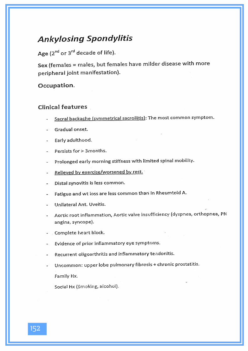
152

153

154
It is important to establish the pattern of joint pain from the following points; so you reach to a
diagnosis more easily:
Articular vs non-articular (joint itself or surrounding structures such as tendons)
Inflammatory vs non-inflammatory (e.g. rheumatoid vs osteoarthritis)
Acute vs chronic
Peripheral vs axial
Additive vs migratory (joints improve and new ones become involved) vs palindromic
(recurrences and relapses)
Inflammatory monoarthritis vs oligoarthritis vs polyarthritis
Joint pain is a pain! So:
I. Simply start with SOCRATES! 1- Site:
Firstly, make sure that the pain is joint pain (i.e. arthralgia)
not pain originating from other structures.
Secondly, ask the patient about the total number of the
joints causing the pain so that you know is it
mono/oligo/polyarthritis as this will guide you to the differential.
Notice whether the involved joints are mainly small joints, large. Peripheral vs
axial. Is it symmetrical involvement or asymmetrical.
2- Onset:
Is it chronic and gradual OR acute (which goes more with septic, gouty arthritis or
traumatic)
3- Character.
4- Radiation: radiation of pain is most likely a sign of nerve involvement.
5- Associated symptoms:
I. Morning stiffness: Ask about the presence of early-morning stiffness
and for how long stiffness lasts: is it for few minutes osteoarthritis ,
less than 30 minutes inflammatory arthritis, around 1 hourRA.
II. Redness and hotness: may be seen in septic joint or inflammatory
arthritis.
III. Swelling: can be inflammatory, septic or may be seen in hemarthrosis.
6- Timing (frequency, duration and periodicity of symptoms)
A history of several weeks of pain, early-morning stiffness and loss of
function is likely to be an inflammatory arthritis.
Approach to joint pain
One joint is a monoarthritis.
2–4 joints oligoarthritis.
>4 is polyarthritis

155
‘Flitting’ pain starting in one joint and moving to others over a period of days
is a feature of rheumatic fever and gonococcal arthritis.
If intermittent with resolution between episodes, it is likely to be palindromic
rheumatism.
7- Exacerbating and relieving factors:
Is it relieved by moving the joint inflammatory?
Is it exacerbated by moving the joint of non-inflammatory (most likely
osteoarthritis)?
Pain is there upon movement and rest septic arthritis is a possible cause.
8- Severity.
Then ask about:
II. Ask about loss of function.
III. Deformities.
IV. History of trauma or injection into a joint.
V. Ask about history of same complaint.
VI. Drugs: NSAID and thiazides (both associated with gout).
Here is a table summarizing the pattern of joint pain with other symptoms according to
the differential -most common ones-.

156
Look at the following table , just be familiar with important causes of joint pain

157
Case: A 40 year-old lady has a history of joint pain for the last 2 months. Examine
her hands for signs suggestive of the diagnosis.
-Greet your patient, Introduce yourself and ask for permission to perform the
examination.
-Wash your hands and ensure adequate privacy, warmth and illumination.
-Position the pt. seated upright, expose both hands up to the elbows and rest the hands
on a pillow.
-Ask for any site of pain to avoid touching or moving it.
Look • Look at the palmar aspect then at the dorsal aspect of both hands.
• At the palmar aspect look for palmar erythema and visible muscle wasting of the
thenar, hypothenar and small muscles of the hand.
• Look for psoriatic nail changes, onycholysis and telangiectasias at nail folds.
• Look for obvious deformities such as Arachnodactyly, Ulnar deviation, Anterior
displacement of the wrist and Duputyren’s contracture.
• Examine the lateral side of the fingers for Mallet finger deformity, Boutonniere
deformity, Swan Neck deformity and Z-thumb.
• Look at the finger tips for calcinosis suggestive of systemic sclerosis.
• Look at the interphalangeal joints for Gouty Tophi of Gouty arthritis.
• Look for Bouchard’s nodes (at PIPs) Heberden’s nodes (at DIPs) and squaring of
the wrist, all suggestive of Osteoarthritis.
• Look for swelling at metacarpophalangeal (MCP) joints and interphalangeal (IP)
joints. Ask the pt. to make a fist and look for hill-valley-hill-valley aspect of the
knuckles which is lost in Rheumatoid Arthritis (RA).
• Ask the pt. to flex the fingers pointing toward the scaphoid bone to look for
Rotational deformity.
• Note: if you detect any deformity, support the patient’s hand against a hard surface
and try to reverse it. If the deformity is reversible it’s due to SLE (Jaccoud
Arthropathy). If it’s not reversible it’s due to RA.
Feel • Examine the dorsum of the patient’s hand with the dorsum of yours to assess
temperature. Compare bilateral hands and assess the temperature of swellings –if
present-.
• Feel the muscle bulk of the thenar and hypothenar eminences for muscle wasting.
• Feel the consistency of swellings –if present- and determine if hard or soft.
• Squeeze with your thumb and index across each IP joint to detect sponginess
and/or tenderness.
Hand Examination

158
• Squeeze with your thumb and index across each MCP joint to detect sponginess
and/ or tenderness. Ask the pt to move his fingers while you do so as to detect
crepitations.
• Palpate the flexor tendon sheaths for swelling or tenderness.
• Feel the extensor surface of the forearm for Rheumatoid Nodules.
Move (Always start with active movement, then examine passively if the pt. can’t move
actively)
• Ask the pt. to make a fist then fully extend his fingers.
• Ask the pt. to perform Prayer’s sign (to assess extension of the wrist) and Reverse
Prayer’s sign (to assess flexion of the wrist), Ulnar deviation (to assess adduction )
and Radial deviation (to assess abduction).
• Test the patient’s grip by asking him to squeeze your index and middle finger and
compare bilaterally.
• To detect triggering, move one joint while fixating the other joints, one at a time.
Calcinosis in systemic sclerosis
Gouty Tophus
Loss of Hill-Valley-Hill-Valley aspect in RA Telangiectasias at nail fold
Rotational deformity

159
Bouchard’s nodes (at PIP joints), Heberden’s node (at DIP joints) and Squaring of the wrist in OA
Rheumatoid nodules at extensor surfaces
Z thumb deformity in RA

160
***This subject is more likely to be in the orthopedic rotation not in the internal
medicine OSCE. But it was included here just in case.***
-Greet your patient, Introduce yourself and ask for permission to perform the
examination.
-Wash your hands and ensure adequate privacy, warmth and illumination.
-Start your examination with the patient standing then in-supine position, expose both
lower limbs up to iliac crests.
-Ask for any site of pain to avoid touching or moving it.
Look
• While the patient is standing examine his gait by asking him to walk a straight path
head and forth.
• Inspect while the pt. is standing looking for Valgus or Varus deformity, Patellar
hollow, obvious muscle wasting (Vastus medialis, calf muscles and quadriceps),
Housemaid’s knee and Baker’s cyst.
Knee Examination
Housemaid’s knee
Baker’s cyst

161
• Ask the patient to lie in a supine position, stand to the right side of the pt. and
comment on the position of the patella, redness, hotness, scars, sinuses, hair
distribution, asymmetry, swelling and sutures.
Feel
• Feel the temperature of the joint with the dorsum of your hand and compare both
limbs.
• Feel the patella, the femoral condyles, Joint lines, Head of fibula, the patellar
tendon and the quadriceps tendon, the tibial tuberosity and comment on tenderness.
• Feel the posterior aspect of the knee and the hamstring muscles.
Move
• Start by examining the active range of motion then examine passively.
• Ask the pt. to flex his knee. Normal range of flexion is 0-140o. Abnormal range of
flexion is when the movement stops before the calf touches the thigh.
• Ask the pt. to extend his knee. Full extension is to 0 degrees.
• Listen for crepitus when the pt. flexes or extends the joint.
Special Tests
• McMurray’s test (for medial and lateral menisci)
Flex the knee to 90o, feel the joint lines with your left hand (between the femoral
condyles and the tibial head) and hold the foot with your right hand.
To examine the lateral meniscus, flex the knee then extend while internally
rotating the foot and place the knee in a varus position (Figure A). A sensation of
clunk on the lateral side of the joint is a positive test.
To examine the medial meniscus, flex the knee then extend while externally
rotating the foot and place the knee in a valgus position (Figure B). A sensation of
clunk on the medial side of the joint is a positive test.
Repeat the flexion-extension cycles a number of times, and reduce the degree of
flexion each time.
Figure A Figure B

162
• Medial and Lateral collateral test
Extend the knee fully, hold the foot and place it between your elbow and waist.
Place one hand on the lateral joint line and the other hand on the medial line.
To test the medial collateral ligament: Move your body to force a valgus stress
and feel for an opening in the medial joint line. Repeat while flexing the knee to
30o. If an opening could still be felt it indicates a defected medial collateral
ligament.
To test the lateral collateral ligament: Move your body to force a varus stress and
feel for an opening in the lateral joint line. Repeat while flexing the knee to 30o. If
an opening could still be felt it indicates a defected lateral collateral ligament.
• Drawer’s test (For Anterior and Posterior Cruciate Ligaments/ACL and PCL)
Flex the patient’s knee to 90o ask for their permission to sit on the foot to prevent
it from rotating. Place both hands the tibia by placing your thumbs on the tibial
tuberosity and your fingers along the popliteal fossa.
Perform the posterior drawer test first by moving the tibia away from you, any
translocation indicates possible PCL rupture.
Look for posterior sag before performing the anterior drawer test, then move the
tibia towards you, any translocation that is >5mm indicates possible ACL rupture.

163
• Lachman’s test (for ACL)
Ask the patient to extend his knee and relax it. Place your knee on the bed under
the patient’s knee and hold the distal end of femur with your left hand and the
proximal end of tibia with your right hand then try to translocate the tibia on the
femur anteriorly. You should feel a stiff end point that indicates normal ACL.
Absence of abrupt stiffness indicates possible ACL rupture (positive test).
Testing for Knee effusion:
• The Patellar Tap:
With the patient’s knee extended, empty the suprapatellar pouch by sliding your left
hand down the thigh until you reach the upper edge of the patella.

164
Keep your hand there and, with the fingertips of your right hand, press down briskly
and firmly over the patella. In a moderate-sized effusion you will feel a tapping
sensation as the patella strikes the femur. You may feel a fluid impulse in your left
hand.
• The ‘Bulge or Ripple test’:
With the patient’s knee extended and the quadriceps muscles relaxed, empty the
medial side of the joint by milking the fluid to the suprapatellar pouch. Now empty the
suprapatellar pouch as for the patellar tap. Stroke the lateral side of the joint. Watch
the medial side for a bulge or ripple as fluid reaccumulates on that side.

165
Chapter 8: Infectious Disease
1- Normal body temperature:
• The ‘normal’ oral or ear temperature is 37°C but may range between 35.8°C and
37.2°C (98–99°F).
➢ Peripheral methods of monitoring temperature are: tympanic membrane, temporal
artery, axillary, and oral thermometry.
➢ Central methods are: pulmonary artery catheter, urinary bladder, esophageal,
and rectal thermometry).
➢ Peripheral methods of monitoring temperature are not as accurate as central methods,
but central methods are less practical than peripheral methods.
2- Definition of Symptoms:
• Fever (pyrexia) is a body temperature >99th percentile of the healthy adult
maximum. Fever is defined as a single spike of temperature above 38oc per 24 hours,
or two spikes of temperature between 37.2oc and 38oc per 24 hours, or sustained
temperature between 37.2oc and 38oc for at least 2 hours per 24 hours.
➢ Fever is a sign, which is the objective measure, the subjective measure (symptom) of
fever is “feeling of hotness”.
➢ A patient with a fever is described to be febrile.
➢ Fever occur due to elevation of the thermostat setting in the hypothalamic
thermoregulatory center.
• Chills & Rigors: Most resources uses these terms interchangeably, but they are not
the same:
➢ Rigors: are bouts of uncontrollable muscular shaking, often with ‘chattering’ teeth,
lasting for minutes. They are associated with rapid temperature rises and may be
caused by cytokines and acute-phase proteins resetting the hypothalamic temperature
set point. Subjectively the patient feels cold and unwell and the episode may be
followed by sweating. A rigor may be associated with bacteraemia or malaria and is
of poor diagnostic value.
➢ Chills (goose skin) seen more commonly with viral infections.
Fever, Hyperthermia & Hyperpyrexia
Note: most of temperature cut points written here are controversial and differ according to
the resource. The cut points that are mentioned here are according to Dr. Jamal, so
memorize these values for the sake of the exam.
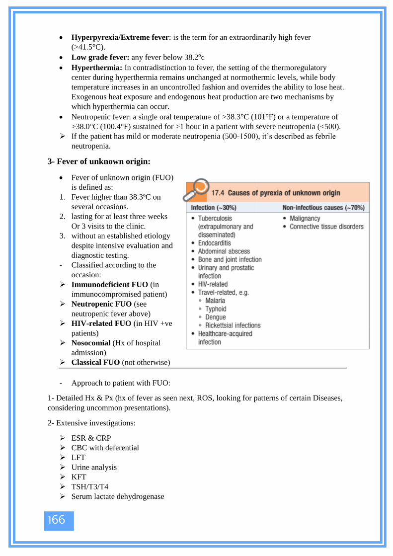
166
• Hyperpyrexia/Extreme fever: is the term for an extraordinarily high fever
(>41.5°C).
• Low grade fever: any fever below 38.2oc
• Hyperthermia: In contradistinction to fever, the setting of the thermoregulatory
center during hyperthermia remains unchanged at normothermic levels, while body
temperature increases in an uncontrolled fashion and overrides the ability to lose heat.
Exogenous heat exposure and endogenous heat production are two mechanisms by
which hyperthermia can occur.
• Neutropenic fever: a single oral temperature of >38.3°C (101°F) or a temperature of
>38.0°C (100.4°F) sustained for >1 hour in a patient with severe neutropenia (<500).
➢ If the patient has mild or moderate neutropenia (500-1500), it’s described as febrile
neutropenia.
3- Fever of unknown origin:
• Fever of unknown origin (FUO)
is defined as:
1. Fever higher than 38.3ºC on
several occasions.
2. lasting for at least three weeks
Or 3 visits to the clinic.
3. without an established etiology
despite intensive evaluation and
diagnostic testing.
- Classified according to the
occasion:
➢ Immunodeficient FUO (in
immunocompromised patient)
➢ Neutropenic FUO (see
neutropenic fever above)
➢ HIV-related FUO (in HIV +ve
patients)
➢ Nosocomial (Hx of hospital
admission)
➢ Classical FUO (not otherwise)
- Approach to patient with FUO:
1- Detailed Hx & Px (hx of fever as seen next, ROS, looking for patterns of certain Diseases,
considering uncommon presentations).
2- Extensive investigations:
➢ ESR & CRP
➢ CBC with deferential
➢ LFT
➢ Urine analysis
➢ KFT
➢ TSH/T3/T4
➢ Serum lactate dehydrogenase

167
➢ Tuberculin skin test or interferon-gamma release assay
➢ HIV antibody assay and HIV viral load for patients at high risk.
➢ Three routine blood cultures drawn from different sites over a period of at least
several hours without administering antibiotics, if not already performed
➢ Rheumatoid factor
➢ Creatine phosphokinase
➢ Heterophile antibody test in children and young adults
➢ Antinuclear antibodies
➢ Serum protein electrophoresis.
➢ Chest X-ray + CT
➢ Abdominal CT with contrast
➢ Echocardiogram.
3- If all these came negative, the patient is Saied to have FUO, what to do here?
➢ Give the patient antipyretics + hydration.
➢ Follow up every week (complete Hx or Px, to check for new complaints), no need to
repeat the work up unless there are new findings.
➢ These follow ups continue until the fever is gone or after 16 Weeks.
➢ At week number 16, do extensive investigations, if not thing is found, repeat the
cycle.
Hx of fever
⸎Patient’s profile: Age, marital state, occupation & residency all give clues about the cause.
⸎Chief complaint: Feeling of hotness for X days duration.
⸎HPI:
➢ How do the patient know they had pyrexia?
➢ Documented (measured) or not? What was the temperature?
➢ Onset (how it started)?
➢ Pattern (Day-night variation, frequency, progression & previous attacks):
Note: In FUO you’re looking for
needle in a haystack, so you should
have very low threshold to think
about a diagnosis.
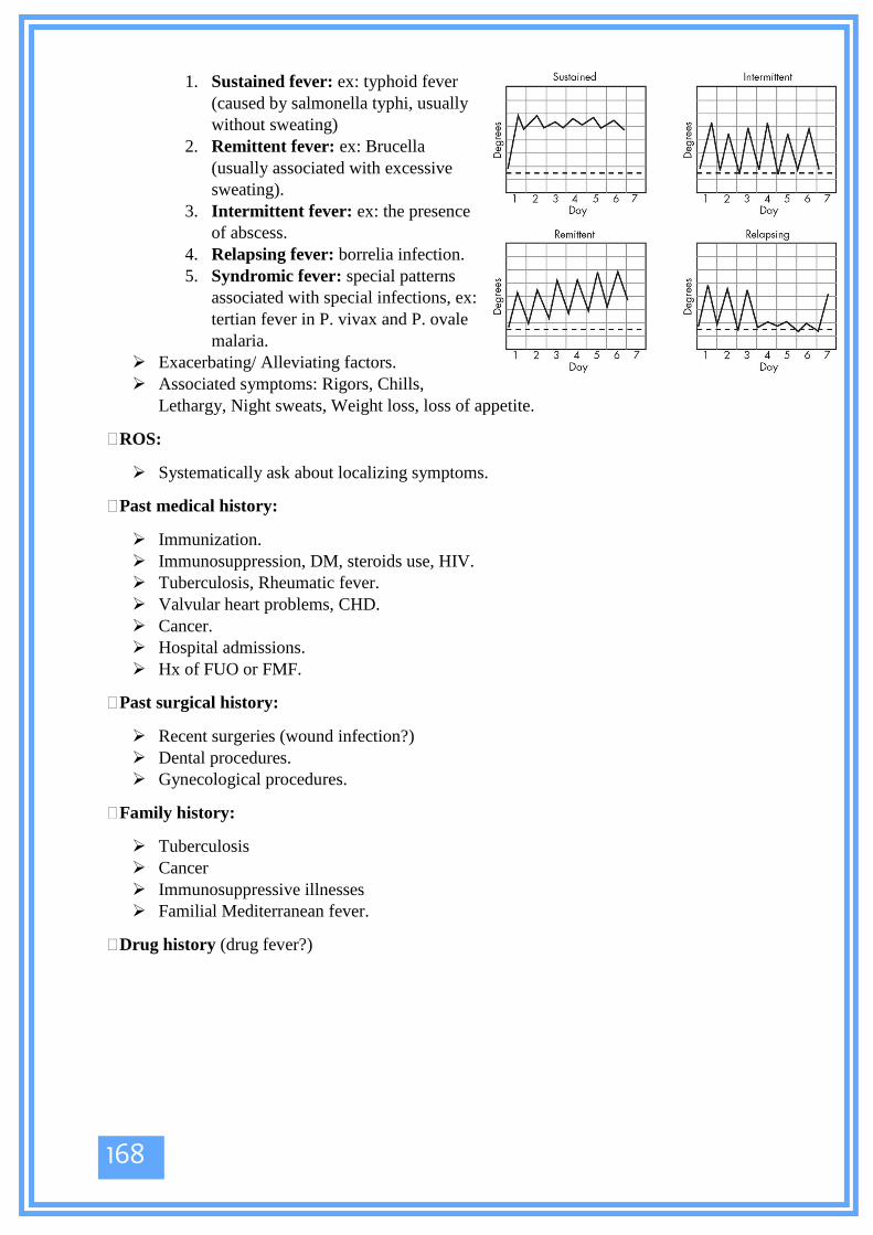
168
1. Sustained fever: ex: typhoid fever
(caused by salmonella typhi, usually
without sweating)
2. Remittent fever: ex: Brucella
(usually associated with excessive
sweating).
3. Intermittent fever: ex: the presence
of abscess.
4. Relapsing fever: borrelia infection.
5. Syndromic fever: special patterns
associated with special infections, ex:
tertian fever in P. vivax and P. ovale
malaria.
➢ Exacerbating/ Alleviating factors.
➢ Associated symptoms: Rigors, Chills,
Lethargy, Night sweats, Weight loss, loss of appetite.
⸎ROS:
➢ Systematically ask about localizing symptoms.
⸎Past medical history:
➢ Immunization.
➢ Immunosuppression, DM, steroids use, HIV.
➢ Tuberculosis, Rheumatic fever.
➢ Valvular heart problems, CHD.
➢ Cancer.
➢ Hospital admissions.
➢ Hx of FUO or FMF.
⸎Past surgical history:
➢ Recent surgeries (wound infection?)
➢ Dental procedures.
➢ Gynecological procedures.
⸎Family history:
➢ Tuberculosis
➢ Cancer
➢ Immunosuppressive illnesses
➢ Familial Mediterranean fever.
⸎Drug history (drug fever?)

169
⸎ Social history:
➢ Alcohol, smoking, illicit drug use.
➢ Recent travel history:
Where/when/what country?
Food, water, restaurants?
Did others on holiday have same symptoms?
Swimming in rivers, at coasts or in possible contaminated waters?
Insect/tick bites?
➢ Recent diet.
➢ Recent contact with animals or pits.
➢ Sexual history (if appropriate and only after signposting clearly)
⸎Brucellosis has a very high incidence in Mediterranean regions.
➢ It’s transmitted by Ingestion of non-pasteurized dairy products; most common route,
Ingestion of infected tissue or Slaughterhouse workers exposed to animal meat
through abraded skin.
➢ Fever is the most common symptom and sign, the infection spreads then and it can
cause a focal disease (Spondylodiscitis, endocarditis, CNS infections, epididymo-
orchitis)
⸎Patient profile: residency (rural area), occupation (Farmer, butcher, Diary products
production)
⸎Chief complaint: Fever for X duration.
⸎History of presenting illness:
➢ Fever Hx (as seen before): usually gradually onset remittent fever, worst at night, with
excessive sweating, chills, rigors, myalgia, Wight loss & loss of appetite.
➢ Focal symptoms (ROS):
1. CNS involvement: Headache, photophobia, neck stiffness, seizures &
confusion. (meningitis).
2. MSS: Low back pain (Spondylodiscitis/sacroiliitis), bone & joint pain.
3. GU: testicular swelling, Tenderness, painful/bloody ejaculation. (epididymo-
orchitis)
4. CVS & RS reviews, especially visitations (systemic thromboembolic events),
that indicates endocarditis.
Brucella (Malta fever)
170
⸎previous medical & surgical Hx (DM, AIDS, malignancy, malnutrition, vitamin
deficiency, immunosuppression state .. etc)
⸎Family history of Brucellosis.
⸎Social history:
➢ Pets/animals.
➢ Feeding habits (unpasteurized milk, uncooked meat)
➢ Travel history.
⸎Physical examination:
➢ General weakness, may be cachectic.
➢ Lymph node enlargement.
➢ GI exam: hepatosplenomegaly.
➢ MSS exam: Joint and bone tenderness, swelling, restriction of movement.
➢ CNS exam: signs of meningitis.
➢ CVS: new onset murmurs, janeway lesions, osler’s nodes, roth's spots.
➢ GU: testicular swelling & tenderness.
⸎DDx:
➢ TB.
➢ Typhoid fever.
➢ Endocarditis.
➢ Malignancy (Ex: lymphoma & multiple myeloma).
➢ HIV
⸎Work up:
➢ CBC, LFT.
➢ Blood cultures (positive in 50% of cases)
➢ Serology (Standard tube agglutination or ELISA) titers above 1/160 considered
+Ve.
➢ PCR
➢ Echocardiogram (to rule out endocarditis).

171
⸎ TB is caused by MTB mycobacterium tuberculosis.
➢ It is transmitted via an airborne droplet nucleus.
➢ Primary TB usually starts in the middle lobes, it has one of 3 fates, it either regress &
calcifies, or becomes latent TB or progresses into Primary pulmonary TB.
➢ Secondary TB: Either exogenous (new infection) or endogenous infection
(reactivation of a dormant primary lesion) in a person who has been sensitized by
earlier exposure.
➢ Miliary TB: Following a massive blood-borne dissemination of MTB, the
microorganism will infiltrate any organ.
➢ Extra-pulmonary TB: TB meningitis, MSS involvement (Pott’s disease), GI lesions
(mimics Crohn’s disease), Lymphadenitis, pericardial involvement, and rarely, GU &
Skin involvement.
⸎Patient profile: Age (more common in children), residency & occupation (crowded places)
⸎Chief complaint: A Respiratory symptom, ex: cough for 3 months duration.
⸎History of presenting illness:
➢ Ask about all respiratory symptoms (check the respiratory section):
1. Cough
2. Sputum production
3. Hemoptysis
4. Dyspnea
5. Wheezing
6. Chest pain
➢ Ask about the constitutional symptoms:
7. Fever.
8. Night sweats.
9. Malaise.
10. Fatigue.
11. Anorexia.
12. Wight loss.
➢ Focal symptoms (ROS):
1. CNS involvement: Headache, photophobia, neck stiffness, seizures &
confusion (Note: TB meningitis usually presents in a chronic pattern).
2. MSS: Bone & joint pain, pathologic fractures (fracture with a trivial trauma),
deformities (Kyphosis).
Tuberculosis (TB)

172
3. CVS: symptoms of constrictive pericarditis (Fatigue, chest pain, dyspnea,
prominent veins in the neck, hepatomegaly, without lower limb edema).
4. GI: Intestinal obstruction (Constipation, vomiting, abdominal distension, with
colicky abdominal pain), Anorectal ulcerations (anal fissure like symptoms;
tearing pain worst after defecation and persists for hours), generally mimics
Chron’s disease.
5. GU: Dysuria, hematuria, loin pain, testicular swelling, painful ejaculation,
bloody ejaculation.
⸎previous medical & surgical Hx (Childhood TB, DM, AIDS, malignancy, malnutrition,
vitamin deficiency, immunosuppression state .. etc)
⸎Family history of TB & malignancy.
⸎Social history:
➢ Travel history (if the patient is immigrant, or traveled to an endemic area)
➢ If he was a prisoner.
⸎DDx:
➢ According to the system involved (malignancy, Brucella, etc ..)
➢ Hint: TB can present in any way that you can think about, so it’s a part of any DDx
list :p
➢ DDx for pulmonary TB (cavitary lesions in X-ray):
1. Lung abscess/pneumonia
2. Lung cancer
3. Pulmonary infarction
4. Wegner’s granumalotosis
5. Progressive massive fibrosis
⸎Work up:
➢ Sputum stain & culture: 3 early morning samples or every 12 hours:
1. Ziehl-Neelsen stain
2. Culture: solid medium (Lowenstein-Jensen agar)
3. PCR: to differentiate it from atypical mycobacteria.
➢ CXR.
➢ Bronchial biopsy.

173
Characteristic symptoms of cystitis
symptoms of the risk factors
(prostatic hypertrophy for example)
⸎The urinary tract is sterile, if it’s colonized by bacteria this is UTI.
➢ UTIs require anatomical problem to occur, it’s the short urethra in females. In males,
however, it always indicates an underlaying problem (obstruction, strictures, stones)
➢ UTIs are either Cystitis (Urgency, frequency, Dysuria & suprapubic pain) or
pyelonephritis (all of the above symptoms + Fever & loin pain), the differentiation
between these infections is critical, as pyelonephritis carries higher risk for the
development od sepsis & MOF.
➢ The most common cause of UTI is E. Coli.
⸎Patient profile: Sex (more common in females)
⸎Chief complaint: dysuria for X days duration.
⸎History of presenting illness:
➢ Lower urinary symptoms (cystitis):
1. Dysuria.
2. Suprapubic pain?
3. Frequency?
4. Urgency?
5. Pyuria (pus in urine? smell in urine?)
6. Hematuria (can be seen in cystitis, pyelonephritis, stones, or tumors, so it’s
not specific)
7. Hesitancy.
8. Intermittency.
9. Poor stream.
10. Terminal drippling.
11. Feeling of incomplete voiding.
➢ Upper urinary symptoms (pyelonephritis)
1. Flank (loin) pain? Unilateral or bilateral? SOCRATES?
2. Fever
3. Rigors
4. Chills
5. Confusion
➢ ROS
UTI
174
⸎Our goal now is to ask about the risk factors (all these may be absent in females, as
being female is the most important risk factor)
➢ Past medical:
1. Kidney stones (ask about fluid intake as well).
2. History of prostatic hypertrophy and its symptoms (already covered
above).
3. History of DM & immunosuppression.
4. History of constipation (independent risk factor for UTI), appendicitis
(pelvic appendicitis may cause UTI) & fistulas (diverticulitis & Chron’s
disease may lead to colo-vesical fistula, resulting in fecalurea, pneumaturia
& recurrent UTI).
5. History of STDs (itching and burning sensation).
6. History of spinal cord injury, cauda equina, urinary retention.
➢ Past surgical:
1. History of instrumentation (Foley’s catheter, urethral surgery, etc ..)
strictures.
➢ Drugs history:
1. steroids, immunosuppressants.
2. Cyclophosphamide (may cause hemorrhagic cystitis)
➢ Family history: of UTIs, Kidney stones, malignancy.
➢ Social history:
1. Genital hygiene.
2. Sexual history:
• Some birth control devices increase the risk of UTI: spermicidal
& Diaphragms.
• Relationship between the intercourse & UTIs: some females
develop “honeymoon” UTI”.
• Oral or anal intercourse: increases the risk for UTI, especially in
males.

175
The
End

176
References:
1- Macleod Clinical Examination, 13th edition.
2- Step-up to Medicine 4th Edition.
3- Macleod Clinical Diagnosis 1st edition.
4- The Old OSCE dossier.
5- Medstudy 17th edition.