Embed Size (px)
Citation preview

INSIDE THIS ISSUE
5 Collaborating to Improve Spinal Infection Control
8 Highlights of Mayo Clinic Contributions to AAOS 2015 Programs
Current trends from the Department of Orthopedic Surgery
OrthopedicSurgeryUpdateVol. 9, No. 2, 2015
Leadership and Innovation in Oncological Reconstructions
Restoring pain-free mobility that improves a patient’s quality of life is chief among the satisfactions of orthopedic surgery. For Matthew P. Abdel, M.D., an orthopedic surgeon at Mayo Clinic in Rochester, Minnesota, this is especially true for reconstruction surgery after oncologic tumor resections.
The most grateful patients“Cancer is a difficult experience for many patients,” Dr. Abdel says. “The fact that we can most often make improvements they never expected is such a motivating and reward-ing experience. These are the most grateful patients in the world because they go from being bedridden, fighting an often end-stage, debilitating disease to bearing weight on their
legs the day after surgery, allowing for more quality time to spend with family.”
Innovation and expertiseThe team’s special emphasis is on periacetabu-lar tumors. Often these require radical recon-structions that lack natural anatomy to build upon (Figures 1A-D).
Mayo’s successful innovation in the field is based on the leadership of the orthopedic surgery team of Franklin H. Sim, M.D., and David G. Lewallen, M.D., at Mayo Clinic’s campus in Min-nesota. Their pioneering expertise and refined techniques helped develop team-based protocols and designed structural elements that include highly porous metal components such as cups and augments reinforced with cages (Figure 2).
Figure 1A-D. Imaging and planning steps in a complex acetabular reconstruction with highly porous metals for primary chondrosarcoma. 1A. Preoperative oblique radiograph depicting periacetabular chondrosarcoma tumor in a 39-year-old male. 1B. Preoperative coronal MRI sec-tion of periacetabular chondrosarcoma tumor. 1C. Model depicting planned resection of the periacetabular chondrosarcoma. 1D. Illustration of the planned resection and possible remaining host bone.
B C DA

“A key reason we maintain a high-level oncology and reconstructive practice is our ability to work as a team to develop surgical techniques and implants to improve patient care and outcomes. Our group continues to care for patients from around the U.S. and around the world with challenging pelvic cancers,” explains Peter S. Rose, M.D., an orthopedic surgeon at Mayo Clinic’s campus in Minnesota, and Dr. Abdel’s teammate who performs the tumor resections.
Reconstruction as jazzTo Dr. Abdel, orthopedic surgery procedures such as primary total hip or knee arthroplas-ties are akin to symphonic performances of Beethoven. “In a primary total hip or knee, you execute 100 steps in the exact manner, and you do it the exact same way every time — the same way professional musicians perform Beethoven in the same way every time. No variation,” he says. “That’s how you get the best patient outcomes in those situations.”
Oncological reconstructions are different; they are improv. In that way, they are more like playing jazz. The reconstruction surgeon responds creatively and originally to cues — in this case, anatomic circumstances and biome-chanical constraints as they are revealed in real time — in an individual effort to support an ensemble outcome. “Until the resection is over, the tumor is removed, and the margins are
established, you just don’t know what you have to work with,” Dr. Abdel explains (Figure 3). “Every case is highly individualized. You have to react in real time with creative resourceful-ness to what you have before you — and what you can contribute to it. It’s the need to be cre-ative on the spot, adjusting for each patient’s individual anatomy and circumstances that I find so compelling about reconstructive surger-ies. Every patient’s reconstruction is strikingly different” (Figures 4 and 5).
The narrow windowPatients who need oncological reconstruction have an extremely narrow window of opportu-nity during which they can safely undergo the surgery. They must stop chemotherapy treat-ment of their cancer, and stay off it for a short period of time after surgery to avoid jeopardiz-ing the success of the reconstruction with side effects of the powerful drugs. Mayo care teams balance each patient’s constitutional readiness to benefit maximally from the procedure with the possibly life-endangering effects of delay-ing chemotherapy. When they determine that balance, the window for reconstruction opens. “I just get a call and it’s time — I never really know precisely in advance when the surgery will happen,” Dr. Abdel says.
Goals of reconstructionWhile scheduling the reconstruction may be
2 MAYO CLINIC | OrthopedicSurgeryUpdate
Matthew P. Abdel, M.D.
Franklin H. Sim, M.D.
Figure 2. Illustration of acetabular reconstruction with highly porous metals and augments.

MAYO CLINIC | OrthopedicSurgeryUpdate 3
Figure 3. Intraoperative images after periacetabular resection of the chondrosarcoma showing remaining host bone and reconstruction with highly porous metal cup and augments.
Figure 4. Final anteroposterior and lateral radiographs (right). Illustration of com-plex acetabular reconstruction (left).
highly variable, the surgical goals are clear and well-established: to secure rigid fixation on compromised — or nonexistent — bone and to achieve biologic fixation on some distant host bone that supports ingrowth of bone into metallic reconstruction components. Failure to achieve these two goals puts the patient at risk of developing major complications from the reconstruction surgery.
Explains Dr. Abdel: “The fixation to host bone has to be rock solid. We often use mul-tiple fanning screws and a reinforcement cage to make it rigid, grabbing onto host bone that is often far away from the ‘traditional’ hip joint.”
Elements of successSuccess in this kind of complex orthopedic surgery is based in Mayo Clinic’s hallmark
David G. Lewallen, M.D.
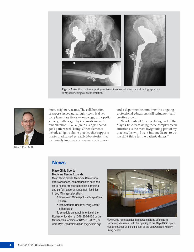
Figure 5. Another patient’s postoperative anteroposterior and lateral radiographs of a complex oncological reconstruction.
4 MAYO CLINIC | OrthopedicSurgeryUpdate
Peter S. Rose, M.D.
interdisciplinary teams. The collaboration of experts in separate, highly technical yet complementary fields — oncology, orthopedic surgery, pathology, physical medicine and rehabilitation — all align in a single shared goal: patient well-being. Other elements include a high-volume practice that supports mastery, advanced research laboratories that continually improve and evaluate outcomes,
and a department commitment to ongoing professional education, skill refinement and creative growth.
Says Dr. Abdel: “For me, being part of the Mayo Clinic team doing these complex recon-structions is the most invigorating part of my practice. It’s why I went into medicine: to do the right thing for the patient, always.”
NewsMayo Clinic Sports Medicine Center ExpandsMayo Clinic Sports Medicine Center now offers advanced, comprehensive care and state-of-the-art sports medicine, training and performance-enhancement facilities in two Minnesota locations:
• Downtown Minneapolis at Mayo Clinic Square
• Dan Abraham Healthy Living Center in Rochester
To schedule an appointment, call the Rochester location at 507-266-9100 or the Minneapolis location at 612-313-0520, or visit https://sportsmedicine.mayoclinic.org.
Mayo Clinic has expanded its sports medicine offerings in Rochester, Minnesota, with the opening of the Mayo Clinic Sports Medicine Center on the third floor of the Dan Abraham Healthy Living Center.

MAYO CLINIC | OrthopedicSurgeryUpdate 5
Collaborating to Improve Spinal Infection Control
Figure 1A. Standard radiographic imaging of the lumbosacral spine depicts an implant-associated spine infection with failure of fixa-tion, deformity and osteomyelitis. The patient was hospitalized and underwent surgical removal and debridement of surgical site. Upon laboratory examination, the implants grew out of Staphylococcus aureus infection, and the patient underwent treatment with IV antibi-otics and had subsequent resolution of his symptoms.
Figure 1B. MRI shows lumbar L2-L3 disk space infection with extension of the infection inside the spinal canal.
Vertebral body and disk joint infections are difficult to diagnose because a patient with infection typically presents with vague clinical symptoms such as generalized back pain, often with indolent progression that can be confused with more-routine musculoskeletal back pain. This is also true for patients who became infected after spine procedures and for those who have not undergone surgery.
Spinal infection rate reduction: 5.9 to 1.8 percentDiagnosing and managing infections early is important because if untreated, they can become paralyzing or fatal. “When you emphasize that the stakes are very high, that paralysis and death are very real possible outcomes of an untreated spinal infection, it gets physicians’ attention,” explains Paul M. Huddleston III, M.D., an ortho-pedic spine surgeon at Mayo Clinic in Rochester, Minnesota. Adds his Mayo Clinic colleague Elie F. Berbari, M.D., Infectious Diseases, who spe-cializes in orthopedic infections: “As a result of our multidisciplinary effort, we have been able to reduce the risk of wound infection in patients undergoing complex spine surgery from 5.9 to 1.8 percent over the last 6 years.”
Red-flag responsesDr. Huddleston emphasizes that spinal infec-tions can result from procedures involving the spine in the neck or back — an injection or sur-gical procedure — or from distant sites of origin such as an infected heart implant or urinary tract infection that travels through the bloodstream to the spine. He urges physicians to remain vigilant by critically assessing red-flag patient responses that initially don’t respond to routine treatments of brief rest, physiotherapy and anti-inflammatory medications. “You must always consider the possibility of diskitis or vertebral osteomyelitis in patients who have night pain, fever or risk factors for vertebral osteomyelitis. Many people with serious spinal infections can suffer for many months before it is diagnosed. That needs to change.”
One of the greatest allies in effecting this change is a high index of clinical suspicion. At the Department of Orthopedic Surgery, the effort to reduce spinal infections involves multi-disciplinary collaborations with specialists across many disciplines: surgeons, physicians, phar-macists, infection prevention and control staff, anesthesiologists, and clinical nurse specialists.

6 MAYO CLINIC | OrthopedicSurgeryUpdate
Figure 2. Staphylococcus aureus biofilm on an indwelling catheter is shown. Photo credit: Centers for Disease Control and Prevention, Rodney M. Donlan, Ph.D., Janice Haney Carr.
Vertebral osteomyelitis databaseWorking with Dr. Berbari, Dr. Huddleston and colleagues investigate topics such as optimal treatment methods for orthopedic hardware infections. In 1969, Mayo Clinic developed the prosthetic joint registry that has information on more than 4,000 patients. Explains Dr. Berbari: “This registry has information on host factors and management and outcomes of patients with prosthetic joint infections. It is a significant source of information for improving practice, and can be a potential source for many future clinical projects.”
Recently the Mayo team developed the vertebral osteomyelitis database. The database has information on more than 500 patients with vertebral osteomylitis. “With this resource we are able to study the utility of imaging techniques in the diagnosis and follow-up of patients with spine infection,” Dr. Berbari explains (Figures 1A and 1B).
Aggressive protocolThis interdisciplinary partnership has developed new protocols for treating spinal infections with a high rate of success. Elements of the team’s approach may consist of:
• Surgical debridement or removal or both of infected hardware
• A prolonged course of intravenous antibiotic• An extended course of oral antibiotic
therapy for certain infections
Explains Dr. Huddleston: “People are surprised because this is a whole different level of intervention. It seems so aggressive. But our data show that’s what it takes to subdue this difficult problem.”
Multilevel, long-term treatmentHe adds that it is also counter to many sur-geons’ training. “The most common factors contributing to an infection becoming uncon-trolled are that a surgeon doesn’t recognize that he or she needs to take the patient back into surgery for debridement or hardware removal or both and that it takes IV antibiotic along with antibiotic pills for an extended period of time, along with diligent follow-up. It’s a multilevel, long-term response to the infection that’s required to stabilize the spine.”
At Mayo Clinic, patients with spine infection are seen at regular intervals by the spine care team starting at the two-week wound-healing check, and out to two years, and as needed after that. At one year, selected patients can undergo an “antibiotic-free challenge” and take a break from the oral antibiotics to see how they feel and to undergo laboratory tests to detect signs of infection. If clinical symptoms recur and lab reports show active infection, they resume oral antibiotics or undergo implant resection and further debridement, as needed to clean the surgical site.
Paul M. Huddleston III, M.D.

MAYO CLINIC | OrthopedicSurgeryUpdate 7
Figure 3. Gram-negative infection of the lumbar L3 disk space is shown. Standard radiographic imaging and MRI reveal an infection of the third lumbar disk space and adjacent vertebra. As reported in Spine in 2013, the patient was a 42-year-old female with insidious onset of back pain for two months after undergoing a routine colo-noscopy. The arrows demonstrate early erosion of the vertebral end plate on the plain radiograph to the left and early inflammatory changes and abscess formation emanat-ing from the disk space on the right.
Elie F. Berbari, M.D.
Persistent biofilmsSpine surgeries are hardware-intense, involving many screws and plates. Metal implants in the body present a potential problem because metal is an ideal habitat on which biofilms develop. Biofilms are adherent forms of bacterial growth that are one of the most persistent and serious challenges to successful outcomes of implanta-tion surgeries, such as spinal fusion.
Not only are biofilms difficult to diagnose, but data show that traditional means detect biofilm infection about 10 percent of the time. Mayo Clinic has developed advanced methods to greatly improve this detection rate (Figure 2).
Strategies for reducing infectionThe Mayo spine team continues to research bio-film infection prevention and management for spine patients, and members are investigating new threats as well. “Right now the big bully on the block is methicillin-resistant Staphylococcus
aureus (MRSA) infection,” Dr. Huddleston says.Investigators at Mayo Clinic are assessing
novel strategies to reduce the risk of infection, including MRSA after spine surgery. A multi-disciplinary effort has been able to reduce the risk of wound infection in patients undergoing complex spine surgery from 5.9 to 1.8 percent over the last 6 years.
The rise of gram-negative infections is also of concern (Figure 3). Working with the infec-tion-control surveillance team at Mayo Clinic in Rochester, Minnesota, the section of spine surgery will continue to monitor the infection rate and to assess the impact of interventions performed.
For more informationAmanda BS, et al. Diagnosis and treatment of fusobacterium nucleatum discitis and vertebral osteomyelitis: Case report and review of the literature. Spine. 2013;38:E120.

8 MAYO CLINIC | OrthopedicSurgeryUpdate
Mayo Clinic orthopedic specialists demon-strated their long-standing commitment to the ever-expanding knowledge demands of the practice with a robust suite of program offerings at the American Academy of Ortho-paedic Surgeons’ (AAOS) annual meeting in Las Vegas in March. Mayo Clinic surgeons and researchers presented 61 scientific papers and 32 scientific posters and served as moderators and faculty for 11 symposia and for 44 instruc-tional course lectures across all subspecialties of orthopedic surgery.
Their effort reflects the department’s priori-ties of creative collaboration to advance patient care, surgical leadership, research excellence,
and continual orthopedic medical and surgical education.
Two high-profile AAOS presentations are summarized:
• The first is the winner of the prestigious Chitranjan Ranawat Award of the Knee Society. It is published in the journal Clini-cal Orthopaedics and Related Research as part of its 2015 Symposium: Knee Society Proceedings coverage.
• The second offering was part of a podium presentation at AAOS on the novel finding of evidence suggesting a role for epigenetics in patient vulnerability to developing avascular necrosis.
Highlights of Mayo Clinic Contributions to AAOS 2015 Programs
Running Subcuticular Closure Technique Provides Most Perfusion to Support Wound Closure after TKA
Total knee arthroplasty (TKA) is one of most commonly performed orthopedic surgeries in the U.S. A 2013 report in The Journal of Bone & Joint Surgery estimated 4 million adults were living with TKAs. Successful outcomes of TKA are linked to wound closure technique that sup-ports optimal perfusion.
Mayo’s pioneering studyDespite consensus that this linkage exists, and multiple investigations to evaluate technique, no study has yet produced convincing data of a
superior wound closure technique for optimizing perfusion — until now. In a randomized clini-cal trial conducted by Mayo Clinic Department of Orthopedic Surgery, investigators conducted a pioneering study to compare three closure techniques and evaluate their effects on perfusion. Their work is the first to provide quantitative evi-dence showing that running subcuticular closure enables the most robust blood flow to the incision site among commonly used closure techniques in reconstructive knee surgery (Figure 1).
Explains Robert T. Trousdale, M.D., an
Figure 1. Three suture techniques compared. From left: running subcuticular closure, vertical mattress closure, skin staple closure.
Robert T. Trousdale, M.D.

MAYO CLINIC | OrthopedicSurgeryUpdate 9
Figure 2. The same image as Figure 1 with overlay of laser-assisted indocyanine green angiography fluores-cence values indicating perfusion levels. Study results show the running subcuticular closure enables the most physiologic robust blood flow, which may improve wound healing and avoid complications.
orthopedic surgeon at Mayo Clinic in Rochester, Minnesota: “Skin and soft tissue complications can lead to a devastating outcome for patients after reconstructive knee surgery. Perfusion status is perhaps the most important factor in appropriate wound healing; however, to date no study has adequately determined which surgical closure technique optimizes this parameter. This investigation determined unequivocally that running subcuticular closure was superior to vertical mattress and staple closure with regard to perfusion status.”
Because closure technique is an important modifiable factor in determining blood flow to the wound, Dr. Trousdale believes surgeons can now use this information to provide patients with the best possible opportunity for excellent postoperative wound healing.
Comparing 3 techniquesIn the study, the Mayo Clinic team compared three wound closure techniques in 45 patients undergoing TKA using the same surgical approach. Patients all lacked comorbidities or other risk factors for wound healing complica-tions and were prospectively randomized to one of three wound closure techniques. The three techniques compared were:
• Running subcuticular• Vertical mattress• Skin staple closure The team’s research objective was to deter-
mine which closure technique enables the most
robust wound perfusion postoperatively.
Novel measurement methodTo assess perfusion, investigators used a novel method that assesses dynamic tissue properties through the use of laser-assisted indocyanine green angiography (LA-ICGA). The device and software system quantifies fluorescence to indi-cate perfusion, with higher rankings indicating greater blood flow.
“As a technology, LA-ICGA is several decades old, but it has recently been revived by plastic surgeons for its ability to provide point-of-care, real-time definition of blood flow to skin and soft tissue that is both dynamic and precise,” explains plastic and reconstructive surgeon Steven R. Jacobson, M.D., at Mayo Clinic’s campus in Min-nesota, and one of Dr. Trousdale’s Mayo Clinic collaborators in the study (Figure 2). He adds that a recently published work he co-authored shows that LA-ICGA has reduced complications in patients undergoing breast reconstruction at Mayo Clinic, providing more-encouraging evidence for the surgical utility of this technology.
ResultsResearchers concluded that the method of clo-sure can influence skin and soft tissue perfusion after TKA and that running subcuticular closure enables the most physiologic robust blood flow, which may improve wound healing.
While further studies of this approach are needed to include patients at higher risk of
Steven R. Jacobson, M.D.

wound complications, the team believes the results are valid and generalizable across patient populations. “We excluded comorbidities for a good reason: to create experimental isola-tion of closure technique as the primary factor driving differences in perfusion,” Dr. Trousdale says. “Despite lack of data on patients with comorbidities at this time, we feel the results are extremely helpful in guiding surgical decision-making about wound closure.”
For more informationWyles CC, et al. The Chitranjan Ranawat Award: Running subcuticular closure enables the most robust perfusion after TKA: A randomized clinical trial. Clinical Orthopaedics and Related Research. In press.
Weinstein AM, et al. Estimating the burden of total knee replacement in the United States. The Journal of Bone & Joint Surgery. 2013;95:385.
10 MAYO CLINIC | OrthopedicSurgeryUpdate
Avascular necrosis (AVN) of the femoral head occurs in an estimated 20,000 new patients every year in the U.S., typically in patients younger than age 40 — a younger age than is characteristically seen among patients with primary osteoarthritis.
The condition develops when the osteocytes of trabecular bone necrose. In most patients, this is progressive and leads to collapse of the femoral head. Once this occurs, patients require definitive treatment with total hip arthroplasty (THA) for pain relief and improvement in daily function.
The pathophysiology of avascular necro-sis (AVN) is not clear. The suspicion that the tendency to develop AVN is driven by patient genetic predisposition or epigenetic sensitivity to steroids is prompted by two findings:
• AVN has multiple etiologies, yet a signifi-cant proportion of AVN burden is charac-terized as idiopathic.
• While use of steroids is commonly con-sidered a primary risk factor for AVN, data show a minority of patients on high-dose steroid regimen actually develop AVN.
Epigenetics and steroid-sensitive subtypeThis curiosity was the topic of a podium presen-tation by a Mayo Clinic team at the American Academy of Orthopaedic Surgeons (AAOS) annual meeting in March 2015. Conducting the world’s largest study of genotype database
patients with AVN compared with matched controls, the team looked for differentially expressed single nucleotide polymorphisms (SNPs) between patients with AVN and matched controls. Results reveal a link between epigenetics and steroid use that may predispose patients to develop a steroid-sensitive subtype of AVN. These data may also point the way to developing a new therapeutic risk-stratification screening prior to steroid use that could reduce the possibility of developing AVN.
Explains Rafael J. Sierra, M.D., an orthopedic surgeon at Mayo Clinic in Rochester, Minnesota: “In both cohorts, we identified multiple differentially expressed SNPs that maintained high levels of statistical significance. These markers may provide a novel platform for diagnosing genetic predisposi-tion to AVN or epigenetic sensitivity to steroids, which may increase a patient’s risk of developing AVN. As a possible practical application, if vali-dated, these findings could be used to develop a simple screening test to stratify risk in patients prior to therapy to avoid AVN complications.”
World’s largest study of AVNThe research collaboration included investiga-tors from Mayo Clinic Department of Ortho-pedic Surgery, Mayo Clinic Biobank and Mayo Clinic Genome Consortium. Mayo Biobank is a tissue repository that allowed genotyping of 88 AVN cases and 176 matched controls. Mayo Genome Consortium is a database of previously
Highlights of Mayo Clinic Contributions to AAOS 2015 Programs (Continued)
Largest Study of Genotype Database of Patients with Avascular Necrosis and Matched Controls Suggests AVN Subtype Linked to Steroids
Rafael J. Sierra, M.D.
MAYO CLINIC | OrthopedicSurgeryUpdate 11
Andre Terzic, M.D., Ph.D.
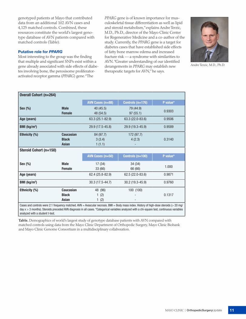
genotyped patients at Mayo that contributed data from an additional 102 AVN cases and 4,125 matched controls. Combined, these resources constitute the world’s largest geno-type database of AVN patients compared with matched controls (Table).
Putative role for PPARGMost interesting to the group was the finding that multiple and significant SNPs exist within a gene already associated with side effects of diabe-tes involving bone, the peroxisome proliferator-activated receptor gamma (PPARG) gene. “The
PPARG gene is of known importance for mus-culoskeletal tissue differentiation as well as lipid and steroid metabolism,” explains Andre Terzic, M.D., Ph.D., director of the Mayo Clinic Center for Regenerative Medicine and a co-author of the study. Currently, the PPARG gene is a target for diabetes cases that have established side effects of fatty bone marrow edema and increased fracture risk — a syndrome with similarities to AVN. “Greater understanding of our identified derangements in PPARG may establish new therapeutic targets for AVN,” he says.
Overall Cohort (n=264)
AVN Cases (n=88) Controls (n=176) P value*
Sex (%) MaleFemale
40 (45.5)48 (54.5)
79 (44.9)97 (55.1) 0.9303
Age (years) 63.3 (25.1-82.9) 63.3 (22.0-83.6) 0.9506
BMI (kg/m2) 29.9 (17.5-45.8) 29.9 (19.3-45.9) 0.9589
Ethnicity (%) CaucasianBlackAsian
84 (97.7)3 (3.4)1 (1.1)
172 (97.7)4 (2.3)
-0.3140
Steroid Cohort (n=150)AVN Cases (n=50) Controls (n=100) P value*
Sex (%) MaleFemale
17 (34)33 (66)
34 (34)66 (66) 1.000
Age (years) 62.4 (25.8-82.9) 62.5 (22.0-83.6) 0.9871
BMI (kg/m2) 30.3 (17.5-44.7) 30.2 (19.3-45.9) 0.9760
Ethnicity (%) CaucasianBlackAsian
48 (96)1 (2)1 (2)
100 (100)--
0.1317
Cases and controls were 2:1 frequency matched. AVN = Avascular necrosis. BMI = Body mass index. History of high-dose steroids (> 20 mg/day x > 3 months). Steroids preceded AVN diagnosis in all cases. *Categorical variables analyzed with a chi-square test, continuous variablesanalyzed with a student t-test.
Table. Demographics of world’s largest study of genotype database patients with AVN compared with matched controls using data from the Mayo Clinic Department of Orthopedic Surgery, Mayo Clinic Biobank and Mayo Clinic Genome Consortium in a multidisciplinary collaboration.

MC6247-0515
Education OpportunitiesFor more information or to register for courses, visit https://ce.mayo.edu, call 800-323-2688 (toll-free) or email [email protected].
Radiology — Helping You Care for Your Patients 2015Aug. 15-16, 2015, in Rochester, Minn.
This course is intended for a broad audience of health care professionals, including those with a need to understand orthopedic imaging. Its goal is to review and discuss contemporary imaging approaches to common diagnostic problems in primary care medicine and surgery.
A case-based approach will describe common patient signs and symptoms in outpatient and emergency settings and indicate best imaging practices for evaluation of these patients considering appropriateness, cost and safety. Emphasis will be placed on algorithmic approaches to radiologic tests when multiple imaging examinations become necessary. Information presented will be organized by differential diagnoses to keep it relevant to daily medical practice.
The course is designed especially for nurse practitioners, physician assistants and primary care physicians who want to improve the quality of their patient care by increasing the value of the radiologic examinations they order.
25th Annual Mayo Clinic Symposium on Sports Medicine 2015Nov. 13-14, 2015, in Rochester, Minn.
This case-oriented program provides an integrated approach to caring for the injured athlete. Through case presentations, lectures and video demonstrations, course offerings are designed to engage a broad spectrum of sports medicine practitioners with the latest in evidence-based clinical practice.
It is appropriate for a continuum of health care professionals:• Those with established sports medicine practices• Those expanding into this clinical specialty as part of an overall wellness focus• Athletic trainers helping patients with conditioning, recovery and rehabilitation
MAYO CLINIC Orthopedic Surgery Update
Medical Editor: Mark W. Pagnano, M.D.
Mayo Clinic Orthopedic Surgery Update is written for
physicians and should be relied upon for medical
education purposes only. It does not provide a
complete overview of the topics covered and should
not replace the independent judgment of a physician
about the appropriateness or risks of a procedure
for a given patient.
Contact UsMayo Clinic welcomes inquiries and referrals, and a request to a specific physician is not required to refer a patient.
Phoenix/ Scottsdale, Arizona866-629-6362 (toll-free)
Jacksonville, Florida800-634-1417 (toll-free) Rochester, Minnesota800-533-1564 (toll-free)
ResourcesMayoClinic.org/medicalprofs
Clinical trials, CME, Grand Rounds, scientific videos and online referrals
August 15-16, 2015 Mayo ClinicPhillips Hall, Siebens Medical Education BuildingRochester, Minnesota
Radiology - Helping You Care for Your Patients
Department of Radiology
REGISTER TODAY!www.ce.mayo.edu/radiology
A Course for Physician Assistants, Nurse Practitioners and Primary Care Physicians