Embed Size (px)
Citation preview
Original
Enfermedades tiroideas en adolescentes con diabetes mellitus tipo 1
M.ª del Carmen Valdés Alonsoa, José M.ª Basain Valdésb, Lucía Llopiz Herrerac, Adriana Li de la Rosad, Aimée Álvarez Álvareze
aServicio de Endocrinología. Hospital Pediátrico Docente Juan Manuel Márquez. La Habana. Cuba • bServicio de Endocrinología. Policlínico Universitario Carlos Manuel Portuondo Lambert.
La Habana. Cuba • cServicio de Pediatría. Hospital Pediátrico Docente Juan Manuel Márquez. La Habana. Cuba • dServicio de Laboratorio. Hospital Pediátrico Docente Juan Manuel Márquez. La Habana. Cuba
• eServicio de Laboratorio. Instituto Nacional de Endocrinología. La Habana. Cuba.
Publicado en Internet:11-septiembre-2017
M.ª del Carmen Valdés Alonso: [email protected]
Palabras clave: Diabetes
mellitus tipo 1 Enfermedades
de la tiroides Tiroiditis autoinmune
Resu
men
Abs
trac
t
Thyroid diseases in adolescents with diabetes mellitus type 1
Introducción: la diabetes mellitus tipo 1 se relaciona con alteraciones tiroideas. Objetivo: determinar el comporta-miento de enfermedades tiroideas en pacientes pediátricos con diabetes mellitus tipo 1. Material y método: estudio descriptivo, transversal, en 56 adolescentes con diagnóstico de diabetes mellitus tipo 1 atendidos en el Servicio de Endocrinología del Hospital Juan Manuel Márquez (La Habana, Cuba) en el periodo de octubre de 2015 a octubre de 2016. Las variables en estudio fueron: edad, sexo, tiempo de evolución de la diabetes mellitus, estado nutricional, función tiroidea y enfermedad tiroidea autoinmune. Las variables cualitativas se describieron estadísticamente mediante frecuencias absolutas y relativas, la asociación entre las variables categó-ricas se exploró con el test χ2 y la probabilidad exacta de Fisher. En todas las pruebas estadísticas se consideró un nivel de significación de α igual a 0,05. Resultados: el 46,29% de los pacientes estudiados presentaron enfermedad tiroidea autoinmune; de ellos, el 33,33% eran del sexo femenino. El mayor grado de disfunción tiroidea se presentó en pacientes con mayores edades (36,0%), normopesos (68,0%) y con tiempo de evolución de la diabetes mellitus de entre 5-9 años (52,0%). El 32,0 frente al 80,0% de los pacientes presentaron respectivamente anticuerpos antitiroglobulina y an-titiroperoxidasa positivos, relacionándose este último con la edad y el tiempo de evolución de la diabetes mellitus. Conclusiones: se presentó disfunción tiroidea en los pacientes con enfermedad tiroidea autoinmune relacionado con la edad y el tiempo de evolución de la diabetes mellitus tipo 1. Los anticuerpos antitiroperoxidasa se relaciona-ron con la edad y el tiempo de evolución de la diabetes mellitus tipo 1.
Introduction: the type 1 diabetes mellitus is related with thyroid alterations. Objective: to determine the characteristics of thyroid diseases in pediatric patients with type 1 diabetes mellitus.Material and method: descriptive cross-sectional study in 56 adolescents with diagnostic of type 1 diabe-tes mellitus assisted in the service of Endocrinology of the Juan Manuel Márquez hospital (La Habana, Cuba) in the period between October 2015 and October 2016. The variables in study were: age, sex, time of evolu-tion of the diabetes mellitus, nutritional state, thyroid function and autoimmune thyroid disease. The qualita-tive variables were statistically described by absolute and relative frequencies; the association among the categorical variables was explored with the χ2 test and Fisher’s exact probability. In all statistical tests, it was considered a a significance level of alfa equal to 0.05. Results: 46.29% of the studied patients presented autoimmune thyroid disease; of them, 33.33% were fe-male. The highest degree of grade of thyroid dysfunction was present in older patients (36.0%), with normal weigh (68.0%) and time of evolution of the diabetes mellitus among 5-9 years (52.0%). 32.0% vs 80.0% of the patients presented antithyroglobulin antibodies and antithyroperoxidase antibodies respectively, being related this last one with the age and the time of evolution of the diabetes mellitus.Conclusions: thyroid dysfunction was present in the patients with autoimmune thyroid disease related to the age and the time of evolution of the type 1 diabetes mellitus. The antithyroperoxidase antibodies were re-lated with the age and the time of evolution of the type 1 diabetes mellitus.
Key words: Diabetes
mellitus type 1 Thyroid diseases
Thyroiditis, autoimmune
Cómo citar este artículo: Valdés Alonso MC, Basain Valdés JM, Llopiz Herrera L, Li de la Rosa A, Álvarez Álvarez A. Enfermedades tiroideas en adolescentes con diabetes mellitus tipo 1. Rev Pediatr Aten Primaria. 2017;19:249-57.
Rev Pediatr Aten Primaria. 2017;19:249-57ISSN: 1139-7632 • www.pap.es
249

INTRODUCCIÓN
La diabetes mellitus tipo 1 (DM1) es la enfermedad
crónica endocrinológica más frecuente en la edad
pediátrica y la segunda en la infancia después del
asma en países desarrollados1. Es un síndrome he-
terogéneo que se caracteriza por hiperglucemia
crónica de origen multifactorial como consecuen-
cia de una alteración en la secreción y/o acción de
la insulina, con repercusión en el metabolismo
de los carbohidratos, lípidos y proteínas y que de-
sarrolla a corto plazo complicaciones agudas, ame-
nazantes para la vida, y a largo plazo complicacio-
nes crónicas, graves e inhabilitantes, que en
ocasiones puede causar la muerte2. El punto cardi-
nal en la fisiopatología de la DM1 es la deficiencia
absoluta de insulina, que predispone a la cetoaci-
dosis. Esta deficiencia se debe a una destrucción
de origen autoinmune de las células β del pán-
creas3, diagnosticándose más frecuentemente en-
tre los 10 y 14 años, lo que se considera que puede
tener relación con los cambios endocrinos de la
pubertad; sin embargo, en los últimos 20 años se
ha informado un incremento en la población de
entre 0 y 4 años, lo que parece ser debido a los
cambios en el estilo de vida de los niños, o bien por
incorporación de procedimientos de diagnósticos
más sensibles, aunque también pudiera tener rela-
ción con infecciones virales por virus de Coxsackie,
de Epstein-Barr o rubeola, entre otros4.
De manera similar a la DM1, las alteraciones tiroi-
deas son frecuentes en la adolescencia, y su preva-
lencia es creciente. Aunque la mayoría de casos,
como el bocio difuso, cursan con función tiroidea
normal, el hipo- o el hipertiroidismo no son infre-
cuentes y son, a menudo, infradiagnosticados en
este grupo de edad5.
Dentro de las alteraciones tiroideas, el hipotiroidis-
mo subclínico (tirotropina [TSH] elevada, triyodoti-
ronina [T3], tiroxina [T4] total y T4 libre normales)
a menudo no es diagnosticado, aunque su preva-
lencia va en aumento debido al creciente cribado
en poblaciones de riesgo (obesidad, hiperlipide-
mias, diabetes mellitus, etc.) y a la determinación
relativamente reciente de la TSH. La mayoría son
idiopáticos, y se evidencian por controles analíticos
rutinarios en ausencia de patología o desencade-
nantes asociados. En los restantes, la tiroiditis au-
toinmune es la causa más frecuente6.
La etiología más frecuente del hipotiroidismo ad-
quirido es la tiroiditis linfocitaria crónica (autoin-
mune, de Hashimoto), con un predominio femeni-
no 2:1, y, en segundo lugar, el bocio endémico por
déficit de yodo. Los casos de origen hipofisario o
hipotalámico son muy raros5. La tiroiditis autoin-
mune es un proceso inflamatorio del tiroides ca-
racterizado por presencia de bocio, anticuerpos
circulantes y alteraciones histológicas. La enferme-
dad es más frecuente en la infancia y adolescencia
y se asocia a determinados antígenos HLA de la
clase II (alelos DR3, DR4 y DR5), algunos presentes
en la DM1. La tiroiditis autoinmunitaria se asocia a
otros procesos autoinmunes como la DM1 (20%),
la insuficiencia suprarrenal, el hipoparatiroidismo
y enfermedades cromosómicas como el síndrome
de Down (4,3%) y síndrome de Turner (3,8%). For-
ma parte de los síndromes pluriglandulares au-
toinmunes tipo I y II, siendo el primero el más fre-
cuente en la infancia7.
La asociación de diabetes mellitus y enfermedades
tiroideas se presenta con más frecuencia en pa-
cientes genéticamente predispuestos, como son
los portadores del haplotipo HLA-DR3. Un porcen-
taje importante de pacientes con DM1 presenta
anticuerpos antitiroideos positivos. De ellos, hasta
un 50% progresa a enfermedad tiroidea autoinmu-
ne, fundamentalmente a hipotiroidismo primario,
el cual se halla presente en un 2-5% de los pacien-
tes con DM1 8.
El hipertiroidismo de origen autoinmune también
se ha asociado con la DM1, aunque con una preva-
lencia bastante menor, de alrededor del 1-2%. En el
caso del hipotiroidismo primario, este incremento
parece relacionarse con la presencia de dislipide-
mia, aumento de la presión arterial diastólica y
disfunción endotelial8.
A pesar de la frecuente asociación de DM1 y enfer-
medad tiroidea autoinmune, son escasos los estu-
dios de prevalencia de ambos procesos en España
y otros países8.
M.ª del Carmen Valdés Alonso, et al. Enfermedades tiroideas en adolescentes con diabetes mellitus tipo 1
Rev Pediatr Aten Primaria. 2017;19:249-57ISSN: 1139-7632 • www.pap.es
250

M.ª del Carmen Valdés Alonso, et al. Enfermedades tiroideas en adolescentes con diabetes mellitus tipo 1
Rev Pediatr Aten Primaria. 2017;19:249-57ISSN: 1139-7632 • www.pap.es
251
El objetivo de la presente investigación fue deter-
minar el comportamiento de las enfermedades del
tiroides en adolescentes con DM1 atendidos en el
Hospital Pediátrico Docente Juan Manuel Márquez
(La Habana, Cuba) en el periodo comprendido de
2015 a 2016.
MATERIAL Y MÉTODOS
Se realizó un estudio descriptivo de corte transver-
sal en 54 pacientes adolescentes con diagnóstico de
DM1 según criterios de la Organización Mundial de
la Salud (n = 54), de ambos sexos, atendidos en el
Servicio de Endocrinología del Hospital Pediátrico
Docente Juan Manuel Márquez (La Habana, Cuba)
en el periodo comprendido de octubre de 2015 a
octubre de 2016. Se excluyeron aquellos pacientes
tratados con fármacos inmunosupresores y/o con
presencia de otras enfermedades intercurrentes.
Las variables en estudio fueron: edad, sexo, tiempo
de evolución de la DM1, estado nutricional, función
tiroidea y enfermedad autoinmune tiroidea. De las
historias clínicas de los pacientes se obtuvieron los
siguientes datos: edad, sexo y tiempo de evolución
de la DM1. A todos los pacientes se les realizaron las
mediciones antropométricas de peso y talla, y pos-
teriormente se les indicó TSH, T4 total, anticuerpos
antitiroglobulina y anticuerpos antitiroperoxidasa.
A partir de los valores de peso y talla se ubicó al pa-
ciente según sexo en la tabla cubana de percentiles
de peso para la talla correspondiente9,10 y se deter-
minó el percentil al que correspondía, interpretán-
dose según lo establecido en Cuba. Las muestras de
laboratorio fueron obtenidas por punción venosa
en el laboratorio clínico, después de 12 horas de
ayuno. Las determinaciones hormonales (TSH, T3 y
T4 total) fueron realizadas en el laboratorio de dicha
institución mediante el principio del análisis inmu-
norradiométrico en fase sólida; mientras que las
determinaciones de los anticuerpos (antitiroglobu-
lina y antiperoxidasa) se realizaron en el Instituto
Nacional de Endocrinología mediante técnica de
inmunoensayo de electroquimioluminiscencia.
Para el procesamiento de la información, los datos
fueron incluidos en una base de datos automatizada
con la hoja de cálculo electrónica Excel® 2003. Las
variables cualitativas y las variables cuantitativas
llevadas a escala ordinal se describieron estadísti-
camente mediante frecuencias absolutas y relati-
vas. La asociación entre las variables categóricas se
exploró con el test χ2 y la probabilidad exacta de
Fisher. En todas las pruebas estadísticas se consi-
deró un nivel de significación de α de 0,05.
El estudio estuvo debidamente avalado desde el
punto de vista ético por las siguientes razones: 1)
se aplicaron planillas de consentimiento informa-
do por escrito, explicándole a cada paciente, padre
y/o tutor legal la importancia de la investigación;
2) se respetó la integridad de los participantes en
la investigación, asegurando la confidencialidad
de todos los datos obtenidos, y 3) el Consejo Cien-
tífico y el Comité de Ética de la Investigación del
Hospital Pediátrico Docente Juan Manuel Márquez
avalaron la presente investigación.
RESULTADOS
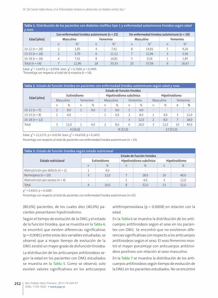
La distribución de los pacientes con DM1 y enfer-
medad autoinmune tiroidea según edad y sexo se
muestran en la Tabla 1. Se observó que 25 (46,29%)
pacientes con DM1 presentaron enfermedad tiroi-
dea autoinmune, de los cuales 18 (33,33%) eran
del sexo femenino.
La comparación del estado de la función tiroidea
en pacientes con enfermedad tiroidea autoinmu-
ne según edad y sexo se muestra en la Tabla 2. Al
comparar el estado de la función tiroidea según la
edad se encontró que los pacientes con mayores
edades presentaron mayor grado de disfunción ti-
roidea que los pacientes con menores edades, don-
de, de los 13 pacientes con hipotiroidismo, nueve
(36,0%) pacientes eran mayores de 16 años de edad,
cuatro (16,0%) tenían entre 13 y 15 años de edad y
ningún paciente tenía entre 10 y 12 años de edad.
Con respecto al sexo, predominó el sexo femenino,
con 17 (68,0%) pacientes.
En la Tabla 3 se muestra el estado de la función ti-
roidea según el estado nutricional. Predominaron
los pacientes con normopeso, con un total de 20
M.ª del Carmen Valdés Alonso, et al. Enfermedades tiroideas en adolescentes con diabetes mellitus tipo 1
Rev Pediatr Aten Primaria. 2017;19:249-57ISSN: 1139-7632 • www.pap.es
252
(80,0%) pacientes, de los cuales diez (40,0%) pa-
cientes presentaron hipotiroidismo.
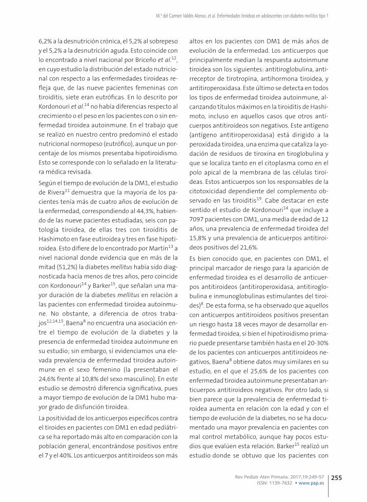
Según el tiempo de evolución de la DM1 y el estado
de la función tiroidea, que se muestra en la Tabla 4,
se encontró que existen diferencias significativas
(p = 0,0081) entre estas dos variables estudiadas, se
observó que a mayor tiempo de evolución de la
DM1 existió un mayor grado de disfunción tiroidea.
La distribución de los anticuerpos antitiroideos se-
gún la edad en los pacientes con DM1 estudiados
se muestra en la Tabla 5. Como se observó, solo
existen valores significativos en los anticuerpos
antitiroperoxidasa (p = 0,0008) en relación con la
edad.
En la Tabla 6 se muestra la distribución de los anti-
cuerpos antitiroideos según el sexo en los pacien-
tes con DM1. Se encontró que no existieron dife-
rencias significativas con respecto a los anticuerpos
antitiroideos según el sexo. El sexo femenino mos-
tró el mayor porcentaje con anticuerpos antitiroi-
deos positivos con relación al sexo masculino.
En la Tabla 7 se muestra la distribución de los anti-
cuerpos antitiroideos según tiempo de evolución de
la DM1 en los pacientes estudiados. No se encontró
Tabla 1. Distribución de los pacientes con diabetes mellitus tipo 1 y enfermedad autoinmune tiroidea según edad y sexo
Edad (años)Con enfermedad tiroidea autoinmune (n = 25) Sin enfermedad tiroidea autoinmune (n = 29)
Masculino Femenino Masculino Femeninon %* n %* n %* n %*
10-12 (n = 18) 1 1,85 4 7,41 8 14,81 5 9,2613-15 (n = 18) 2 3,70 6 11,11 7 12,96 3 5,5616-18 (n = 18) 4 7,41 8 14,81 5 9,26 1 1,85Total (n = 54) 7 12,96 18 33,33 20 37,04 9 16,67
Edad: χ2 = 5,6479; p = 0,0594. Sexo: χ2 = 6,7000; p = 0,3495.*Porcentaje con respecto al total de la muestra (n = 54).
Tabla 2. Estado de función tiroidea en pacientes con enfermedad tiroidea autoinmune según edad y sexo
Edad (años)
Estado de función tiroideaEutiroidismo Hipotiroidismo subclínico Hipotiroidismo
Masculino Femenino Masculino Femenino Masculino Femeninon % n % n % n % n % n %
10-12 (n = 5) 2 8,0 1 4,0 1 4,0 1 4,0 - - - -13-15 (n = 8) 1 4,0 - - 1 4,0 2 8,0 1 4,0 3 12,016-18 (n = 12) - - - - - - 3 12,0 2 8,0 7 28,0Total 3 12,0 1 4,0 2 8,0 6 24,0 3 12,0 10 40,0
4 (16,0) 8 (32,0) 13 (52,0)
Edad: χ2 = 12,2175; p = 0,0158. Sexo: χ2 = 14,6528; p = 0,1452.
Porcentaje con respecto al total de pacientes con enfermedad tiroidea autoinmune (n = 25).
Tabla 3. Estado de función tiroidea según estado nutricional
Estado nutricionalEstado de función tiroidea
Eutiroidismo Hipotiroidismo subclínico Hipotiroidismon % n % n %
Malnutrición por defecto (n = 1) 1 4,0 - - - -Normopeso (n = 20) 3 12,0 7 28,0 10 40,0Malnutrición por exceso (n = 4) - - 1 4,0 3 12,0Total 4 16,0 8 32,0 13 52,0
χ2 = 6,4423; p = 0,1685.
Porcentaje con respecto al total de pacientes con enfermedad tiroidea autoinmune (n=25).

M.ª del Carmen Valdés Alonso, et al. Enfermedades tiroideas en adolescentes con diabetes mellitus tipo 1
Rev Pediatr Aten Primaria. 2017;19:249-57ISSN: 1139-7632 • www.pap.es
253
Tabla 4. Estado de función tiroidea según tiempo de evolución de la diabetes mellitus tipo 1
Tiempo de evolución (años)
Estado de función tiroidea
Eutiroidismo Hipotiroidismo subclínico Hipotiroidismo
n % n % n %
0-4 (n = 8) 3 12,0 5 20,0 - -
5-9 (n = 14) 1 4,0 3 12,0 10 40,0
≥ 10 (n = 3) - - - - 3 12,0
Total (n = 25) 4 16,0 8 32,0 13 52,0
χ2 = 13,7577; p = 0,0081.
Porcentaje con respecto al total de pacientes con enfermedad tiroidea autoinmune (n = 25).
Tabla 5. Distribución de los anticuerpos antitiroideos según la edad en pacientes con diabetes mellitus tipo 1
Edad (años)
Anticuerpos antitiroideos
Antitiroglobulina (n = 25) Antitiroperoxidasa (n = 25) Ambos (n = 25)
Positivo Negativo Positivo Negativo Positivo Negativo
n %* n % n %* n %* n %* n %*
10-12 (n = 5) 3 12,0 2 8,0 1 4,0 4 16,0 1 11,11 1 11,11
13-15 (n = 8) 2 8,0 6 24,0 8 32,0 - - 1 11,11 - -
15-18 (n = 12) 3 12,0 9 36,0 11 44,0 1 4,0 4 44,44 2 22,22
Total 8 32,0 17 68,0 20 80,0 5 20,0 6 66,67 3 33,33
χ2 = 2,2518; p = 0,3244 χ2 = 14,2708; p = 0,0008 χ2 = 0,7500; p = 0,6873
*Porcentaje con respecto al total de pacientes con enfermedad tiroidea autoinmune (n = 25) según tipo de anticuerpos antitiroideos.
Tabla 6. Distribución de los anticuerpos antitiroideos según el sexo en pacientes con diabetes mellitus tipo 1
Sexo
Anticuerpos antitiroideos
Antitiroglobulina Antitiroperoxidasa Ambos
Positivo Negativo Positivo Negativo Positivo Negativo
n %* n %* n %* n % n %* n %*
Masculino (n = 8) 4 16,0 4 16,0 7 28,0 1 4,0 4 44,44 2 22,22
Femenino (n = 17) 4 16,0 13 52,0 13 52,0 4 16,0 2 22,22 1 11,11
Total (n = 25) 8 32,0 17 78,0 20 80,0 5 20,0 6 66,67 3 33,33
χ2 = 1,7029; p = 0,1919 χ2 = 0,4416; p = 0,5064 χ2 = 0,0000; p = 1,0000
*Porcentaje con respecto al total de pacientes con enfermedad tiroidea autoinmune (n = 25) según tipo de anticuerpos antitiroideos.
Tabla 7. Distribución de los anticuerpos antitiroideos según tiempo de evolución de la diabetes mellitus tipo 1
Tiempo de evolución (años)
Anticuerpos antitiroideos
Antitiroglobulina (n = 25) Antitiroperoxidasa (n = 25) Ambos (n = 25)
Positivo Negativo Positivo Negativo Positivo Negativo
n % n % n % n % n % n %
0-4 (n = -8) 2 8,0 6 24,0 5 20,0 3 12,0 1 11,11 1 11,11
5-9 (n- = 14) 4 16,0 10 40,0 14 56,0 - - 4 44,44 - -
≥ 10 (n = 3) 1 4,0 2 8,0 2 8,0 1 4,0 1 11,11 2 22,22
Total 7 28,0 18 72,0 20 84,0 5 16,0 6 66,67 3 33,33
χ2 = 0,0803; p = 0,9606 χ2 = 6,0888; p = 0,0476 χ2 = 3,7500; p = 0,1534
Porcentaje con respecto al total de pacientes con enfermedad tiroidea autoinmune (n = 25) según tipo de anticuerpos antitiroideos.

M.ª del Carmen Valdés Alonso, et al. Enfermedades tiroideas en adolescentes con diabetes mellitus tipo 1
Rev Pediatr Aten Primaria. 2017;19:249-57ISSN: 1139-7632 • www.pap.es
254
asociación entre los anticuerpos antitiroglobulina
y ambos anticuerpos positivos con el tiempo de
evolución de la DM1. Al analizar los anticuerpos
antitiroperoxidasa, se pudo observar que existie-
ron valores más significativos de dichos anticuer-
pos (p = 0,0476) en relación con el mayor tiempo
de evolución de la diabetes mellitus (el 100% de los
pacientes entre cinco y nueve años de evolución de
la DM1 presentaban estos anticuerpos positivos,
mientras que el 66,67% de los diabéticos tipo 1 con
diez años o más de evolución de la diabetes pre-
sentaron estos anticuerpos positivos.
DISCUSIÓN
La DM1 se asocia con cierta frecuencia a otras en-
fermedades de etiología inmunitaria, siendo la
más prevalente la enfermedad tiroidea autoinmu-
ne. Ambas son enfermedades de glándulas endo-
crinas originadas por la estimulación de células T
órgano-específicas. La asociación de ambas se pre-
senta con más frecuencia en pacientes genética-
mente predispuestos, como son los portadores del
haplotipo HLA-DR3, donde un porcentaje impor-
tante de pacientes con DM1 presenta anticuerpos
antitiroideos positivos. De ellos, hasta un 50% pro-
gresa a enfermedad tiroidea autoinmune, funda-
mentalmente a hipotiroidismo primario, el cual se
halla presente en un 2-5% de los pacientes con
DM1 8. La tiroiditis crónica autoinmune se caracte-
riza por la presencia de anticuerpos antitiroideos
específicos en suero, los cuales son positivos en el
10-12% en la población general y entre niños con
DM1, de acuerdo con el grado de disfunción tiroi-
dea, entre 3 y 50%11,12.
El estudio de Baena8 muestra una elevada preva-
lencia de enfermedad tiroidea autoinmune en pa-
cientes con DM1 (17%). No obstante, este resulta-
do es similar a los publicados por otros autores11,12
en estudios internacionales, la mayoría de ellos
realizados en población pediátrica, en los que la
prevalencia de enfermedad tiroidea autoinmune
se encuentra entre el 15,8 y el 43,7%8. En el estudio
de Rivera11 se obtiene que, del total de pacientes
diabéticos, hay una predominancia porcentual del
género femenino con un 52,6% (p < 0,019) y con
una mayoría significativa de los adolescentes, del
52,6% (p < 0,001), lo cual guarda relación con la bi-
bliografía nacional12,13 e internacional14,15 que
apoya dicha investigación. En el estudio de Rivera11
se diagnosticaron nueve pacientes de enfermedad
tiroidea autoinmune para un 9,3%, concentrándo-
se mayormente en el grupo de los adolescentes. De
las nueve pacientes con enfermedad tiroidea au-
toinmune, seis presentaban tiroiditis de Hashimo-
to en fase eutiroidea y tres en fase hipotiroidea, lo
cual difiere en lo descrito por Briceño et al.12 a nivel
nacional y Barker et al.15 a nivel internacional, que
encuentran predominio estadísticamente signifi-
cativo del hipotiroidismo.
Baena et al.8, en un estudio de 90 pacientes con
DM1, encontró que el 17,8% del total de pacientes
estudiados presentaban enfermedad tiroidea au-
toinmune conocida que, por orden de frecuencia,
se correspondía con hipotiroidismo primario
(9,9%), hipotiroidismo subclínico (7,1%) y enferme-
dad de Graves (0,8%). En el estudio de Miraval
León16, la disfunción tiroidea más frecuente es el
hipotiroidismo clínico, con un 84,4%, lo que supera
en frecuencia a otros estudios14,16, pero coincide
con la literatura médica internacional17.
El porcentaje de hipotiroidismo subclínico fue del
2,5% de todo aquel con despistaje hormonal, me-
nor en cantidad en comparación con otros estu-
dios14,16. Johnson et al.18 reportan una prevalencia
de hasta un 50%, Gronich17 encuentra un 8,6% de
casos de hipotiroidismo subclínico en mujeres aus-
tralianas a pesar de que el hipotiroidismo subclínico
no fue la principal alteración tiroidea encontrada.
En el estudio realizado en nuestro hospital se en-
contró que la enfermedad tiroidea autoinmune
que predominó fue el hipotiroidismo, siguiéndole
en orden de frecuencia el hipotiroidismo subclíni-
co en pacientes adolescentes y en el sexo femeni-
no, lo cual coincide con lo descrito en la literatura
médica revisada. No se presentaron casos de hi-
perfunción tiroidea (hipertiroidismo).
En relación con el estado nutricional de los pacien-
tes con DM1, que estudia Rivera11, el 77,2% corres-
pondió al tipo eutrófico, el 6,2% a la obesidad, el
M.ª del Carmen Valdés Alonso, et al. Enfermedades tiroideas en adolescentes con diabetes mellitus tipo 1
Rev Pediatr Aten Primaria. 2017;19:249-57ISSN: 1139-7632 • www.pap.es
255
6,2% a la desnutrición crónica, el 5,2% al sobrepeso
y el 5,2% a la desnutrición aguda. Esto coincide con
lo encontrado a nivel nacional por Briceño et al.12,
en cuyo estudio la distribución del estado nutricio-
nal con respecto a las enfermedades tiroideas re-
fleja que, de las nueve pacientes femeninas con
tiroiditis, siete eran eutróficas. En lo descrito por
Kordonouri et al.14 no había diferencias respecto al
crecimiento o el peso en los pacientes con o sin en-
fermedad tiroidea autoinmune. En el trabajo que
se realizó en nuestro centro predominó el estado
nutricional normopeso (eutrófico), aunque un por-
centaje de los mismos presentaba hipotiroidismo.
Esto se corresponde con lo señalado en la literatu-
ra médica revisada.
Según el tiempo de evolución de la DM1, el estudio
de Rivera11 demuestra que la mayoría de los pa-
cientes tenía más de cuatro años de evolución de
la enfermedad, correspondiendo al 44,3%, habien-
do de las nueve pacientes estudiadas, seis con pa-
tología tiroidea, de ellas tres con tiroiditis de
Hashimoto en fase eutiroidea y tres en fase hipoti-
roidea. Esto difiere de lo encontrado por Martín13 a
nivel nacional donde evidencia que en más de la
mitad (51,2%) la diabetes mellitus había sido diag-
nosticada hacía menos de tres años, pero coincide
con Kordonouri14 y Barker15, que señalan una ma-
yor duración de la diabetes mellitus en relación a
las pacientes con enfermedad tiroidea autoinmu-
ne. No obstante, a diferencia de otros traba-
jos12,14,15, Baena8 no encuentra una asociación en-
tre el tiempo de evolución de la diabetes y la
presencia de enfermedad tiroidea autoinmune en
su estudio; sin embargo, sí evidenciamos una ele-
vada prevalencia de enfermedad tiroidea autoin-
mune en el sexo femenino (la presentaban el
24,6% frente al 10,8% del sexo masculino). En este
estudio se demostró diferencia significativa, pues
a mayor tiempo de evolución de la DM1 hubo ma-
yor grado de disfunción tiroidea.
La positividad de los anticuerpos específicos contra
el tiroides en pacientes con DM1 en edad pediátri-
ca se ha reportado más alto en comparación con la
población general, encontrándose positivos entre
el 7 y el 40%. Los anticuerpos antitiroideos son más
altos en los pacientes con DM1 de más años de
evolución de la enfermedad. Los anticuerpos que
principalmente median la respuesta autoinmune
tiroidea son los siguientes: antitiroglobulina, anti-
rreceptor de tirotropina, antihormona tiroidea, y
antitiroperoxidasa. Este último se detecta en todos
los tipos de enfermedad tiroidea autoinmune, al-
canzando títulos máximos en la tiroiditis de Hashi-
moto, incluso en aquellos casos que otros anti-
cuerpos antitiroideos son negativos. Este antígeno
(antígeno antitiroperoxidasa) está dirigido a la
peroxidada tiroidea, una enzima que cataliza la yo-
dación de residuos de tiroxina en tiroglobulina y
que se localiza tanto en el citoplasma como en el
polo apical de la membrana de las células tiroi-
deas. Estos anticuerpos son los responsables de la
citotoxicidad dependiente del complemento ob-
servado en las tiroiditis19. Cabe destacar en este
sentido el estudio de Kordonouri14 que incluye a
7097 pacientes con DM1, una media de edad de 12
años, una prevalencia de enfermedad tiroidea del
15,8% y una prevalencia de anticuerpos antitiroi-
deos positivos del 21,6%.
Es bien conocido que, en pacientes con DM1, el
principal marcador de riesgo para la aparición de
enfermedad tiroidea es el desarrollo de anticuer-
pos antitiroideos (antitiroperoxidasa, antitiroglo-
bulina e inmunoglobulinas estimulantes del tiroi-
des)8. De esta forma, se ha observado que aquellos
con anticuerpos antitiroideos positivos presentan
un riesgo hasta 18 veces mayor de desarrollar en-
fermedad tiroidea, si bien el hipotiroidismo prima-
rio puede presentarse también hasta en el 20-30%
de los pacientes con anticuerpos antitiroideos ne-
gativos, Baena8 obtiene datos muy similares en su
estudio, en el que el 25,6% de los pacientes con
enfermedad tiroidea autoinmune presentaban an-
ticuerpos antitiroideos negativos. Por otro lado, si
bien parece que la prevalencia de enfermedad ti-
roidea aumenta en relación con la edad y con el
tiempo de evolución de la diabetes, no se ha docu-
mentado una mayor prevalencia en pacientes con
mal control metabólico, aunque hay pocos estu-
dios que evalúen esta relación. Barker15 realizó un
estudio donde se obtuvo que los pacientes con
M.ª del Carmen Valdés Alonso, et al. Enfermedades tiroideas en adolescentes con diabetes mellitus tipo 1
Rev Pediatr Aten Primaria. 2017;19:249-57ISSN: 1139-7632 • www.pap.es
256
anticuerpos antitiroideos positivos eran con más
frecuencia mujeres (58%; p < 0,0001), adolescen-
tes (16,1 años; p < 0,0013).
En el estudio Rivera11, de los pacientes a los que les
realiza determinación de anticuerpos antitirope-
roxidasa, solo seis resultaron positivos para un
11,2%, cuatro (66,7%) con tiroiditis de Hashimoto
en fase eutiroidea y dos (33,3%) en fase hipotiroi-
dea. En el ámbito nacional, Briceño12 evidencia en
relación con los anticuerpos antitiroideos algo si-
milar; en 126 pacientes (86,3%) resultan negativos
y positivos en 12,7%. A nivel internacional, Glas-
tras20 detectó anticuerpos antitiroperoxidasa posi-
tivos en 13 de 166 pacientes (7,8%); seis de los 13
pacientes (46,2%) con anticuerpos antitiroperoxi-
dasa positivos en el momento del diagnóstico de-
sarrollaron una patología tiroidea, frente a solo el
3,6% con anticuerpos antitiroperoxidasa negati-
vos; datos diferentes se obtuvieron del estudio de
Baena8 donde un 41,1% resultaron antitioperoxi-
dasa positivos y un 25,6% negativos; en el 33,3%
no se dispuso de datos.
En nuestro estudio, de los anticuerpos antitiroi-
deos, solo los anticuerpos antitiroperoxidasa resul-
taron positivos y se relacionaron con el tiempo de
evolución de la enfermedad tiroidea autoinmune,
no así en el caso de los anticuerpos antitiroglobu-
lina o la presencia de ambos anticuerpos, donde no
hubo significación estadística con el tiempo de
evolución de la enfermedad. El sexo femenino
mostró un mayor porcentaje de anticuerpos antiti-
roperoxidasa positivos, aunque no resultó signifi-
cativo. Nuestros resultados son similares a los des-
critos en la literatura médica revisada.
CONCLUSIONES
Existió enfermedad tiroidea autoinmune en los
adolescentes con diabetes mellitus tipo 1.
Se presentó disfunción tiroidea en los adolescen-
tes con enfermedad tiroidea autoinmune expresa-
da por hipotiroidismo clínico y subclínico relacio-
nado con la edad y el tiempo de evolución de la
DM1.
Los anticuerpos antitiroperoxidasa se relacionaron
con la edad y el tiempo de evolución de la diabetes
mellitus tipo 1, no así con el sexo. No existió rela-
ción de los anticuerpos antitiroglobulina con las
variables estudiadas.
CONFLICTO DE INTERESES
Los autores declaran no presentar conflictos de intereses en relación con la preparación y publicación de este artículo.
ABREVIATURAS
DM1: diabetes mellitus tipo 1 T3: triyodotironina T4: ti-roxina TSH: tirotropina.
BIBLIOGRAFÍA
1. Camacho B, Manzanares A, Espino R. Debut de diabe-
tes mellitus tipo 1 en el área hospitalaria de Valme.
Vox Paediatr. 2012;19:9-13.
2. Asenjo S, Muzzo S, Pérez MV, Ugarte F, Willshaw ME.
Consenso en el diagnóstico y tratamiento de la dia-
betes tipo 1 del niño y del adolescente. Rev Chil
Pediatr. 2007;78:534-41.
3. Soltész G. La diabetes en niños: tendencias cambian-
tes dentro de una epidemia emergente. Diabetes
Voice. 2007;52:13-5.
4. Braverman Bronstein A, Rendón Macías ME, Iglesias
Leboreiro J, Bernárdez Zapata I, Antillón Ferreira C.
Características clínicas y de laboratorio en niños con
diabetes mellitus. Rev Mex Pediatr. 2013;80;200-5.
5. Curell Aguilá N. Hipotiroidismo en adolescentes.
Adolescere. 2013;12:24-31.
6. Chueca Guindulain M, Berrade Zubiri S, Dura Travé T,
Oyarzábal Irigoyen M. Hipotiroidismo subclínico en
la infancia y adolescencia. Rev Esp Endocrinol Pediatr.
2014;5:49-57.
7. Valdés Alonso MC. Síndrome poliglandular autoinmu-
ne. En: Coto Hermosilla C. Reumatología Pediátrica. La
Habana: Editorial Ciencias Médicas; 2012. p. 412-36.

M.ª del Carmen Valdés Alonso, et al. Enfermedades tiroideas en adolescentes con diabetes mellitus tipo 1
Rev Pediatr Aten Primaria. 2017;19:249-57ISSN: 1139-7632 • www.pap.es
257
8. Baena MG, Carral F, Roca MM, Cayón M, Ortego J,
Aguilar-Diosdado M. Prevalencia de la enfermedad
tiroidea autoinmune en pacientes con diabetes me-
llitus tipo 1. Av Diabetol. 2010;26:42-6.
9. Jordan JR. Desarrollo humano en Cuba. La Habana:
Editorial Científico-Técnica; 1979.
10. Esquivel M, Rubí A. Curvas nacionales de peso y talla.
Su interpretación y uso en la valoración del estado
nutricional. Rev Cubana Pediatr. 1985;57:377-83.
11. Rivera N. Enfermedad tiroidea autoinmune en pa-
cientes con diabetes mellitus tipo 1. Consulta de
Endocrinología Pediátrica. Hospital de Niños Dr.
Jorge Lizarraga. Valencia, junio 2009-2012. Valencia,
Venezuela: Universidad de Carabobo; 2013.
12. Briceño Y, Paoli M, Maulino N, Gaffaro L, Marcano H,
Pérez M. Dislipidemia y disfunción tiroidea en niños
y adolescentes con diabetes mellitus tipo1: relación
con el control metabólico e índice de masa corporal.
Rev Venez Endocrinol Metab. 2009;7:23-8.
13. Martín R. Frecuencia de trastornos de la función ti-
roidea en escolares y adolescentes con diabetes me-
llitus tipo1. Consulta de Endocrinología. Hospital
Pediátrico Dr. Agustín Zubillaga. Barquisimeto,
Venezuela: Universidad Centro Occidental Lisandro
Alvarado; 2007.
14. Kordonouri O, Klinghammer A, Lang EB, Gruters-
Kieslich A, Grabert M, Holl RW. Thiyroid autoimmuni-
ty in children and adolescents with type 1 diabetes: a
multicenter survey. Diabetes Care. 2002;25:1346-50.
15. Barker JM, Yu J, Yu L, Wang J, Miao D, Bao F, et al.
Autoantibody “subspecificity” in type 1 diabetes: risk
for organ specific autoimmunity clusters in distinct
groups. Diabetes Care. 2005;28:850-5.
16. Miraval León LJ. La disfunción tiroidea en pacientes
con diabetes mellitus tipo 2. Hospital Nacional Dos de
Mayo 2013-2015. Tesis. Lima: Universidad Nacional
Mayor de San Marcos; 2016.
17. Gronich N, Deftereos SN, Lavi I, Persidis AS, Abernethy
DR, Rennert G. Hypothyroidism is a risk factor for
new-onset diabetes: a cohort study. Diabetes Care.
2015;38:1657-64.
18. Johnson J. Diabetes control in thyroid disease.
Diabetes Spectrum. 2006;19:148-53.
19. Rivero Escalante H, Dorantes Álvarez LM, García
Morales L, Coyote Estrada N, Martínez Duncker C,
Palafox Vázquez H, et al. Frecuencia de enfermedad
tiroidea autoinmune en niños con diabetes mellitus
tipo 1. Bol Med Hosp Infant Mex. 2001;58:627-34.
20. Glastras SJ, Craig ME, Verge CF, Chan AK, Cusumano
JM, Donaghue KC. The role of autoimmunity at diag-
nosis of type 1 diabetes in the development of
thyroid and celiac disease and microvascular compli-
cations. Diabetes Care. 2005;28:2170-5.

Original Paper
Thyroid diseases in adolescents with type 1 diabetes mellitus
M.ª del Carmen Valdés Alonsoa, José M.ª Basain Valdésb, Lucía Llopiz Herrerac, Adriana Li de la Rosad, Aimée Álvarez Álvareze
aServicio de Endocrinología. Hospital Pediátrico Docente Juan Manuel Márquez. La Habana. Cuba • bServicio de Endocrinología. Policlínico Universitario Carlos Manuel Portuondo Lambert.
La Habana. Cuba • cServicio de Pediatría. Hospital Pediátrico Docente Juan Manuel Márquez. La Habana. Cuba • dServicio de Laboratorio. Hospital Pediátrico Docente Juan Manuel Márquez. La Habana. Cuba
• eServicio de Laboratorio. Instituto Nacional de Endocrinología. La Habana. Cuba.
Published online:11-september-2017
M.ª del Carmen Valdés Alonso: [email protected]
Palabras clave: Diabetes
mellitus tipo 1 Enfermedades
de la tiroides Tiroiditis autoinmune
Resu
men
Abs
trac
t
Enfermedades tiroideas en adolescentes con diabetes mellitus tipo 1
Introducción: la diabetes mellitus tipo 1 se relaciona con alteraciones tiroideas. Objetivo: determinar el com-portamiento de enfermedades tiroideas en pacientes pediátricos con diabetes mellitus tipo 1. Material y método: estudio descriptivo, transversal, en 56 adolescentes con diagnóstico de diabetes mellitus tipo 1 atendidos en el Servicio de Endocrinología del Hospital Juan Manuel Márquez (La Habana, Cuba) en el periodo de octubre de 2015 a octubre de 2016. Las variables en estudio fueron: edad, sexo, tiempo de evolución de la diabetes mellitus, estado nutricional, función tiroidea y enfermedad tiroidea autoinmune. Las variables cualitativas se describieron estadísticamente mediante frecuencias absolutas y relativas, la asociación entre las variables categóricas se exploró con el test χ2 y la probabilidad exacta de Fisher. En todas las pruebas esta-dísticas se consideró un nivel de significación de α igual a 0,05. Resultados: el 46,29% de los pacientes estudiados presentaron enfermedad tiroidea autoinmune; de ellos, el 33,33% eran del sexo femenino. El mayor grado de disfunción tiroidea se presentó en pacientes con mayores edades (36,0%), normopesos (68,0%) y con tiempo de evolución de la diabetes mellitus de entre 5-9 años (52,0%). El 32,0 frente al 80,0% de los pacientes presentaron respectivamente anticuerpos antitiroglobulina y antitiroperoxidasa positivos, relacionándose este último con la edad y el tiempo de evolución de la diabetes mellitus. Conclusiones: se presentó disfunción tiroidea en los pacientes con enfermedad tiroidea autoinmune relacio-nado con la edad y el tiempo de evolución de la diabetes mellitus tipo 1. Los anticuerpos antitiroperoxidasa se relacionaron con la edad y el tiempo de evolución de la diabetes mellitus tipo 1.
Introduction: the type 1 diabetes mellitus is related with thyroid alterations. Objective: to determine the characteristics of thyroid diseases in pediatric patients with type 1 diabetes mellitus.Material and method: descriptive cross-sectional study in 56 adolescents with diagnostic of type 1 diabe-tes mellitus assisted in the service of Endocrinology of the Juan Manuel Márquez hospital (La Habana, Cuba) in the period between October 2015 and October 2016. The variables in study were: age, sex, time of evolu-tion of the diabetes mellitus, nutritional state, thyroid function and autoimmune thyroid disease. The qualita-tive variables were statistically described by absolute and relative frequencies; the association among the categorical variables was explored with the χ2 test and Fisher’s exact probability. In all statistical tests, it was considered a a significance level of alfa equal to 0.05. Results: 46.29% of the studied patients presented autoimmune thyroid disease; of them, 33.33% were fe-male. The highest degree of grade of thyroid dysfunction was present in older patients (36.0%), with normal weigh (68.0%) and time of evolution of the diabetes mellitus among 5-9 years (52.0%). 32.0% vs 80.0% of the patients presented antithyroglobulin antibodies and antithyroperoxidase antibodies respectively, being related this last one with the age and the time of evolution of the diabetes mellitus.Conclusions: thyroid dysfunction was present in the patients with autoimmune thyroid disease related to the age and the time of evolution of the type 1 diabetes mellitus. The antithyroperoxidase antibodies were re-lated with the age and the time of evolution of the type 1 diabetes mellitus.
Key words: Diabetes
mellitus type 1 Thyroid diseases
Thyroiditis, autoimmune
How to cite this article: Valdés Alonso MC, Basain Valdés JM, Llopiz Herrera L, Li de la Rosa A, Álvarez Álvarez A. Enfermedades tiroideas en adolescentes con diabetes mellitus tipo 1. Rev Pediatr Aten Primaria. 2017;19:249-57.
Rev Pediatr Aten Primaria. 2017;19:249-57ISSN: 1139-7632 • www.pap.es
249

INTRODUCTION
Type 1 diabetes mellitus (T1D) is the most preva-
lent endocrine chronic disease in the paediatric
age group, and the second most frequent in chil-
dren in developed countries following asthma.1 It
is a heterogeneous syndrome characterised by
chronic hyperglycaemia of a multifactorial aetiolo-
gy that results from changes in the secretion and/
or activity of insulin affecting the metabolism of
carbohydrates, lipids and proteins, and presents
with acute life-threatening complications in the
short-term and with chronic, severe, disabling and
potentially fatal complications in the long-term.2
The key point in the pathophysiology of T1D is ab-
solute insulin deficiency, which predisposes the
individual to ketoacidosis. This deficiency is due to
the autoimmune destruction of the β cells of the
pancreas3 and is diagnosed most frequently bet-
ween ages 10 and 14 years, which suggests an as-
sociation with the endocrine changes that take
place in puberty; however, in the past 20 years the-
re has been evidence of an increase in its incidence
in children aged 0 to 4 years, which may be due to
changes in lifestyle in this population or to the in-
troduction of diagnostic methods that are more
sensitive, although it could also be associated with
viral infection by Coxsackievirus, Epstein-Barr virus
or rubella, among others.4
Similarly to T1D, thyroid abnormalities are fre-
quent in adolescence, and their prevalence is in-
creasing. Although in most cases, for instance tho-
se of diffuse goitre, patients present with normal
thyroid function, hypo- or hyperthyroidism are not
infrequent and are commonly underdiagnosed in
this age group.5
Among the thyroid abnormalities, subclinical
hypothyroidism (elevated thyroid-stimulating hor-
mone [TSH] with normal triiodothyronine [T3] and
free and total thyroxine [T4] levels) is often not
diagnosed, although its reported prevalence has
been rising due to increased screening in risk po-
pulations (with obesity, hyperlipidaemia, diabetes
mellitus, etc) and the relatively recent TSH test.
Most cases are idiopathic and are identified by
routine tests in the absence of associated manifes-
tations or triggers. In the remaining cases, autoim-
mune thyroiditis is the most common cause.6
The most frequent aetiology of acquired hypothyroi-
dism is chronic lymphocytic thyroiditis (autoimmu-
ne, Hashimoto disease), with a 2:1 female to male
ratio, followed by endemic goitre due to iodine de-
ficiency. Cases with a pituitary or hypothalamic ae-
tiology are very rare.5 Autoimmune thyroiditis is an
inflammatory process of the thyroid characterised
by the presence of goitre, circulating antibodies
and histologic changes. This disease is most fre-
quent in children and adolescents and is associated
with certain class II HLA antigens (DR3, DR4 and
DR5 alleles), some of which are present in T1D. Au-
toimmune thyroiditis is associated with other au-
toimmune processes, such as T1D (20%), adrenal
insufficiency, hypoparathyroidism and chromoso-
mal disorders such as Down syndrome (4.3%) and
Turner syndrome (3.8%). It is a feature of type I and
II autoimmune polyendocrine syndromes, the for-
mer of which is more prevalent in children.7
The association of diabetes mellitus with thyroid
disorders is found more frequently in patients with
a genetic predisposition, for instance HLA-DR3 ha-
plotype carriers. A considerable percentage of pa-
tients with T1D test positive for antithyroid anti-
bodies. Up to 50% of these patients progress to
autoimmune thyroid disease, most frequently pri-
mary hypothyroidism, which is found in 2% to 5%
of patients with T1D.8
Autoimmune hyperthyroidism has also been asso-
ciated with T1D, although with a considerably
lower prevalence of 1% to 2%. In the case of pri-
mary hypothyroidism, this increase seems to be
associated with the presence of dyslipidaemia,
high diastolic blood pressure and endothelial dys-
function.8
Despite the frequent association of T1D and au-
toimmune thyroid disease, few studies have been
conducted on the prevalence of this comorbidity in
Spain and other countries.8
The aim of this study was to establish patterns in
thyroid disorders in adolescents with T1D managed
M.ª del Carmen Valdés Alonso, et al. Thyroid diseases in adolescents with type 1 diabetes mellitus
Rev Pediatr Aten Primaria. 2017;19:249-57ISSN: 1139-7632 • www.pap.es
250
M.ª del Carmen Valdés Alonso, et al. Thyroid diseases in adolescents with type 1 diabetes mellitus
Rev Pediatr Aten Primaria. 2017;19:249-57ISSN: 1139-7632 • www.pap.es
251
at the Hospital Pediátrico Docente Juan Manuel
Márquez (Havana, Cuba) between 2015 and 2016.
MATERIALS AND METHODS
We conducted a cross-sectional descriptive study
in 54 patients of both sexes with a diagnosis of
T1D based on the criteria established by the World
Health Organization (n = 54) managed at the en-
docrinology department of the Hospital Pediátrico
Docente Juan Manuel Márquez (Havana, Cuba)
between October 2015 and October 2016. We ex-
cluded patients treated with immunosuppres-
sants and/or with intercurrent disease. The varia-
bles under study were: age, sex, duration of T1D,
nutritional status, thyroid function and autoim-
mune thyroid disease. We collected the following
data from the medical records of the patients: age,
sex and duration of T1D. All patients underwent
anthropometric measurements of their weight
and height, followed by determination of TSH, to-
tal T4, and antithyroglobulin and antithyroperoxi-
dase antibody levels. Based on their measured
weight and height, patients were placed in the
corresponding Cuban weight-for-height percentile
table for their sex9,10 to obtain their percentile,
which was interpreted according to the criteria es-
tablished for Cuba. Test samples were obtained by
venipuncture at the clinical laboratory after 12
hours of fasting. Hormone levels (TSH, total T3 and
T4) were measured at the laboratory of the hospi-
tal by solid-phase radioimmunoassay, while anti-
body levels (antithyroglobulin and antithyrope-
roxidase) were measured at the Instituto Nacional
de Endocrinología (National Institute of Endocri-
nology) by means of electrochemiluminescence
immunoassay.
To process the data, we entered them in a digital
database using Excel® 2003 worksheets. Qualitati-
ve and quantitative variables converted to an ordi-
nal scale were described statistically by means of
absolute and relative frequencies. We studied the
association between categorical variables using
the χ2 and Fisher exact tests. We set the α level of
significance for all tests at 0.05.
We ensured that the study complied with ethical
standards with the following: 1) Use of written in-
formed consent forms explaining to each patient,
parent and/or legal guardian the relevance of the
research; 2) the study respected the integrity of
participants and ensured the confidentiality of all
the collected data, and 3) the Scientific Council
and the Research Ethics Committee of the Hospital
Pediátrico Docente Juan Manuel Márquez appro-
ved the study presented here.
RESULTS
Table 1 shows the distribution of autoimmune
thyroid disease in patients with T1D by age and
sex. We found that 25 patients with T1D (46.29%)
had autoimmune thyroid disease, of who 18
(33.33%) were female.
Table 2 compares thyroid function in patients with
autoimmune thyroid disease by age and sex. In the
comparison by age, we found that older patients
had a greater degree of thyroid dysfunction com-
pared to younger patients, with 9 (36.0%) of the 13
patients with hypothyroidism aged more than 16
years, 4 (16.0%) aged 13 to 15 years, and none aged
10 to 12 years. When it came to sex, there was a
predominance of female patients, who amounted
to 17 of these patients (68.0%).
Table 3 presents current thyroid functioning in pa-
tients by nutritional status. The majority of pa-
tients, amounting to 20 (80.0%), had normal
weight, and 10 of them (40.0%) presented with
hypothyroidism.
As regards the duration of T1D and current thyroid
functioning, for which data are shown in Table 4,
we found a significant association in the differen-
ces in these two variables (P = .0081), with the de-
gree of thyroid impairment increasing with longer
duration of T1D.
The distribution of antithyroid antibodies by age in
the studied sample of patients with T1D is shown
in Table 5. We only found a significant association
with age for antithyroperoxidase antibodies (P =
.0008).

M.ª del Carmen Valdés Alonso, et al. Thyroid diseases in adolescents with type 1 diabetes mellitus
Rev Pediatr Aten Primaria. 2017;19:249-57ISSN: 1139-7632 • www.pap.es
252
Table 6 shows the distribution of antithyroid anti-
bodies by sex in patients with T1D. We found that
there were no statistically significant differences
in antithyroid antibodies based on sex. There was
a greater proportion of female patients with posi-
tive antithyroid antibody test results compared to
male patients.
Table 7 shows the distribution of antithyroid anti-
bodies based on the duration of T1D in the pa-
tients under study. We did not find an association
between positive results for antithyroglobulin an-
tibodies or both antibodies and the duration of
T1D. When we analysed the antithyroperoxidase
antibody results, we observed that its levels were
significantly greater (P = .0476) with greater dura-
tion of T1D (100% of patients with T1D duration of
5 to 9 years tested positive for these antibodies, as
did 66.67% of patients with 10 or more years of
T1D).
Table 1. Distribution of autoimmune thyroid disease in patients with type 1 diabetes mellitus by age and sex
Age (years)With autoimmune thyroid disease (n = 25) Without autoimmune thyroid disease (n = 29)
Male Female Male Femalen %* n %* n %* n %*
10-12 (n = 18) 1 1.85 4 7.41 8 14.81 5 9.2613-15 (n = 18) 2 3.70 6 11.11 7 12.96 3 5.5616-18 (n = 18) 4 7.41 8 14.81 5 9.26 1 1.85Total (n = 54) 7 12.96 18 33.33 20 37.04 9 16.67
Age: χ2 = 5.6479; P = .0594. Sex: χ2 = 6.7000; P = .3495.*Percentage of the total sample (n = 54).
Table 2. Thyroid function in patients with autoimmune thyroid disease by age and sex
Age (years)
Thyroid functionNormal function Subclinical hypothyroidism Hypothyroidism
Male Female Male Female Male Femalen % n % n % n % n %
10-12 (n = 5) 2 8.0 1 4,0 1 4.0 1 4.0 - - - -13-15 (n = 8) 1 4.0 - - 1 4.0 2 8.0 1 4.0 3 12.016-18 (n = 12) - - - - - - 3 12.0 2 8.0 7 28.0Total 3 12.0 1 4,0 2 8.0 6 24.0 3 12.0 10 40.0
4 (16.0) 8 (32.0) 13 (52.0)
Age: χ2 = 12.2175; P = .0158. Sex: χ2 = 14.6528; P = .1452.
Percentage of all patients with autoimmune thyroid disease (n = 25).
Table 3. Thyroid function by nutritional status
Nutritional status
Thyroid functionNormal function Subclinical
hypothyroidismHypothyroidism
n % n % n %Underweight (n = 1) 1 4.0 - - - -Normal weight (n = 20) 3 12.0 7 28.0 10 40.0Excess weight (n = 4) - - 1 4.0 3 12.0Total 4 16.0 8 32.0 13 52.0
χ2 = 6.4423; P = .1685.
Percentage of all patients with autoimmune thyroid disease (n = 25).

M.ª del Carmen Valdés Alonso, et al. Thyroid diseases in adolescents with type 1 diabetes mellitus
Rev Pediatr Aten Primaria. 2017;19:249-57ISSN: 1139-7632 • www.pap.es
253
Table 4. Thyroid function by duration of type 1 diabetes mellitus
Disease duration (years)
Thyroid function
Normal function Subclinical hypothyroidism
Hypothyroidism
n % n % n %
0-4 (n = 8) 3 12.0 5 20.0 - -
5-9 (n = 14) 1 4.0 3 12.0 10 40.0
≥ 10 (n = 3) - - - - 3 12.0
Total (n = 25) 4 16.0 8 32.0 13 52.0
χ2 = 13.7577; P = .0081.
Percentage of all patients with autoimmune thyroid disease (n = 25).
Table 5. Distribution of antithyroid antibodies by age in patients with type 1 diabetes mellitus
Age (years)
Antithyroid antibodies
Antithyroglobulin (n = 25) Antithyroperoxidase (n = 25) Both (n = 25)
Positive Negative Positive Negative Positive Negative
n %* n % n %* n %* n %* n %*
10-12 (n = 5) 3 12.0 2 8.0 1 4.0 4 16.0 1 11.11 1 11.11
13-15 (n = 8) 2 8.0 6 24.0 8 32.0 - - 1 11.11 - -
15-18 (n = 12) 3 12.0 9 36.0 11 44.0 1 4.0 4 44.44 2 22.22
Total 8 32.0 17 68.0 20 80.0 5 20.0 6 66.67 3 33.33
χ2 = 2.2518; P = .3244 χ2 = 14.2708; P = .0008 χ2 = .7500; P = .6873
* Percentage of all patients with autoimmune thyroid disease (n = 25) by type of antithyroid antibody.
Table 6. Distribution of antithyroid antibodies by sex in patients with type 1 diabetes mellitus
Sex
Antithyroid antibodies
Antithyroglobulin Antithyroperoxidase Both
Positive Negative Positive Negative Positive Negative
n %* n %* n %* n % n %* n %*
Male (n = 8) 4 16.0 4 16.0 7 28.0 1 4.0 4 44.44 2 22.22
Female (n = 17) 4 16.0 13 52.0 13 52.0 4 16.0 2 22.22 1 11.11
Total (n = 25) 8 32.0 17 78.0 20 80.0 5 20.0 6 66.67 3 33.33
χ2 = 1.7029; P = .1919 χ2 = 0,4416; P = .5064 χ2 = .0000; P = 1.0000
* Percentage of all patients with autoimmune thyroid disease (n = 25) by type of antithyroid antibody.
Table 7. Distribution of antithyroid antibodies by duration of type 1 diabetes mellitus
Disease duration (years)
Antithyroid antibodies
Antithyroglobulin (n = 25) Antithyroperoxidase (n = 25) Both (n = 25)
Positive Negative Positive Negative Positive Negative
n % n % n % n % n % n %
0-4 (n =-8) 2 8.0 6 24.0 5 20.0 3 12.0 1 11.11 1 11.11
5-9 (n-= 14) 4 16.0 10 40.0 14 56.0 - - 4 44.44 - -
≥ 10 (n = 3) 1 4.0 2 8.0 2 8.0 1 4.0 1 11.11 2 22.22
Total 7 28.0 18 72.0 20 84.0 5 16.0 6 66.67 3 33.33
χ2 = .0803; P = .9606 χ2 = 6.0888; P = .0476 χ2 = 3.7500; P = .1534
Percentage of all patients with autoimmune thyroid disease (n = 25) by type of antithyroid antibody.

M.ª del Carmen Valdés Alonso, et al. Thyroid diseases in adolescents with type 1 diabetes mellitus
Rev Pediatr Aten Primaria. 2017;19:249-57ISSN: 1139-7632 • www.pap.es
254
DISCUSSION
Type 1 diabetes is often associated to other disea-
ses of immune aetiology, the most prevalent of
which is autoimmune thyroid disease. Both are
endocrine gland diseases caused by the stimula-
tion of organ-specific T-cells. The association of
these two diseases is most frequently found in pa-
tients with a genetic predisposition, such as ca-
rriers of the HLA-DR3 haplotype, among who an
important number of those with T1D test positive
for antithyroid antibodies. Of the latter, up to 50%
progress to autoimmune thyroid diseases, most
frequently to primary hypothyroidism, which is
found in 2% to 5% of patients with T1D.8 Chronic
autoimmune thyroiditis is characterised by the
presence of specific antithyroid antibodies in
serum, which are detected in 10% to 12% of the
general population and in 3% to 50% of children
with T1D depending on the degree of thyroid dys-
function.11,12
The study by Baena8 showed a high prevalence of
autoimmune thyroid disease in patients with T1D
(17%). However, this result is similar to the fin-
dings reported by other authors11,12 in internatio-
nal studies, most of them performed in the pae-
diatric population, in which the prevalence of
autoimmune thyroid disease ranges between 15.8
and 43.7%.8 The study by Rivera11 showed that out
of all the patients with diabetes, there was a hig-
her proportion of the female sex of 52.6% (P < .019)
and a significant predominance of adolescents,
who amounted to 52.6% (P < .001), which is consis-
tent with the national 12,13 and international14,15
literature, which supports the findings of that stu-
dy. In the study by Rivera,11 nine patients, or 9.3%,
received a diagnosis of autoimmune thyroid disea-
se, a majority of who were adolescents. Of the nine
patients with autoimmune thyroid disease, six
had Hashimoto thyroiditis in the euthyroid phase,
and three in the hypothyroid phase, which diver-
ges from the findings of Briceño et al.12 at the na-
tional level and Barker et al.15 at the international
level, who find a statistically significant predomi-
nance of hypothyroidism.
In a study of 90 patients with T1D, Baena et al.8
found that 17.8% of the total had a known autoim-
mune thyroid disease which corresponded, in or-
der of decreasing frequency, to primary hypothyroi-
dism (9.9%), subclinical hypothyroidism (7.1%) and
Graves disease (0.8%). In the study by Miraval
León,16 the most frequent thyroid disorder was cli-
nical hypothyroidism, with a prevalence of 84.4%,
which was higher than the prevalence reported in
other studies14,16 but consistent with the interna-
tional medical literature.17
The percentage of subclinical hypothyroidism in
patients that underwent a hormone screen was
2.5%, lesser than that reported in other studies.14,16
Johnson et al.18 reported a prevalence of up to 50%,
and Gronich17 found a percentage of subclinical
hypothyroidism of 8.6% in Australian women, des-
pite the fact that this was not the thyroid disorder
found most frequently in the study.
In the study conducted in our hospital, we found
that hypothyroidism was the most frequent au-
toimmune thyroid disease, followed by subclinical
hypothyroidism in adolescent and in female pa-
tients, which was consistent with the reviewed
medical literature. We found no cases of increased
thyroid activity (hyperthyroidism).
As for the association with the nutritional status
of patients with T1D, which was analysed by Rive-
ra,11 77.2% had normal weight, 6.2% obesity, 6.2%
chronic malnutrition, 5.2% overweight and 5.2%
acute malnutrition. This was consistent with the
description at the national level of Briceño et al.,12
whose study of the distribution of nutritional sta-
tus based on the presence of thyroid disease
showed that out of 9 female patients with thyroi-
ditis, 7 had normal weight. Kordonouri et al.14
found no differences in growth or weight between
patients with and without autoimmune thyroidi-
tis. In the study conducted in our hospital, the ma-
jority of patients had normal weight, although
part of them had hypothyroidism. This was consis-
tent with what had been described in the reviewed
medical literature.
In terms of the duration of T1D, the study by Rive-
ra11 found a duration of at least 4 years in the ma-

M.ª del Carmen Valdés Alonso, et al. Thyroid diseases in adolescents with type 1 diabetes mellitus
Rev Pediatr Aten Primaria. 2017;19:249-57ISSN: 1139-7632 • www.pap.es
255
jority of patients, which corresponded to 44.3%; of
the 9 patients under study, 6 had a thyroid disor-
der, Hashimoto thyroiditis, in the euthyroid phase
in 3 and the hypothyroid phase in the other 3. This
diverged from the findings of the nationwide stu-
dy by Martín,13 which found that diabetes mellitus
had been diagnosed less than 3 years prior in more
than half of the patients (51.2%), but was consis-
tent with the findings of Kordonouri14 and Bar-
ker,15 who reported a longer duration of diabetes
mellitus in patients with autoimmune thyroid di-
sease. However, in opposition to other works,12,14,15
the study by Baena8 did not find an association
between duration of diabetes and the presence of
autoimmune thyroid disorder; although it did find
an increased prevalence of autoimmune thyroid
disease in female patients (24.6% compared to
10.8% of male patients). This study found a signifi-
cant difference, with an increased prevalence of
thyroid dysfunction with increasing T1D duration.
Positive detection of specific antithyroid antibodies
has been reported in a larger proportion of paedia-
tric patients with T1D compared to the general po-
pulation, with the percentage of positive results
ranging from 7% to 40%. Antithyroid antibody titres
are higher in patients with more years of T1D. The
main antibodies that mediate the autoimmune
thyroid response are the following: antithyroglobu-
lin, anti-TSH receptor, anti-thyroid hormone and an-
tithyroperoxidase. The latter is found in every type
of autoimmune thyroid disease, reaching the hig-
hest titres in Hashimoto thyroiditis, even in cases in
which tests for other antithyroid antibodies are ne-
gative. This antigen (antithyroperoxidase antigen)
targets thyroid peroxidase, an enzyme that cataly-
ses the iodination of tyrosine residues in thyroglo-
bulin and that is located both in the cytoplasm and
in the apical membrane of thyroid cells. These anti-
bodies are responsible for the complement-depen-
dent cytotoxicity observed in thyroiditis.19 In rela-
tion to this, we ought to highlight the study by
Kordonouri14 that included 7097 patients with T1D,
with a mean age of 12 years, a 15.8% prevalence of
thyroid disease and a 21.6% prevalence of positive
antithyroid antibody detection.
It is well known that in patients with T1D, the
main marker of risk of future thyroid disease is the
development of antithyroid antibodies (antithyro-
peroxidase, antithyroglobulin and thyroid-stimu-
lating immunoglobulins).8 Thus, there is evidence
that patients who test positive for antithyroid an-
tibodies have a risk up to 18 times greater of futu-
re thyroid disease, although primary hypothyroi-
dism can also occur in up to 20% to 30% of patients
with negative results for antithyroid antibodies;
the study by Baena8 obtained very similar data,
with 25.6% of patients with autoimmune thyroid
disease having negative antithyroid antibody test
results. On the other hand, while it seems that the
prevalence of thyroid disease increases with age
and with duration of diabetes, there is no publis-
hed evidence of an increased prevalence in pa-
tients with poor metabolic control, although few
studies have analysed this association. Barker15
conducted a study that found that antithyroid an-
tibody test results were most frequent in female
patients (58%; P < .0001) and adolescents (16.1
years; P < .0013).
In the study by Rivera,11 of the patients that un-
derwent measurement of antithyroperoxidase le-
vels, only 6 (11.2%) had positive results, of who 4
(66.7%) had Hashimoto thyroiditis in the euthyroid
phase and 2 (33.3%) in the hypothyroid phase. At
the national level, Briceño12 obtained similar fin-
dings in relation to antithyroid antibodies, which
were negative in 126 patients (86.3%) and positive
in 12.7%. At the international level, Glastras20
found positive antithyroperoxidase antibodies in
13 out of 166 patients (7.8%); 6 of the 13 patients
(46.2%) who tested positive for antithyroperoxida-
se antibodies at the time of diagnosis developed
thyroid disease, compared to only 3.6% of patients
with negative results; the study by Baena8 found
different results, with only 41.1% of patients tes-
ting positive for antithyroperoxidase antibodies
and 25.6% testing negative; there were no data for
the remaining 33.3% of patients.
When it came to antithyroid antibody tests in our
study, the results were only positive and associa-
ted with the duration of autoimmune thyroid di-
M.ª del Carmen Valdés Alonso, et al. Thyroid diseases in adolescents with type 1 diabetes mellitus
Rev Pediatr Aten Primaria. 2017;19:249-57ISSN: 1139-7632 • www.pap.es
256
sease for antithyroperoxidase levels, which was
not the case for antithyroglobulin levels or the de-
tection of both antibodies, for which we did not
find a statistically significant association with the
duration of disease. There was a higher proportion
of the female sex in patients with positive an-
tithyroperoxidase results, although this predomi-
nance was not statistically significant our results
were similar to those reported in the reviewed me-
dical literature.
CONCLUSIONS
We found autoimmune thyroid disease in adoles-
cents with type 1 diabetes mellitus.
Thyroid dysfunction in adolescents with autoim-
mune thyroid disease manifested with clinical and
subclinical hypothyroidism and was associated
with age and duration of T1D.
The presence of antithyroperoxidase antibodies
was associated with age and duration of T1D, but
not with sex. We found no association between
antithyroglobulin levels and any of the variables
under study.
CONFLICTS OF INTEREST
The authors have no conflicts of interest to declare in rela-tion to the preparation or publication of this article.
ABBREVIATIONS
T1D: type 1 diabetes mellitus T3: triiodothyronine T4: thyroxine TSH: thyroid-stimulating hormone.
REFERENCES
1. Camacho B, Manzanares A, Espino R. Debut de diabe-
tes mellitus tipo 1 en el área hospitalaria de Valme.
Vox Paediatr. 2012;19:9-13.
2. Asenjo S, Muzzo S, Pérez MV, Ugarte F, Willshaw ME.
Consenso en el diagnóstico y tratamiento de la dia-
betes tipo 1 del niño y del adolescente. Rev Chil Pe-
diatr. 2007;78:534-41.
3. Soltész G. La diabetes en niños: tendencias cambian-
tes dentro de una epidemia emergente. Diabetes
Voice. 2007;52:13-5.
4. Braverman Bronstein A, Rendón Macías ME, Iglesias
Leboreiro J, Bernárdez Zapata I, Antillón Ferreira C.
Características clínicas y de laboratorio en niños con
diabetes mellitus. Rev Mex Pediatr. 2013;80;200-5.
5. Curell Aguilá N. Hipotiroidismo en adolescentes.
Adolescere. 2013;12:24-31.
6. Chueca Guindulain M, Berrade Zubiri S, Dura Travé T,
Oyarzábal Irigoyen M. Hipotiroidismo subclínico en
la infancia y adolescencia. Rev Esp Endocrinol Pediatr.
2014;5:49-57.
7. Valdés Alonso MC. Síndrome poliglandular autoinmu-
ne. In: Coto Hermosilla C. Reumatología Pediátrica.
Havana: Editorial Ciencias Médicas; 2012. p. 412-36.
8. Baena MG, Carral F, Roca MM, Cayón M, Ortego J,
Aguilar-Diosdado M. Prevalencia de la enfermedad
tiroidea autoinmune en pacientes con diabetes me-
llitus tipo 1. Av Diabetol. 2010;26:42-6.
9. Jordan JR. Desarrollo humano en Cuba. Havana: Edi-
torial Científico-Técnica; 1979.
10. Esquivel M, Rubí A. Curvas nacionales de peso y talla.
Su interpretación y uso en la valoración del estado
nutricional. Rev Cubana Pediatr. 1985;57:377-83.
11. Rivera N. Enfermedad tiroidea autoinmune en pa-
cientes con diabetes mellitus tipo 1. Consulta de En-
docrinología Pediátrica. Hospital de Niños Dr. Jorge
Lizarraga. Valencia, junio 2009-2012. Valencia, Vene-
zuela: Universidad de Carabobo; 2013.
12. Briceño Y, Paoli M, Maulino N, Gaffaro L, Marcano H,
Pérez M. Dislipidemia y disfunción tiroidea en niños
y adolescentes con diabetes mellitus tipo1: relación
con el control metabólico e índice de masa corporal.
Rev Venez Endocrinol Metab. 2009;7:23-8.
13. Martín R. Frecuencia de trastornos de la función ti-
roidea en escolares y adolescentes con diabetes me-
llitus Consulta de Endocrinología. Hospital Pediátri-
co Dr. Agustín Zubillaga. Barquisimeto, Venezuela:
Universidad Centro Occidental Lisandro Alvarado;
2007.

M.ª del Carmen Valdés Alonso, et al. Thyroid diseases in adolescents with type 1 diabetes mellitus
Rev Pediatr Aten Primaria. 2017;19:249-57ISSN: 1139-7632 • www.pap.es
257
14. Kordonouri O, Klinghammer A, Lang EB, Gruters-Kies-
lich A, Grabert M, Holl RW. Thiyroid autoimmunity in
children and adolescents with type 1 diabetes: a mul-
ticenter survey. Diabetes Care. 2002;25:1346-50.
15. Barker JM, Yu J, Yu L, Wang J, Miao D, Bao F, et al. Au-
toantibody “subspecificity” in type 1 diabetes: risk
for organ specific autoimmunity clusters in distinct
groups. Diabetes Care. 2005;28:850-5.
16. Miraval León LJ. La disfunción tiroidea en pacientes
con diabetes mellitus tipo 2. Hospital Nacional Dos
de Mayo 2013-2015. Thesis. Lima: Universidad Na-
cional Mayor de San Marcos; 2016.
17. Gronich N, Deftereos SN, Lavi I, Persidis AS, Aber-
nethy DR, Rennert G. Hypothyroidism is a risk factor
for new-onset diabetes: a cohort study. Diabetes
Care. 2015;38:1657-64.
18. Johnson J. Diabetes control in thyroid disease. Diabe-
tes Spectrum. 2006;19:148-53.
19. Rivero Escalante H, Dorantes Álvarez LM, García Mo-
rales L, Coyote Estrada N, Martínez Duncker C, Pala-
fox Vázquez H, et al. Frecuencia de enfermedad tiroi-
dea autoinmune en niños con diabetes mellitus tipo
1. Bol Med Hosp Infant Mex. 2001;58:627-34.
20. Glastras SJ, Craig ME, Verge CF, Chan AK, Cusumano
JM, Donaghue KC. The role of autoimmunity at diag-
nosis of type 1 diabetes in the development of
thyroid and celiac disease and microvascular compli-
cations. Diabetes Care. 2005;28:2170-5.


![Incertidumbre[1] diapositivas mias](https://img.dokumen.tips/doc/110x75/55873dabd8b42a86098b46ca/incertidumbre1-diapositivas-mias.jpg)


![[Team MIAS] Initial Presentation](https://img.dokumen.tips/doc/110x75/58aa04d21a28abec248b6927/team-mias-initial-presentation.jpg)

