Embed Size (px)
Citation preview

1086
Scholars Journal of Applied Medical Sciences (SJAMS) ISSN 2320-6691 (Online)
Sch. J. App. Med. Sci., 2016; 4(3F):1086-1105 ISSN 2347-954X (Print) ©Scholars Academic and Scientific Publisher
(An International Publisher for Academic and Scientific Resources) www.saspublisher.com
Original Research Article
Study of Clinico radiological Profile and Treatment Modalities in Interstitial
Lung Disease Dr Abhishek Tiwari
1, Dr Kuldeep Kumar
2, Dr. Bharat Bhushan
2, Dr. Nirmal Chand Kajal
2, Dr. Sandeep Gupta
2,
Dr. Daljit Singh2
1JR, pursuing MD degree in TB & Respiratory Diseases, Final Year, Baba Farid University of Health Sciences, Faridkot 2Department of TB & Chest, Government Medical College Amritsar, near Radha swami satsang, Mall Avenue, Amritsar,
Punjab-143001, India
*Corresponding author Dr. Abhishek Tiwari
Email: [email protected]
Abstract: The main objective is to study the clinical and radiological presentation of interstitial lung disease and also various type of treatment modalities used in intestinal lung disease. The method in present study was conducted on 50
diagnosed patients of interstitial lung diseases attending OPD/Indoor of Tuberculosis & Chest Department, Govt. medical
college, Amritsar. In results & conclusion of the study group of 50 patients with interstitial lung disease, Sarcoidosis is
the most common cause of ILD consists of 18 patients forming 36% of the study group. Average duration of symptoms
in ILD patients in this study is 2.5 years. On an average age at presentation in the study group came out to be 48.8 yrs.
Age distribution of the cases in the study group shows age group 40-60 yrs form 80% of patients consisting of 40 patients. In the present study of 50 patients, 27 are females (54%) & 23 are males (46%). Cough & Dyspnoea are the
most common feature at presentation on our study group present in 45 (90%) & 40 patients (80%) respectively. Most
common Chest X ray feature in our study group is reticular/ reticulo nodular opacity which was present in 41 patients
(82%), followed by hilar lymphadenopathy in 10 patients (20%) and honey combing in 3 patients (6%).
Keywords: Interstitial lung disease, Sarcoidosis, Age, Symptom duration, Cough, Chest X-ray, CT scan etc
INTRODUCTION
Interstitial lung disease (ILD) is a heterogeneous
group of disorders that are characterized by varying
degrees of fibrosis and inflammation of lung
parenchyma leading to restrictive pathology and
common clinical, radiological, physiological and
pathological manifestations [1]. Establishing an
accurate diagnosis of ILD can be challenging for clinicians as there are more than 200 different subtypes.
Patients with ILD often report progressive shortness of
breath, exercise intolerance and a pervasive dry cough.
Fine crepitations may be appreciated on chest
auscultation. Signs of pulmonary hypertension and right
heart failure may also be present, particularly in
advanced disease. Oxygen denaturation commonly
occurs during exertion and is associated with poorer
long-term survival [2, 3].
Determination of the disease subtype requires
consideration of the patient‘s history of exposures,
specific clinical features, serology, and radiological
pattern and, in some cases, lung biopsy during
multidisciplinary discussions. Surgical lung biopsy carries an inherent risk of morbidity and mortality and
only a fraction of patients are deemed suitable [4]. Over
the past decade, ILDs have been reclassified in
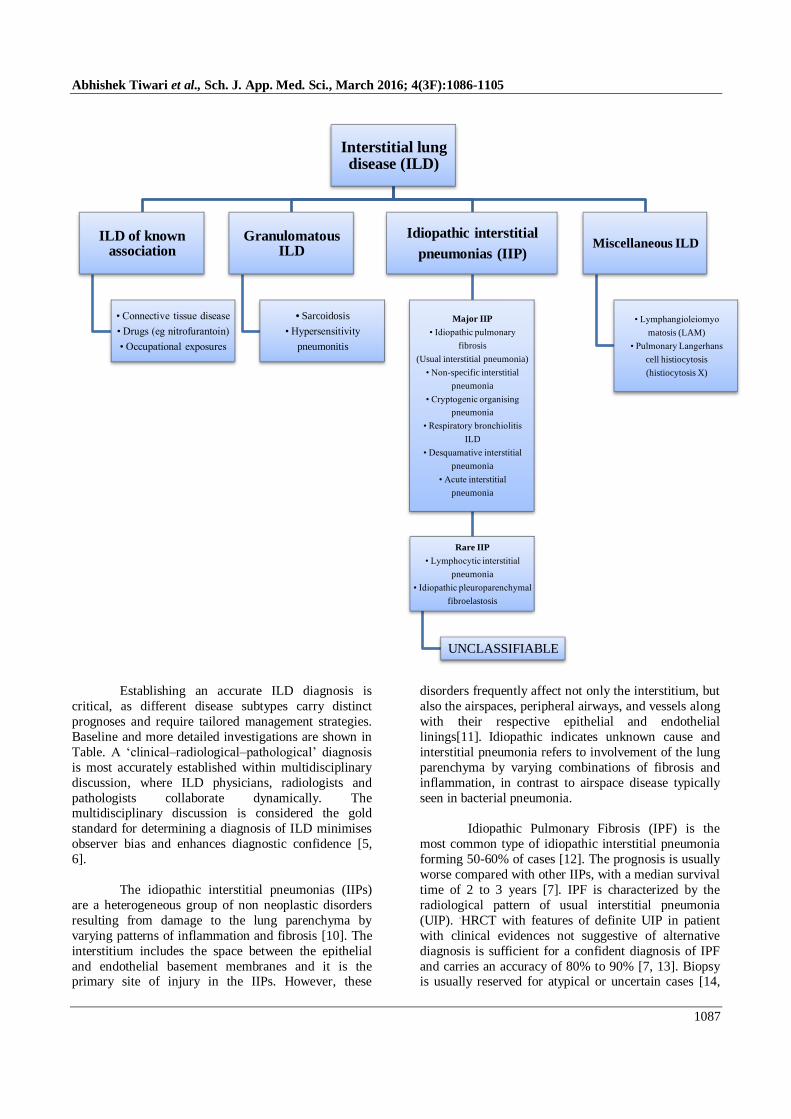
comprehensive international consensus statements [6-
9]. The major subgroups of ILD (Figure) are broadly
defined as:
Abhishek Tiwari et al., Sch. J. App. Med. Sci., March 2016; 4(3F):1086-1105
1087
Establishing an accurate ILD diagnosis is
critical, as different disease subtypes carry distinct
prognoses and require tailored management strategies.
Baseline and more detailed investigations are shown in
Table. A ‗clinical–radiological–pathological‘ diagnosis
is most accurately established within multidisciplinary
discussion, where ILD physicians, radiologists and
pathologists collaborate dynamically. The multidisciplinary discussion is considered the gold
standard for determining a diagnosis of ILD minimises
observer bias and enhances diagnostic confidence [5,
6].
The idiopathic interstitial pneumonias (IIPs)
are a heterogeneous group of non neoplastic disorders
resulting from damage to the lung parenchyma by
varying patterns of inflammation and fibrosis [10]. The
interstitium includes the space between the epithelial
and endothelial basement membranes and it is the primary site of injury in the IIPs. However, these
disorders frequently affect not only the interstitium, but
also the airspaces, peripheral airways, and vessels along
with their respective epithelial and endothelial
linings[11]. Idiopathic indicates unknown cause and
interstitial pneumonia refers to involvement of the lung
parenchyma by varying combinations of fibrosis and
inflammation, in contrast to airspace disease typically
seen in bacterial pneumonia.
Idiopathic Pulmonary Fibrosis (IPF) is the
most common type of idiopathic interstitial pneumonia
forming 50-60% of cases [12]. The prognosis is usually
worse compared with other IIPs, with a median survival
time of 2 to 3 years [7]. IPF is characterized by the
radiological pattern of usual interstitial pneumonia
(UIP). .HRCT with features of definite UIP in patient
with clinical evidences not suggestive of alternative
diagnosis is sufficient for a confident diagnosis of IPF
and carries an accuracy of 80% to 90% [7, 13]. Biopsy is usually reserved for atypical or uncertain cases [14,
Interstitial lung disease (ILD)
ILD of known association
• Connective tissue disease
• Drugs (eg nitrofurantoin)
• Occupational exposures
Granulomatous ILD
• Sarcoidosis
• Hypersensitivity
pneumonitis
Idiopathic interstitial
pneumonias (IIP)
Major IIP
• Idiopathic pulmonary
fibrosis
(Usual interstitial pneumonia)
• Non-specific interstitial
pneumonia
• Cryptogenic organising
pneumonia
• Respiratory bronchiolitis
ILD
• Desquamative interstitial
pneumonia
• Acute interstitial
pneumonia
Rare IIP
• Lymphocytic interstitial
pneumonia
• Idiopathic pleuroparenchymal
fibroelastosis
UNCLASSIFIABLE
Miscellaneous ILD
• Lymphangioleiomyo
matosis (LAM)
• Pulmonary Langerhans
cell histiocytosis
(histiocytosis X)
Abhishek Tiwari et al., Sch. J. App. Med. Sci., March 2016; 4(3F):1086-1105
1088
15]. The chest radiograph is normal in most patients
with early disease. In advanced disease, the chest
radiograph shows decreased lung volumes and sub
pleural reticular opacities that increase from the apex to
the bases of the lungs [16]. This apico-basal gradient is
even better seen on high-resolution CT images. Together with sub pleural reticular opacities and
macrocystic honeycombing combined with traction
bronchiectasis, the apicobasal gradient represents a trio
of signs that is highly suggestive of UIP [17, 18].
Ground glass abnormality is minimal or absent, never
being the predominant pattern. Many patients with IPF
may show atypical pattern of UIP on HRCT, with
overlapping features of nonspecific interstitial
pneumonia (NSIP), chronic HP, or sarcoidosis; in these
patients open lung biopsy is usually necessary to
establish a confident diagnosis [19]. The incidence of
the disease increases with older age, with presentation
typically occurring in the sixth and seventh decades [20, 21, 22]. IPF should be considered in all adult patients
with unexplained chronic exertional dyspnea, and
commonly presents with cough, bibasilar inspiratory
crackles, and finger clubbing [23, 24, 25]. More men
have been reported with IPF than women, and the
majority of patients have a history of cigarette smoking
[26], Exposure, medication, or systemic disease.
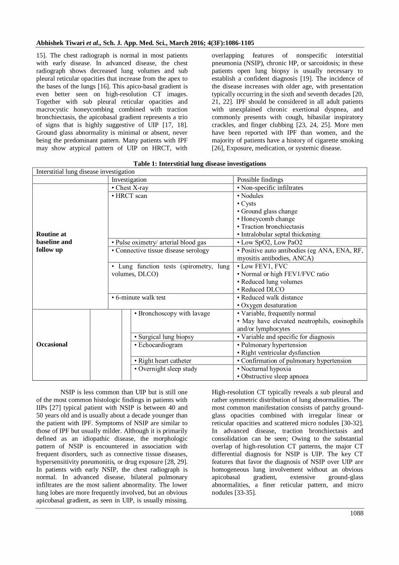
Table 1: Interstitial lung disease investigations
Interstitial lung disease investigation
Investigation Possible findings
Routine at
baseline and
follow up
• Chest X-ray • Non-specific infiltrates
• HRCT scan • Nodules
• Cysts
• Ground glass change • Honeycomb change
• Traction bronchiectasis
• Intralobular septal thickening
• Pulse oximetry/ arterial blood gas • Low SpO2, Low PaO2
• Connective tissue disease serology • Positive auto antibodies (eg ANA, ENA, RF,
myositis antibodies, ANCA)
• Lung function tests (spirometry, lung
volumes, DLCO)
• Low FEV1, FVC
• Normal or high FEV1/FVC ratio
• Reduced lung volumes
• Reduced DLCO
• 6-minute walk test • Reduced walk distance
• Oxygen desaturation
Occasional
• Bronchoscopy with lavage • Variable, frequently normal
• May have elevated neutrophils, eosinophils
and/or lymphocytes
• Surgical lung biopsy • Variable and specific for diagnosis
• Echocardiogram • Pulmonary hypertension
• Right ventricular dysfunction
• Right heart catheter • Confirmation of pulmonary hypertension
• Overnight sleep study • Nocturnal hypoxia • Obstructive sleep apnoea
NSIP is less common than UIP but is still one
of the most common histologic findings in patients with
IIPs [27] typical patient with NSIP is between 40 and
50 years old and is usually about a decade younger than
the patient with IPF. Symptoms of NSIP are similar to
those of IPF but usually milder. Although it is primarily
defined as an idiopathic disease, the morphologic
pattern of NSIP is encountered in association with
frequent disorders, such as connective tissue diseases,
hypersensitivity pneumonitis, or drug exposure [28, 29].
In patients with early NSIP, the chest radiograph is normal. In advanced disease, bilateral pulmonary
infiltrates are the most salient abnormality. The lower
lung lobes are more frequently involved, but an obvious
apicobasal gradient, as seen in UIP, is usually missing.
High-resolution CT typically reveals a sub pleural and
rather symmetric distribution of lung abnormalities. The
most common manifestation consists of patchy ground-
glass opacities combined with irregular linear or
reticular opacities and scattered micro nodules [30-32].
In advanced disease, traction bronchiectasis and
consolidation can be seen; Owing to the substantial
overlap of high-resolution CT patterns, the major CT
differential diagnosis for NSIP is UIP. The key CT
features that favor the diagnosis of NSIP over UIP are
homogeneous lung involvement without an obvious apicobasal gradient, extensive ground-glass
abnormalities, a finer reticular pattern, and micro
nodules [33-35].

Abhishek Tiwari et al., Sch. J. App. Med. Sci., March 2016; 4(3F):1086-1105
1089
Cryptogenic organizing pneumonia (COP),
previously known as bronchiolitis obliterans organizing
pneumonia is the idiopathic form of organizing
pneumonia. On HRCT, the two most frequently seen
features include bilateral, multifocal, patchy
consolidation (present in upto 90% of cases) and ground glass abnormality [36]. The lung volumes are generally
preserved, COP tends to preferentially involve the sub
pleural and bronchovascular regions of the lung
parenchyma [37].
Respiratory bronchiolitis (RB)-ILD is a part of the
spectrum of smoking related lung diseases. The
predominant finding on HRCT is ground-glass
abnormality and preferentially involves the upper lobes.
The ground glass abnormality of RB-ILD has been
shown to represent areas of macrophage accumulation
in the distal airspaces [38].
DIP (desquamative interstitial pneumonia) is a rare
form of ILD. The usual age of presentation is 40-50
years, with men affected more than women
(male/female >2:1). The disease predominantly affects
smokers (90%) cases, but can also be seen secondary to
lung infections, organic dust exposure, and marijuana
smoke inhalation. HRCT typically shows a ground glass
pattern, which is caused by diffuse macrophage
infiltration of the alveoli along with interstitial septal
thickening; this is generally present in all cases of DIP [42]. The ground glass pattern can either be patchy or
diffuse, with a predilection for peripheral and basal lung
zones [40].
Acute interstitial pneumonia (AIP) is notable for its
acute presentation. On HRCT, the most common
finding includes ground glass abnormalities, traction
bronchiectasis, and architectural distortion. The ground
glass pattern is patchy in most cases, with areas of
lobular sparing; however some cases may show a more
diffuse distribution [41].
ILDs can be associated with various occupational
lung diseases (e.g. asbestosis, silicosis, coal worker
pneumoconiosis, HP, berylliosis) [30, 31] and
connective tissue disorders (e.g. rheumatoid arthritis,
systemic sclerosis, systemic lupus erythematosus,
mixed connective tissue disease).
Interstitial lung diseases of some specific type
also show gender as well as age predilection in their
prevalence. As women are more likely to have collagen
vascular associated interstitial lung diseases due to increased risk of autoimmune diseases. Women are also
almost exclusively affected by lymphangioleio
myomatosis and tuberous sclerosis-associated lung
diseases [33]. Particular ancestory also increases the
likelihood of some interstitial lung diseases. Sarcoidosis
occurs 10-to 12-fold more in blacks than in their white
counterparts [42].
Patients with ILD frequently present with
exertional dyspnoea which has been shown to be
closely related to quality of life [34-36]. The mechanisms through which ILD produces dyspnoea
include ventilation perfusion de-arrangements, diffusion
impairment, neuro-mechanical dissociation,
physiological restriction (due to reduced compliance
and decreased elastic recoil), circulatory and
cardiovascular limitation, anxiety and depression as
well as skeletal and ventilatory muscle weakness [44].
The optimal therapy for interstitial lung diseases is an
area of intense investigation. Current medical regimens
have not shown to improve survival but nevertheless are
routinely prescribed with hope of slowing of progression of disease. Immunosuppressive anti-
inflammatory agents are used to treat various forms of
ILD [45, 46]. There is reasonably compelling evidence
that the administration of agents such as corticosteroids
is strongly associated with improvement or even
clearing of lung pathology for many forms of ILD. This
is particularly the case for disorders such as cryptogenic
organizing pneumonia (COP), eosinophilic pneumonia,
sarcoidosis, or cellular non-specific interstitial
pneumonia (NSIP) [45]. Traditional therapies that were
suggested to benefit patients with IPF included corticosteroids and cytotoxic drugs (e.g. azathioprine,
cyclophosphamide) [47].
Regarding the role of anti fibrotic therapy in
patients with interstitial lung disease, Following
evaluation in Phase II and Phase III clinical trials in
patients with IPF, [48-50] pirfenidone was approved by
the European Commission in February 2011.
Pirfenidone is indicated for the treatment of patients
with mild-to-moderate IPF.
AIMS AND OBJECTIVES
To study the clinical features of interstitial
lung disease.
To study the radiological presentation of
interstitial lung disease.
To study the treatment modalities in interstitial
lung disease.
MATERIALS AND METHODS
The present study was carried out on patients who
were attending the outpatient department and/or
admitted in Tuberculosis & Chest Department, Govt. Medical College, Amritsar, after taking approval from
the ethical committee.
Study was conducted on 50 diagnosed patients
of interstitial lung diseases attending OPD/Indoor of
Tuberculosis & Chest Department, Govt. medical

Abhishek Tiwari et al., Sch. J. App. Med. Sci., March 2016; 4(3F):1086-1105
1090
college, Amritsar after taking approval from ethical
committee and informed consent from patient.
Inclusion criteria
Diagnosed case of interstitial lung disease
Exclusion criteria
1) Patient not willing to give consent
2) Age less than 12 years
3) Pregnant ladies
4) Patients with obstructive lung disease such as
COPD or asthma or active coronary disease or
other co-morbid illness precluding
performance of 6 min walk test.
Descriptive type of study of patients diagnosed as
ILDs was done. Patient diagnosed as ILD based on
clinical, radiological and PFT findings will be included in the study. Each patient was explained the purpose of
the study and need for complete co-operation
emphasized. Those who satisfied the inclusion and
exclusion criteria were interviewed according to
prepared pro forma. Interview was conducted in a well
lit and ventilated examination room.
Detailed clinical history, physical examination,
routine investigations (Hemoglobin, total leucocyte
count, differential leucocyte count, erythrocyte
sedimentation rate, random blood sugar, renal function
tests, liver function tests), sputum examinations (for acid fast bacilli, malignant cells), chest radiography,
HRCT, PFT, ECG, investigations for connective tissue
diseases according to patient's clinical profile, and 6
minute walk test was done on all the patients.
ECHO and related other cardiac investigations
were done for selected patients, as and when required.
Treatment details were also noted.
OBSERVATIONS & RESULTS
Fifty patients diagnosed as ILD based on clinical,
radiological and PFT findings attending OPD and/or
admitted in Chest and TB hospital, Amritsar were included in the study. They were studied according to
their demographic features, clinical characteristics,
Radiological findings and treatment modalities and the
following observations were made which have been
depicted in tabular form.
Aetiology of Interstitial Lung Disease:
Table 1: Aetiology Distribution of Patients with Interstitial Lung Disease
Total
(N)
Sarcoidosis
(N)
IPF
(N)
NSIP
(N)
HSP
(N)
RA-
ILD
(N)
SS-ILD
(N)
SLE
(N)
LIP
(N)
DIP
(N)
Number of
subjects 50 18 13 12 2 1 1 1 1 1
Fig 1: Pie Chart Showing Aetiology Distribution of Patients with Interstitial Lung Disease
Of the study group of 50 patients with interstitial
lung disease, Sarcoidosis is the most common cause of
ILD consists of 18 patients forming 36% of the study
group. IPF is the second most common cause with 13
patients (26%) followed by NSIP with 12 patients
(24%).
18
13
12
2
11 1 1 1
SARCOIDOSIS
IPF
NSIP
HSP
RA-ILD
SS-ILD
SLE-ILD
LIP
DIP

Abhishek Tiwari et al., Sch. J. App. Med. Sci., March 2016; 4(3F):1086-1105
1091
Duration of Symptoms:
Table 2: Duration of Symptoms in ILD Patients
Overall Sarcoidosis IPF NSIP HSP RA-
ILD
SS-
ILD
SLE LIP DIP
Avg.
Duration of
symptoms
(yrs)
2.5 2.2 2.2 2.5 2.8 4.5 3.5 3 4.2 3.5
Fig 2: Bar Diagram Showing Duration of Symptoms in ILD Patients
Average duration of symptoms in ILD patients
in this study is 2.5 years. Sarcoidosis and IPF both
having average duration of symptoms 2.2 yrs which is
minimum in the study group while RA-ILD patient
have duration of symptoms for 4.5 yrs, maximum in the
study group.
AGE AT PRESENTATION:
Table 3: Age at Presentation in ILD Patients
Fig 3: Bar Diagram Showing Age at Presentation in ILD Patients
00.5
11.5
22.5
33.5
44.5
2.52.2 2.2
2.52.8
4.5
3.53
4.23.5
AVG. DURATION OF SYMPTOMS(YRS)
0
20
40
60 45.854.9 48.7 45
52 4639 42
49.3
AGE AT PRESENTATION
Overall
(yrs)
Sarcoidosis
(yrs)
IPF
(yrs)
NSIP
(yrs)
HP
(yrs)
RA-
ILD
(yrs)
SS-
ILD
(yrs)
SLE
(yrs)
LIP
(yrs)
DIP
(yrs)
Average Age at
presentation 48.8 45.8 54.9 48.7 45 52 46 39 42 49.3
Abhishek Tiwari et al., Sch. J. App. Med. Sci., March 2016; 4(3F):1086-1105
1092
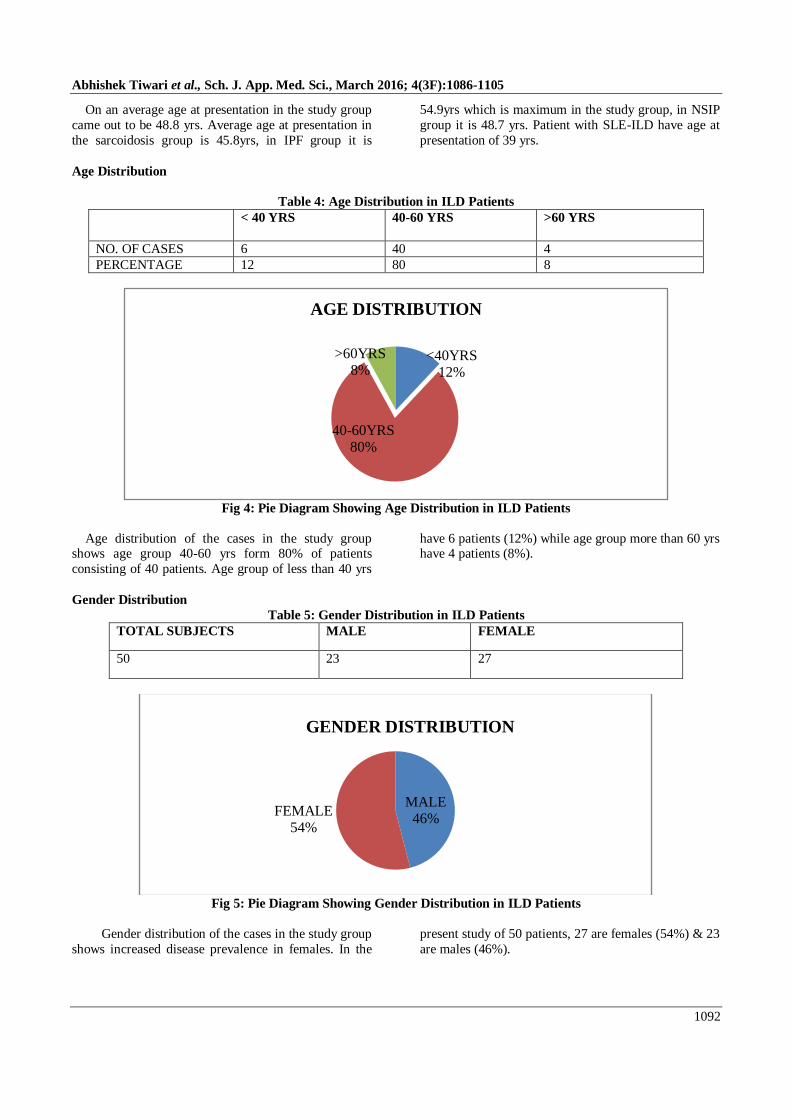
On an average age at presentation in the study group
came out to be 48.8 yrs. Average age at presentation in
the sarcoidosis group is 45.8yrs, in IPF group it is
54.9yrs which is maximum in the study group, in NSIP
group it is 48.7 yrs. Patient with SLE-ILD have age at
presentation of 39 yrs.
Age Distribution
Table 4: Age Distribution in ILD Patients
< 40 YRS 40-60 YRS >60 YRS
NO. OF CASES 6 40 4
PERCENTAGE 12 80 8
Fig 4: Pie Diagram Showing Age Distribution in ILD Patients
Age distribution of the cases in the study group shows age group 40-60 yrs form 80% of patients
consisting of 40 patients. Age group of less than 40 yrs
have 6 patients (12%) while age group more than 60 yrs have 4 patients (8%).
Gender Distribution
Table 5: Gender Distribution in ILD Patients
TOTAL SUBJECTS MALE FEMALE
50 23 27
Fig 5: Pie Diagram Showing Gender Distribution in ILD Patients
Gender distribution of the cases in the study group
shows increased disease prevalence in females. In the
present study of 50 patients, 27 are females (54%) & 23
are males (46%).
<40YRS12%
40-60YRS80%
>60YRS8%
AGE DISTRIBUTION
MALE46%
FEMALE54%
GENDER DISTRIBUTION

Abhishek Tiwari et al., Sch. J. App. Med. Sci., March 2016; 4(3F):1086-1105
1093
Smoking Pattern:
Table 6: Smoking Pattern in ILD Patients
TOTAL SUBJECTS SMOKER NON SMOKER
50 8 42
Fig 6: Pie Diagram Showing Smoking Pattern in ILD Patients
Smoking pattern in the study group shows that
only 8 patients out of 50 patients are smoker (16%),
While 42 patients are non smokers (84%).
Clinical Profile of ILD Patients
Total
(N)
Sarcoidosis
(N)
IPF
(N)
NSIP
(N)
HSP
(N)
RA-
ILD
(N)
SS-
ILD(N)
SLE
(N)
LIP
(N)
DIP
(N)
Number of subjects 50 18 13 12 2 1 1 1 1 1
Mean age (yrs) 48.8 45.8 54.9 48.7 45 52 46 39 42 49.3
Male/Female 23/27 7/11 7/6 5/7 2/0 1/0 0/1 0/1 0/1 1/0
Smoking 8 2 3 2 - - - - - 1
Duration of symptoms(yrs) 2.5 2.2 2.2 2.5 2.8 4.5 3.5 3 4.2 3.5
Cough 45 16 11 11 2 1 1 1 1 1
Dyspnoea 40 15 10 10 1 1 1 0 1 1
Haemoptysis 2 1 - 1 - - - - - -
Fever 8 2 2 1 - 1 - 1 1 -
Joint symptoms 6 2 1 1 - 1 - 1 - -
ATT intake 9 4 2 1 1 1 - - - -
Clubbing 6 1 4 1 - - - - - -
Desaturation on 6 MWT (SPo2 <
88% or fall of 4% from
baseline)
17 5 4 6 1 1 - - - -
Table 7: Gender Distribution in Patient With ILD
NO OF SUBJECTS MALE FEMALE
SARCOIDOSIS 18 7 11
IPF 13 7 6
NSIP 12 5 7
HSP 2 2 0
RA-ILD 1 1 0
SS-ILD 1 0 1
SLE-ILD 1 0 1
LIP 1 0 1
DIP 1 1 0
SMOKER16%
NON SMOKER
84%
SMOKING PATTERN

Abhishek Tiwari et al., Sch. J. App. Med. Sci., March 2016; 4(3F):1086-1105
1094
Fig 7: Bar Diagram Showing Gender Distribution in Patient with ILD
Gender distribution in the ILD cases with
different etiology shows that, Out of 18 patients of
sarcoidosis 11 were females (61%) & 7 were males
(39%). In IPF group of 13 patients, 6 were females
(46%) & 7 were males (54%). In NSIP group out of 12
patients, 7 were female (58%) & 5 were males (42%).
Signs & Symptoms at Presentation
Table 8: Signs & Symptoms Of Patients With ILD At Presentation
Symptom Number of cases Percentage
Cough 45 90.0
Dyspnoea 40 80.0
Haemoptysis 2 4.0
Fever 8 16.0
Joint symptoms 6 12.0
Clubbing 6 12.0
Desaturation at 6 MWT 17 34.0
Fig 8: Bar Diagram Showing Signs & Symptoms of Patients with ILD At Presentation
Cough & Dyspnoea are the most common feature
at presentation on our study group present in 45 (90%)
& 40 patients (80%) respectively. Other features at
presentation include fever, haemoptysis, joint
symptoms, clubbing etc.
7 75
2 1 0 0 0 1
11
67
00 1 1 00
2468
101214161820
FEMALE
MALE
0
10
20
30
40
5045
40
2
8 6 6
17
COUGH DYSPNOEA HAEMOPTYSISFEVER JOINT SYMPTOMS CLUBBINGDESATURATION AT 6MWT

Abhishek Tiwari et al., Sch. J. App. Med. Sci., March 2016; 4(3F):1086-1105
1095
Frequency of Cough & Dyspnoea
Table 9: Frequency of Cough & Dyspnoea in ILD Patients
CAUSE OF ILD NO OF CASES PRESENCE OF
COUGH
PRESENCE OF
DYSPNOEA
SARCOIDOSIS 18 16 15
IPF 13 11 10
NSIP 12 11 10
HSP 2 2 1
RA-ILD 1 1 1
SS-ILD 1 1 1
SLE-ILD 1 1 0
LIP 1 1 1
DIP 1 1 1
Fig 9: Bar Diagram Showing Frequency of Cough & Dyspnoea in ILD Patients
History of ATT Intake:
Table 10: History of ATT Intake in ILD Patients
NO OF CASES POSITIVE HISTORY OF ATT
INTAKE
OVERALL 50 9
SARCOIDOSIS 18 4
IPF 13 2
NSIP 12 1
HSP 2 1
RA-ILD 1 1
SS-ILD 1 0
SLE-ILD 1 0
LIP 1 0
DIP 1 0
0
5
10
15
20 18
1312
21 1 1 1 1
16
11 11
21 1 1 1 1
15
10 10
1 1 10
1 1
TOTAL CASES COUGH DYSPNOEA

Abhishek Tiwari et al., Sch. J. App. Med. Sci., March 2016; 4(3F):1086-1105
1096
Fig 10: Bar Diagram Showing History of ATT Intake in ILD Patients
As the radiological presentation of ILD some
time simulate TB, there is significant number of
patients have history of ATT intake. 9 (18%) of total 50
patients have history of ATT intake, most significant in
sarcoidosis group in which 4 (22%) out of 18 patients
have history of ATT intake.
Radiological Profile of ILD Patients
Total
(N)
Sarcoidosis
(N)
IPF
(N)
NSIP
(N)
HSP(N) RA-
ILD
(N)
SS-
ILD
(N)
SLE
(N)
LIP
(N)
DIP
(N)
CHEST X-RAY FINDINGS
Reticular/Reticulo-
nodular
41 13 11 11 1 1 1 1 1 1
Hilar lymph-
adenopathy
10 7 - 2 - 1 - - - -
Honey combing 3 1 2 - - - - - - -
HRCT FINDINGS
Fibrosis 26 3 13 7 - 1 1 1 - -
Honey combing 17 1 13 2 - - - - - 1
Ground glass opacity 19 7 1 7 1 - 1 - 1 1
Interstitial infiltrate 20 6 - 8 2 1 - 1 1 1
Sub pleural opacity 18 2 13 3 - - - - - -
Traction
Bronchiectasis
9 - 6 2 - - 1 - - -
Lymphadenopathy 10 8 - 2 - - - - - -
Pleural opacity 3 2 - - - 1 - - - -
Nodules 3 2 - - 1 - - - - -
Cysts - - - - - - - - - -
50
1813 12
2 1 1 1 1 1
94 2 1 1 1 0 0 0 0
0
10
20
30
40
50
60
CASES H/O ATT PRESENT

Abhishek Tiwari et al., Sch. J. App. Med. Sci., March 2016; 4(3F):1086-1105
1097
Chest X-ray Features:
Table 10: Chest X Ray Features of ILD Patients
PATTERN NO OF CASES PERCENTAGE OF CASES
RETICULAR/RETICULONODULAR 41 82.0
HILAR LYMPHADENOPATHY 10 20.0
HONEY COMBING 3 6.0
Fig 10: Bar Diagram Showing Chest X ray Features Of ILD Patients
Most common Chest X ray feature in our study
group is reticular/ reticulo nodular opacity which was
present in 41 patients (82%), followed by hilar
lymphadenopathy in 10 patients (20%) and honey
combing in 3 patients (6%).
HRCT Features of ILD Patients
Table 11: Table Showing HRCT Pattern in ILD Patients
HRCT PATTERN NO OF CASES PERCENTAGE OF
CASES
FIBROSIS 26 52.0
HONEYCOMBING 17 34.0
GROUND GLASS OPACITY 19 38.0
INTERSTITIAL INFILTRATES 20 40.0
SUBPLEURAL OPACITY 18 36.0
TRACTION BRONCHIECTASIS 9 18.0
LYMPHADENOPATHY 10 20.0
PLEURAL OPACITY 3 6.0
NODULES 3 6.0
0
10
20
30
40
50 41
10
3
RETICULAR/RETICULONODULAR HILAR LYMPHADENOPATHY
HONEY COMBING

Abhishek Tiwari et al., Sch. J. App. Med. Sci., March 2016; 4(3F):1086-1105
1098
Fig 11: Bar Diagram Showing HRCT Pattern in ILD Patients
Fibrosis (52%) is the most common HRCT
feature in our study group followed by interstitial
infiltrate (40%), ground glass opacity (38%), sub
pleural opacity (36%), honey combing (34%),
lymphadenopathy (20%), traction bronchiectasis (18%),
pleural opacity (6%), and nodules (6%).
Spirometry Pattern
Table 12: Spirometric Parameters in ILD Patients
SPIROMETRY
Total
(N)
Sarcoidosis
(N)
IPF
(N)
NSIP
(N)
HSP
(N)
RA-
ILD
(N)
SS-
ILD
(N)
SLE
(N)
LIP
(N)
DIP
(N)
FEV1% (mean %
predicted) 60 65 55 60 75 58 53 43 53 68
FVC%(mean %
predicted) 66 71 58 68 79 68 59 52 58 76
FEV1/FVC (mean) 87 86 91 87 90 80 95 71 87 95
Fig 12: Bar Diagram Showing Spirometric Parameters in ILD Patients
0
5
10
15
20
25
30 26
1719 20
18
9 10
3 3
0102030405060708090
100
6065
5560
75
5853
4353
686671
5868
79
6859
5258
76
87 8691 87 90
80
95
71
8795
FEV1% FVC% FEV1/FVC%
Abhishek Tiwari et al., Sch. J. App. Med. Sci., March 2016; 4(3F):1086-1105
1099
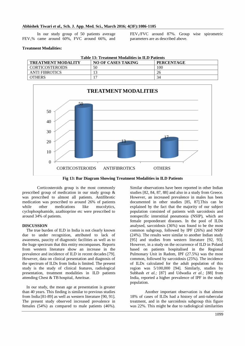
In our study group of 50 patients average
FEV1% came around 60%, FVC around 66%, and
FEV1/FVC around 87%. Group wise spirometric
parameters are as described above.
Treatment Modalities:
Table 13: Treatment Modalities in ILD Patients
TREATMENT MODALITY NO OF CASES TAKING PERCENTAGE
CORTICOSTEROIDS 50 100
ANTI FIBROTICS 13 26
OTHERS 17 34
Fig 13: Bar Diagram Showing Treatment Modalities in ILD Patients
Corticosteroids group is the most commonly
prescribed group of medication in our study group &
was prescribed to almost all patients. Antifibrotic
medication was prescribed to around 26% of patients
while other medications like mucolytics,
cyclophosphamide, azathioprine etc were prescribed to
around 34% of patients.
DISCUSSION The true burden of ILD in India is not clearly known
due to under recognition, attributed to lack of
awareness, paucity of diagnostic facilities as well as to
the huge spectrum that this entity encompasses. Reports
from western literature show an increase in the
prevalence and incidence of ILD in recent decades [79].
However, data on clinical presentation and diagnosis of
the spectrum of ILDs from India is limited. The present
study is the study of clinical features, radiological
presentation, treatment modalities in ILD patients
attending Chest & TB hospital, Amritsar.
In our study, the mean age at presentation is greater
than 40 years. This finding is similar to previous studies
from India [81-89] as well as western literature [90, 91].
The present study observed increased prevalence in
females (54%) as compared to male patients (46%).
Similar observations have been reported in other Indian
studies [82, 84, 87, 88] and also in a study from Greece.
However, an increased prevalence in males has been
documented in other studies [85, 87].This can be
explained by the fact that the majority of our subject
population consisted of patients with sarcoidosis and
nonspecific interstitial pneumonia (NSIP), which are
female preponderant diseases. In the pool of ILDs
analysed, sarcoidosis (36%) was found to be the most common subgroup, followed by IPF (26%) and NSIP
(24%). The results were similar to another Indian study
[95] and studies from western literature [92, 93].
However, in a study on the occurrence of ILD in Poland
based on patients hospitalised in the Regional
Pulmonary Unit in Radom, IPF (27.5%) was the most
common, followed by sarcoidosis (25%). The incidence
of ILDs calculated for the adult population of this
region was 5/100,000 [94]. Similarly, studies by
Subhash et al.; [87] and Udwadia et al.; [88] from
India, reported a higher prevalence of IPF in the study
population.
Another important observation is that almost
18% of cases of ILDs had a history of anti-tubercular
treatment, and in the sarcoidosis subgroup this figure
was 22%. This might be due to radiological similarities
0
10
20
30
40
50
CORTICOSTEROIDS ANTIFIBROTICS OTHERS
50
1317
TREATMENT MODALITIES
Abhishek Tiwari et al., Sch. J. App. Med. Sci., March 2016; 4(3F):1086-1105
1100
between ILD and pulmonary tuberculosis and a lack of
awareness and paucity of diagnostic facilities in remote
areas.
The current study included 18 (36%) cases of
pulmonary sarcoidosis, 11 (61.1%) being females. The higher prevalence in females is coherent with findings
in western literature [93]. The mean age at presentation
was 48.8 years with the average duration of symptoms
being 2.5 years; the majority were non-smokers (84%).
This data is similar to other Indian studies [95, 96]. In
contradiction with the literature [93] clubbing (12%)
was an uncommon finding in our study. 6MWT showed
significant desaturation (SPo2< 88% or 4 % fall from
the baseline) in 17 (34%) cases. The plausible
explanation of this could be the advanced stage of the
disease at presentation.
Idiopathic pulmonary fibrosis is a specific type
of ILD, with characteristic radiological features and
histopathology. In the present study we had 13 (26%)
cases of IPF, and it was the second most common
subgroup in the pool of ILDs. In contrast, IPF was
observed as the most common ILD in other Indian [86-
88] and western studies [97]. In the current study, mean
age at presentation was 54.9 years, male to female ratio
was 7:6 and 23.07% of cases were smokers. The current
study agrees with data from western literature in terms
of age at presentation with disease typically occurring in 6th–7th decade of life. The literature [92] shows
more men being diagnosed with IPF than women and
the majority being smokers. However, our study
reported around equal prevalence in male and female
subjects and a more prevalence of smoking. In another
Indian study by Subhash et al.; [87] out of 33 cases of
IPF, 16 were females and smoking was present in only
18% of all IPF cases. Another point that merits mention
is that diagnostic criteria vary across studies, leading to
differences in epidemiological parameters of IPF. In our
study, on 6MWT, 34% of cases showed significant desaturation (SPo2< 88% or 4 % fall from the baseline)
at presentation. This finding has clinical implications as
studies have advocated that desaturation (i.e. a decline
in oxygen saturation to below 88%) during 6MWT is a
marker for increased risk of mortality[100].
In the present study, we had 12 (24%) cases
with a diagnosis of NSIP based on clinical, radiological
and pathological features, but we were unable to
elucidate the cause. The mean age at presentation was
48.7 years, 7 were females and only 16.6% were
smokers. The review of literature shows NSIP has a mean age of 52 years and is more common in females
and never smokers [101].
Of 3 cases diagnosed as CTD -associated interstitial
lung disease (CTD-ILD), lung involvement at
presentation was observed in 1 case each of rheumatoid
arthritis (RA), scleroderma & SLE. The prevalence of
RA-ILD varies from 5–58% [102] and ILD in systemic
sclerosis is observed in 40–80% of cases [103]. The
prevalence of CTD–ILD in India has been reported as
ranging from 5.6% to 50.8% in various studies [87, 91].
Hypersensitivity pneumonitis (HP) was
diagnosed in 2 (4%) cases; all were associated with
pigeon exposure, with duration of exposure ranging
from 3–6 years. In a previous study from India,
Udwadia et al.; [94] reported HP in 15 (6%) from a
total of 273 cases. Other ILDs diagnosed as per clinico-
radio-pathological criteria in the current study were
desquamative interstitial pneumonia (DIP) 1 case,
lymphocytic interstitial pneumonia (LIP) 1 case.
Regarding treatment modalities patients using
in our study group, Corticosteroids is the most commonly used group, with all 50 patients (100%)
taking this medication, followed by anti fibrotic
medication in 13 patients (26%), other less commonly
used medications like mucolytics, cyclophosphamide,
azathioprine etc in 17 patients (34%)
SUMMARY AND CONCLUSION
In present study, fifty patients diagnosed as ILD
based on clinical, radiological and PFT findings
attending OPD and/or admitted in Chest and TB
hospital, Amritsar are included in the study. They were studied according to their demographic features, clinical
characteristics, Radiological findings and treatment
modalities.
1. There were 23 males and 27 females yielding a
male: female ratio of 23:27.
2. 40 patients (83.3%) were between 40-60 years of
age. The mean overall age of patients was 48.8
years.
3. 8 patients (16%) were smokers and 42 patients
(84%) non smokers, yielding a smoker: non-
smoker ratio of 4:21. All smokers are of male gender.
4. Sarcoidosis came out as most common cause of
ILD with 18 patients (36%) followed by IPF (26%)
& NSIP (24%) respectively.
5. Mean duration of symptoms of illness at
presentation overall came out around 2.5 years.
6. The common symptoms at presentation included
cough (90%), breathlessness (80%) followed by
fever (16%), joint symptoms (12%), hemoptysis
(4%).
7. There is history of ATT intake in 9 patients (18%),
more frequently in sarcoidosis group (22%). 8. Of the total 50 patients 17 (34%) were showing
significant desaturation on 6 minute walk test, that
is SPo2 less than 88% or more than 4% fall in
baseline SPo2.

Abhishek Tiwari et al., Sch. J. App. Med. Sci., March 2016; 4(3F):1086-1105
1101
9. Common Chest X ray findings were
reticular/reticulo nodular pattern (82%), hilar
lymphadenopathy (10%), honeycombing (6%) etc.
10. Common HRCT findings were fibrosis (52%),
interstitial infiltrates (40%), ground glass opacity
(38%), sub pleural opacity (36%), honeycombing (34%), lymphadenopathy (20%), traction
bronchiectasis (18%), pleural opacity (6%),
nodules (6%) etc.
11. Average FEV1, FVC, FEV1/FVC came around 60,
66, and 87 respectively. SLE-ILD group has worst
pulmonary function test.
12. All 50 patients were taking corticosteroids (100%),
13 patients were on anti fibrotic & anti
inflammatory drugs (26%), 17 patients were on
other medications (34%).
This study describes the spectrum of ILDs prevalent in patients presenting to outdoor & indoor department
of Chest & TB hospital, Govt. Medical College,
Amritsar (Punjab). Diagnosis of ILDs at an early stage
is paramount to prevent/delay progression to
irreversible damage to the lungs, especially in
treatment-responsive ILDs like sarcoidosis. Hence, in a
developing country like India, with high prevalence of
pulmonary tuberculosis, education and awareness of
general practitioners and physicians about ILDs
deserves special attention.
REFERENCES 1. American Thoracic Society, European Respiratory
Society. American Thoracic Society/European
Respiratory Society International Multidisciplinary
Consensus Classification of the Idiopathic Interstitial
Pneumonias. Am J Respir Crit Care Med 2002;
165:277–304.
2. Flaherty KR, Andrei AC, Murray S, Fraley C,
Colby T.V, Travis WD et al.; Idiopathic pulmonary
fibrosis: prognostic value of changes in physiology
and six-minute-walk test. Am J Respir Critic Care Med 2006; 174(7):803–09.
3. Lama VN, Flaherty KR, Toews GB, Colby, T.V,
Travis W.D, Long Q et al.; Prognostic value of
desaturation during a 6-minute walk test in
idiopathic interstitial pneumonia. Am J Respir
Critic Care Med 2003; 168(9):1084–90.
4. Park JH, Kim DK, Kim DS, Koh Y, Lee S.D, Kim
W.S et al.; Mortality and risk factors for surgical
lung biopsy in patients with idiopathic interstitial
pneumonia. Eur J Cardio thorac Surg 2007;
31(6):1115–19.
5. Flaherty KR, King TE Jr, Raghu G, Lynch III J.P, Colby T.V, Travis W.D et al.; Idiopathic interstitial
pneumonia: What is the effect of a
multidisciplinary approach to diagnosis? Am J
Respir Critic Care Med 2004; 170(8):904–10.
6. Travis WD, Costabel U, Hansell DM, King Jr T.E,
Lynch D.A, Nicholson A.G et al.; An official
American Thoracic Society/ European Respiratory
Society statement: Update of the international
multidisciplinary classification of the idiopathic
interstitial pneumonias. Am J Respir Critic Care
Med 2013; 188(6):733–48.
7. Raghu G, Collard HR, Egan JJ, Martinez F.J, Behr J, Brown K.K et al.; An official ATS/ERS/JRS/
ALAT statement: Idiopathic pulmonary fibrosis:
evidence-based guidelines for diagnosis and
management. Am J Respir Critic Care Med 2011;
183(6):788–824.
8. Bradley B, Branley HM, Egan JJ; Interstitial lung
disease guideline: The British Thoracic Society in
collaboration with the Thoracic Society of
Australia and New Zealand and the Irish Thoracic
Society. Thorax 2008; 63 Suppl 5:v1-58.
9. American Thoracic Society, European Respiratory
Society. American Thoracic Society/European Respiratory Society International Multidisciplinary
Consensus Classification of the Idiopathic
Interstitial Pneumonias. This joint statement of the
American Thoracic Society (ATS), and the
European Respiratory Society (ERS) was adopted
by the ATS board of directors, June 2001 and by
the ERS Executive Committee, June 2001. Am J
Respir Critic Care Med 2002; 165:277–304.
10. American Thoracic Society; European Respiratory
Society. Idiopathic pulmonary fibrosis: diagnosis
and treatment: international consensus statement. Am J Respir Crit Care Med 2000; 161:646–664.
11. Cushley MJ, Davison AG, du Bois RM, Egan J,
Flower CD, Gibson GJ, et al.; The diagnosis,
assessment and treatment of diffuse parenchymal
lung disease in adults. Thorax 1999; 54:S1–S30.
12. Travis WD, King TE Jr, Bateman ED, Lynch D.A,
Capron F, Center D et al.; American Thoracic
Society/European Respiratory Society international
multidisciplinary consensus classification of the
idiopathic interstitial pneumonias. Am J Respir Crit
Care Med 2002; 165(2):277. 13. Tsubamato M, Muller NL, Johkoh T, Ichikado K,
Taniguchi H, Kondoh Y et al.; Pathologic
subgroups of nonspecific interstitial pneumonia:
differential diagnosis from other idiopathic
interstitial pneumonias on high resolution
computed tomography. J Comput Assist Tomogr
2005; 29(6):793.
14. Hunninghake GW, Zimmerman MB, Schwartz DA,
KING JR T.E, Lynch J, Hegele R et al.; Utility of a
lung biopsy for the diagnosis of idiopathic
pulmonary fibrosis. Am J Respir Crit Care Med
2001; 164(2):193-196. 15. Raghu G, Mageto YN, Lockhart D, Schmidt R.A,
Wood D.E, Godwin J.D; The accuracy of the
clinical diagnosis of new-onset idiopathic
pulmonary fibrosis and other interstitial lung
disease: a prospective study. Chest 1999;
116(5):1168-74.
Abhishek Tiwari et al., Sch. J. App. Med. Sci., March 2016; 4(3F):1086-1105
1102
16. Chandler PW, Shin MS, Friedman SE, Myers JL,
Katzenstein AL; Radiographic manifestations of
bronchiolitis obliterans with organizing pneumonia
vs usual interstitial pneumonia. AJR Am J
Roentgenol 1986; 147:899–906.
17. Hunninghake GW, Lynch DA, Galvin JR, Gross B.H, Muller N, Schwartz D.A et al.; Radiologic
findings are strongly associated with a pathologic
diagnosis of usual interstitial pneumonia. Chest
2003; 124(4):1215–1223.
18. Johkoh T, Muller NL, Cartier Y, Kavanagh P.V,
Hartman T.E, Akira M et al.; Idiopathic interstitial
pneumonias: diagnostic accuracy of thin-section
CT in 129 patients. Radiology 1999; 211(12):555–
560.
19. Sverzellati N, Wells AU, Tomassetti S, Desai S.R,
Copley S.J, Aziz Z.A et al.; Biopsy-proved
idiopathic pulmonary fibrosis: spectrum of non-diagnostic thin section CT diagnoses. Radiology
2010; 254(3):957.
20. Scott J, Johnston I, Britton J; What causes
cryptogenic fibrosing alveolitis? A case-control
study of environmental exposure to dust. BMJ
1990; 301:1015–1017.
21. Mannino DM, Etzel RA, Parrish RG; Pulmonary
fibrosis deaths in the United States, 1979–1991: an
analysis of multiple-cause mortality data. Am J
Respir Crit Care Med 1996; 153:1548–1552.
22. Raghu G, Freudenberger TD, Yang S, Curtis JR, Spada C, Hayes J, et al.; High prevalence of
abnormal acid gastro-oesophageal reflux in
idiopathic pulmonary fibrosis. Eur Respir J 2006;
27:136–142.
23. Douglas WW, Ryu JH, Schroeder DR; Idiopathic
pulmonary fibrosis: Impact of oxygen and
colchicine, prednisone, or no therapy on survival.
Am J Respir Crit Care Med 2000; 161:1172–1178.
24. King TE Jr, Tooze JA, Schwarz MI, Brown KR,
Cherniack RM; Predicting survival in idiopathic
pulmonary fibrosis: scoring system and survival model. Am J Respir Crit Care Med 2001;
164:1171– 1181.
25. Gribbin J, Hubbard RB, Le Jeune I, Smith CJ, West
J, Tata LJ; Incidence and mortality of idiopathic
pulmonary fibrosis and sarcoidosis in the UK.
Thorax 2006; 61:980–985.
26. Iwai K, Mori T, Yamada N, Yamaguchi M, Hosoda
Y; Idiopathic pulmonary fibrosis: epidemiologic
approaches to occupational exposure. Am J Respir
Crit Care Med 1994; 150:670–675.
27. Travis WD, Matsui K, Moss J, Ferrans VJ;
Idiopathic nonspecific interstitial pneumonia: prognostic significance of cellular and fibrosing
patterns— survival comparison with usual
interstitial pneumonia and desquamative interstitial
pneumonia. Am J Surg Pathol 2000; 24:19–33.
28. Kim EA, Lee KS, Johkoh T, Kim T.S, Suh G.Y,
Kwon O.J et al.; Interstitial lung diseases
associated with collagen vascular diseases:
radiologic and histopathologic findings. Radio
Graphics 2002; 22(1):S151–S165.
29. Rossi SE, Erasmus JJ, McAdams HP, Sporn TA,
Goodman PC; Pulmonary drug toxicity: radiologic
and pathologic manifestations. RadioGraphics 2000; 20:1245–1259.
30. Johkoh T, Muller NL, Colby TV, Ichikado K,
Taniguchi H, Kondoh Y et al.; Nonspecific
interstitial pneumonia: correlation between thin-
section CT findings and pathologic subgroups in 55
patients. Radiology 2002; 225(1):199–204.
31. Akira M, Inoue G, Yamamoto S, Sakatani M; Non-
specific interstitial pneumonia: findings on
sequential CT scans of nine patients. Thorax 2000;
55:854–859.
32. Kim EY, Lee KS, Chung MP, Kwon OJ, Kim TS,
Hwang JH; Nonspecific interstitial pneumonia with fibrosis: serial high-resolution CT findings with
functional correlation. AJR Am J Roentgenol 1999;
173: 949–953.
33. MacDonald SL, Rubens MB, Hansell DM, Copley
SJ, Desai S.R, du Bois R.M et al.; Nonspecific
interstitial pneumonia and usual interstitial
pneumonia: comparative appearances at and
diagnostic accuracy of thin-section CT. Radiology
2001;221(3):600–605.
34. Flaherty KR, Thwaite EL, Kazerooni EA, Gross
B.H, Toews G.B, Colby T.V et al.; Radiological versus histological diagnosis in UIP and NSIP:
survival implications. Thorax 2003; 58(2): 143–
148.
35. Do KH, Lee JS, Colby TV, Kitaichi M, Kim DS;
Nonspecific interstitial pneumonia versus usual
interstitial pneumonia: differences in the density
histogram of high-resolution CT. J Comput Assist
Tomogr 2005;29:544–548.
36. Jara-palomares L, Gomez-Izquierdo L, Gonzalez-
Vergara D, Rodriguez-Becerra E, Marquez-Martin
E, Barrot-Cortés E, et al.; Utility of high-resolution computed tomography and BAL in cryptogenic
organizing pneumonia. Respir Med 2010;
104(11):1706-1711.
37. Lee KS, Kullnig P, Hartman TE, Müller N.L;
Cryptogenic organizing pneumonia: CT findings in
43 patients. AJR Am J Roentgenol 1994; 162: 543-
546.
38. Remy-Jardin M, Remy J, Boulenguez C, et al.;
Morphologic effects of cigarette smoking on
airways and pulmonary parenchyma in healthy
adult volunteers: CT evaluation and correlation
with pulmonary function tests. Radiology 1993; 186:107.
39. Johkoh T, Muller NL, Cartier Y, Kavanagh P.V,
Hartman T.E, Akira M et al.; Idiopathic interstitial
pneumonias: diagnostic accuracy of thin section
CT in 129 patients. Radiology 1999; 211(2):555.

Abhishek Tiwari et al., Sch. J. App. Med. Sci., March 2016; 4(3F):1086-1105
1103
40. Hartman TE, Primack SL, Swensen SJ, Hansell D,
McGuinness G, Müller N.L; Desquamative
interstitial pneumonia: thin section CT findings in
22 patients. Radiology 1993; 187(3):787-790.
41. Primack SL, Hartman TE, Ikezoe J, Akira M,
Sakatani M, Müller N.L; Acute interstitial pneumonia: radiographic and CT findings in nine
patients. Radiology 1993; 188(3):817.
42. Michael A. Nead, David G Morris, et al.;
Interstitial Lung Disease: A Clinical Overview and
General Approach. In. Fisherman‘s Pulmonary
Diseases and Disorders. 2008; 4: 1105-7.
43. Hansen JE, Wasserman K; Pathophysiology of
activity limitation in patients with interstitial lung
disease. Chest 1996; 109:1566–76.
44. Schwarz M, King TE Jr.; Physiology of interstitial
lung disease. In: Schwarz M, King TE Jr.
Interstitial lung disease. 2003; 4:54–74. 45. Kim R, Meyer KC; Therapies of interstitial lung
disease—past, present, and future. Therapeutic
Advances Respir Dis 2008, 2:319–338.
46. Meyer KC, Bierach J; Immunosuppressive therapy
for autoimmune lung diseases. Immunol Allergy
Clin North Am 2012, 32:633–639.
47. American Thoracic Society: Idiopathic pulmonary
fibrosis: diagnosis and treatment. International
consensus statement. American Thoracic Society
(ATS) and the European Respiratory Society
(ERS). Am J Respir Crit Care Med 2000, 161:646–664.
48. Azuma A, Nukiwa T, Tsuboi E, Suga M, Abe S,
Nakata K et al.; Double-blind, placebo-controlled
trial of pirfenidone in patients with idiopathic
pulmonary fibrosis. Am J Respir Crit Care Med
2005, 171(9):1040-1047.
49. Taniguchi H, Ebina M, Kondoh Y, Ogura T,
Azuma A, Suga M et al.; Pirfenidone in idiopathic
pulmonary fibrosis. Eur Respir J 2010; 35(4):821-
829.
50. Noble PW, Albera C, Bradford WZ, Costabel U, Glassberg M.K, Kardatzke D et al.; Pirfenidone in
patients with idiopathic pulmonary fibrosis
(CAPACITY): two randomised trials. Lancet 2011,
377(9779):1760-1769.
51. Coultas DB, Zumwalt RE, Black WC, Sobonya
RE; The epidemiology of interstitial lung diseases.
Am J Respiratory Critical Care Medicine 1994;
150:967-72.
52. A U Wells, N Hirani; Interstitial lung disease
guideline: the British thoracic society in
collaboration with the Thoracic society of Australia
and New Zealand and the Irish thoracic society. Thorax 2008; 63:1-9.
53. Travis WD, Costabel U, Hansell DM, King Jr T.E,
Lynch D.A, Nicholson A.G et al.; An official
American Thoracic Society/European Respiratory
Society statement: update of the international
multidisciplinary classification of the idiopathic
interstitial pneumonias. Am J Respir Crit Care Med
2013; 188(6): 733–748.
54. Thomeer M, Demedts M, Behr J, Buhl R, Costabel
U, Flower C.D.R et al.; Multidisciplinary
interobserver agreement in the diagnosis of
idiopathic pulmonary fibrosis. Eur Respir J 2008; 31(3): 585–591.
55. Meyer KC, Raghu G; Patient evaluation. In
Interstitial Lung Disease: A Practical Approach.
Secondth edition. Edited by Baughman RP, Du
Bois RM. New York: Springer; 2011:3–16.
56. Meyer KC; Interstitial lung disease in the elderly:
pathogenesis, diagnosis and management.
Sarcoidosis Vasc Diffuse Lung Dis 2011; 28:3–17.
57. Raghu G, Weycker D, Edelsberg J, Bradford W.Z,
Oster G; Incidence and prevalence of idiopathic
pulmonary fibrosis. Am J Respir Crit Care Med.
2006, 174(7): 810-816. 58. Raghu G, Mageto YN, Lockhart D, Schmidt R.A,
Wood D.E, Godwin J.D; The accuracy of the
clinical diagnosis of new-onset idiopathic pulmonary
fibrosis and other interstitial lung disease: A
prospective study. Chest. 1999, 116(5): 1168-1174.
59. Hunninghake GW, Zimmerman MB, Schwartz DA,
KING JR T.E, Lynch J, Hegele R et al.; Utility of a
lung biopsy for the diagnosis of idiopathic
pulmonary fibrosis. Am J Respir Crit Care Med.
2001; 164(2): 193-196.
60. Martinez FJ, Safrin S, Weycker D, Starko K.M, Bradford W.Z, King T.E et al.; The clinical course
of patients with idiopathic pulmonary fibrosis. Ann
Intern Med. 2005, 142(12_part_1): 963-967.
61. Collard HR, King TE Jr, Bartelson BB, Vourlekis
J.S, Schwarz M.I, Brown KK; Changes in clinical
and physiologic variables predict survival in
idiopathic pulmonary fibrosis. Am J Respir Crit
Care Med. 2003, 168(5): 538-542.
62. Richeldi L, Davies HR, Ferrara G, Franco F;
Corticosteroids for idiopathic pulmonary fibrosis.
Cochrane Database Syst Rev 2003; (3): CD002880. 63. Davies HR, Richeldi L, Walters EH;
Immunomodulatory agents for idiopathic
pulmonary fibrosis. Cochrane Database Syst Rev
2003 ;( 3): CD003134.
64. Collard HR, Ryu JH, Douglas WW, Schwarz M.I,
Curran-Everett D, King T.E et al.; Combined
corticosteroid and cyclophosphamide therapy does
not alter survival in idiopathic pulmonary fibrosis.
Chest. 2004, 125(6): 2169-2174.
65. Martinez FJ; Idiopathic interstitial pneumonias:
Usual interstitial pneumonia versus nonspecific
interstitial pneumonia. Proc Am Thorac Soc. 2006, 3: 81-95.
66. Cha SI, Fessler MB, Cool CD, Schwarz M.I,
Brown K.K; Lymphoid interstitial pneumonia:
Clinical features, associations and prognosis. Eur
Respir J. 2006, 28(2): 364-369.

Abhishek Tiwari et al., Sch. J. App. Med. Sci., March 2016; 4(3F):1086-1105
1104
67. Strange C, Highland KB; Interstitial lung disease in
the patient who has connective tissue disease. Clin
Chest Med. 2004, 25: 549-559.
68. Tanaka N, Newell JD, Brown KK, Cool C.D,
Lynch D.A; Collagen vascular disease-related lung
disease: High-resolution computed tomography findings based on the pathologic classification. J
Comput Assist Tomogr. 2004, 28(3): 351-360.
69. Tashkin DP, Elashoff R, Clements PJ, Goldin J,
Roth M.D, Furst D.E et al.; Cyclophosphamide
versus placebo in scleroderma lung disease. N Engl
J Med. 2006, 354(25): 2655-2666.
70. Swigris JJ, Olson AL, Fischer A, Lynch D.A,
Cosgrove G.P, Frankel SK et al.; Mycophenolate
mofetil is safe, well tolerated, and preserves lung
function in patients with connective tissue disease-
related interstitial lung disease. Chest. 2006, 130(1):
30-36. 71. Baughman RP; Pulmonary sarcoidosis. Clin Chest
Med. 2004, 25: 521-530.
72. Shorr AF, Torrington KG, Hnatiuk OW;
Endobronchial involvement and airway hyper
reactivity in patients with sarcoidosis. Chest. 2001,
120: 881-886.
73. Paramothayan NS, Lasserson TJ, Jones PW;
Corticosteroids for pulmonary sarcoidosis.
Cochrane Database Syst Rev 2005; (2): CD001114.
74. Selman M; Hypersensitivity pneumonitis: A
multifaceted deceiving disorder. Clin Chest Med. 2004, 25: 531-547.
75. Monkare S; Influence of corticosteroid treatment
on the course of farmer's lung. Eur J Respir Dis.
1983, 64: 283-293.
76. Camus P, Bonniaud P, Fanton A, Camus C,
Baudaun N, Foucher P; Drug-induced and
iatrogenic infiltrative lung disease. Clin Chest Med.
2004, 25(3): 479-519.
77. American Thoracic Society. Diagnosis and initial
management of non-malignant diseases related to
asbestos. Am J Respir Crit Care Med. 2004, 170: 691-715.
78. Schwartz DA, Davis CS, Merchant JA, Bunn W.B,
Galvin J.R, Van Fossen D.S et al.; Longitudinal
changes in lung function among asbestos-exposed
workers. Am J Respir Crit Care Med. 1994, 150(5):
1243-1249.
79. Kornum J.B., Christensen S., Grijota M, Pedersen
L, Wogelius P, Beiderbeck A et al.; The incidence
of interstitial lung disease 1995–2005: a Danish
nationwide population-based study. BMC Pulm.
Med. 2008; 8(1): 1.
80. American Thoracic Society, European Respiratory Society, World Association of sarcoidosis and
Other Granulomatous Disorders. Statement on
sarcoidosis. Am. J. Respir. Crit. Care Med. 1999;
160: 736-55.
81. Shah J.R; Diffuse interstitial pulmonary fibrosis:
course and prognosis. Indian J. Chest Dis. Allied
Sci. 1974; 21: 174–179.
82. Jindal S.K., Malik S.K., Deodhar S.D., Sharma
B.K; Fibrosing alveolitis: a report of 61 cases seen
over the past five years. Indian J. Chest Dis. Allied Sci. 1979; 21: 174–179.
83. Mahasur A.A., Dave K.M., Kinare S.G., Kamat
S.R., Shetye V.M., Kolhatkar V.P; Diffuse
fibrosing alveolitis-an Indian experience. Lung
India 1983; 5: 171–179.
84. Sharma S.K., Pande J.N., Guleria J.S; Diffuse
interstitial pulmonary fibrosis. Indian J. Chest Dis.
Allied Sci. 1984; 26: 214–219.
85. Sharma S.K., Pande J.N., Verma K., Guleria J.S;
Bronchoalveolar lavage fluid (BALF) analysis in
interstitial lung diseases. Indian J. Chest Dis. Allied
Sci. 1989; 31: 187–196. 86. Kalra S., D`Souza G., Bhusnuramth B., Jindal S.K;
Transbronchial lung biopsy in diffuse lung disease.
Indian J. Chest Dis. Allied Sci. 1989; 31: 265–270.
87. Subhash H.S., Ashwin I., Solomon S.K., David T.,
Cherian A.M., Thomas K; A comparative study on
idiopathic pulmonary fibrosis and secondary
diffuse parenchymal lung disease. Indian Journal of
Medical Science 2004; 58: 185–190.
88. Sen T., Udwadi Z.F; Retrospective Study of
Interstitial Lung Disease in a Tertiary Care Centre
in India. Indian J. Chest Dis. Allied Sci. 2010; 52: 207–211.
89. Gagiya A.K., Suthar H.N., Bhagat G.R; Clinical
profile of interstitial lung diseases cases. Natl. J.
Med. Res. 2012; 2: 2–4.
90. Raghu G., Nyberg F., Morgan G; The
epidemiology of interstitial lung disease and its
association with lung cancer. Br. J. Cancer. 2004;
91: S3–10.
91. Kornum J.B., Christensen S., Grijota M, Pedersen
L, Wogelius P, Beiderbeck A et al.; The incidence
of interstitial lung disease 1995–2005: a Danish nationwide population-based study. BMC Pulm.
Med. 2008; 8(1): 1.
92. Karakatsani A., Papakosta D., Rapti A, Antoniou
K.M, Dimadi M, Markopoulou A et al.; Hellenic
Interstitial Lung Diseases Group. Epidemiology of
interstitial lung diseases in Greece. Respir. Med.
2009; 103(8): 1122–1129.
93. Schweisfurth H., Kieslich C., Satake N,
Loddenkemper R, Schönfeld N, Mäder I et al.;
How are interstitial lung diseases diagnosed in
Germany? Results of the scientific registry for the
exploration of interstitial lung diseases (―Fibrosis registry‖) of the WATL. Pneumologie 2003; 57(7):
373–382.
94. Szafrański W; Interstitial lung diseases among
patients hospitalized in the Department of
Respiratory Medicine in Radom District Hospital

Abhishek Tiwari et al., Sch. J. App. Med. Sci., March 2016; 4(3F):1086-1105
1105
during the years 2000–2009. Pneumonol. Alergol.
Pol. 2012; 80: 523–532.
95. Sharma S.K, Mohan A; Sarcoidosis in India: not so
rare! J. Indian Acad. Clin. Med. 2004; 5: 12–21.
96. Sharma S.K, Mohan A, Guleria J.S; Clinical
characteristics, pulmonary function abnormalities and outcome of prednisolone treatment in 106
patients with sarcoidosis. J. Assoc. Physicians India
2001; 49: 697–704.
97. Xaubet A., Ancochea J., Morell F, Rodriguez-Arias
J.M, Villena V, Blanquer R et al.; Report on the
incidence of interstitial lung diseases in Spain.
Sarcoidosis Vasc. Diffuse Lung Dis.2004; 21(1):
64–70.
98. Scott J, Johnston I, Britton J; What causes
cryptogenic fibrosing alveolitis? A case-control
study of environmental exposure to dust. BMJ
1990; 301: 1015–1017. 99. Mannino D.M, Etzel R.A, Parrish R.G; Pulmonary
fibrosis deaths in the United States, 1979–1991: an
analysis of multiple- cause mortality data. Am. J.
Respir. Crit. Care Med. 1996; 153: 1548–1552.
100. Lama V.N., Flaherty K.R., Toews G.B, Colby T.V,
Travis W.D, Long Q et al.; Prognostic value of
desaturation during a 6-minute walk test in
idiopathic interstitial pneumonia. Am. J. Respir.
Crit. Care Med. 2003; 168(9): 1084–1090.
101. Travis W.D, Hunninghake G, King T.E. Jr, Lynch
D.A, Colby T.V, Galvin JR, et al.; Idiopathic nonspecific interstitial pneumonia: report of an
American Thoracic Society project. Am. J. Respir.
Crit. Care Med. 2008; 177(12): 1338–1347.
102. Fischer A, West S.G, Swigris J.J, Brown K.K, du
Bois R.M; Connective tissue disease-associated
interstitial lung disease: a call for clarification.
Chest. 2010; 138: 251–256.
103. Gabbay E., Tarala R., Will R, Carroll G, Adler B,
Cameron D et al.; Interstitial lung disease in recent
onset rheumatoid arthritis. Am. J. Respir. Crit. Care
Med. 1997; 156(2): 528–535.