Embed Size (px)
Citation preview
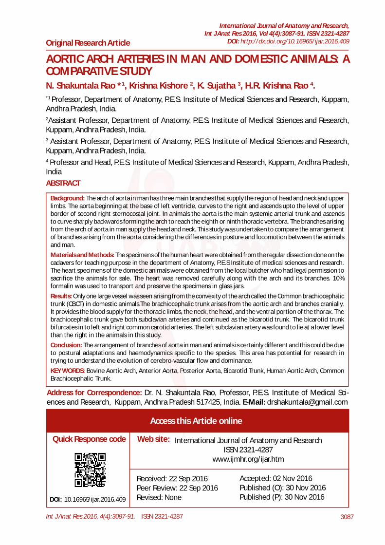
Int J Anat Res 2016, 4(4):3087-91. ISSN 2321-4287 3087
Original Research Article
AORTIC ARCH ARTERIES IN MAN AND DOMESTIC ANIMALS: ACOMPARATIVE STUDYN. Shakuntala Rao *1, Krishna Kishore 2, K. Sujatha 3, H.R. Krishna Rao 4.
ABSTRACT
Address for Correspondence: Dr. N. Shakuntala Rao, Professor, P.E.S. Institute of Medical Sci-ences and Research, Kuppam, Andhra Pradesh 517425, India. E-Mail: [email protected]
Background: The arch of aorta in man has three main branches that supply the region of head and neck and upperlimbs. The aorta beginning at the base of left ventricle, curves to the right and ascends upto the level of upperborder of second right sternocostal joint. In animals the aorta is the main systemic arterial trunk and ascendsto curve sharply backwards forming the arch to reach the eighth or ninth thoracic vertebra. The branches arisingfrom the arch of aorta in man supply the head and neck. This study was undertaken to compare the arrangementof branches arising from the aorta considering the differences in posture and locomotion between the animalsand man.Materials and Methods: The specimens of the human heart were obtained from the regular dissection done on thecadavers for teaching purpose in the department of Anatomy, P.E.S Institute of medical sciences and research.The heart specimens of the domestic animals were obtained from the local butcher who had legal permission tosacrifice the animals for sale. The heart was removed carefully along with the arch and its branches. 10%formalin was used to transport and preserve the specimens in glass jars.Results: Only one large vessel was seen arising from the convexity of the arch called the Common brachiocephalictrunk (CBCT) in domestic animals.The brachiocephalic trunk arises from the aortic arch and branches cranially.It provides the blood supply for the thoracic limbs, the neck, the head, and the ventral portion of the thorax. Thebrachiocephalic trunk gave both subclavian arteries and continued as the bicarotid trunk. The bicarotid trunkbifurcates in to left and right common carotid arteries. The left subclavian artery was found to lie at a lower levelthan the right in the animals in this study.Conclusion: The arrangement of branches of aorta in man and animals is certainly different and this could be dueto postural adaptations and haemodynamics specific to the species. This area has potential for research intrying to understand the evolution of cerebro-vascular flow and dominance.KEY WORDS: Bovine Aortic Arch, Anterior Aorta, Posterior Aorta, Bicarotid Trunk, Human Aortic Arch, CommonBrachiocephalic Trunk.
International Journal of Anatomy and Research,Int J Anat Res 2016, Vol 4(4):3087-91. ISSN 2321-4287
DOI: http://dx.doi.org/10.16965/ijar.2016.409
Access this Article online
Quick Response code Web site: International Journal of Anatomy and ResearchISSN 2321-4287
www.ijmhr.org/ijar.htm
DOI: 10.16965/ijar.2016.409
*1 Professor, Department of Anatomy, P.E.S. Institute of Medical Sciences and Research, Kuppam,Andhra Pradesh, India.2Assistant Professor, Department of Anatomy, P.E.S. Institute of Medical Sciences and Research,Kuppam, Andhra Pradesh, India.3 Assistant Professor, Department of Anatomy, P.E.S. Institute of Medical Sciences and Research,Kuppam, Andhra Pradesh, India.4 Professor and Head, P.E.S. Institute of Medical Sciences and Research, Kuppam, Andhra Pradesh,India
Received: 22 Sep 2016Peer Review: 22 Sep 2016Revised: None
Accepted: 02 Nov 2016Published (O): 30 Nov 2016Published (P): 30 Nov 2016
Int J Anat Res 2016, 4(4):3087-91. ISSN 2321-4287 3088
N. Shakuntala Rao, Krishna Kishore, K. Sujatha, H.R. Krishna Rao. AORTIC ARCH ARTERIES IN MAN AND DOMESTIC ANIMALS: ACOMPARATIVE STUDY.
BACKGROUNDThe ascending aorta is 5 cm long and begins atthe base of the left ventricle. It begins at thelevel of the lower border of the 3rd left costalcartilage. It ascends obliquely, curving to theright, behind the left of the sternum, to the levelof upper border of the second left costal carti-lage. Anterior to the ascending aorta in its lowerpart is the infundibulum and initial segment ofthe pulmonary trunk and posterior to it lie theright pulmonary artery, left atrium and principalbronchus.The arch of aorta arises at the level of the up-per border of second right sternocostal joint. Itends level with the sternal end of the secondleft costal cartilage. The summit of the arch is2.5 cm below the superiosternal border.The archof aorta gives three branches in humans, theyare Brachiocephalic trunk, left Common Carotid,left Subclavian artery.In the domestic mammals the aorta is the mainsystemic arterial trunk. It begins at the base ofthe left ventricle and is almost median at itsorigin.Its first part, the ascending aorta (aorta ascends)passes upwards and forwards between the pul-monary artery on the left and right atrium onthe right. It then curves sharply backward anddorsally inclines somewhat to the left, formingthe arch of aorta (arcus aortae) and reaches theventral surface of the spine at the eight or ninththoracic vertebrae. After passing backward alongthe ventral aspect of the bodies of the verte-brae and between the lungs, it traverses the hia-tus aorticus and enters the abdominal cavity.The branches arising from the arch of aorta inman supply the head and neck. Man’s erect pos-ture has a bearing on the number of branchesand the direction of blood flow towards the struc-tures in the head and neck. Where as in ani-mals with a posture that varies with their loco-motion and feeding habits like reaching out tobranches of trees or bowing low to feed fromthe ground or ruminating with head upwards theblood flow should definitely have a distinct ar-rangement of vessels that arise from the heart.Therefore the study was undertaken to observeand compare the differences in the branchingpattern of the arch of aorta in man and other
domestic mammals. Only one large vesselarises from the convexity of the arch called theCommon brachiocephalic trunk (CBCT).(fig 1,2,3)The brachiocephalic trunk arises from the aor-tic arch and branches cranially. It provides theblood supply for the thoracic limbs, the neck,the head, and the ventral portion of the thorax.The brachiocephalic trunk gives the leftsubclavian (left axillary) artery and the rightsubclavian (right axillary) artery and continuesas bicarotid trunk. The bicarotid trunk bifurcatesin to left and right common carotid arteries.Thusit was observed that the common brachiocepha-lic trunk gives origin to the left subclavianartery (a.subclaviasinistra), Right subclavianartery (a.subclaviadextra),and the Bicarotidtrunk (truncusbicaroticus).The bicarotid trunk is a short common trunk,which arises from the common brachiocephalictrunk,extends cranially and branches in to theleft and right common carotid arteries.Theresults might help us understand the functionsof the great vessels of the heart and theirarrangement in relation to the evolutionarypostural changes and adaptations to the envi-ronment.
The specimens of the human heart wereobtained from the regular dissection done onthe cadavers for teaching purpose in thedepartment of Anatomy, P.E.S Institute of medicalsciences and research.The heart specimens of the domestic animalswere obtained from the local butcher who hadlegal permission to sacrifice the animals for sale.The heart was removed carefully along with thearch and its branches.10% formalin was used to transport and preservethe specimens in glass jars.
MATERIALS AND METHODS
RESULTS
Arch Of Aorta In Domestic Mammals (Cow, Goat&Sheep): The aorta is the main systemic arte-rial trunk. (fig-1,2,3) In the cow there were twomain divisions from the arch.which was veryshort. First part, the ascending aorta (aorta as-cends) passes upwards and forwards betweenthe pulmonary artery on the left and right atrium
Int J Anat Res 2016, 4(4):3087-91. ISSN 2321-4287 3089
N. Shakuntala Rao, Krishna Kishore, K. Sujatha, H.R. Krishna Rao. AORTIC ARCH ARTERIES IN MAN AND DOMESTIC ANIMALS: ACOMPARATIVE STUDY.
on the right. It then curves sharply backward anddorsally inclines somewhat to the left, formingthe arch of aorta (arcus aortae) and reaches theventral surface of the spine at the eight or ninththoracic vertebrae. After passing backward alongthe ventral aspect of the bodies of the verte-brae and between the lungs,it traverses the hia-tus aorticus and enters the abdominal cavity.Branches Of The Aortic Arch (Cow,Goat&Sheep): Only one large vessel arises from theconvexity of the arch called the Commonbrachiocephalic trunk (CBCT).(fig 1,2,3)The brachiocephalic trunk arises from theaortic arch and branches cranially. It providesthe blood supply for the thoracic limbs, the neck,
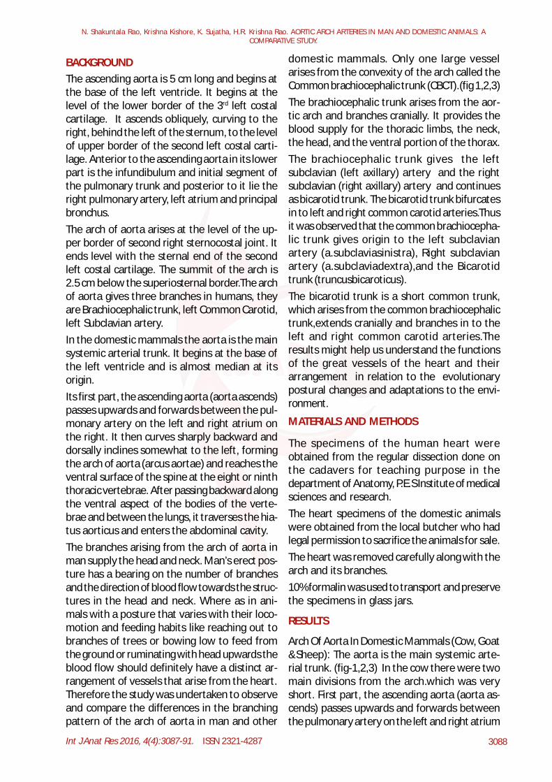
Fig. 1: Arch of aorta in the cow.
Right subclavian artery
Common brachiocephalic trunk
Arch of aorta
Bi carotid trunk
Left subclavian artery
Common carotid artery
the head, and the ventral portion of the thorax.The brachiocephalic trunk gives the leftsubclavian (left axillary) artery and the rightsubclavian (right axillary) artery and continuesas bicarotid trunk. The bicarotid trunk bifurcatesin to left and right common carotid arteries.Thusit was observed that the common brachiocepha-lic trunk gives origin to the left subclavianartery (a.subclaviasinistra), Right subclavianartery (a.subclaviadextra),and the Bicarotidtrunk (truncusbicaroticus).The bicarotid trunk is a short commontrunk,which arises from the common brachio-cephalic trunk,extends cranially and branchesin to the left and right common carotid arteries.
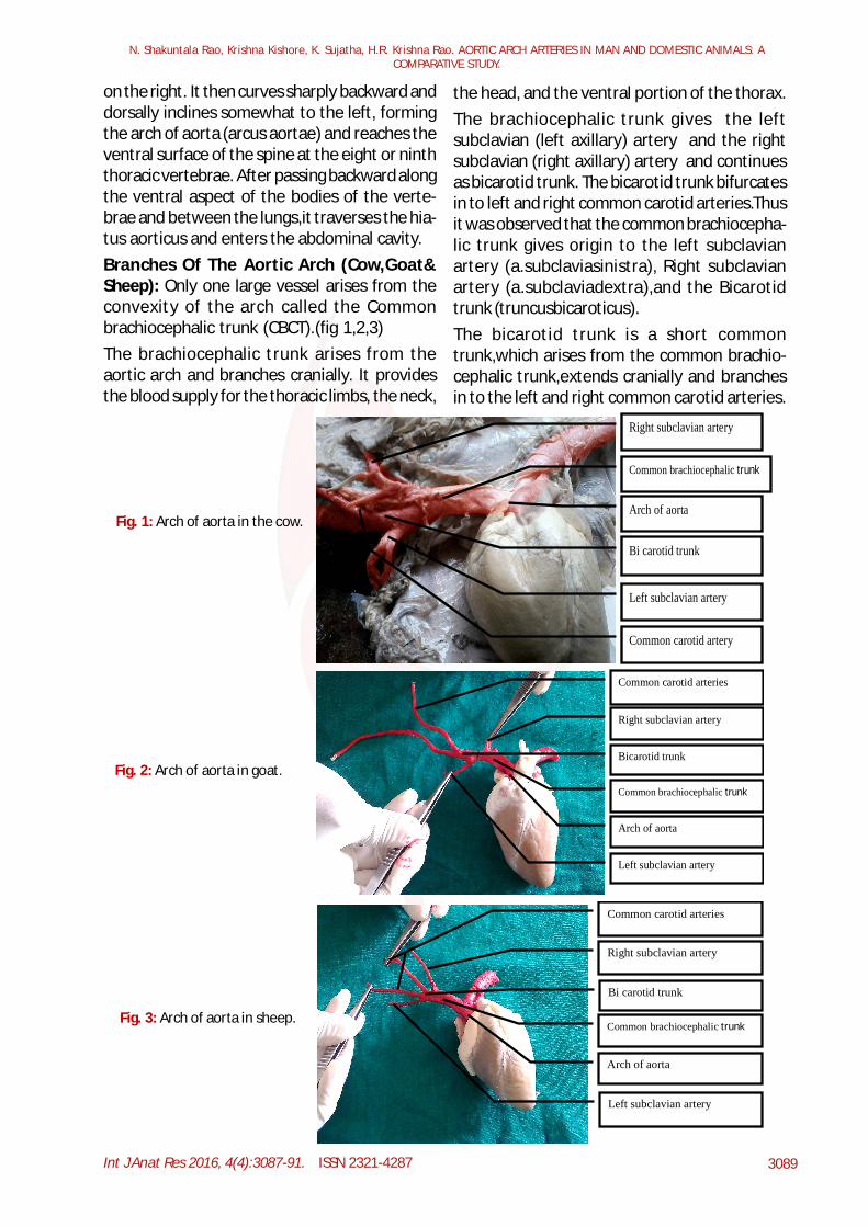
Fig. 2: Arch of aorta in goat.
Common carotid arteries
Right subclavian artery
Bicarotid trunk
Common brachiocephalic trunk
Arch of aorta
Left subclavian artery
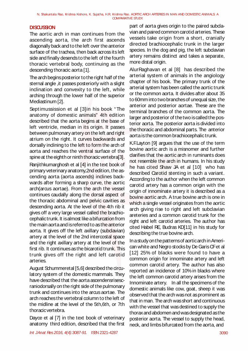
Common carotid arteries
Right subclavian artery
Bi carotid trunk
Common brachiocephalic trunk
Arch of aorta
Left subclavian artery
Fig. 3: Arch of aorta in sheep.
Int J Anat Res 2016, 4(4):3087-91. ISSN 2321-4287 3090
N. Shakuntala Rao, Krishna Kishore, K. Sujatha, H.R. Krishna Rao. AORTIC ARCH ARTERIES IN MAN AND DOMESTIC ANIMALS: ACOMPARATIVE STUDY.
DISCUSSION part of aorta gives origin to the paired subcla-vian and paired common carotid arteries. Thesevessels take origin from a short, craniallydirected brachiocephalic trunk in the largerspecies. In the dog and pig, the left subclavianartery remains distinct and takes a separate,more distal origin.AlurRaghavan et al [8] has described thearterial system of animals in the angiologychapter of his book. The primary trunk of thearterial system has been called the aortic trunkor the common aorta. It divides after about 35to 60mm into two branches of unequal size, theanterior and posterior aortae. These are theterminal branches of the common aorta. Thelarger and posterior of the two is called the pos-terior aorta. The posterior aorta is divided intothe thoracic and abdominal parts. The anterioraorta is the common brachiocephalic trunk.K.F.Layton [9] argues that the use of the termbovine aortic arch is a misnomer and furtherclarifies that the aortic arch in ruminants doesnot resemble the arch in humans. In his studyhe has cited Shaw JA et al [10] who hasdescribed Carotid stenting in such a variant.According to the author when the left commoncarotid artery has a common origin with theorigin of innominate artery it is described as abovine aortic arch. A true bovine arch is one inwhich a single vessel originates from the aorticarch giving rise to right and left subclavianareteries and a common carotid trunk for theright and left carotid arteries. The author hascited Habel RE, Budras KD[11] in his study fordescribing the true bovine arch.In a study on the patterns of aortic arch in Ameri-can white and Negro stocks by De Garis CF et al[12] 25% of blacks were found to have acommon origin for innominate artery and leftcommon carotid artery. The author has alsoreported an incidence of 10% in blacks wherethe left common carotid artery arises from theInnominate artery. In all the specimens of thedomestic animals like cow, goat, sheep it wasobserved that the arch was not as prominent asthat in man. The arch was short and continuouswith the vessel that was destined to supply thethorax and abdomen and was designated as theposterior aorta. The vessel to supply the head,neck, and limbs bifurcated from the aorta, and
The aortic arch in man continues from theascending aorta, the arch first ascendsdiagonally back and to the left over the anteriorsurface of the trachea, then back across its leftside and finally desends to the left of the fourththoracic vertebral body, continuing as thedescending thoracic aorta [1].The arch begins posterior to the right half of thesternal angle ,it passes posteriorly with a slightinclination and convexity to the left, whilearching through the lower half of the superiorMediastinum [2].Septimussission et al [3]in his book “Theanatomy of domestic animals” 4th editiondescribed that the aorta begins at the base ofleft ventricle, median in its origin. It passesbetween pulmonary artery on the left and rightatrium on the right. It curves backwards anddorsally inclining to the left to form the arch ofaorta and reaches the ventral surface of thespine at the eighth or ninth thoracic vertebra[3].Ranjithkumarghosh et al [4] in the text book ofprimary veterinary anatomy,2nd edition, the as-cending aorta (aorta ascends) inclines back-wards after forming a sharp curve, the aorticarch(arcus aortae). From the arch the vesselcontinues caudally along the dorsal aspect ofthe thoracic abdominal and pelvic cavities asdescending aorta. At the level of the 4th rib itgives off a very large vessel called the brachio-cephalic trunk. It is almost like a bifurcation fromthe main aorta and is referred to as the anterioraorta. It gives off the left axillary (subclavian)artery at the level of the 2nd intercostal spaceand the right axillary artery at the level of thefirst rib. It continues as the bicarotid trunk. Thistrunk gives off the right and left carotidarteries.August Schummeretal [5,6] described the circu-latory system of the domestic mammals. Theyhave described that the aorta ascendensrisesc-raniodorsally on the right side of the pulmonarytrunk and continues into the arcus aortae. Thearch reaches the vertebral column to the left ofthe midline at the level of the 5th,6th, or 7ththoracic vertebra.Dayce et al [7] in the text book of veterinaryanatomy third edition, described that the first
Int J Anat Res 2016, 4(4):3087-91. ISSN 2321-4287 3091
turned cranially to reach the various structures.What is notable here is that unlike in man wherewe have three different branches, in the animalswe have one large trunk which gives offbranches to the head and neck. This commontrunk has been named by some authors theanterior aorta. Another feature to note is thatthe subclavian arteries are given off by thebrachiocephalic trunk to the right and left limbs.In man only the right subclavian artery arisesfrom the brachiocephalic trunk and the leftsubclavian artery arises directly from the archof aorta. Both the carotids have again acommon trunk in animals.In a study by Christophe C et al [13] thoracicaorta and its branches were studied in mice theyexplained that the geometry of the branches ofthe murine aortic arch was similar to that of menand both these species had a sigmoidal curveof the first part of aorta comprising of ascend-ing aorta,aortic arch and superior part ofdescending aorta. In both the ascending anddescending aorta do not lie in a single verticalplane. Such an arrangement describes anon-planar aortic geometry. They concludedthat this arrangement is in contrast to theplanar aortic pattern in domestic mammalswhere the ascending and dscending aorta lie ina single vertical plane.
CONCLUSION
The observation in this study shows theremarkable branching of arteries from the aortaaccording to the needs of the species. Thepresence of so many arteries serves the survivalfunctions of the animals like searching for preyand feeding and later ruminating in leisure. Inman the sharing of a common trunk to give awaythe subclavian and carotid on the right side canbe given a second thought. The developmentof carotids is from the 3rd arch arteries. The leftsubclavian artery is not a pharyngeal archartery. It forms from the left seventh interseg-mental artery. It shifts its origin close to the leftcommon carotid artery due to differential growth.The left subclavian artery was found to lie at alower level than the right in the animals in thisstudy. This point can be further studied on thebasis of cerebral dominance in man. Is the rightsubclavian artery supplying the right upper limb
sharing the trunk with right common carotid forany reason still unknown? Does this kind ofarrangement of vessels have any bearing ofsignificance to determine cerebral dominancein humans ? If the individual has a variation inthe branches of the arches, might there be anyvariation in his brain dominance? Can it becorrelated? This observation has potential forresearch in this area.
Conflicts of Interests: None
REFERENCES
[1]. Susan Standring. The Anatomical basis of ClinicalPractice: Elsevier Churchill Livingstone 2008; 984-985.
[2]. G.J.Romanes. Cunningham’s manual of practicalanatomy: Volume-2 Thorax and abdomen.15th edi-tion Oxford University Press. pp 59-60.
[3]. Sisson and Grossman’s the Anatomy of the Domes-tic Animals. the anatomy of the domestic animals,4th edition, page no: 641– 645.
[4]. Ranjithkumar Ghosh et al. primary veterinaryanatomy,2nd edition, page no: 215 – 220.
[5]. August Schummer et al The viscera of the domesticmammals. 2ndedition, page no:198-201.
[6]. August schummer, Helmet Wilkens, Berndvollmer-hans and Karl-Heinz Habermehl. The circulatorysystem, the skin and the cutaneous organs of thedomestic mammals. page no:71-77.
[7]. Dayce et al. Text book of Veterinary Anatomy, 3rd
edition, page no:237-242.[8]. AlurRaghavan et al. Text book of Veterinary Anatomy,
4th edition, page no:465-468 & 491-498.[9]. K.F.Layton, D.F. Kallmes, H.J Cloft, E.P.Lindell and V.S
Cox. Bovine Aortic Arch Variant in Humans: Clarifi-cation of a Common Misnomer AJNR August 200627: 1541-1542
[10]. Shaw JA, Gravereaux EC,Eisenhauer AC. Carotidstenting in the bovine arch. Catheter CardiovascInterv 2003; 60: 566-69.
[11]. Habel RE,Budras KD. Thoracic cavity. In: BovineAnatomy: An Illustrated Text. Hanover, Germany:Schlutersche GmbH & Co; 2003: 62-65.
[12]. De Garis CF, Black IB,Riemenschneider EA. Patternsof the aortic arch in American white and Negrostocks, with comparative notes on certain othermammals. J Anat 1933; 67:599-618.
[13]. Christophe Casteleyn, Bram Trachet, Denis Van Looand Daniel G H Devos. Validation of the murine aor-tic arch as a model to study human vasculardiseases.J Anat.2010 May; 216(5): 563-571.
How to cite this article: N. Shakuntala Rao, KrishnaKishore, K. Sujatha, H.R. Krishna Rao. AORTIC ARCHARTERIES IN MAN AND DOMESTIC ANIMALS: ACOMPARATIVE STUDY. Int J Anat Res 2016;4(4):3087-3091. DOI: 10.16965/ijar.2016.409
N. Shakuntala Rao, Krishna Kishore, K. Sujatha, H.R. Krishna Rao. AORTIC ARCH ARTERIES IN MAN AND DOMESTIC ANIMALS: ACOMPARATIVE STUDY.