Embed Size (px)
Citation preview

Injuries to the Foot, Ankle and Lower Leg
Original Author:Sabino Sports MedicineConnie Rauser, Instructor

Bony Anatomy
• Tibia• Fibula• Tarsals•Metatarsals• Phalanges• Sesamoid Bones• Calcaneus
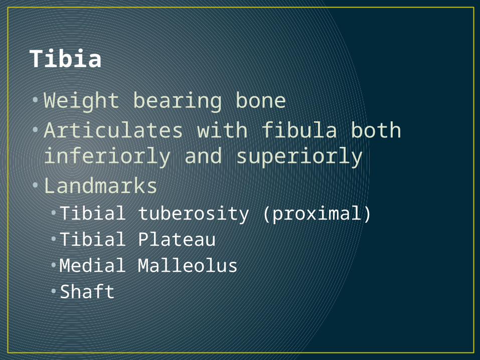
Tibia
• Weight bearing bone• Articulates with fibula both inferiorly
and superiorly• Landmarks•Tibial tuberosity (proximal)•Tibial Plateau•Medial Malleolus•Shaft

Fibula
• Non-weight bearing bone• Extends down past calcaneus
providing bony support to prevent eversion• Serves as site for muscle
attachments• Landmarks•Head of fibula (proximal)•Lateral malleolus

Tarsals
• Talus—articulates with the tibia/fibula• Calcaneus• Navicular• Cuboid• Medial, intermediate and
lateral cuneiforms

Joints
• Tibiofibular joint--syndesmosis• Ankle joint (talocrural)
Ankle mortise• Subtalar joint• Metatarsalphalangeal
joints (MP)• Interphalangeal joints •PIP•DIP

Arches• Transverse: proximal across tarsals• Medial longitudinal arch: from calcaneus to 1st
metatarsal• Strengthened by spring ligament (plantar
calcaneonavicular ligament)
• Lateral longitudinal arch: from calcaneus to 5th metatarsal
• Metatarsal arch: shaped by distal heads of metatarsals

Muscles of lateral compartment
• Peroneus longus• Peroneus brevis•Both do eversion and plantarflex
• Peroneus tertius•Dorsiflex and evert

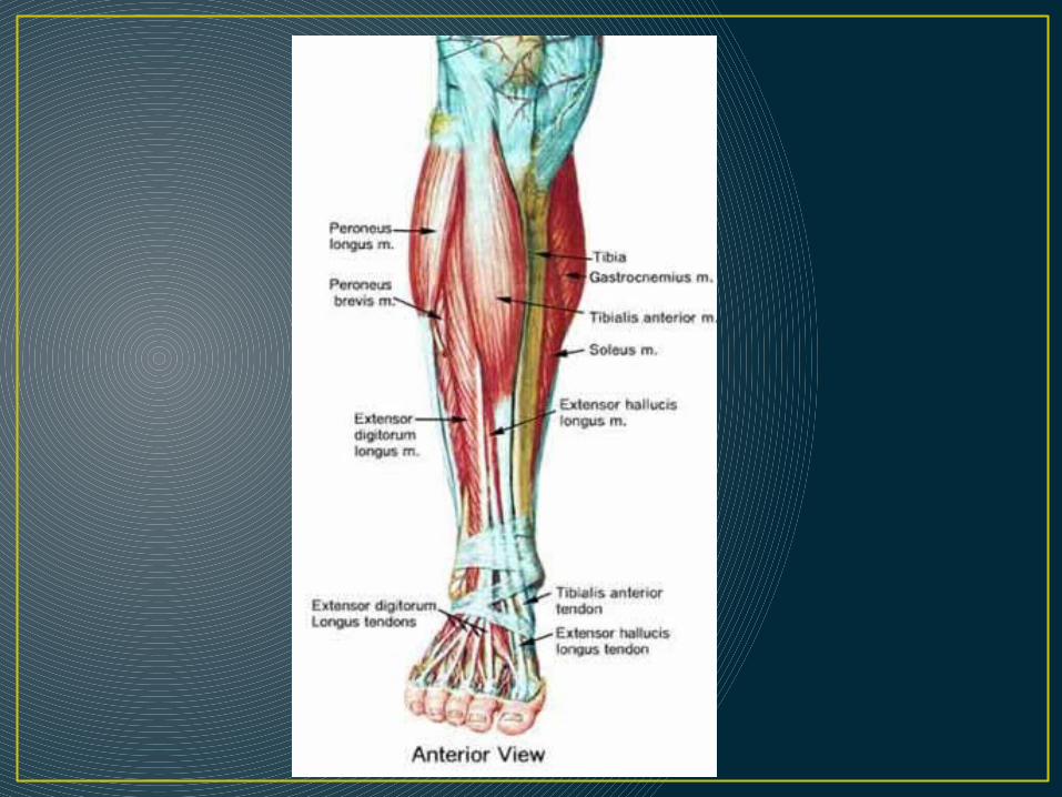
Muscles of the anterior compartment
• Tibialis Anterior• Extensor Digitorum Longus• Extensor Hallicus Longus• All do dorsiflexion and some inversion• EDL—extension of toes 2-5• EHL—extension of great toe
• **EDB—extends toes 2-4 • (dorsum of foot)


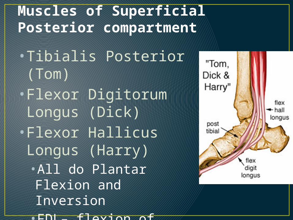
Muscles of Superficial Posterior compartment
• Tibialis Posterior (Tom)• Flexor Digitorum Longus (Dick)• Flexor Hallicus Longus (Harry)•All do Plantar Flexion and Inversion•FDL– flexion of toes 2-5•FHL—flexion of great toe

Muscles of Deep Posterior Compartment• Gastrocnemius—crosses
knee and ankle joint. Knee flexion/plantar flexion
• Soleus---crosses ankle joint. Plantarflexion• Join together at the Achilles tendon
• Plantaris—cross ankle and knee joints. Knee flexion/plantar flexion• Tendon run parallel to the Achilles tendon medially


Miscellaneous• Plantar Fascia• From calcaneus to heads of metatarsals.•Maintain stability of foot and supports medial longitudinal arch
• Interosseus Membrane• Thick connective tissue runs length of tib/fib and holds them together
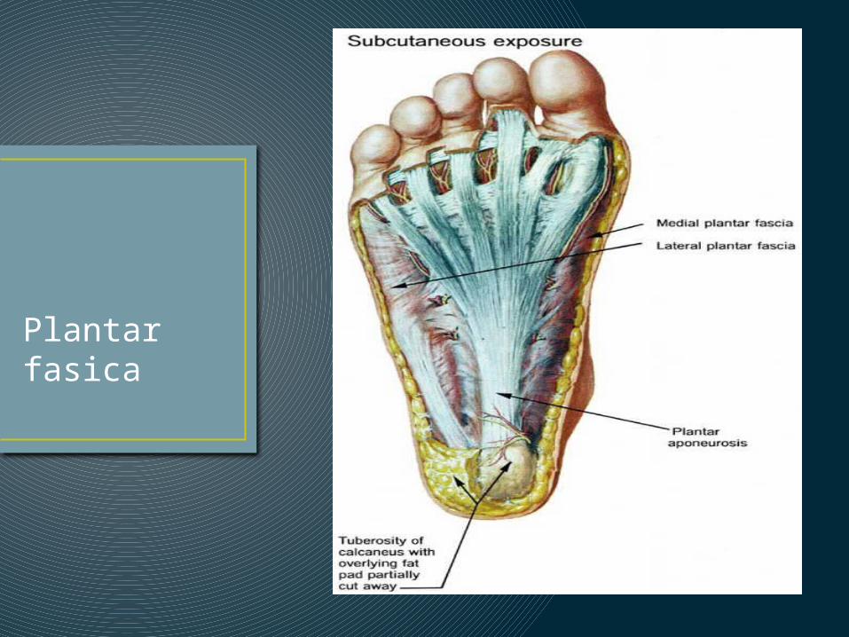
Plantar fasica
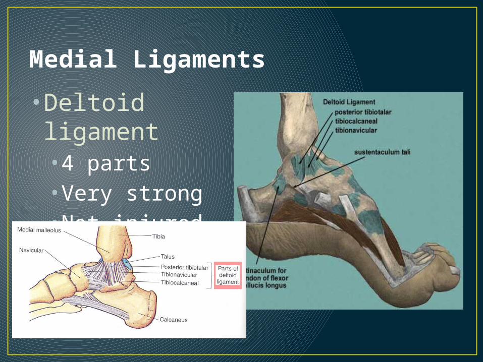
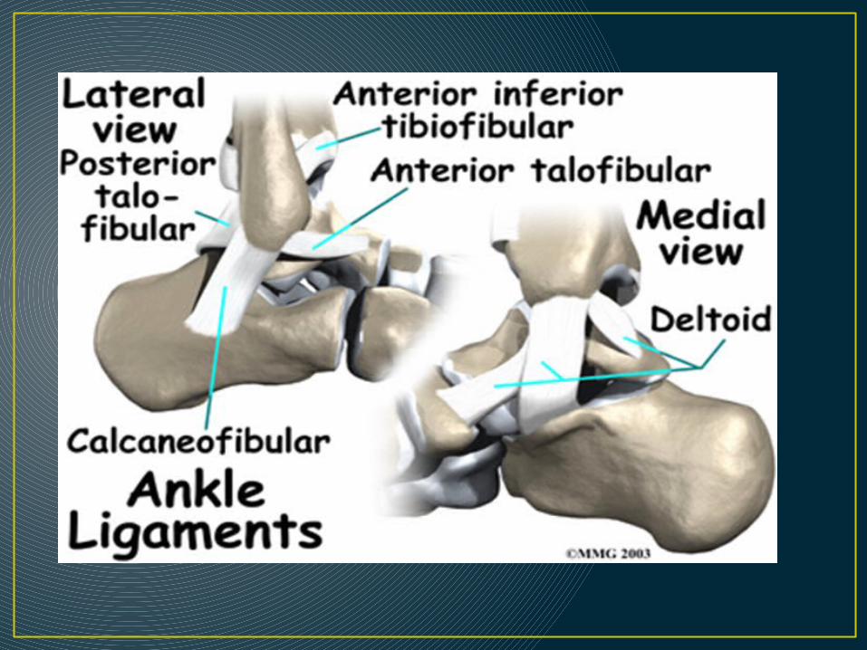
Medial Ligaments
• Deltoid ligament•4 parts•Very strong•Not injured as often
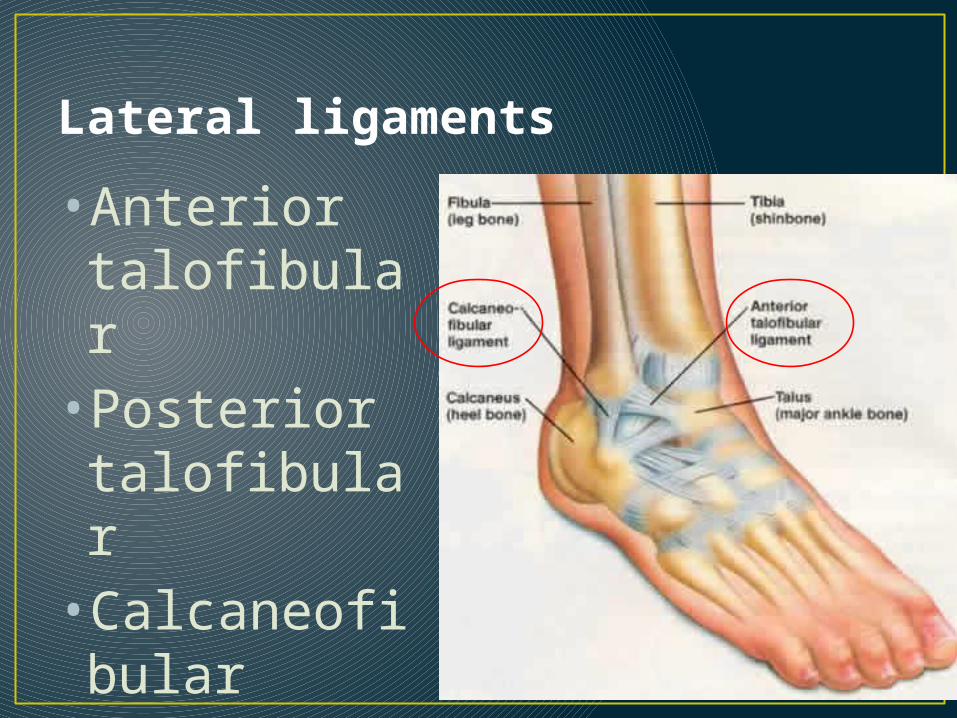
Lateral ligaments
•Anterior talofibular• Posterior talofibular•Calcaneofibular
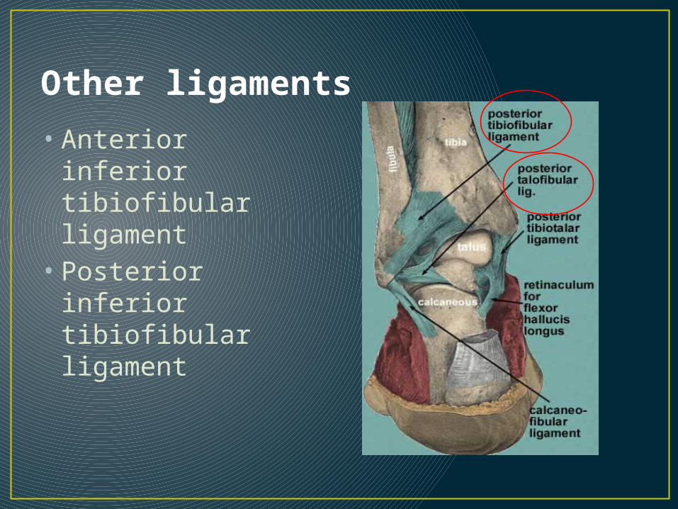
Other ligaments
• Anterior inferior tibiofibular ligament
• Posterior inferior tibiofibular ligament
Prevention of Injuries
• Wear properly fitting shoes• Ankle support• Protective equipment• Maintain adequate strength and
flexibility•Heel cord stretching•Strengthening in inversion, eversion, plantar and dorsiflexion•Proprioception (balance training)
Injuries to the Foot, Ankle and Lower Leg


Heel Bruise (Stone Bruise)
• MOI: Landing on heels, hitting heel on something hard—causing a contusion to the bottom of calcaneus• S/S: Severe pain in heel, difficulty
weight bearing, POT• TX: ice, rest/non weight bearing til
pain subsides, heel cup or doughnut when returning• Complication: inflammation of
periosteum
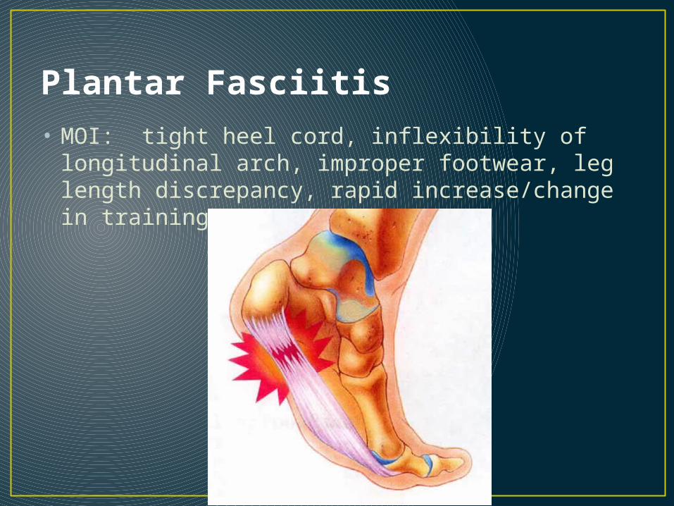
Plantar Fasciitis• MOI: tight heel cord, inflexibility of longitudinal
arch, improper footwear, leg length discrepancy, rapid increase/change in training

• S/S: Pt tender over the anteriomedial calcaneus and plantar fascia, stiffness and pain in AM or after prolonged sitting, pain with passive extension of toes combined with dorsiflexion

• TX: long term—8-12 weeksvigorous heel cord stretching, ice massage, heel cup, taping, ultrasound, NSAIDS,
Last resort: surgery to cut the fascia
Complications: can develop a bone spur if not cared for—surgery to remove it

Metatarsal Fracture
•MOI: direct force or twisting/torsion force or overuse•Most common is the Jone’s fracture—near base of 5th, avulsion (at the base), midshaft

• S/S: Pt. tender over metatarsal, swelling, pain, “pop” or “crack”, possible deformity

• Tx: Ice, Compression wrap, crutches, send to Dr. for x-ray.• Possibly on crutches for 6-8 weeks, non-weight bearing to allow for healing
• Complication: Non union fracture. May require surgery to fix

Longitudinal Arch Strain
•MOI: Unaccustomed stresses/forces placed on foot when in contact with a hard playing surface.•Flattening of the foot (arch) when in midsupport phase•May occur suddenly or over a longer period of time

• S/S: Pain felt just distal to the medial malleolus when running •Swelling and Pt. tender along the calcaneonavicular ligament (spring ligament) and the first cuneiform•Pt. tender over the FHL tendon as a result of compensation for stress on ligament
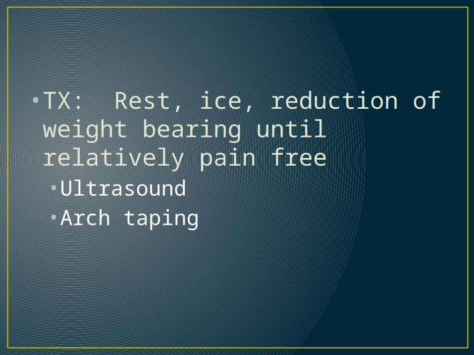
• TX: Rest, ice, reduction of weight bearing until relatively pain free•Ultrasound•Arch taping

Turf Toe• Sprain of the MP joint of the great toe• MOI: Hyperextension of great toe—trauma
or overuse• Usually occurs on an unyielding surface such as turf• Kicking an unyielding object
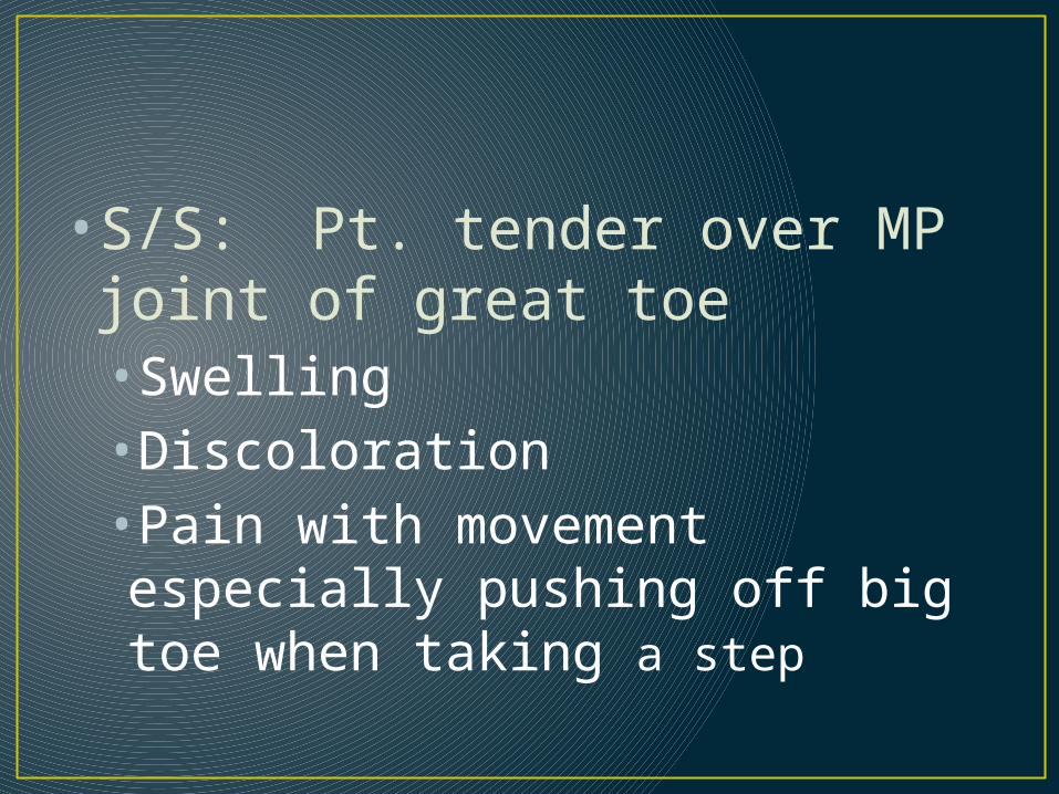
•S/S: Pt. tender over MP joint of great toe•Swelling•Discoloration•Pain with movement especially pushing off big toe when taking a step

• TX: Rest, ice, compression• Insert a hard insole into shoe to prevent hyperextension of MP joint•Tape for hyperextension
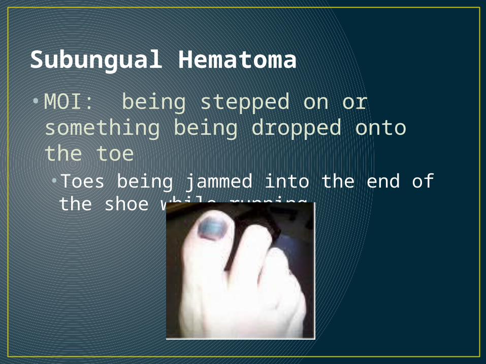
Subungual Hematoma
• MOI: being stepped on or something being dropped onto the toe•Toes being jammed into the end of the shoe while running
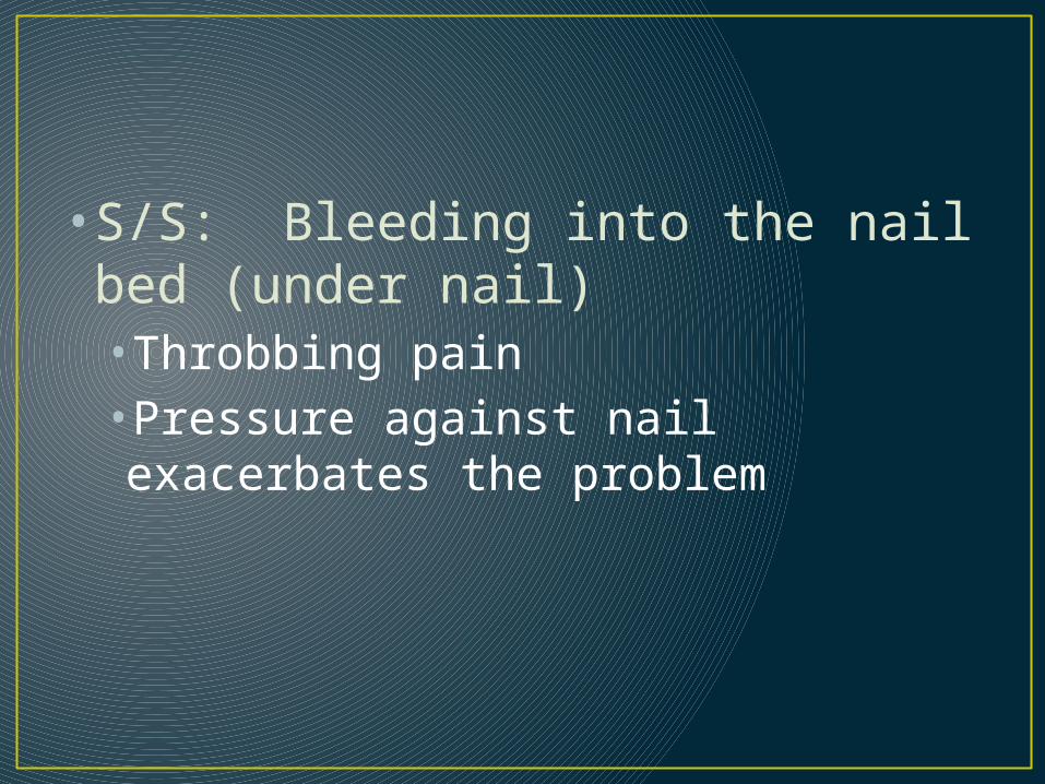
• S/S: Bleeding into the nail bed (under nail)•Throbbing pain•Pressure against nail exacerbates the problem
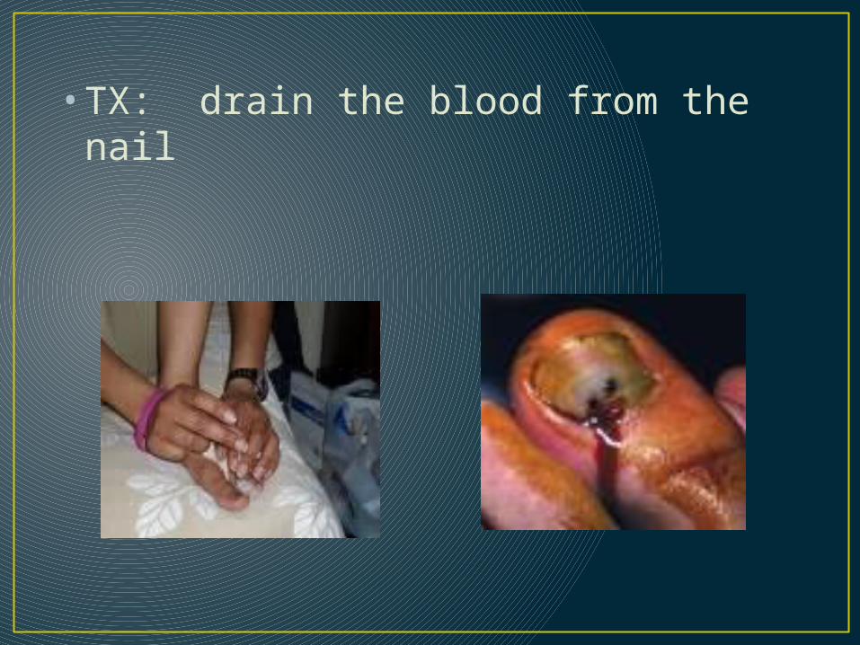
• TX: drain the blood from the nail

Blisters• MOI: shearing force on the skin that
causes fluid to accumulate below top layer of skin•May be clear, bloody or become infected
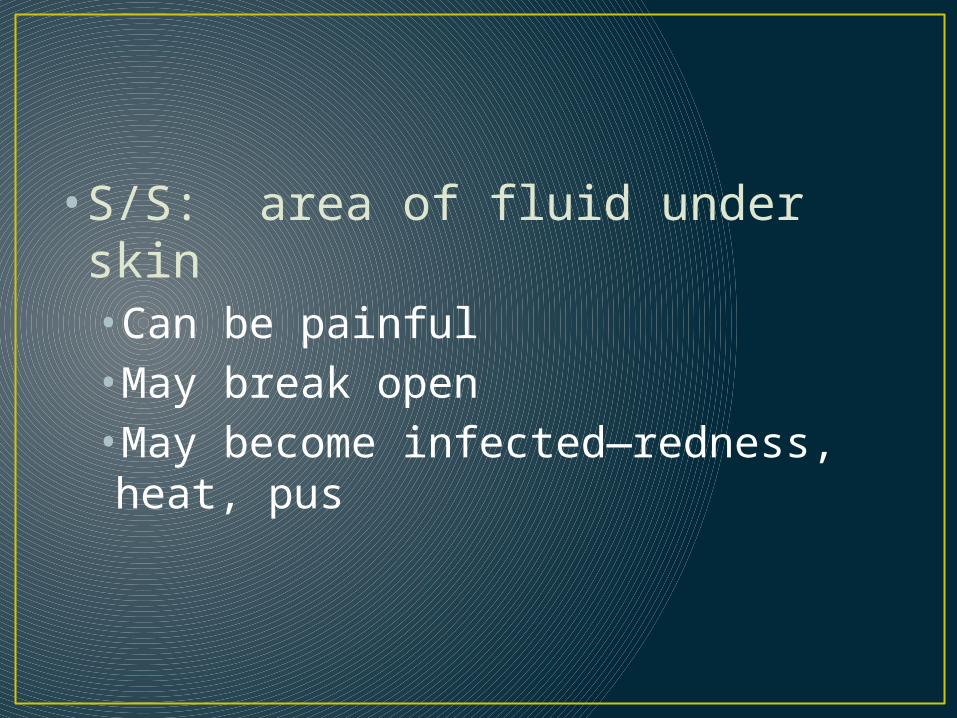
• S/S: area of fluid under skin•Can be painful•May break open•May become infected—redness, heat, pus

• TX: cover with skin lube, bandage, foam or felt doughnut around it.• If large, then drain, but clean it and treat as open wound•Cover prior to practices/competitions

Ankle Sprains
• Inversion• Eversion• (Syndesmoti
c) High Ankle Sprain
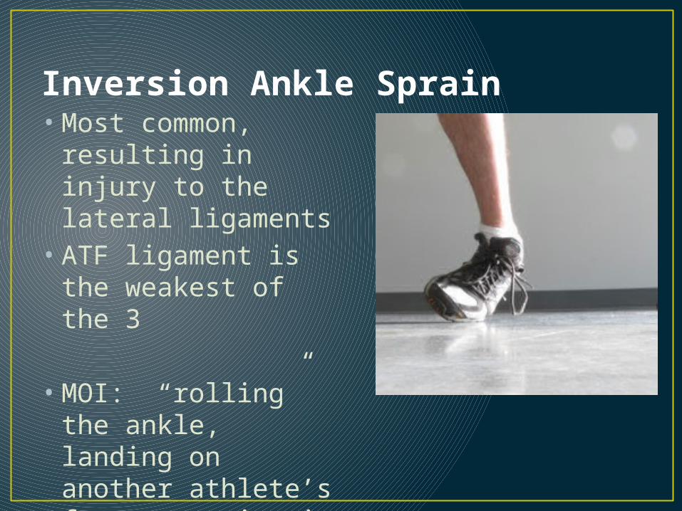
Inversion Ankle Sprain• Most common,
resulting in injury to the lateral ligaments
• ATF ligament is the weakest of the 3
• MOI: “rolling” the ankle, landing on another athlete’s foot, stepping in a hole, etc. • Inversion/plantar flexion

The inversion MOI

Structures injured• ATF lig. injured with the plantar
flexion/inversion MOI• Calcaneofibular lig. and posterior
talofibular lig. injured when then inversion force is increased

3rd degree Lateral Ankle sprain

• S/S: Pain, Swelling, discoloration, Pt. tender over the sinus tarsi, the distal end of the lateral malleolus and posterior of the lateral malleolus, joint instability, joint stiffness, decreased ROM, “+” anterior drawer test
• Will vary with the degree of the injury
• Anterior Drawer Test – Tests ATF • Talar Tilt – Calcaneofib and Deltoid
Ligaments• Kleiger Test – High Ankle • Calcaneus (Bump) Test – Calcaneus Fx

• Tx: RICE, “horseshoe” shaped felt/foam pad fit around the lateral malleolus•Treat for shock (only in severe cases)•crutches if necessary•Medical attention if severe or possibility of fracture

Complications• Avulsion fracture of lateral malleolus• Avulsion fracture of base of 5th metatarsal• Push-off fracture of medial malleolus

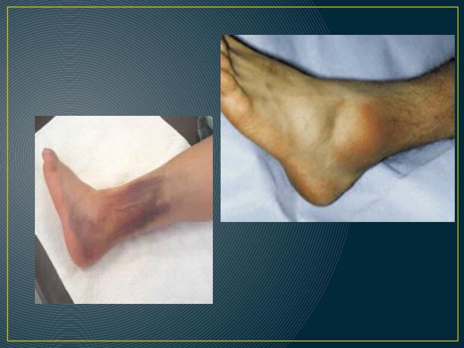
Eversion Ankle Sprain
• Less common due to bony structure of ankle• Deltoid ligament damage (any or all 4
portions)
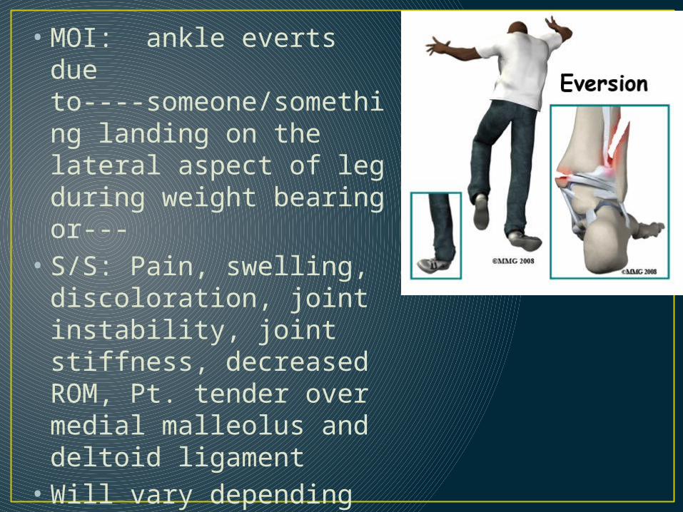
• MOI: ankle everts due to----someone/something landing on the lateral aspect of leg during weight bearing or---
• S/S: Pain, swelling, discoloration, joint instability, joint stiffness, decreased ROM, Pt. tender over medial malleolus and deltoid ligament
• Will vary depending on severity
• Tests:• Talar Tilt

• Tx: RICE, “horseshoe” shaped felt/foam pad, • crutches if necessary• Treat for shock• Medical attention with severe sprain or if fracture is
suspected

Complications
• Avulsion fracture of medial malleolus• Contused deltoid ligament due to
impingement between medial malleolus and calcaneus• Fracture of lateral malleolus
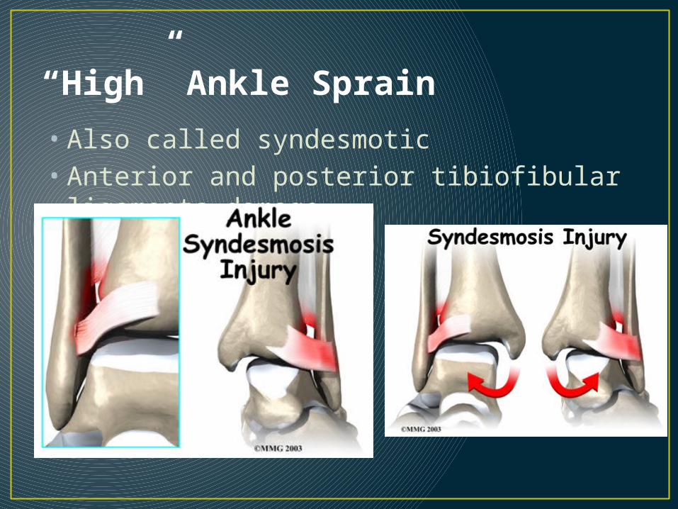
“High” Ankle Sprain
• Also called syndesmotic• Anterior and posterior tibiofibular
ligaments damage

• MOI: forced dorsiflexion or extreme plantar flexion/inversion
• Someone landing on the back of the leg with the foot in contact with the ground (dorsiflexion)
• S/S: may be swelling or not, may have discoloration or not • pain• Pt. tender over ATF and proximal to
that at the junction of the tibia and fibula• painful to bear weight, unable to go
up on toes

• Tx: RICE, Crutches, medical attention if unable to bear weight or if significant swelling occurs• Treat for shock• Hard to treat and can take weeks to heal

Complications
• Fracture to the dome of the talus• Tear of the interosseus membrane

Ankle Fractures and Dislocations
•MOI: similar to those of the ankle sprains but generally more force is applied• Can be open or closed

What do these injuries look like?
After the MOISee the placement of the
foot?

Sliding into base He’s there!
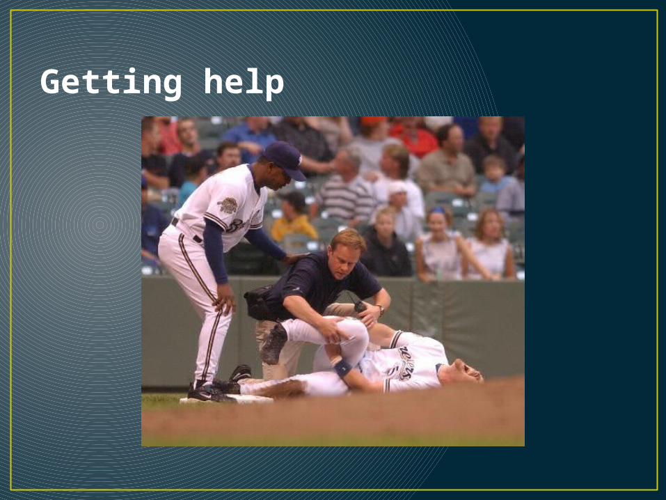
Getting help

And the open ones?
Open Fx/dislocation Open fracture

And some x-rays

• S/S: Immediate swelling • immense pain • possible deformity and/or open
wound• Pt. tender over the bone• + compression and percussion tests

• Tx: Splint in the position you find it• Care for open wound if necessary• Treat for shock• Call 911 if the injury is severe/open• ER visit
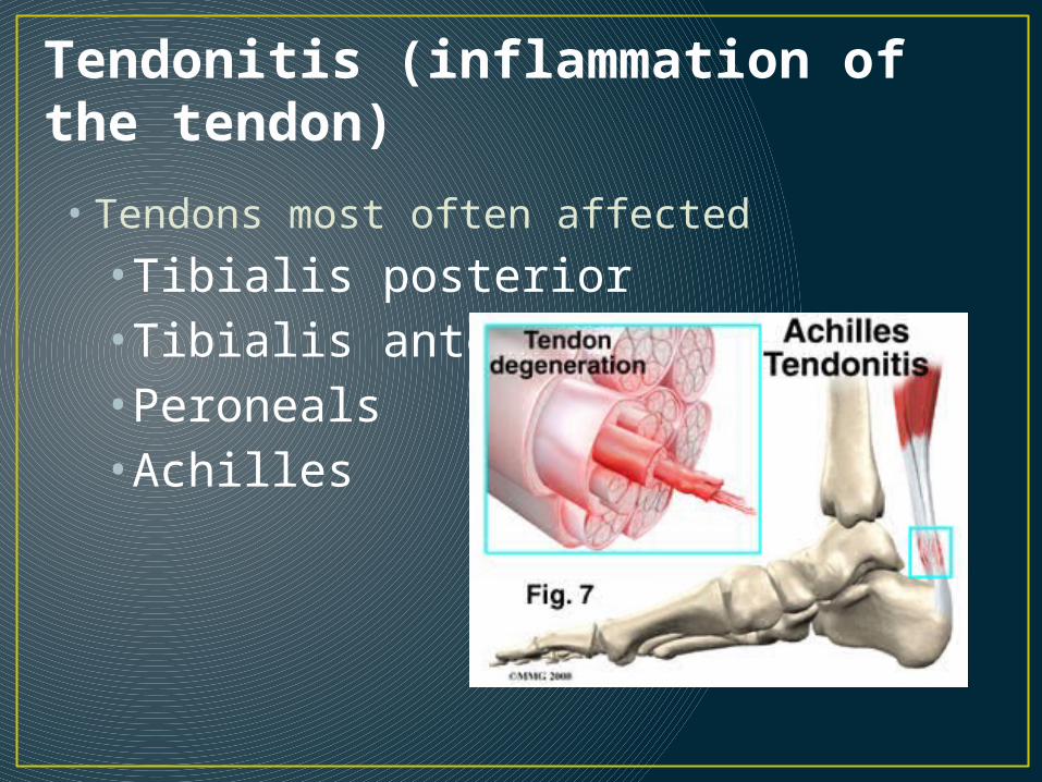
Tendonitis (inflammation of the tendon)
• Tendons most often affected
•Tibialis posterior•Tibialis anterior•Peroneals•Achilles
• MOI: faulty foot biomechanics• Inappropriate or poor/worn footwear•Acute trauma to tendon•Tightness of heel cord•Training errors•Excessive running, jumping, hills
• S/S: pain with active movements and passive stretching•Pt. tender over insertion of tendon•warmth•Crepitus•Thickening of tendon (achilles)•Stiffness and pain following periods of inactivity

• Tx: Rest•Modalities: ice, heat, ultrasound•NSAIDS•Exercise to strengthen muscle(s) involved•Stretching•Orthotics or taping to relieve stress on tendon
Medial Tibial Stress Syndrome
• Shin splints
• What is it?• Theories• Fascia pulling off of the bone (Soleus)• Bone Reaction (bone not being able to keep
up between osteoclasts and osteoblasts)• Posterior tibialis pulling off of the medial
surface of the bone

• MOI: strain of tibialis posterior tendon and its fascial sheath at attachment to periosteum of distal tibia due to running/etc.
• Faulty biomechanics• Improper footwear• Tight heel cord/Achilles tendon• Training errors
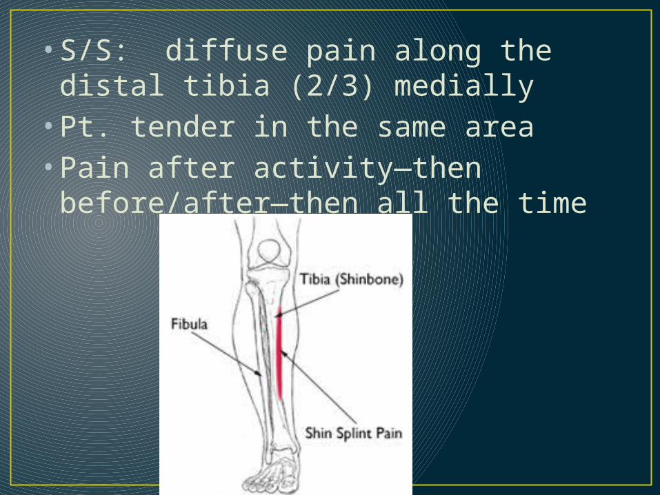
• S/S: diffuse pain along the distal tibia (2/3) medially• Pt. tender in the same area• Pain after activity—then before/after
—then all the time

• Tx: Modify activity• Correct foot biomechanics (orthotics)• Heel cord stretching (slant board)• Strengthening of muscles in Posterior
compartment• Ice massage• Friction massage• Taping—arch support/ankle
• Demonstrate Arch Taping

Tib/Fib fracture
• Tibia is most commonly fractured long bone in the body

•MOI: direct trauma to the tibia/fibula or both• Indirect trauma such as combination rotation/compressive force
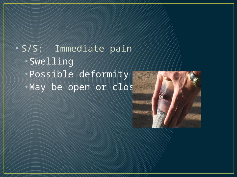
• S/S: Immediate pain•Swelling•Possible deformity•May be open or closed

• Tx: Splint in the position you find it•Treat for shock•Call 911 if necessary•ER visit
Stress Fractures
• Tibial (mid shaft)• Fibular (distal third)•Metatarsal (2nd is most common)

• MOI: repetitive loading during training and conditioning and jumping•Faulty biomechanics combined with excessive/change in training

• S/S: pain with activity• Increase in pain when activity is finished•Gradually gets worse•Pt. tender on one specific point on the bone•Can limit ability to participate
• Tx: stop activity (2-4 weeks)•Alternate conditioning—non weight bearing• Ice•Crutches/protective footwear•Medical referral• Xrays• Bone scan

Compartment Syndromes• Increased pressure in the
compartment(s) of the leg• Causes compression of the muscles &
neurovascular structures • Anterior, lateral, deep posterior
common• 3 types•Acute •Acute exertional•Chronic
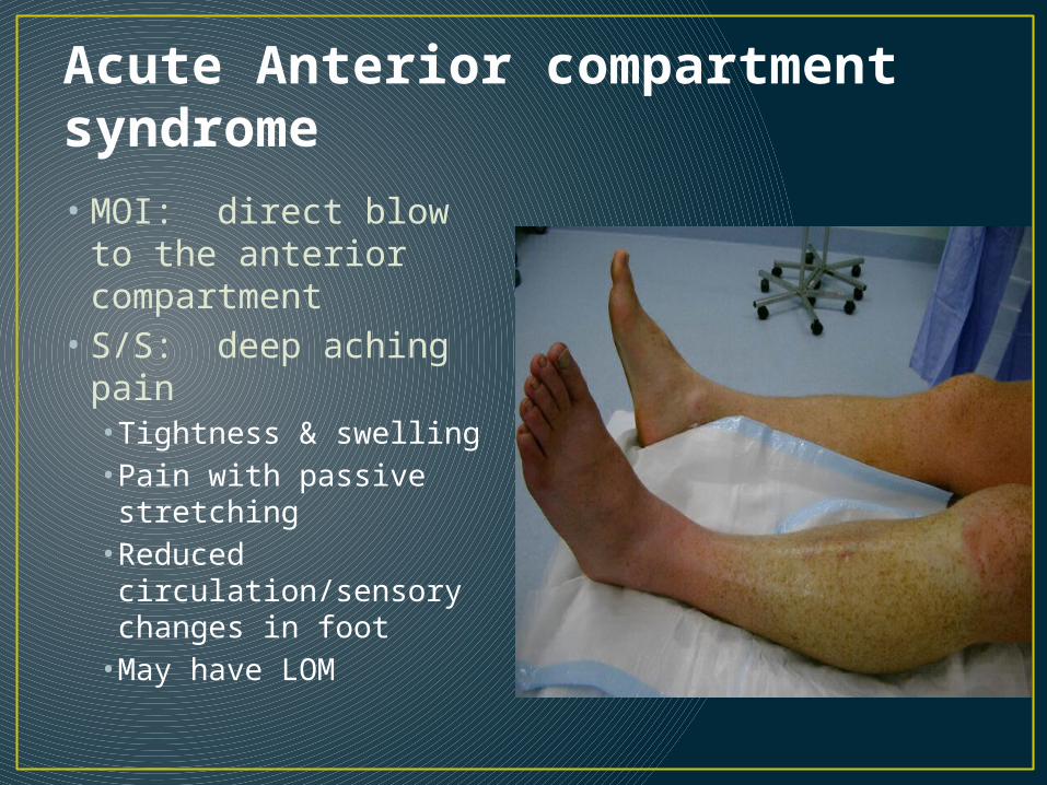
Acute Anterior compartment syndrome
• MOI: direct blow to the anterior compartment
• S/S: deep aching pain• Tightness & swelling• Pain with passive stretching• Reduced circulation/sensory changes in foot•May have LOM
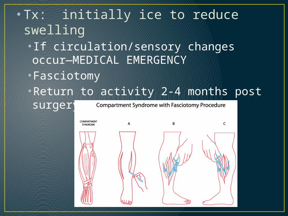
• Tx: initially ice to reduce swelling• If circulation/sensory changes occur—MEDICAL EMERGENCY•Fasciotomy•Return to activity 2-4 months post surgery

Achilles Tendon Rupture• Largest tendon in body• Most common in athletes over 30 yrs• Seen in sports with ballistic movements—
tennis, raquetball, basketball, etc.
• MOI: sudden forceful plantar flexion of ankle

• S/S: felt/heard a “pop” at back of leg (sounds like a twig snap or gun shot)•Felt as is someone hit them with a rock•Pain with plantar flexion/dorsiflexion• Inability to plantar flex•Palpable/visible defect at the achilles tendon•+ Thompson test

Achilles tendon defect
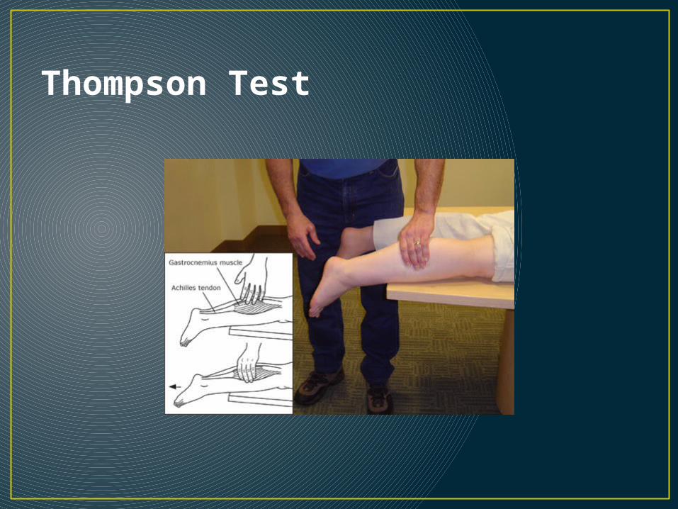
Thompson Test

• Tx: immobilize• ice•Send to ER•Requires surgery w/ 6-8 weeks immobilization•Rehab to regain full ROM/Strength

Open achilles tendon rupture
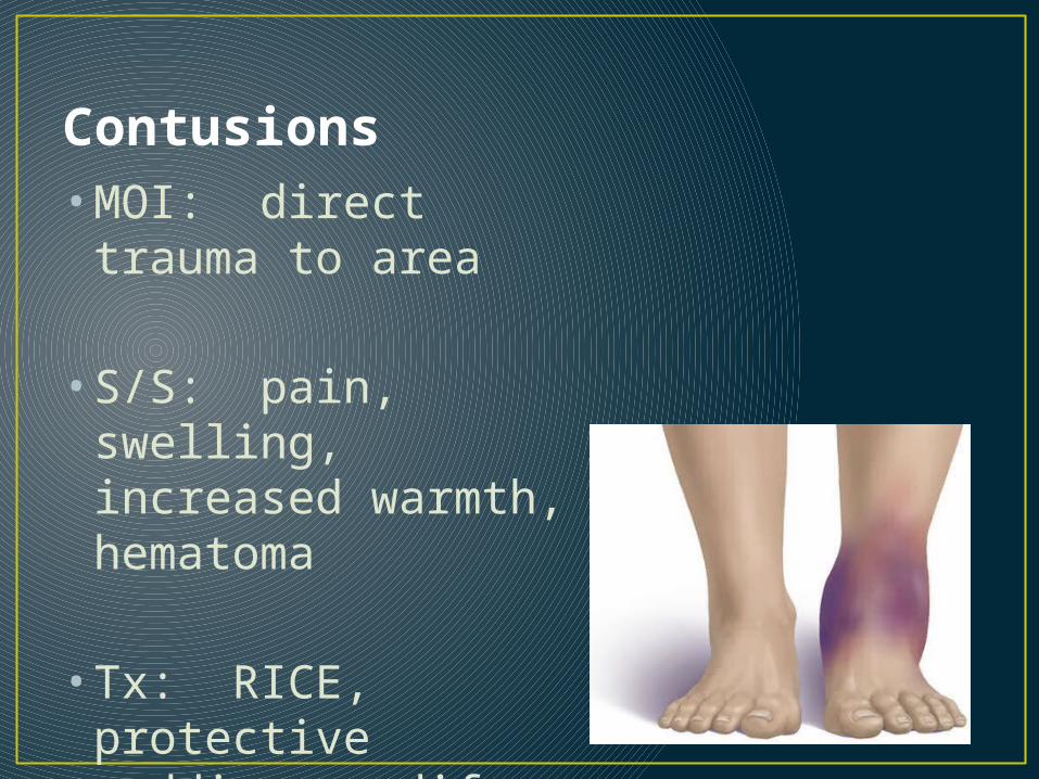
Contusions• MOI: direct trauma
to area
• S/S: pain, swelling, increased warmth, hematoma
• Tx: RICE, protective padding, modify activity if necessary

And other weird things
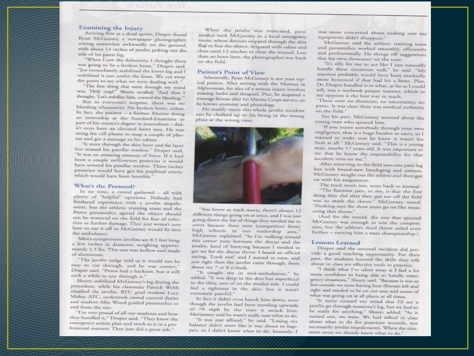

Treatment for this?
• Immoblize object• Cut object at each end to allow for
transport• Treat for shock• Surgery to remove impaled object

Ankle Taping Procedures
• Apply Tuf-Skin• Heel and Lace Pads• Pre-wrap from midfoot to 2 finger widths
below calf belly• 2 anchor strips

• Begin 3 Stirrups• In between each
stirrup is a horseshoe/C strip
• ALWAYS GO MEDIAL TO LATERAL….unless

• Once 3 stirrups and C strips are in place
• 4 heel locks • 2 medial• 2 lateral
• 2 figure 8s

• Once all parts are on the ankle
• Close out• Make it Pretty
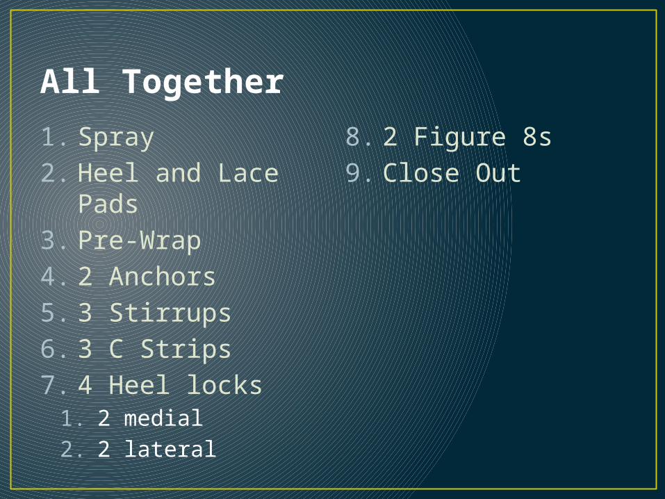
All Together
1. Spray2. Heel and Lace
Pads3. Pre-Wrap4. 2 Anchors5. 3 Stirrups6. 3 C Strips7. 4 Heel locks
1. 2 medial 2. 2 lateral
8. 2 Figure 8s 9. Close Out