Embed Size (px)
Citation preview
Introduction Shin defects of moderate dimension are fre-quently encountered following trauma and in-fection. Flaps of different constituents which have been described are bulky and thus fall short of the desired functional and aesthetic result. The concept of a split fascial flap emerged in search of a thinner flap. Three rele-vant parameters proved adequate vascularity in both the layers of the deep fascia. Thereafter, it was successfully used in 5 clinical cases. The technical intricacies have been mentioned in detail. Long term follow up proved that it is a simple, effective, durable, single stage proce-dure with appropriate aesthetic result.
Materials and method The study began in January and continued till December 2010. The formal approval was taken from the ethical committee of our insti-tute. Following parameters were taken into con-sideration prior to clinical application and its confirmation during surgery: (a) Fresh Cadaveric dissection of split fascial flap; (b) Demonstration of independent live microcirculation in both the layers of deep fascia; (c) Intraoperative fluo-rescein study of split fascial flap as described below: (a) Fresh Cadaveric dissection was carried out on 10 limbs of 5 bodies. The popliteal artery
Int J Burn Trauma 2012;2(2):86-92 www.IJBT.org /ISSN: 2160-2126/IJBT1205001
Original Article Turn over split fascial flap - a refinement for resurfacing shin defect Visweswar Bhattacharya1, Neeraj K Agrawal2, Gaurab R Chaudhuri3, Partha S Barooah4, Tripathi SK5, Rana Birendra6, Siddhartha Bhattacharya7, Dhruva J Deka8 1Department of Plastic Surgery, Institute of Medical Sciences, Banaras Hindu University, Varanasi, U.P., India; 2De-partment of Plastic Surgery, Institute of Medical Sciences, Banaras Hindu University, Varanasi, U.P., India; 3Depart-ment of Plastic Surgery, NRS Medical College and Hospital, Kolkata, West Bengal, India; 4Plastic Surgery Depart-ment, Assam Medical College& Hospital, Dibrugarh, Assam, India; 5Department of Forensic Medicine, Institute of Medical Sciences, Banaras Hindu University, Varanasi, U.P., India; 6Department of General Surgery, Institute of Medi-cal Sciences, Banaras Hindu University, Varanasi, U.P., India; 7Department of General Surgery, IPGME&R & SSKM Hospital, Kolkata. West Bengal, India; 8Department of Plastic Surgery, Institute of Medical Sciences, Banaras Hindu University, Varanasi, U.P., India Received May 5, 2012; accepted August 7, 2012; Epub September 15, 2012; Published September 30, 2012 Abstract: Moderate size defects of the shin of tibia are frequently encountered following trauma and infection. They may be associated with or without a fracture. Such defects require resurfacing by a flap. Many different types of flaps have been described but most of them proved to be more bulky than desired. Although these procedures cover the defects successfully the results they produce are not aesthetically appropriate. The flap looks bulkier because the native subcutaneous tissue is thin over the shin and distal leg. Hence a search for a vascularized tissue of minimal bulk for suitable resurfacing was initiated. A turnover fascial flap fulfilled the requirement. Such a flap can be made thinner by splitting its distal part into two layers while maintaining a common vascular fascial pedicle with both the layers of the fascia. This allowed a larger surface area to be covered. Such refinement is based on the following pa-rameters (a) fresh cadaveric dissection, (b) demonstration of live microcirculation individually in the superficial and deep layers of the deep fascia and (c) intraoperative flourescein study of the split fascial flap. The technique has been used in 5 cases over the upper and middle third of the shin of tibia. The split fascial flap was turned over and inset in the defect and covered with a split skin graft. The donor site was primarily closed. The functional and aes-thetic results were highly satisfactory. The follow up of 18 months proved the durability and usefulness of the flap. Keywords: Split fascial flap, shin defect, lower limb reconstruction
Turn over split fascial flap for resurfacing shin defect
87 Int J Burn Trauma 2012;2(2):86-92
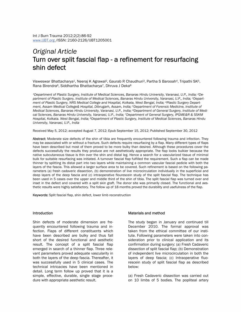
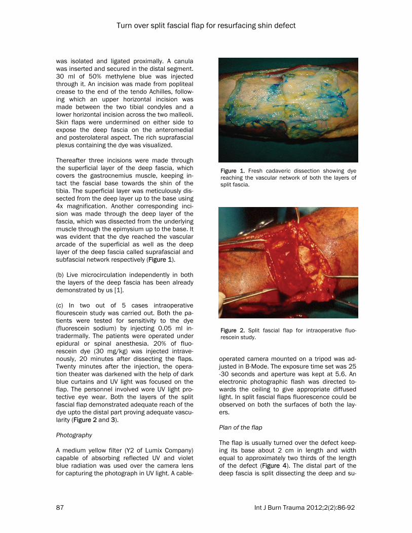
was isolated and ligated proximally. A canula was inserted and secured in the distal segment. 30 ml of 50% methylene blue was injected through it. An incision was made from popliteal crease to the end of the tendo Achilles, follow-ing which an upper horizontal incision was made between the two tibial condyles and a lower horizontal incision across the two malleoli. Skin flaps were undermined on either side to expose the deep fascia on the anteromedial and posterolateral aspect. The rich suprafascial plexus containing the dye was visualized. Thereafter three incisions were made through the superficial layer of the deep fascia, which covers the gastrocnemius muscle, keeping in-tact the fascial base towards the shin of the tibia. The superficial layer was meticulously dis-sected from the deep layer up to the base using 4x magnification. Another corresponding inci-sion was made through the deep layer of the fascia, which was dissected from the underlying muscle through the epimysium up to the base. It was evident that the dye reached the vascular arcade of the superficial as well as the deep layer of the deep fascia called suprafascial and subfascial network respectively (Figure 1). (b) Live microcirculation independently in both the layers of the deep fascia has been already demonstrated by us [1]. (c) In two out of 5 cases intraoperative flourescein study was carried out. Both the pa-tients were tested for sensitivity to the dye (fluorescein sodium) by injecting 0.05 ml in-tradermally. The patients were operated under epidural or spinal anesthesia. 20% of fluo-rescein dye (30 mg/kg) was injected intrave-nously, 20 minutes after dissecting the flaps. Twenty minutes after the injection, the opera-tion theater was darkened with the help of dark blue curtains and UV light was focused on the flap. The personnel involved wore UV light pro-tective eye wear. Both the layers of the split fascial flap demonstrated adequate reach of the dye upto the distal part proving adequate vascu-larity (Figure 2 and 3). Photography A medium yellow filter (Y2 of Lumix Company) capable of absorbing reflected UV and violet blue radiation was used over the camera lens for capturing the photograph in UV light. A cable-
operated camera mounted on a tripod was ad-justed in B-Mode. The exposure time set was 25-30 seconds and aperture was kept at 5.6. An electronic photographic flash was directed to-wards the ceiling to give appropriate diffused light. In split fascial flaps fluorescence could be observed on both the surfaces of both the lay-ers. Plan of the flap The flap is usually turned over the defect keep-ing its base about 2 cm in length and width equal to approximately two thirds of the length of the defect (Figure 4). The distal part of the deep fascia is split dissecting the deep and su-
Figure 1. Fresh cadaveric dissection showing dye reaching the vascular network of both the layers of split fascia.
Figure 2. Split fascial flap for intraoperative fluo-rescein study.
Turn over split fascial flap for resurfacing shin defect
88 Int J Burn Trauma 2012;2(2):86-92
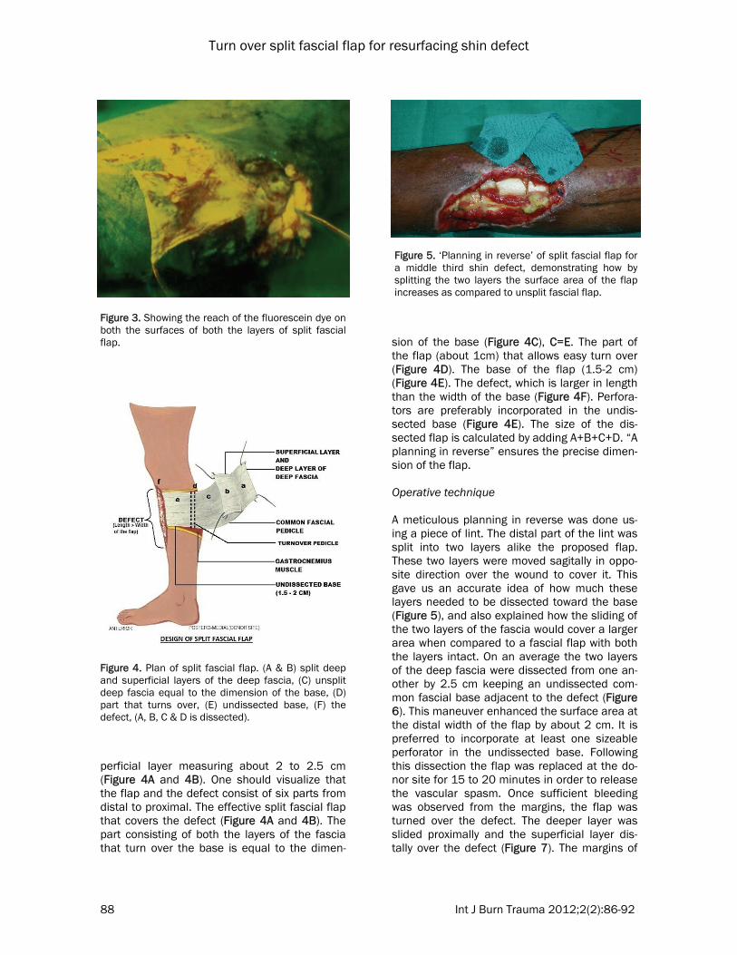
perficial layer measuring about 2 to 2.5 cm (Figure 4A and 4B). One should visualize that the flap and the defect consist of six parts from distal to proximal. The effective split fascial flap that covers the defect (Figure 4A and 4B). The part consisting of both the layers of the fascia that turn over the base is equal to the dimen-
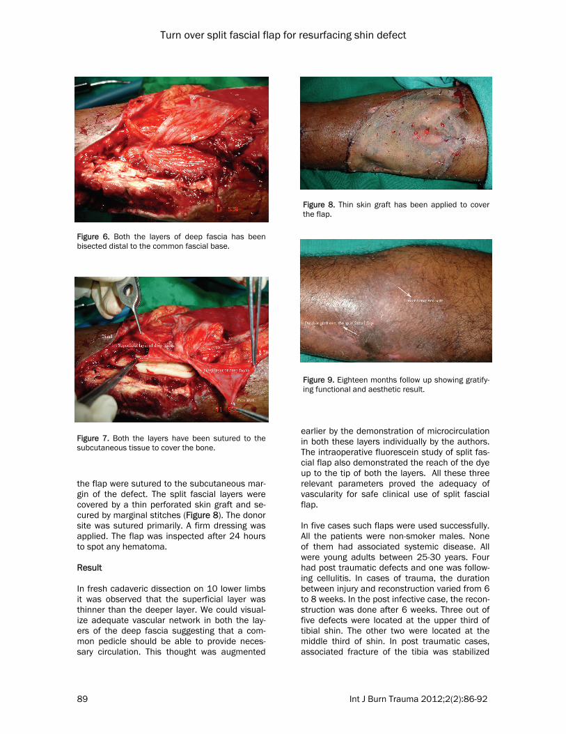
sion of the base (Figure 4C), C=E. The part of the flap (about 1cm) that allows easy turn over (Figure 4D). The base of the flap (1.5-2 cm) (Figure 4E). The defect, which is larger in length than the width of the base (Figure 4F). Perfora-tors are preferably incorporated in the undis-sected base (Figure 4E). The size of the dis-sected flap is calculated by adding A+B+C+D. “A planning in reverse” ensures the precise dimen-sion of the flap. Operative technique A meticulous planning in reverse was done us-ing a piece of lint. The distal part of the lint was split into two layers alike the proposed flap. These two layers were moved sagitally in oppo-site direction over the wound to cover it. This gave us an accurate idea of how much these layers needed to be dissected toward the base (Figure 5), and also explained how the sliding of the two layers of the fascia would cover a larger area when compared to a fascial flap with both the layers intact. On an average the two layers of the deep fascia were dissected from one an-other by 2.5 cm keeping an undissected com-mon fascial base adjacent to the defect (Figure 6). This maneuver enhanced the surface area at the distal width of the flap by about 2 cm. It is preferred to incorporate at least one sizeable perforator in the undissected base. Following this dissection the flap was replaced at the do-nor site for 15 to 20 minutes in order to release the vascular spasm. Once sufficient bleeding was observed from the margins, the flap was turned over the defect. The deeper layer was slided proximally and the superficial layer dis-tally over the defect (Figure 7). The margins of
Figure 3. Showing the reach of the fluorescein dye on both the surfaces of both the layers of split fascial flap.
Figure 4. Plan of split fascial flap. (A & B) split deep and superficial layers of the deep fascia, (C) unsplit deep fascia equal to the dimension of the base, (D) part that turns over, (E) undissected base, (F) the defect, (A, B, C & D is dissected).
Figure 5. ‘Planning in reverse’ of split fascial flap for a middle third shin defect, demonstrating how by splitting the two layers the surface area of the flap increases as compared to unsplit fascial flap.
Turn over split fascial flap for resurfacing shin defect
89 Int J Burn Trauma 2012;2(2):86-92
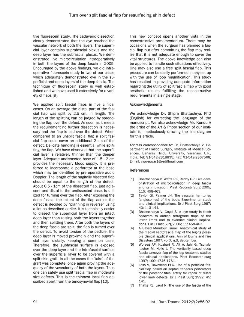
the flap were sutured to the subcutaneous mar-gin of the defect. The split fascial layers were covered by a thin perforated skin graft and se-cured by marginal stitches (Figure 8). The donor site was sutured primarily. A firm dressing was applied. The flap was inspected after 24 hours to spot any hematoma. Result In fresh cadaveric dissection on 10 lower limbs it was observed that the superficial layer was thinner than the deeper layer. We could visual-ize adequate vascular network in both the lay-ers of the deep fascia suggesting that a com-mon pedicle should be able to provide neces-sary circulation. This thought was augmented
earlier by the demonstration of microcirculation in both these layers individually by the authors. The intraoperative fluorescein study of split fas-cial flap also demonstrated the reach of the dye up to the tip of both the layers. All these three relevant parameters proved the adequacy of vascularity for safe clinical use of split fascial flap. In five cases such flaps were used successfully. All the patients were non-smoker males. None of them had associated systemic disease. All were young adults between 25-30 years. Four had post traumatic defects and one was follow-ing cellulitis. In cases of trauma, the duration between injury and reconstruction varied from 6 to 8 weeks. In the post infective case, the recon-struction was done after 6 weeks. Three out of five defects were located at the upper third of tibial shin. The other two were located at the middle third of shin. In post traumatic cases, associated fracture of the tibia was stabilized
Figure 6. Both the layers of deep fascia has been bisected distal to the common fascial base.
Figure 7. Both the layers have been sutured to the subcutaneous tissue to cover the bone.
Figure 8. Thin skin graft has been applied to cover the flap.
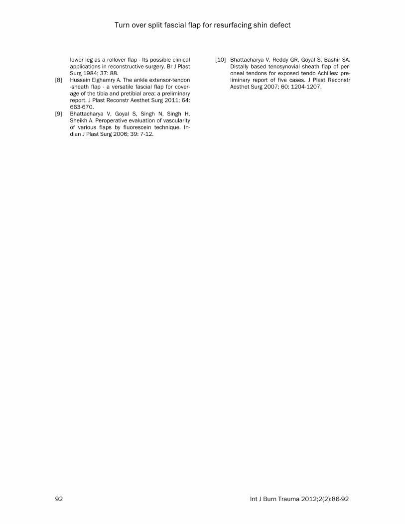
Figure 9. Eighteen months follow up showing gratify-ing functional and aesthetic result.

Turn over split fascial flap for resurfacing shin defect
90 Int J Burn Trauma 2012;2(2):86-92
earlier by internal fixation. The dimension of the defects ranged from 4 to 6 cm in length and 2 to 4 cm in width (Table 1). The dimension of the flap ranged from 6 to 8 cm in length and 2 to 4 cm in width. The addition of both the layers af-ter splitting formed the width of the flap. Four flaps were medially based on the posterior tibial axis and one was laterally based on the per-oneal axis. Prior to reconstruction the wounds were dressed with normal saline and antiseptic lotion. Prior to resurfacing the wound swab was sent regularly till the wound became healthy and free of infection. All the five cases healed uneventfully. The graft ‘take’ was complete in every case. The average hospital stay was of 14 days. The donor site healed with a linear scar. A follow up extending up to eighteen months proved the durability of the flap with gratifying aesthetic result (Figure 9). It did not produce any extra bulk over the shin and merged perfectly with the contour. Fol-lowing points were taken into account for as-sessing the result. (a) Contour, matching with the adjacent area. (b) Durability proved by following up. (c) Minimal linear scar at the donor site. (d) Satisfaction of the patient. Discussion Moderate size shin defects of the tibia are fre-quently encountered and the commonest cause is trauma. Several methods have been de-scribed depending upon the size and contour of the defect. Both these aspects are of significant consideration for functional and aesthetic re-sults. The loco regional flaps are preferred wher-ever non traumatic adjacent tissue of adequate dimension is available. Whenever the contour of the defect is deep, a thick flap of different con-
stituents is indicated in the form of musculocu-taneous, muscle, fasciocutaneous, adipofascial and fascial flaps. The flap selection forms an essential part of reconstruction, otherwise the flap may successfully cover the exposed vital structures but may not be acceptable aestheti-cally by the patient or even by the surgeon. Hence, there has been a constant search for refined flaps to meet these requirements. This problem is more prominent when the defect is over the shin or distal third of leg as these areas have less subcutaneous tissue. In such a situa-tion a split fascial flap with skin graft produces a good result with a linear scar at the donor site. To the best of our knowledge split fascial flaps have not been used clinically. Cadaveric dissec-tion forms an important part of proving the vas-cular basis of flaps of different constituents in-cluding fascial flaps [2-4]. Fascial flaps incorpo-rating both the layers of gastrocnemius fascia have been used as vertically based turnover flaps [5] or for distal leg defects [6, 7]. Elghamry AH has described fascial flap from the outer layer of tendon sheath of the ankle extensors. The flap is nourished by the sepal perforators. It was successfully used as a proximally based turn over flap with skin graft for tibial and pretibial area at the distal part of the leg. His flap is deep to deep fascia [8]. Our flap is har-vested as a split fascial flap of the deep fascia over the gastrocnemius muscle. Having under-stood that the deep fascia over the gastrocne-mius muscle consists of two distinct layers; we planned to use them clinically. There were two reasons for splitting the fascia (a) to cover a larger surface area of the defect as compared to a fascial flap, incorporating both the layers. (b) To have a more refined thin flap to maintain the contour of the shin. To prove the rationality of split fascial flaps we conducted fresh ca-daveric dissection with dye, demonstrated live circulation in individual layers and intraopera-
Table 1. The anatomical location, dimension of the defect and flap with result Patient Site of defect over
shin Dimension of defect L x W in cm
Dimension of flap L x W in cm
Location of base
Results
1 Upper third 4 × 6 6 × 6 PT Healed
2 Upper third 6 × 5 8× 6 PT Healed
3 Middle third 5 × 4 8 × 5 PT Healed
4 Middle third 4 × 4 6 × 5 PR Healed
5 Upper third 6 × 6 8 × 6 PT Healed PT- Posterior tibial axis, PR- Peroneal axis.
Turn over split fascial flap for resurfacing shin defect
91 Int J Burn Trauma 2012;2(2):86-92
tive fluorescein study. The cadaveric dissection clearly demonstrated that the dye reached the vascular network of both the layers. The superfi-cial layer contains suprafascial plexus and the deep layer has the subfascial plexus. We dem-onstrated live microcirculation intraoperatively in both the layers of the deep fascia in 2005. Encouraged by the above findings, we did intra-operative fluorescein study in two of our cases which adequately demonstrated dye in the su-perficial and deep layers of the deep fascia. The technique of fluorescein study is well estab-lished and we have used it extensively for a vari-ety of flaps [9]. We applied split fascial flaps in five clinical cases. On an average the distal part of the fas-cial flap was split by 2.5 cm, in length. The length of the splitting can be judged by spread-ing the flap over the defect. As soon as it meets the requirement no further dissection is neces-sary and the flap is laid over the defect. When compared to an unsplit fascial flap a split fas-cial flap could cover an additional 2 cm of the defect. Delicate handling is essential while split-ting the flap. We have observed that the superfi-cial layer is relatively thinner than the deeper layer. Adequate undissected base of 1.5 - 2 cm provides the necessary blood supply. It is pre-ferred to incorporate a perforator at the base which may be identified by pre operative audio Doppler. The length of the sagitally bisected flap should be equal to the length of the defect. About 0.5 - 1cm of the dissected flap, just adja-cent and distal to the undissected base, is util-ized for turning over the flap. After exposing the deep fascia, the extent of the flap across the defect is decided by “planning in reverse” using a lint as described earlier. It is technically easier to dissect the superficial layer from an intact deep layer than raising both the layers together and then splitting them. After both the layers of the deep fascia are split, the flap is turned over the defect. To avoid torsion of the pedicle, the deep layer is moved proximally and the superfi-cial layer distally, keeping a common base. Therefore, the subfascial surface is exposed over the deep layer and the intrafascial surface over the superficial layer to be covered with a split skin graft. In all the cases the ‘take’ of the graft was complete, once again proving the ade-quacy of the vascularity of both the layers. Thus one can safely use split fascial flap in moderate size defects. This is the thinnest local flap de-scribed apart from the tenosynovial flap [10].
This new concept opens another vista in the reconstructive armamentarium. There may be occasions when the surgeon has planned a fas-cial flap but after committing the flap may real-ize that it is not adequate enough to cover the vital structures. The above knowledge can also be applied to handle such situations effectively. One may also use a free split fascial flap. This procedure can be easily performed in any set up with the use of loop magnification. This study has resulted in providing adequate information regarding the utility of split fascial flap with good aesthetic results fulfilling the reconstructive requirements in a single stage. Acknowledgements We acknowledge Dr. Shipra Bhattachrya, PhD (English) for correcting the language of the manuscript. We also acknowledge Mr. Kundu A the artist of the Art & Photo section of our insti-tute for meticulously drawing the line diagram for this article. Address correspondence to: Dr. Bhattacharya V, De-partment of Plastic Surgery, Institute of Medical Sci-ences, Banaras Hindu University, Varanasi, U.P., India. Tel: 91-542-2318835; Fax: 91-542-2367568; E-mail: [email protected] References [1] Bhattacharya V, Watts RK, Reddy GR. Live dem-
onstration of microcirculation in deep fascia and its implication. Plast Reconstr Surg 2005; 115: 458-463.
[2] Taylor GI, Palmer JH. The vascular territories (angiosomes) of the body: Experimental study and clinical implications. Br J Plast Surg 1987; 40: 113-141.
[3] Bhattacharya V, Goyal S. A dye study in fresh cadavers to outline retrograde flaps of the lower limbs and to examine clinical implica-tions. Eur J Plast Surg 2006; 11: 458-463.
[4] AI-Sayed Mandour Ismail. Anatomical study of the medial septofascial flap of the leg-its possi-ble clinical applications. Ann of Burns and Fire Disasters 1997; vol X: n.3, September.
[5] Worseg AP, Kuzbari R, Alt A, Jahl G, Tschab-itscher M, Holle J. The vertically based deep fascia turnover flap of the leg: Anatomic studies and clinical applications. Plast Reconstr surg 1997; 100: 1746-1761.
[6] Less V, Townsend PLG. Use of a pedicled fas-cial flap based on septocutaneous perforators of the posterior tibial artery for repair of distal lower limb defects. Br J Plast Surg 1992; 45: 141.
[7] Thatte RL, Laud N. The use of the fascia of the
Turn over split fascial flap for resurfacing shin defect
92 Int J Burn Trauma 2012;2(2):86-92
lower leg as a rollover flap - Its possible clinical applications in reconstructive surgery. Br J Plast Surg 1984; 37: 88.
[8] Hussein Elghamry A. The ankle extensor-tendon-sheath flap - a versatile fascial flap for cover-age of the tibia and pretibial area: a preliminary report. J Plast Reconstr Aesthet Surg 2011; 64: 663-670.
[9] Bhattacharya V, Goyal S, Singh N, Singh H, Sheikh A. Peroperative evaluation of vascularity of various flaps by fluorescein technique. In-dian J Plast Surg 2006; 39: 7-12.
[10] Bhattacharya V, Reddy GR, Goyal S, Bashir SA. Distally based tenosynovial sheath flap of per-oneal tendons for exposed tendo Achilles: pre-liminary report of five cases. J Plast Reconstr Aesthet Surg 2007; 60: 1204-1207.