Embed Size (px)
Citation preview

Int J Clin Exp Pathol 2016;9(8):7836-7853www.ijcep.com /ISSN:1936-2625/IJCEP0027017
Original Article T-cell lymphoma complicated by myelofibrosis: report of three cases and review of literature
Yasunobu Sekiguchi1, Syuichi Shirane2, Haruko Takizawa1, Mutsumi Wakabayashi1, Keiji Sugimoto1, Shigeki Tomita3, Hiroshi Izumi3, Noriko Nakamura4, Tomohiro Sawada4, Yasunori Ohta5, Norio Komatsu2, Masaaki Noguchi1
Departments of 1Hematology, 3Pathology, 4Clinical Laboratory, Juntendo University Urayasu Hospital, Urayasu, Japan; 2Department of Hematology, Juntendo University Hospital, Tokyo, Japan; 5Department of Pathology, Institute of Medical Science, University of Tokyo, Tokyo, Japan
Received March 1, 2016; Accepted June 29, 2016; Epub August 1, 2016; Published August 15, 2016
Abstract: Primary myelofibrosis (MF) is a rare cancerous condition that disrupts normal blood cell production and causes scar tissue to accumulate within the bone marrow. MF complicating malignant lymphoma is rare and its cause often remains unknown, but, like primary MF, its pathogenesis is likely related to cytokines produced by proliferating megakaryocytes, monocytes, and histiocytes. We have experienced three cases of peripheral T-cell lymphoma (PTCL) complicated by MF. In each case, no evidence of central nervous system infiltration was found on admission; however, bone marrow infiltration was present. We carried out a chronological analysis of the cytokine production by malignant cells in these three patients by means of lymph node and bone marrow immunostaining. Malignant cells from lymph nodes and bone marrow sampled before treatment stained positive for cytokines: plate-let-derived growth factor (PDGF) and tumor necrosis factor-α (TNF-α) in case 1; PDGF in case 2; and PDGF, TNF-α, and transforming growth factor-β in case 3. These cytokines were considered to have been the cause of MF in each case. A literature search indicated that 16 cases of T-cell lymphoma complicated by MF (including our three cases) have been reported to date. Data on cytokine production by malignant cells given in these reports are summarized. Much remains to be clarified concerning the role of cytokines in the development of MF. Consequently, we plan to carry out chronological examinations of cytokines in the serum and immunostaining for cytokines in cases of PTCL and malignant lymphoma, both those complicated and not complicated by MF.
Keywords: T-cell lymphoma, bone marrow fibrosis, cytokine
Introduction
Primary myelofibrosis (MF) is a rare cancerous condition that disrupts normal blood cell pro-duction and causes the slow build up of scar tissue within the bone marrow. The pathogen-esis of primary MF is likely related the produc-tion of cytokines such as transforming growth factor-β (TGF-β), basic fibroblast growth factor (b-FGF), vascular endothelial growth factor (VEGF), and tumor necrosis factor-α (TNF-α) by proliferating megakaryocytes, monocytes, and histiocytes [1-3]. MF complicating malignant lymphoma is rare and its cause remains unknown in many cases. However, several pre-viously published articles [4-12] and some of our previous studies suggest that MF in patients
with malignant lymphoma is also attributable to cytokines such as TGF-β [13-16], b-FGF [2, 17], platelet-derived growth factor (PDGF) [2, 14-16], and/or TNF-α [16], and that its patho-genesis is similar to that of primary MF.
At Juntendo University Urayasu Hospital, Japan, we have encountered three cases of peripheral T-cell lymphoma (PTCL) complicated by MF (including two cases reported by us in 2013 and 2015 [2, 16]). To help clarify the cause of MF for each case, we carried out a chronologi-cal analysis of the cytokine production by malig-nant cells by means of lymph node and bone marrow immunostaining. Immunohistochemi- stry yielded positive staining for PDGF and TNF-α in case 1; for PDGF alone in case 2; and
T-cell lymphoma complicated by MF
7837 Int J Clin Exp Pathol 2016;9(8):7836-7853
for PDGF, TNF-α, and TGF-β in case 3. Marrow infiltration was noted in all three cases. Therefore, in all three of our-cases, MF seems to have been induced by PDGF, TNF-α, and/or TGF-β produced by lymphoma cells infiltrating the bone marrow. The investiga-tions and treatments carried out in our three cases will now be described.
Case presentation
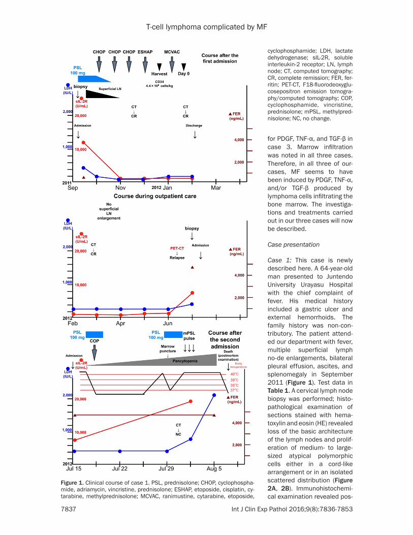
Case 1: This case is newly described here. A 64-year-old man presented to Juntendo University Urayasu Hospital with the chief complaint of fever. His medical history included a gastric ulcer and external hemorrhoids. The family history was non-con-tributory. The patient attend-ed our department with fever, multiple superficial lymph no-de enlargements, bilateral pleural effusion, ascites, and splenomegaly in September 2011 (Figure 1). Test data in Table 1. A cervical lymph node biopsy was performed; histo-pathological examination of sections stained with hema-toxylin and eosin (HE) revealed loss of the basic architecture of the lymph nodes and prolif-eration of medium- to large-sized atypical polymorphic cells either in a cord-like arrangement or in an isolated scattered distribution (Figure 2A, 2B). Immunohistochemi- cal examination revealed pos-
Figure 1. Clinical course of case 1. PSL, prednisolone; CHOP, cyclophospha-mide, adriamycin, vincristine, prednisolone; ESHAP, etoposide, cisplatin, cy-tarabine, methylprednisolone; MCVAC, ranimustine, cytarabine, etoposide,
cyclophosphamide; LDH, lactate dehydrogenase; sIL-2R, soluble interleukin-2 receptor; LN, lymph node; CT, computed tomography; CR, complete remission; FER, fer-ritin; PET-CT, F18-fluorodeoxyglu-cosepositron emission tomogra-phy/computed tomography; COP, cyclophosphamide, vincristine, prednisolone; mPSL, methylpred-nisolone; NC, no change.

T-cell lymphoma complicated by MF
7838 Int J Clin Exp Pathol 2016;9(8):7836-7853
Table 1. Test data for Case 1Blood count WBC 9700/μL↑ Aty Ly 2.5%↑ Neut 76.5%↑ Hb 12.7 g/dL↓ Lym 13.5%↓ MCV 87.3 fl Mono 5.0%↑ MCH 30.6 pg Eos 2.0% Plt 301,000/μL Bas 0.5%Aggregation PT activity 84% FDP 11.3 μg/mL↑ PT-INR 1.10 DD 4.03 μg/mL↑ APTT 31.4 s AT 3.65%↓ Fbg 222 mg/dLBiochemistry TP 7.9 g/dL AST 25 IU/L ALB 2.6 g/dL↓ ALT 14 IU/L BUN 24 mg/dL↑ LDH 422 IU/L↑ Cr 1.23 mg/dL↑ ALP 327 IU/L Na 135 mM/L↓ γ-GTP 19 IU/L Cl 100 mM/L Ch-E 121 IU/L↓ K 4.3 mM/L AMY 33 IU/L↓ Ca (corrected) 10.4 g/dL↑ CRP 1.2 mg/dL↑ Glu 96 mg/dL FER 361.8 ng/mL T-Bil 0.5 mg/dLImmunoserology IgG 3671 mg/dL↑ HTLV-1Ab (-) IgA 319 mg/dL HIV1/2Ab (-) IgM 934 mg/dL↑ Soluble IL-2 receptor 11,600 IU/L↑↑, upper limit; ↓, lower limit. WBC, white blood cells; Neut, neutrophil; Lym, lymphocyte; Mono, monocyte; Eos, eosinocyte; Bas, basophil; Aty Ly, Atypical lymphocyte; Hb, hemoglobin; MCV, mean corpuscular cell volume; MCH, mean corpuscular cell hemoglobin; Plt, platelets; PT, prothrombin time; PT-INR, pro-thrombin International Normalized Ratio; APTT, activated partial thromboplastin time; Fbg, fibrinogen; FDP, fibrin/fibrinogen degradation products; DD, D-dimer; AT, antithrombin; TP, total protein; ALB, albumin; BUN, blood urea nitrogen; Cr, creatinine; Glu, glucose; T-Bil, total bilirubin; AST, aspartate aminotransferase; ALT, alanine aminotransferase; LDH, lactate dehydrogenase; ALP, alkaline phos-phatase; γ GTP, P-guanosine triphosphate; Ch-E, cholinesterase; AMY, amylase; CRP, C-reactive protein; FER, ferritin; IgG, immunoglobulin G; IgA, immunoglobu-lin A; IgM, immunoglobulin M; HTLV, human T-cell leukemia virus; HIV, human immunodeficiency virus; sIL-2R, soluble interleukin-2 receptor.
itive staining of the tumor cells for cluster of dif-ferentiation (CD) 3, CD4, programmed cell death (PD) 1, and chemokine (C-X-C motif) ligand 13 (CXCL13) (Figure 2C-F), and the chro-matic response of CD21 suggested marked hyperplasia of the follicular dendritic cells around the capillaries (Figure 2G). Staining for CD10 was negative (Figure 2H). Positive stain-ing for Ki-67, a cellular marker for proliferation, was noted in a large percentage of cells (Figure 2I). On the basis of these findings, the patient
abdomen, and pelvis; and splenomegaly. In July 2012, a right inguinal lymph node biopsy was performed, and the results confirmed relapse of the disease. After CHOP therapy, the patient’s condition was complicated by hemophagocytic syndrome. Steroid pulse therapy was institut-ed; however, the patient showed no improve-ment and died of his disease in August 2012. The bone marrow was free of AITL infiltration (Figure 3C27, 3C28) and MF (Figure 3C29, 3C30) at the time of relapse.
was diagnosed as having angio-immunoblastic lymphoma (AITL).
Cerebrospinal fluid examination revealed no signs of central ner-vous system infiltration. Exami- nation of a bone marrow sample obtained by aspiration revealed infiltration of the marrow by AITL (Figure 3B13, 3B14), and silver impregnation staining revealed MF (Figure 3B15, 3B16). The marrow blood tested negative for the Janus kinase 2 (JAK-2) V617F mutation (data not shown). The clinical stage of the disease was judged as IVB, and the Prognos- tic Index for PTCL-unspecified (PIT) [18] was group 4. The pa- tient was started on cyclophos-phamide, adriamycin, vincristine, and prednisolone (CHOP) therapy in early October 2011. Complete remission was confirmed by a computed tomography (CT) scan carried out at the end of the third course of CHOP therapy in mid-November 2011. Marrow testing was not performed at this time because the patient refused con-sent for the procedure. Up front autologous peripheral blood ste- m cell transplantation (auto-PB- SCT) was performed. Complete remission was confirmed again by CT in February 2012. Positron emission tomography/computed tomography was performed in June 2012. This investigation revealed bilateral lymph node enlargement in the neck, axillae, and inguinal regions; enlarged lymph nodes in the mediastinum,

T-cell lymphoma complicated by MF
7839 Int J Clin Exp Pathol 2016;9(8):7836-7853
Case 2: This case was reported by us in 2013 [2]. A 65-year-old man presented to Juntendo University Urayasu Hospital with the chief com-plaints of fever and rash. His medical history and family history were non-contributory. The fever and rash developed in March 2012 (Figure 4). In April 2012, multiple superficial lymph node enlargements became evident. The patient was admitted to our department in May 2012. Test data in Table 2.
A left axillary lymph node biopsy was per-formed. Histopathological examination of se-ctions stained with HE revealed loss of the basic architecture of the lymph node and the proliferation of medium- to large-sized aty- pical polymorphiccells either in a cord-like arrangement (Figure 5A, 5B) or in an isolated scattered distribution. Immunohistochemical
staining of the tumor cells were positive for CD3, CD4, PD1, and CXCL13 (Figure 5C-F), and the chromatic response of CD21 suggested marked hyperplasia of follicular dendritic cells around the capillaries (Figure 5G). Staining for CD10 was negative (Figure 5H). Positive stain-ing for Ki-67 was observed in a large percent-age of cells (Figure 5I). On the basis of these findings, the patient was diagnosed as having AITL. Cerebrospinal fluid examination revealed no signs of central nervous system infiltration. Because no sample could be obtained by aspi-ration, a bone marrow biopsy was performed. Examination of HE-stained specimens re- vealed AITL infiltration of the marrow (Figure 6B13, 6B14). Silver impregnation staining revealed intense fibrosis (Figure 6B15, 6B16). The marrow blood tested negative for the JAK-2 V617F mutation (data not shown). The clinical
Figure 2. Histopathological findings of cervical lymph node biopsy Tin case 1. A. HE stain (×40), B. HE stain (×600): the basic architecture of the lymph node is lost and proliferation of medium- to large-sized polymorphic atypical cells is seen in a cord-like arrangement or in an isolated scattered distribution; C. CD3 positive (×600); D. CD4 positive (×600); E. PD1 positive (×600); F. CXCL13 positive (×600); G. CD21: marked hyperplasia of follicular dendritic cells is visible around the capillaries (×600); H. CD10 negative (×600); I. Ki-67 positive in a high percentage of cells (×600).
T-cell lymphoma complicated by MF
7840 Int J Clin Exp Pathol 2016;9(8):7836-7853
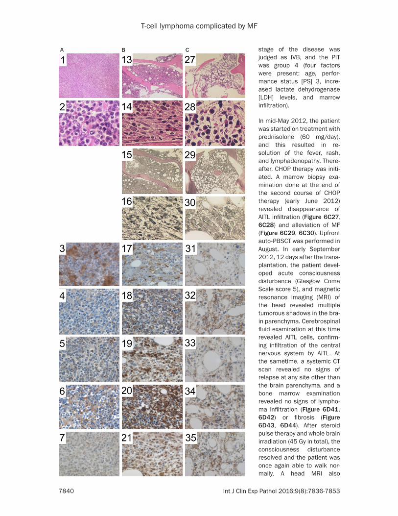
stage of the disease was judged as IVB, and the PIT was group 4 (four factors were present: age, perfor-mance status [PS] 3, incre- ased lactate dehydrogenase [LDH] levels, and marrow infiltration).
In mid-May 2012, the patient was started on treatment with prednisolone (60 mg/day), and this resulted in re- solution of the fever, rash, and lymphadenopathy. There- after, CHOP therapy was initi-ated. A marrow biopsy exa- mination done at the end of the second course of CHOP therapy (early June 2012) revealed disappearance of AITL infiltration (Figure 6C27, 6C28) and alleviation of MF (Figure 6C29, 6C30). Upfront auto-PBSCT was performed in August. In early September 2012, 12 days after the trans-plantation, the patient devel-oped acute consciousness disturbance (Glasgow Coma Scale score 5), and magnetic resonance imaging (MRI) of the head revealed multiple tumorous shadows in the bra- in parenchyma. Cerebrospinal fluid examination at this time revealed AITL cells, confirm-ing infiltration of the central nervous system by AITL. At the sametime, a systemic CT scan revealed no signs of relapse at any site other than the brain parenchyma, and a bone marrow examination revealed no signs of lympho-ma infiltration (Figure 6D41, 6D42) or fibrosis (Figure 6D43, 6D44). After steroid pulse therapy and whole brain irradiation (45 Gy in total), the consciousness disturbance resolved and the patient was once again able to walk nor-mally. A head MRI also

T-cell lymphoma complicated by MF
7841 Int J Clin Exp Pathol 2016;9(8):7836-7853
2013, relapse of the central nervous system infiltration was detected, and the patient died.
Case 3: This case was report-ed by us in 2015 [16]. A 68-year-old man presented at Juntendo University Urayasu Hospital with the chief com-plaints of pancytopenia, ery- throderma, and multiple su- perficial/deep lymph node en- largements. His medical and family histories contained no- thing noteworthy. The patient developed generalized ede- ma, erythroderma, and pan-cytopenia in June 2014 (Figure 7). He visited the Department of Dermatology of our hospital in July 2014. At that time, a skin biopsy led to the diagnosis of psoriatic erythroderma. Treatment wi- th prednisolone (15 mg/day) was started, and this resulted in alleviation of the systemic edema and erythroderma. However, the pancytopenia worsened, and multiple sup- erficial/deep lymph node en- largements also became evi-dent. The patient was there-fore admitted to our depart-ment in August 2014. Test data in Table 3. A left axillary lymph node biopsy was car-ried out, and histopathologi-cal examination of sections stained with HE revealed dis-appearance of the basic ar- chitecture of the lymph node (Figure 8A) and proliferation of medium-sized atypical cells with irregular nuclei (Figure 8B). Immunohistoche- mical staining of the tumor cells were positive for CD2, CD3, and C-C chemokine receptor type 4 (CCR4) (Figure 8C, 8D, 8P). The staining
Figure 3. Histopathological findings and immunohistochemical staining for cytokines in case 1. A1-12: Lymph node biopsy before treatment, B13-26: Bone marrow biopsy before treatment, C27-40: Bone marrow biopsy on re-lapse. A1. HE stain (×40), A2. HE stain (×600): the basic architecture of the lymph node is lost and proliferation of medium- to large-sized atypical poly-morphic cells are seen in a cord-like arrangement or in an isolated scattered distribution. A3. PDGF positive (×400); A4. b-FGF weakly positive (×400); A5. VEGF weakly positive (×400); A6. TNF-α positive (×400); A7. IFN-γ weakly positive (×400); A8. IL-1β weakly positive (×400); A9. IL-2 weakly positive (×400); A10. TGF-β weakly positive (×400); A11. IL-6 weakly positive (×400); A12. FN negative (×400); B13. HE stain (×40), B14. HE stain (×600): AITL infiltration visible; B15. Silver impregnation stain (×40), B16. Silver impreg-nation stain (×600): MF visible; B17. PDGF positive (×400); B18. b-FGF posi-tive (×400); B19. VEGF positive (×400); B20. TNF-α positive (×400); B21. IFN-γ positive (×400); B22. IL-1β positive (×400); B23. IL-2 positive (×400); B24. TGF-β positive (×400); B25. IL-6 positive (×400); B26. FN weakly posi-tive (×400); C27. HE stain (×40), C28. HE stain (×600): no evidence of AITL infiltration; C29. Silver impregnation stain (×40), C30. Silver impregnation stain (×600): no evidence of MF; C31. PDGF weakly positive (×400); C32. b-FGF weakly positive (×400); C33. VEGF weakly positive (×400); C34. TNF-α weakly positive (×400); C35. IFN-γ weakly positive (×400); C36. IL-1β weakly positive (×400); C37. IL-2 weakly positive (×400); C38. TGF-β weakly positive (×400); C39. IL-6 weakly positive (×400); C40. FN negative (×400).
revealed improvement, and the patient was discharged in October 2012. However, in May
results were negative for CD4, CD5, CD7, CD8, CD10, CD20, CD56, Epstein-Barr virus-encod-

T-cell lymphoma complicated by MF
7842 Int J Clin Exp Pathol 2016;9(8):7836-7853
ed small RNA (EBER), Granzyme B, and PD1 (Figure 8E-J, 8L-O). The chromatic response of CD21 did not suggest hyperplasia of the follicu-lar dendritic cells (Figure 8K). On the basis of these findings, the patient was diagnosed as having peripheral T-cell lymphoma, not other-wise specified (PTCL-NOS). Cerebrospinal fluid examination did not reveal evidence of central nervous system infiltration. Only a very small amount of bone marrow could be collected by puncture and aspiration. Therefore, a bone marrow biopsy was performed. Analysis of the sample revealed infiltration by PTCL-NOS (Fi- gure 9B13, 9B14). Silver impregnation staining revealed MF (Figure 9B15, 9B16). The marrow blood tested negative for the JAK-2 V617F mutation (data not shown). The clinical disease stage was IVB, and the PIT was group 4 (four factors were present: age, PS3, increased LDH levels, and marrow infiltration).
In early September 2014, the patient was sta- rted on the first course of CHOP therapy. Bone marrow examination was conducted and reve- aled a marked decrease of the PTCL-NOS infil-tration (Figure 9C27, 9C28) and alleviation of MF (Figure 9C29, 9C30). In early October 2014, the patient was started on the second course of CHOP therapy. At this time, a bone marrow
days 1-10 (CEPP therapy). This resulted in tem-porary improvement of the patient’s condition. However, the lymphoma relapsed, and the patient died in January 2015.
Results
The data acquired in the treatment of these three patients allowed the time profiles of cyto-kine production in the PTCL cells to be analyzed.
Case 1: Immunohistochemical staining of the lymph node for cytokines was performed before CHOP therapy and waspositive for PDGF and TNF-α (Figure 3A3, 3A6; Table 4). Before the CHOP therapy was started, the bone marrow showed AITL infiltration (Figure 3B13, 3B14) and MF (Figure 3B15, 3B16) was observed. Strongly positive staining was observed for all cytokines except for fibronectin (FN), which was only weakly positive (Figure 3B17-26). No im- munohistochemical staining of the bone mar-row was performed at the time of remission. On- relapse, the bone marrow showed no infiltra-tion (Figure 3C27, 3C28) and complete resolu-tion of MF (Figure 3C29, 3C30). Immunostaining was negative for FN only (Figure 3C40) and we- akly positive for all other cytokines (Figure 3C31- 39). The results are summarized in Table 4.
Figure 4. Clinical course of case 2. IT, intrathecal; WBI, whole brain irradia-tion; CSF, cerebrospinal fluid; MRI, magnetic resonance imaging; CNS, cen-tral nervous system; ESHAP, etoposide, cisplatin, cytarabine, methylprednis-olone; WBI, whole brain irradiation.
examination revealed aggra-vation of the PTCL-NOS infil-tration (Figure 9D41, 9D42) and MF (Figure 9D43, 9D44). The patient was thus judged to be refractory to CHOP ther-apy, and treatment was swi- tched to mogamulizumab the- rapy (1 mg/kg) in mid-Novem-ber. Two courses of this thera-py were applied; however, fur-ther elevation of LDH and sol-uble interleukin-2 receptor (sIL-2R) was noted, accompa-nied by increased superficial lymph node enlargement. In early December 2014, the therapy was switched again from mogamulizumab to cy- clophosphamide 600 mg/m2, days 1 and 8; etoposide 70 mg/m2, days 1-3; procarba-zine 60 mg/m2, days 1-10; and prednisolone 60 mg/m2,

T-cell lymphoma complicated by MF
7843 Int J Clin Exp Pathol 2016;9(8):7836-7853
Table 2. Test data for case 2Blood count WBC 3400/μL↓ Hb 10.5 g/dL↓ Band 9.0% Ht 31.8%↓ Seg 68.0%↓ MCV 85.1% Lym 13.0%↓ MCH 28.2% Mono 10.0%↑ Plt 10.0×10⁴/μL↓ RBC 374×104/μL↓ Retic 0.1%↓Biochemistry TP 6.3 g/dL↓ γ-GTP 46 IU/L Alb 2.5 g/dL↓ T-Bil 0.4 mg/dL AST 17 IU/L BUN 14 mg/dL ALT 24 IU/L Cr 0.82 mg/dL LDH 259 IU/L↓ CRP 1.0 mg/dL↑ ALP 161 IU/L Ferritin 544.2 ng/mL↑Immunoserological findings IgG 2588 mg/dL↑ sIL-2R 9140 U/mL↑ IgA 297 mg/dL Direct Coomb’s negative IgM 196 mg/dL Haptoglobin 211 mg/dL↓ Antinuclear antibody 1:40 HTLV-1 antibody Negative PAIgG 83 ng/107 cell↑ HIV antibody NegativeCoagulation profile PT 69%↓ APTT 31.1 s Cytokines PDGF-AB 11,400 pg/mL (10, 499-29, 463) High-sensitivity TNF-α 1.9 pg/mL (0.550-2.816) VEGF 89 pg/mL (ND-115) IL-6 4.2↑ pg/mL (≤4.0) TGF-β1 11.7↓ ng/mL (903-1654)↑, upper limit; ↓, lower limit. No abnormality was found on urinalysis. Lymph node and whole blood chromosomes (G-band): 46, XY. On Southern blotting of lymph nodes and whole blood, gene rearrangement noted for TCRCβ1, but not noted for IG (H) JH. Band, banding; Seg, segment; RBC, red blood cells; Ht, hematocrit; Retic, reticulocyte; PAIgG, platelet associated immunoglob-ulin G; PT, prothrombin time; APTT, activated partial thromboplastin time; PDGF, platelet-derived growth factor; VEGF, vascular endothelial growth factor; TGF, transforming growth factor; TNF-α, tumor necrosis factor-α; IL, interleukin; TCRC β1, T-cell recep-tor Cβ1; IG(H)JH, immunoglobulin heavy chain.
Case 2: Immunohistochemical staining of the lymph node for cytokines was performed before CHOP therapy and was positive for PDGF, interleukin (IL)-2, and FN (Figure 6A3, 6A9, 6A12; Table 5). Weakly positive staining was noted for b-FGF and IL-6 (Figure 6A4, 6A11). Staining was negative for VEGF, TNF-α, inter-feron (IFN)-g, IL-1β, and TGF-β (Figure 6A5-8, 6A10). Before the CHOP therapy, bone marrow examination revealed AITL infiltration (Figure 6B13, 6B14) and MF (Figure 6B15, 6B16). Staining was positive for PDGF, b-FGF, and IL-2 (Figure 6B17, 6B18, 6B23). Staining for VEGF and FN was weakly positive (Figure 6B19, 6B26), and staining for TNF-α, IFN-γ, IL-1β, TGF-
β, and IL-6 was negative (Figure 6B20-22, 6B24, 6B25). At the time of remission, the bone marrow no longer showed either infiltra-tion (Figure 6C27, 6C28) or MF (Figure 6C29, 6C30). Strongly positive staining was observed for b-FGF, IL-2, and FN (Figure 6C32, 6C37, 6C40); weakly positive staining for IL-1β (Figure 6C36); and negative staining for PDGF, VEGF, TNF-α, IFN-γ, TGF-β, and IL-6 (Figure 6C31, 6C33-35, 6C38, 6C39). Even after relapse, the bone marrow showed no evidence of infiltration (Figure 6D41, 6D42) or MF (Figure 6D43, 6D44). Strongly positive staining was noted for b-FGF, IL-2, and FN (Figure 6D46, 6D51, 6D54); weakly positive staining for IL-1β, TGF-β, and

T-cell lymphoma complicated by MF
7844 Int J Clin Exp Pathol 2016;9(8):7836-7853
Figure 5. Histopathological findings of axillary lymph node biopsy in case 2. A. HE stain (×40), B. HE stain (×600): the basic architecture of the lymph node is lost and proliferation of medium- to large-sized atypical polymorphiccell-sis seen in a cord-like arrangement or in an isolated scattered distribution; C. CD3 positive (×600); D. CD4 positive (×600); E. PD1 positive (×600); F. CXCL13 positive (×600); G. CD21 marked hyperplasia of the follicular dendritic cells visible around the capillaries (×600); H. CD10 negative (×600); I. Ki-67 positive in a high percentage of cells (×600).

T-cell lymphoma complicated by MF
7845 Int J Clin Exp Pathol 2016;9(8):7836-7853

T-cell lymphoma complicated by MF
7846 Int J Clin Exp Pathol 2016;9(8):7836-7853
Figure 6. Histopathological findings and immunohistochemical staining for cytokines in case 2. A1-12: Axillary lymph node biopsy, B13-26: Bone marrow biopsy before treatment, C27-40: Bone marrow biopsy on complete remission, D41-54: Bone marrow biopsy on relapse. A1. HE stain (×40), A2. HE stain (×600): The basic architecture of the lymph node is lost and proliferation of medium- to large-sized atypical polymorphiccellsis seen in a cord-like ar-rangement or in an isolated scattered distribution; A3. PDGF positive (×400); A4. b-FGF weakly positive (×400); A5. VEGF negative (×400); A6. TNF-α negative (×400); A7. IFN-γ negative (×400); A8. IL-1β negative (×400); A9. IL-2 positive (×400); A10. TGF-β negative (×400); A11. IL-6 weakly positive (×400); A12. FN positive (×400); B13. HE stain (×40), B14. HE stain (×600): AITL infiltration visible; B15. Silver impregnation stain (×40), B16. Silver impregnation stain (×600): MF visible; B17. PDGF positive (×400); B18. b-FGF positive (×400); B19. VEGF weakly positive (×400); B20. TNF-α negative (×400); B21. IFN-γ negative (×400); B22. IL-1β negative (×400); B23. IL-2 positive (×400); B24. TGF-β negative (×400); B25. IL-6 negative (×400); B26. FN weakly positive (×400); C27. HE stain (×40), C28. HE stain (×600): no evidence of AITL infiltration; C29. Silver impregnation stain (×40), C30. Silver impregnation stain (×600): no evidence of MF; C31. PDGF negative (×400); C32. b-FGF positive (×400); C33. VEGF negative (×400); C34. TNF-α negative (×400); C35. IFN-γ negative (×400); C36. IL-1β weakly positive (×400); C37. IL-2 positive (×400); C38. TGF-β negative (×400); C39. IL-6 negative (×400); C40. FN positive (×400); D41. HE stain (×40), D42. HE stain (×600): AITL infiltration absent; D43. Silver impregnation stain (×40), D44. Silver impregnation stain (×600): MF absent; D45. PDGF negative (×400); D46. b-FGF positive (×400); D47. VEGF negative (×400); D48. TNF-α negative (×400); D49. IFN-γ negative (×400); D50. IL-1β weakly positive (×400); D51. IL-2 positive (×400); D52. TGF-β weakly positive (×400); D53. IL-6 weakly positive (×400); D54. FN positive (×400).
Figure 7. Clinical course of case 3. CEPP, cyclophosphamide, eto-poside, procarbazine, predniso-lone. LN, lymph node.
IL-6 (Figure 6D50, 6D52, 6D53); and negative staining for PDGF, VEGF, TNF-α, and IFN-γ (Figure 6D45, 6D47-49). The results are summa-rized in Table 5.
Case 3: Immunohistochemical staining of the lymph node for cytokines was performed be- fore CHOP therapy and was positive for PDGF, b-FGF, VEGF, TNF-α, IFN-γ, IL-1β, IL-2,

T-cell lymphoma complicated by MF
7847 Int J Clin Exp Pathol 2016;9(8):7836-7853
Table 3. Test data for case 1Blood count WBC 500/μL↓ RBC 311×104/μL↓ Band 16.0%↑ Hb 10.3 g/dL↓ Seg 53.0% Ht 32.0%↓ Lym 27.0% MCV 102.9 fl↑ Mono 1.0%↓ MCH 33.1 pg Eos 2.0% Plt 6.8×10⁴/μL↓ Bas 1.0% Retic 1.1% Biochemistry TP 5.1 g/dL↓ γ-GTP 22 IU/L Alb 3.0 g/dL↓ T-Bil 0.6 mg/dL AST 24 IU/L BUN 16 mg/dL ALT 31 IU/L Cr 0.90 mg/dL LDH 398 IU/L↑ CRP 1.9 mg/dL↑ ALP 169 IU/L Ferritin 372.0 ng/mLSerum cytokines PDGF-AB 845 pg/mL↓ (10, 499-29, 463) b-FGF ≤10 pg/mL (≤10) VEGF 33 pg/mL↓ (62-707) IL-6 5.7 pg/mL↑ (≤4.0) TGF-β1 2.40 ng/mL (1.56-3.24) IL-10 125 pg/mL↑ (ND-5) High-sensitivity TNF-α 12.3 pg/mL↑ (0.550-2.816)Coagulation profile PT 80% FDP 8.8 μg/mL↑ APTT 32.8 s DD 3.64 μg/mL↑ Fbg 203 mg/dL AT3 53%↓Immunoserological findings IgG 851 mg/dL↓ sIL-2R 5410 U/mL↑ IgA 156 mg/dL HTLV-1 antibody Negative IgM 25 mg/dL↓ HIV antibody NegativeAntinuclear antibody 1:40 ↑, upper limit ; ↓, lower limit; No abnormality was found on urinalysis. Lymph nodes analysis: G-band - 46, Y, add (X) (q22), del (6) (q?), -9, inv (9) (p12q13), del (11) (q?), -12, add (13) (q22), add (16) (q12.1), add (18) (q21), +mar1, +mar2; TCRCβ1 - gene rearrangement; IG (H) JH - no gene rearrangement; CCR4 immunostaining - positive. Bone marrow analysis: G-Band - poor proliferation; TCRC β1 and IG (H) JH - samples insufficient for testing; JAK2V617F - no mutation (paraffin block sample). b-FGF, basic fibroblast growth factor; add, additional material of unknown origin; del, deletion; mar, marker chromosome; CCR4, C-C chemokine receptor type 4.
and TGF-β (Figure 9A3-10; Table 6) and negative for IL-6 and FN (Figure 9A11, 9A12). Before the start of CHOP therapy, the bone marrow showed PTCL-NOS infiltration (Figure 9B13, 9B14) and MF (Figure 9B15, 9B16). Strongly positive staining for PDGF, b-FGF, VEGF, TNF-α, IFN-γ, IL-1β, IL-2, and TGF-β was noted (Figure 9B17-24), whereas staining was negative for IL-6 and FN (Figure 9B25, 9B26). During the period of partial response (PR) after the first course of CHOP therapy, both infil-tration (Figure 9C27, 9C28) and MF (Figure 9C29, 9C30) were no longer seen in the bone marrow. Positive staining was observed for
b-FGF, IFN-γ, IL-1β, IL-2, and IL-6 (Figure 9C32, 9C35-37, 9C39), whereas staining was ne- gative for PDGF, VEGF, TNF-α, TGF-β, and FN (Figure 9C31, 9C33, 9C34, 9C38, 9C40). When relapse occurred after the second course of CHOP therapy, relapse of infiltration (Figure 9D41, 9D42) and MF (Figure 9D43, 9D44) was noted in the bone marrow, with positive staining for PDGF, b-FGF, TNF-α, IFN-γ, IL-1β, TGF-β, and IL-6 (Figure 9D45, 9D46, 9D48-50, 9D52, 9D53), and negative sta- ining for VEGF, IL-2, and FN (Figure 9D47, 9D51, 9D54). The results are summarized in Table 6.
T-cell lymphoma complicated by MF
7848 Int J Clin Exp Pathol 2016;9(8):7836-7853
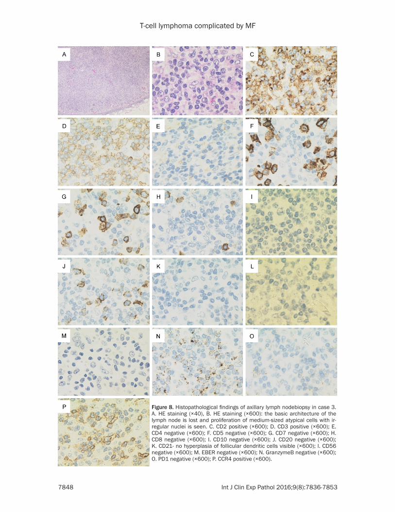
Figure 8. Histopathological findings of axillary lymph nodebiopsy in case 3. A. HE staining (×40), B. HE staining (×600): the basic architecture of the lymph node is lost and proliferation of medium-sized atypical cells with ir-regular nuclei is seen. C. CD2 positive (×600); D. CD3 positive (×600); E. CD4 negative (×600); F. CD5 negative (×600); G. CD7 negative (×600); H. CD8 negative (×600); I. CD10 negative (×600); J. CD20 negative (×600); K. CD21- no hyperplasia of follicular dendritic cells visible (×600); l. CD56 negative (×600); M. EBER negative (×600); N. GranzymeB negative (×600); O. PD1 negative (×600); P. CCR4 positive (×600).

T-cell lymphoma complicated by MF
7849 Int J Clin Exp Pathol 2016;9(8):7836-7853
T-cell lymphoma complicated by MF
7850 Int J Clin Exp Pathol 2016;9(8):7836-7853
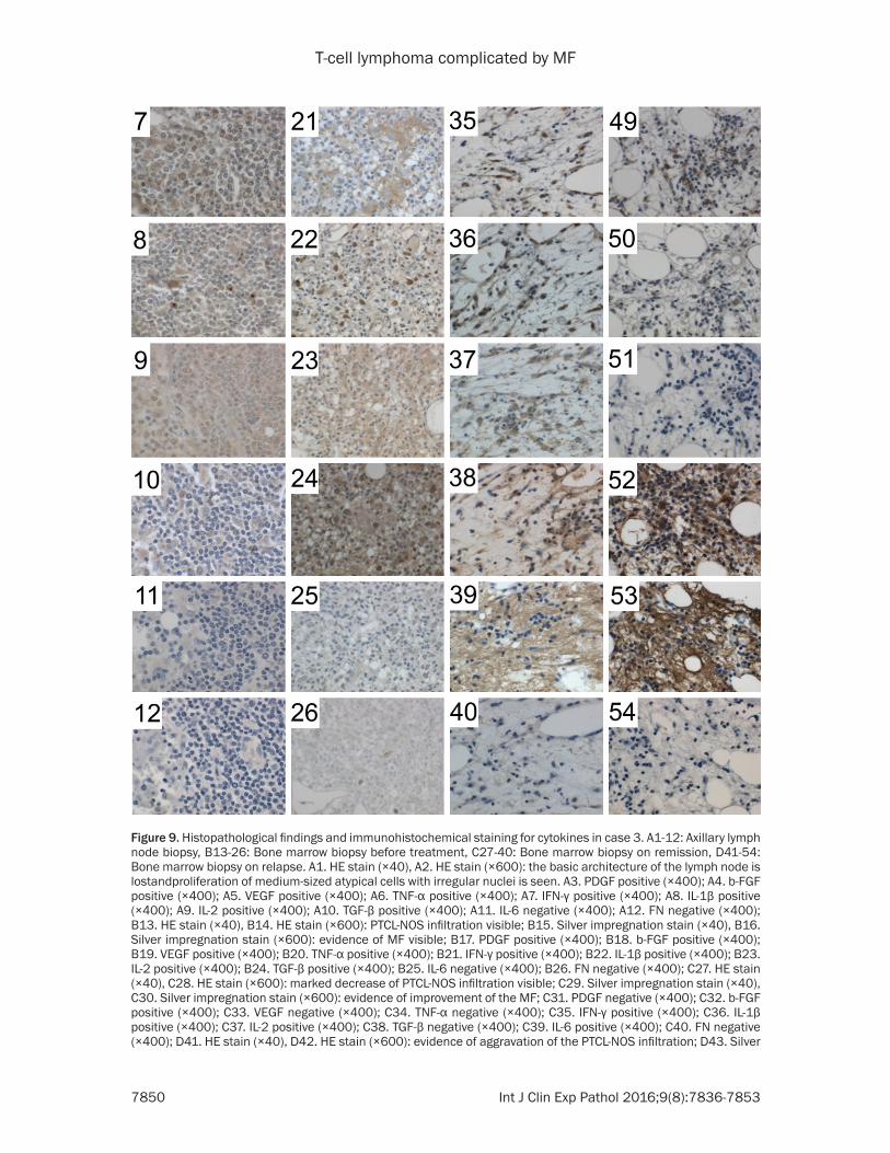
Figure 9. Histopathological findings and immunohistochemical staining for cytokines in case 3. A1-12: Axillary lymph node biopsy, B13-26: Bone marrow biopsy before treatment, C27-40: Bone marrow biopsy on remission, D41-54: Bone marrow biopsy on relapse. A1. HE stain (×40), A2. HE stain (×600): the basic architecture of the lymph node is lostandproliferation of medium-sized atypical cells with irregular nuclei is seen. A3. PDGF positive (×400); A4. b-FGF positive (×400); A5. VEGF positive (×400); A6. TNF-α positive (×400); A7. IFN-γ positive (×400); A8. IL-1β positive (×400); A9. IL-2 positive (×400); A10. TGF-β positive (×400); A11. IL-6 negative (×400); A12. FN negative (×400); B13. HE stain (×40), B14. HE stain (×600): PTCL-NOS infiltration visible; B15. Silver impregnation stain (×40), B16. Silver impregnation stain (×600): evidence of MF visible; B17. PDGF positive (×400); B18. b-FGF positive (×400); B19. VEGF positive (×400); B20. TNF-α positive (×400); B21. IFN-γ positive (×400); B22. IL-1β positive (×400); B23. IL-2 positive (×400); B24. TGF-β positive (×400); B25. IL-6 negative (×400); B26. FN negative (×400); C27. HE stain (×40), C28. HE stain (×600): marked decrease of PTCL-NOS infiltration visible; C29. Silver impregnation stain (×40), C30. Silver impregnation stain (×600): evidence of improvement of the MF; C31. PDGF negative (×400); C32. b-FGF positive (×400); C33. VEGF negative (×400); C34. TNF-α negative (×400); C35. IFN-γ positive (×400); C36. IL-1β positive (×400); C37. IL-2 positive (×400); C38. TGF-β negative (×400); C39. IL-6 positive (×400); C40. FN negative (×400); D41. HE stain (×40), D42. HE stain (×600): evidence of aggravation of the PTCL-NOS infiltration; D43. Silver

T-cell lymphoma complicated by MF
7851 Int J Clin Exp Pathol 2016;9(8):7836-7853
impregnation stain (×40), D44. Silver impregnation stain (×600); evidence of aggravation of the MF. D45. PDGF positive (×400); D46. b-FGF positive (×400); D47. VEGF negative (×400); D48. TNF-α positive (×400); D49. IFN-γ positive (×400); D50. IL-1β positive (×400); D51. IL-2 negative (×400); D52. TGF-β positive (×400); D53. IL-6 posi-tive (×400); D54. FN negative (×400).
Discussion
According to our literature search, 16 cases (including our three cases) of T-cell lymphoma complicated by MF have been reported to date (i.e., 15 previously reported cases and our current case 1) (Table 7). The most frequent histological type was AITL (eightcases), fol-lowed by PTCL (six cases). In addition, there was one case of cytotoxic T-cell lymphoma and one case of T-cell lymphoma. The disease was stage III in 2 cases and stage IV in 13 cases, indicating that in all cases except one, the disease was at an advanced stage. Bone marrow infiltration was seen frequently (pres-ent in 13 cases and absent in 3 cases). All three of the cases encountered at our depart-ment had bone marrow infiltration. Exami- nation of cytokines, which are considered to be the cause of MF, was done in only seven cases. Examination of cytokines was per-formed in the serum only in three cases (cases 6, 9, and 16), immunostaining only was per-formed in three cases (cases 2, 3, and 5), and serum examination and immunostaining was performed in one case (case 1). No exami-nation for cytokines was performed in nine cases (cases 4, 7, 8, and 10-15). Staining for PDGF was positive in fivecases, TGF-β was positive in four cases, TNF-α was positive in two cases, and b-FGF was positive in one case, although some cases showed positive staining for two or more of these cytokines. The possible mechanisms for the onset of MF are (1) induction by cytokines produced by the lymphoma cells infiltrating the bone marrow, (2) induction via by cytokines produced by lymphoma cells remote from the marrow in the absence of infiltration of the bone mar-row by lymphoma cells, or (3) a combination of mechanisms (1) and (2). In the future, we plan to carry out chronological examination for cytokines in the serum and carry out immunos-taining for cytokines not only in cases of T-cell lymphoma but also in cases of malignant lym-phoma complicated and not complicated by MF.
Table 4. Summary of the findings of immunohis-tochemical staining of the lymph nodes and bone marrow for cytokines in case 1Case 1 LN BM BM BM
Before CHOP
Before CHOP
On remission
On relapse
Marrow infiltration ND + ND -BMF ND + ND -PDGF + + ND ±b-FGF ± + ND ±VEGF ± + ND ±TNF-α + + ND ±IFN-γ ± + ND ±IL-1β ± + ND ±IL-2 ± + ND ±TGF-β ± + ND ±IL-6 ± + ND ±FN - ± ND -+, positive; ±, pseudo positive; -, negative; LN, lymph node; BM, bone marrow; CHOP, cyclophosphamide, adriamycin, vincristine, and prednisolone therapy; BMF, bone marrow fibrosis; ND, not done; IFN, interferon; FN, fibronectin.
Table 5. Summary of the findings of immunohis-tochemical staining of the lymph nodes and bone marrow for cytokines in case 2Case 2 LN BM BM BM
Before CHOP
Before CHOP
On remission
On relapse
Marrow infiltration + - -BMF + - -PDGF + + - -b-FGF ± + + +VEGF - ± - -TNF-α - - - -IFN-γ - - - -IL-1β - - ± ±IL-2 + + + +TGF-β - - - ±IL-6 - - - ±FN + ± + ++, positive; ±, pseudo positive; -, negative.

T-cell lymphoma complicated by MF
7852 Int J Clin Exp Pathol 2016;9(8):7836-7853
Table 6. Summary of findings of immunohistochemical staining of the lymph nodes and bone marrow for cytokines in case 3Case 3 LN BM BM BM
Before CHOP Before CHOP On partial remission after first course of CHOP
After 2 courses of CHOP
Marrow infiltration ND + - +BMF ND + - +PDGF + + - +b-FGF + + + +VEGF + + - -TNF-α + + - +IFN-γ + + + +IL-1β + + + +IL-2 + + + -TGF-β + + - +IL-6 - - + +FN - - - -+, positive; ±, pseudo positive; -, negative.
Table 7. Details of the 16 reported cases of T-cell lymphoma complicated by myelofibrosis
Case Age/sex Histological type Stage BM
infiltration Cytokine Ref.
1 64/M AITL IV + PDGF (immunostained) TNF-α
Current article
2 65/M AITL IV + PDGF (immunostained) [2]3 68/M PTCL-NOS IV + PDGF (immunostained)
TNF-α TGF-β
[16]
4 67/F PTCL-NOS IV + NA [4]5 90/M PTCL-U IV + b-FGF (immunostained) [17]6 68/M PTCL-U IV + TGF-β (serum) [13]7 69/M PTCL IV + NA [5]8 46/F T-cell lymphoma (no details) IV + NA [6]9 19/F Cytotoxic T-cell lymphoma IV + PDGF (serum) TGF-β [14]10 65/F PTCL IV + NA [7]11 63/M AITL IV + NA [8]12 NA AITL II or greater – NA [9]13 69/F AITL III – NA [10]14 47/M AITL IV + NA [11]15 55/F AITL IV + NA [12]16 56/M AITL III – PDGF (serum) TGF-β [15]Cases 1-3 in this table are cases 1-3 as described in the current article. BM, bone marrow; PTCL-NOS, peripheral T-cell lymphoma-not otherwise specified; NA, not available; PTCL-U, peripheral T-cell lymphoma-unspecified; AITL, angioimmunoblas-tic T-cell lymphoma.
Disclosure of conflict of interest
None.
Address correspondence to: Yasunobu Sekiguchi, Department of Hematology, Juntendo University Urayasu Hospital, 2-1-1 Tomioka, Urayasu, Chiba,
Japan. Tel: +81-47-353-3111; Fax: +81-47-381-5054; E-mail: [email protected]
References
[1] Tefferi A. Myelofibrosis with myeloid metapla-sia. N Engl J Med 2000; 342: 1255-65.

T-cell lymphoma complicated by MF
7853 Int J Clin Exp Pathol 2016;9(8):7836-7853
[2] Sekiguchi Y, Matsuzawa N, Shimada A, Imai H, Wakabayashi M, Sugimoto K, Nakamura N, Sawada T, Izutsu K, Takeuchi K, Ohta Y, Komatsu N and Noguchi M. Angioimmunobla- stic T-cell lymphoma with intramedullary pro-duction of platelet-derived growth factor and possibly complicating myelofibrosis: report of a case with review of the literature. Int J Hematol 2013; 98: 250-7.
[3] Le Bousse-Kerdilès MC and Martyré MC. Dual implication of fibrogenic cytokines in the pathogenesis of fibrosis and myeloproliferation in myeloid metaplasia with myelofibrosis. Ann Hematol 1999; 78: 437-44.
[4] Jain P, Lin P, Bueso-Ramos C, Verstovsek S and Pemmaraju N. Primary autoimmune myelofi-brosis (MF) with high-grade peripheral T-cell lymphoma (PTCL) NOS. Eur J Haematol 2013; 91: 378-9.
[5] Uehara E, Tasaka T, Matsuhashi Y, Fujita M, Tamura T, Shimoura Y, Mano S, KuwajimaM and Nagai M. Peripheral T-cell lymphoma pre-senting with rapidly progressing myelofibrosis. Leuk Lymphoma 2003; 44: 361-3.
[6] Rao SA, Gottesman SR, Nguyen MC and Braverman AS. T cell lymphoma associated with myelofibrosis. Leuk Lymphoma 2003; 44: 715-8.
[7] Takai K and Sanada M. [Peripheral T-cell lym-phoma initially presenting as secondary myelo-fibrosis]. Rinsho Ketsueki 1989; 30: 2199-204.
[8] Nishida M, Iwanaga T, Irimajiri K, Horiuchi A, Hashimoto S and Furuta I. [An autopsy case of immunoblastic lymphadenopathy associated with pancytopenia and interstitial pneumoni-tis]. Rinsho Ketsueki 1981; 22: 1977-83.
[9] Brenner B, Green J, Rosenbaum H, Ben Arieh Y, Nagler A and Tatarsky I. Severe pancytope-nia due to marked marrow fibrosis associated with angioimmunoblastic lymphadenopathy. Acta Haematol 1985; 74: 43-4.
[10] Saito T, Miyawaki S, Nemoto K, Suga H, Yashiro K, Baba N, Uchida S, Kojima M, Ito H, and Joshita T. [A case report of angioimmunoblas-tic lymphadenopathy accompanying pancyto-penia and bone marrow fibrosis]. Rinsho Ketsueki 1988; 29: 2073-8.
[11] Sato I, Miura A, Yokomichi H, Suzuki C and Ichinohazama R. [Angio-immunoblastic lymph-adenopathy with fibrosis of bone marrow, lymph node, liver and spleen, and proliferation of epithelioid cells in lymph nodes]. Rinsho Ketsueki 1990; 31: 958-62.
[12] Orth T, Treichel U, Mayet WJ, Störkel S and Meyer zum Büschenfelde KH. [Reversible my-elofibrosis in angioimmunoblastic lymphade-nopathy]. Dtsch Med Wochenschr 1994; 119: 694-8.
[13] Okabe S, Miyazawa K, Iguchi T, Sumi M, Takaku T, Ito Y, Kimura Y, Serizawa H, MukaiK and Ohyashiki K. Peripheral T-cell lymphoma to-gether with myelofibrosis with elevated plasma transforming growth factor-beta1. Leuk Lymphoma 2005; 46: 599-602.
[14] Abe Y, Ohshima K, Shiratsuchi M, Honda K, Nishimura J, Nawata H and Muta K. Cytotoxic T-cell lymphoma presenting as secondary my-elofibrosis with high levels of PDGF and TGF-beta. Eur J Haematol 2001; 66: 210-2.
[15] Matsui K, Adachi M, Tominaga T, Shinohara K and Kamei T. Angioimmunoblastic T cell lym-phoma associated with reversible myelofibro-sis. Intern Med 2008; 47: 1921-4.
[16] Sekiguchi Y, Shirane S, Shimada A, Ichikawa K, Wakabayashi M, Sugimoto K, Tomita S, Izumi H, Nakamura N, Sawada T, Ohta Y, Komatsu N and Noguchi M. Peripheral T cell lymphoma, not otherwise specified with myelofibrosis: re-port of a case with review of the literature. Int J Clin Exp Pathol 2015; 8: 4186-203.
[17] Kikukawa M, Umahara T, Kikawada M, Kanaya K, Sakurai H, Shin K, Mori M and Iwamoto T. Peripheral T-cell lymphoma presenting as my-elofibrosis with the expression of basic fibro-blast growth factor. Geriatr Gerontol Int 2009; 9: 395-8.
[18] Gallamini A, Stelitano C, Calvi R, Bellei M, Mattei D, Vitolo U, Morabito F, Martelli M, Brusamolino E, Iannitto E, Zaja F, Cortelazzo S, Rigacci L, Devizzi L, Todeschini G, Santini G, Brugiatelli M and Federico M; Intergruppo Italiano Linfomi. Peripheral T-cell lymphoma unspecified (PTCL-U): a new prognostic model from a retrospective multicentric clinical study. Blood 2004; 103: 2474-9.


![An autopsy case of peripheral T cell lymphoma occurring in ...and one case of PTCL at 3 months after delivery have been reported [1–6], there are no reports of autopsy case of PTCL](https://img.dokumen.tips/doc/110x75/607801e0e3a63a4150305312/an-autopsy-case-of-peripheral-t-cell-lymphoma-occurring-in-and-one-case-of-ptcl.jpg)


