Embed Size (px)
Citation preview

Retinal Microglial Activation and Inflammation Inducedby Amadori-Glycated Albumin in a Rat Model of DiabetesAhmed S. Ibrahim,
1,2,3Azza B. El-Remessy,
1,2,4,5Suraporn Matragoon,
4,5Wenbo Zhang,
6Yogin Patel,
7
Sohail Khan,1Mohammed M. Al-Gayyar,
3,4,5Mamdouh M. El-Shishtawy,
3and Gregory I. Liou
1,2
OBJECTIVE—During diabetes, retinal microglial cells are acti-vated to release inflammatory cytokines that initiate neuronalloss and blood–retinal barrier breakdown seen in diabetic reti-nopathy (DR). The mechanism by which diabetes activatesmicroglia to release those inflammatory mediators is unclearand was therefore elucidated.
RESEARCH DESIGN AND METHODS—Microglia activationwas characterized in streptozocin-injected rats and in isolatedmicroglial cells using immunofluorescence, enzyme-linked immu-nosorbent assay, RT-PCR, and Western blot analyses.
RESULTS—In 8-week diabetic retina, phospho-extracellularsignal–related kinase (ERK) and P38 mitogen-activated proteinkinases were localized in microglia, but not in Mueller cells orastrocytes. At the same time, Amadori-glycated albumin (AGA)-like epitopes were featured in the regions of microglia distribu-tion, implicating a pathogenic effect on microglial activation. Totest this, diabetic rats were treated intravitreally with A717, aspecific AGA-neutralizing antibody, or murine IgG. Relative tonondiabetic rats, diabetic rats (IgG-treated) manifested 3.9- and7.9-fold increases in Iba-1 and tumor necrosis factor (TNF)-amRNAs, respectively. Treatment of diabetic rats with A717 sig-nificantly attenuated overexpression of these mRNAs. Intravitrealinjection of AGA per se in normal rats resulted in increases ofIba-1 expression and TNF-a release. Guided by these results,a cultured retinal microglia model was developed to study micro-glial response after AGA treatment and the mechanistic basisbehind this response. The results showed that formation of re-active oxygen species and subsequent activation of ERK and P38,but not Jun NH2-terminal kinase, are molecular events underpin-ning retinal microglial TNF-a release during AGA treatment.
CONCLUSIONS—These results provide new insights in un-derstanding the pathogenesis of early DR, showing that theaccumulated AGA within the diabetic retina elicits the microglialactivation and secretion of TNF-a. Thus, intervention trials withagents that neutralize AGA effects may emerge as a new thera-peutic approach to modulate early pathologic pathways long be-fore the occurrence of vision loss among patients with diabetes.Diabetes 60:1122–1133, 2011
During the past decade, it has become clear thatinflammation is a key feature in diabetes thatleads to long-term complications in specificorgans, in particular the eye and kidney (1). In
the eye, the major complication is diabetic retinopathy(DR), which is the leading cause of blindness in the westernworld and affects approximately three fourths of diabeticpatients within 15 years after onset of the disease (2). Therecommended treatment for these patients has been laserphotocoagulation, which is an invasive procedure with con-siderable limitations and adverse effects. Therefore, thereis a great need for the development of new noninvasivetherapies to treat those affected by DR. These therapiescan be discovered by unraveling the pathophysiologyof DR.
As a consequence of diabetes, retinal microglia, a sub-type of glial-immune sentinel cells prestationed in the tis-sue, become reactive, leading to the release of solublecytotoxins that contribute to neuronal and vascular celldeath and ultimately the progression of DR (3). However,the underlying mechanism of microglial activation duringdiabetes is still incompletely understood.
In recent years, human and animal studies have eluci-dated that many effects of hyperglycemia are mediated byglycated proteins (4). Amadori-glycated albumin (AGA) isthe prominent form of circulating glycated proteins in vivo,and its concentration is significantly increased after di-abetes, reaching its maximum in 5–7 weeks (5). AGA arisesfrom the nonenzymatic condensation reaction between areducing sugar and susceptible amino groups. This mod-ification confers properties to AGA that are not possessedby the native, nonglycated albumin, such as the promotionof the inflammatory response and the activation of differ-ent mitogen-activated protein kinase (MAPK) cascades inseveral cell types (6–9). These MAPKs, including extracel-lular signal–related kinase (ERK), Jun NH2-terminal kinases(JNKs), and P38, can be independently or simultaneouslyactivated depending on the target cells (8–10).
On the basis of these properties of AGA, a growing bodyof evidence now supports the causal role of AGA in thedevelopment of many complications associated with di-abetes (11–13). In relation to DR, elevated AGA has beendocumented in the retinal capillaries of diabetic patientswith retinopathy (14) and in the retina of diabetic rats (15).Treatment of diabetic db/dbmice with A717 antibody, whichspecifically recognizes AGA, ameliorated retinal basementmembrane thickening (16). Furthermore, treatment of di-abetic rats with 2-(3-chlorophenylamino)-phenylacetic acid,which inhibits the nonenzymatic glycation of albumin, miti-gated vitreous changes in angiogenic cytokines associatedwith the development of DR (17). Therefore, AGA is be-lieved to possess biologic characteristics that are linked to
From the 1Department of Ophthalmology, Medical College of Georgia, Au-gusta, Georgia; the 2Vision Discovery Institute, Medical College of Georgia,Augusta, Georgia; the 3Department of Biochemistry, Faculty of Pharmacy,Mansoura University, Mansoura, Egypt; the 4Program in Clinical and Exper-imental Therapeutics, University of Georgia, Athens, Georgia; the 5VA Med-ical Center, Augusta, Georgia; the 6Vascular Biology Center, Medical Collegeof Georgia, Augusta, Georgia; and the 7Department of Medicine, MedicalCollege of Georgia, Augusta, Georgia.
Corresponding author: Gregory I. Liou, [email protected] 16 August 2010 and accepted 6 January 2011.DOI: 10.2337/db10-1160This article contains Supplementary Data online at http://diabetes.
diabetesjournals.org/lookup/suppl/doi:10.2337/db10-1160/-/DC1.� 2011 by the American Diabetes Association. Readers may use this article as
long as the work is properly cited, the use is educational and not for profit,and the work is not altered. See http://creativecommons.org/licenses/by-nc-nd/3.0/ for details.
1122 DIABETES, VOL. 60, APRIL 2011 diabetes.diabetesjournals.org
ORIGINAL ARTICLE
the DR pathogenesis and might be involved in the activationof retinal microglia. In the present work, we aimed to studythe ability of AGA to induce retinal microglial activation andtheir secretion of inflammatory cytokines both in vivo andin vitro.
RESEARCH DESIGN AND METHODS
All procedures with animals were performed in accordance with the Asso-ciation for Research in Vision and Ophthalmology Statement for the Use ofAnimals in Ophthalmic and Vision Research and Medical College of Georgiaguidelines. Diabetes was induced in male SD rats by intravenous injection ofstreptozocin (STZ) (60 mg/kg) and confirmed by urine-glucose levels .350mg/dL. Eyes were used for immunofluorescence or Western examinationat 8 weeks of diabetes. For intravitreal injections, the procedure was essen-tially the same as previously described (18). The A717 intravitreal injectionscheme was chosen on the basis of certain assumptions regarding the esti-mated amount of AGA to be neutralized. We estimated rat vitreous volume at50 mL (19), with AGA concentration of 1.9 mg/mL (17). This would corre-spond with an approximate total vitreous AGA of 0.09 mg. On a stoichio-metric basis, it would require ;0.2 mg antibody (mol wt ;150 kD) toneutralize ;0.09 mg of AGA (mol wt ;66.5 kD). First, A717 injections weregiven at 10 days, and a booster dose was given at 7 weeks of diabetes. Thesetwo time points were selected presumably because the former representsthe earliest occurrence of AGA, whereas the latter marks its maximumaccumulation (5).Microglia culture. Microglia were isolated from retinas of newborn SD ratsaccording to a previous procedure (20). Cell viability after various drugtreatments was determined by counting the number of trypan blue-excludingcells using a hemocytometer.AGA preparation. AGA was purchased from Sigma-Aldrich Co. (St. Louis,MO), purified by affinity chromatography on phenylboronate resin, concen-trated, and desalted into PBS. This preparation contains 2.2-mol hexose-lysine/mol-albumin, but fluorescent advanced glycation end products (AGEs) werenot detectable and the major AGE N-(carboxymethyl) lysine was present inonly minute amounts (21). The concentration of AGA (500 mg/mL) chosen inour study is close to what has been used previously to study other glycatedalbumin-mediated responses (22) and represents those found in clinicalspecimens (23). The specificity of AGA activity was determined by neutrali-zation with its antibody, A717. Briefly, AGA or BSA was balanced with PBS atpH = 7.4, kept overnight at 4°C on a rocker with A717 antibody or IgG, servedas a control, and followed by the addition of A/G agarose beads. The beads
were then removed by centrifugation, and the supernatant was collected andused for microglia treatment. The endotoxin level was checked by an endo-toxin testing kit (Limulus amebocyte assay, E-Toxate, Sigma-Aldrich Co.).Alternatively, AGA was purchased from Exocell Inc. (Philadelphia, PA). Thisalternative product contains 1-mol hexose-lysine/mol-albumin and was puri-fied by boronate-affinity column and Detoxi-Gel Endotoxin Removal Gel.Viability of cells after AGA-treatment was checked by transferase-mediateddUTP nick-end labeling assay (Trevigen Inc., Gaithersburg, MD) following themanufacturer’s instructions.Immunofluorescence. Dual-labeled immunofluorescence analysis was per-formed using frozen retinal sections. Briefly, sections were fixed in 4% para-formaldehyde, blocked with 10% normal goat serum, and then incubated withprimary antibodies (Table 1) overnight. Thereafter, sections were brieflywashed with PBS and incubated with appropriate secondary antibodies (Table1). Slides were examined by confocal microscopy (LSM 510, Carl Zeiss AG,Oberkochen, Germany). Specificity of the reaction was confirmed by omittingthe primary antibody. Images were collected from five sections per rat of atleast four to six rats per group.RNA interference. Microglial cells were transfected with ERK or controlsmall interfering (si)RNAs (Ambion, Austin, TX) using HiPerFect (Qiagen,Venlo, the Netherlands) per the manufacturer’s instructions.Quantitative real-time PCR. Total RNA was isolated from rat retina usinga Promega kit (Promega Corp., Madison, WI). Subsequently, cDNAs weregenerated from 1 mg of total RNA, using the High-Capacity cDNA ReverseTranscription Kit (Applied Biosystems, Foster City, CA), and subjected to a 40-cycle PCR amplification. The ready-made primer and probe sets were orderedfrom Applied Biosystems (Catalog: Iba-1: Rn01525935_m1; TNF-a: Rn99999017_m1;18S: Hs03003631_g1). Three replicates were run for each gene for each samplein a 96-well plate. 18S was used as the endogenous reference gene.Western blot analysis. Cell or retinal lysates were subjected to Western blotanalysis using antibodies specified in Table 1 according to a previous pro-cedure (20).ELISA assay. Tumor necrosis factor (TNF)-a levels in the rat vitreous orsupernatants of culture media were estimated with ELISA kit (R&D SystemsInc., Minneapolis, MN) per the manufacturer’s instructions (20).DCF assay for reactive oxygen species formation. DCF, the oxidationproduct of the reagent 2`,7`-dichlorofluorescin diacetate, was used as a markerof cellular oxidation according to a previous procedure (20).Data analysis. The results were expressed as mean 6 SD. Differences amongexperimental groups were evaluated by ANOVA, and the significance of dif-ferences between groups was assessed by the post hoc test (Fisher pro-tected least significant difference) when indicated. Significance was definedas P , 0.05.
TABLE 1Primary antibodies used for different experiments in this study
Antibody Catalog no. Vendor Host
Workingdilution
IF WB
Anti-Iba1 019–19741 Wako Rabbit 1:200 —
Anti-OX-42 201801 BioLegend (San Diego, CA) Mouse 1:200 —
Antiphospho-ERK 9106 Cell Signaling Technology Inc. (Danvers, MA) Mouse 1:400 1:2,000Antiphospho-P38 4631 Cell Signaling Technology Inc. Rabbit 1:100 1:1,000Anti-GFAP AB5541 Millipore (Billerica, MA) Chicken 1:500 —
Anti-AGA A717 Exocell Inc. (Philadelphia, PA) Mouse 1:50 1:500Anti-albumin PA1–10298 Thermo Fisher Scientific (Waltham, MA) Rabbit — 1:2,000Antiactin A2066 Sigma-Aldrich Co. (St. Louis, MO) Rabbit — 1:2,000Anti-ERK 9102 Cell Signaling Technology Rabbit — 1:1,000Anti-P38 9212 Cell Signaling Technology Rabbit — 1:1,000Antiphospho-JNK 9255 Cell Signaling Technology Mouse — 1:2,000Anti-JNK 9252 Cell Signaling Technology Rabbit — 1:1,000Anti-rabbit (Texas red or Oregon green) A21072 Invitrogen (Carlsbad, CA) Goat 1:500 —
737677 ChickenAnti-mouse (Texas red or Oregon green) 737674 Invitrogen Chicken 1:500 —
A11017 GoatAntichicken (Cya5) 92590 Millipore Donkey 1:500 —
Anti-rabbit HRP 7074 Cell Signaling Technology Goat — 1:10,000Anti-mouse HRP 7076 Cell Signaling Technology Goat — 1:10,000
HRP, horseradish peroxidase; IF, immunofluorescence; WB, Western blot.
A.S. IBRAHIM AND ASSOCIATES
diabetes.diabetesjournals.org DIABETES, VOL. 60, APRIL 2011 1123

RESULTS
ERK and P38 MAPKs are selectively activated in theretinal microglia during diabetes. Recent evidence hasshown that MAPK cascade is one of the attractive targetsfor intervention in the inflammatory-associated diseases,such as diabetes. Therefore, the role of retinal microgliaand macroglia during diabetes was investigated by de-termining MAPK signaling in frozen eye sections. Asshown in Fig. 1A and B, in the 8-week diabetic retina,numerous Iba-1- or OX-42-positive cells (activated micro-glia) appeared hypertrophic or amoeboid and were ob-served in the outer plexiform layer or ganglion cell layer,or frequently clustered around the perivascular region(Supplementary Fig. 1). In contrast, in the nondiabeticretina, these cells had oval cell bodies with ramified pro-cesses (Supplementary Fig. 2). Double immunofluores-cence showed that pERK- and pP38-labeled cells werecolocalized with Iba-1 or OX-42 antigen, but not with glial
fibrillary acidic protein (GFAP). These results revealedthat diabetes leads to characteristic glial cell changeswithin the retina in which activation of ERK and P38 occurin the activated microglia, but not in astrocytes or Muellercells.AGA is a proinflammatory trigger in diabetes-inducedretinal microglial activation and inflammation. Theinduction of microglial reactivity is associated with a localproinflammatory environment (3,24) that could be trig-gered by various conditions operating in the course of di-abetes. Given the fact that AGA provokes proinflammatoryresponses in many types of cells, including immune cells(7,9,25), we hypothesized that this glycated protein mayalso be implicated in diabetes-induced microglial activa-tion and retinal inflammation. To test this, we first soughtto determine whether this glycated protein is accumulatedin the diabetic retina and whether this accumulationoccurs in the region of microglia distribution. The results
FIG. 1. Activation of ERK and P38 in diabetes occurs in the retinal microglia. Immunolabeling of phospho-ERK (green) (A) or phospho-P38 (green)(B) with Iba-1 (red) or OX-42 (red), markers of activated microglia, or with GFAP (red), a marker of astrocytes or activated Mueller cells, wasmade in the retinas of diabetic (8 weeks after STZ induction) and normal rats. Yellow displayed from merged red and green. Scale bar, 20 mm. GCL,ganglion cell layer. H&E, hematoxylin–eosin stain; OPL, outer plexiform layer. (A high-quality digital representation of this figure is available inthe online issue.)
AGA-INDUCED MICROGLIAL ACTIVATION IN DR
1124 DIABETES, VOL. 60, APRIL 2011 diabetes.diabetesjournals.org

showed that AGA and albumin were increased approxi-mately 4.5- and fourfold in the retina of 8-week diabeticrats compared with nondiabetic retinas (Fig. 2A and B).Moreover, AGA-like epitopes were featured within the ret-inal tissue of 8-week diabetic rats (Fig. 2C) and at 10 daysafter the detection of hyperglycemia (data not shown).These epitopes were found to be colocalized with themicroglial marker (Fig. 2C), implicating a pathogenic ef-fect on microglial function.
Second, to determine whether AGA is involved in thepathogenesis of microglial activation and inflammation
during diabetes, we used two complementary approaches. Inour first approach, the putative biological effect of AGA perse in retinal inflammation was investigated using an experi-mental model in which normal rats were injected intra-vitreally with AGA or nonglycated albumin. As shown inFig. 2D and E, AGA injection induced a significant five- and3.3-fold increase in retinal Iba1 mRNA expression and TNF-arelease, respectively, compared with controls injected withnonglycated albumin.
The second approach was to assess whether neutralizingAGA biological activity in the diabetic retinas with A717
FIG. 2. AGA as a proinflammatory trigger in diabetes-induced retinal microglial activation and inflammation. A and B: AGA and albumin expressionin retina of nondiabetic rats (normal) and 8-week diabetic rats, analyzed by Western blot. Ratio of the intensities of AGA or albumin relative to theactin for the patients with diabetes was compared with normal, which was arbitrarily set at 1.0. Data shown for the comparison of normal anddiabetic rats are the mean 6 SD and representative of four to six animals per group. C: Immunolabeling of AGA (green) with Iba-1 (red) in thenormal and 8-week diabetic rat retinas. D: Activation of retinal microglia by injected AGA in normal rats. Nondiabetic rats were injected intra-vitreally with AGA (500 mg/mL) or equal amounts of nonglycated albumin. Retinal Iba-1 mRNA was determined by real-time PCR 24 h later. Datashown are the mean 6 SD (n = 4–6). E: Proinflammatory effect of injected AGA in normal rat retina. Nondiabetic rats were injected intravitreallywith AGA (500 mg/mL) or equal amounts of BSA. Vitreal TNF-a was determined by ELISA 24 h later. Data shown are the mean 6 SD (n = 4–6).F and G: Attenuation of increased Iba1 and TNF-a gene expression in diabetic rats treated with A717 or IgG. Total RNA was extracted from snap-frozen retinas, and Iba1 and TNF-a mRNAs were then determined by real-time PCR and normalized to normal nondiabetic control. Data shown arethe mean 6 SD (n = 4–6). H: Cyclic threshold values of 18S in the retina of diabetic rats treated with A717 or IgG. Data shown are the mean 6 SD(n = 4–6). Ct, cyclic threshold; GCL, ganglion cell layer; OPL, outer plexiform layer. (A high-quality color representation of this figure is availablein the online issue.)
A.S. IBRAHIM AND ASSOCIATES
diabetes.diabetesjournals.org DIABETES, VOL. 60, APRIL 2011 1125
would attenuate diabetes-induced retinal microglial activa-tion and TNF-a release. As shown in Fig. 2F and G, treat-ment with A717 significantly decreased the elevated retinalIba1 and TNF-a gene expression observed in the IgG-treateddiabetic rats by ;60 and 65%, respectively. Of note, the ex-pression level of 18S was approximately the same among
diabetic groups (Fig. 2H), indicating that the effect of A717 ongene expression is not due to a generalized nonspecific sup-pression of retinal gene expression. Taken together, theseresults demonstrate that the formation of AGA within thediabetic milieu is one of the major contributing factors tomicroglial activation and therefore retinal inflammation.
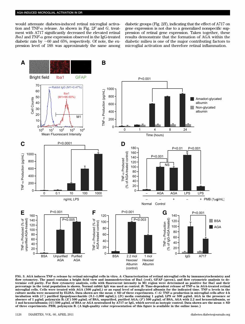
FIG. 3. AGA induces TNF-a release by retinal microglial cells in vitro. A: Characterization of retinal microglial cells by immunocytochemistry andflow cytometry. The panel contains a bright field view and immunodetection of Iba1 (red), GFAP (green), and flow cytometric analysis to de-termine cell purity. For flow cytometry analysis, cells with fluorescent intensity in M1 region were determined as positive for Iba1 and theirpercentage in the total population is shown. Normal rabbit IgG was used as control. B: Time-dependent release of TNF-a in AGA-treated retinalmicroglial cells. Cells were treated with AGA (500 mg/mL) or an equal level of nonglycated albumin for the indicated time. TNF-a levels in theculture media were measured by ELISA. Data shown are the mean 6 SD of three experiments. C–G: TNF-a production in microglial cells after 4-hincubation with (C) purified lipopolysaccharide (0.1–1,000 ng/mL); (D) media containing 1 mg/mL LPS or 500 mg/mL AGA in the presence orabsence of 1 mg/mL polymyxin B; (E) 500 mg/mL of BSA, unpurified, purified AGA; (F) 500 mg/mL of BSA, AGA with 2.2 mol hexose/albumin, or1 mol hexose/albumin; (G) 500 mg/mL of BSA or AGA neutralized by A717 or IgG, which served as isotypic control. Data shown are the mean 6 SDof three experiments. PBM, polymyxin B. (A high-quality color representation of this figure is available in the online issue.)
AGA-INDUCED MICROGLIAL ACTIVATION IN DR
1126 DIABETES, VOL. 60, APRIL 2011 diabetes.diabetesjournals.org

FIG. 4. Phosphorylation of ERK and P38, but not JNK, causes TNF-a release in the activated retinal microglial cells. A: Time-dependent, AGA-induced activation of ERK in retinal microglial cells. Cells were treated with 500 mg/mL AGA for 1–8 h. Phospho (p)ERK and its total protein in celllysate were determined by Western analysis. Ratios of the intensities of phospho-proteins relative to total proteins for each time point werecompared with control, which was arbitrarily set at 1.0. Data shown are the mean 6 SD of three experiments (*P < 0.001). B: Dose-dependentinhibition of AGA-induced TNF-a release in retinal microglial cells by inhibitor for ERK (U0126). Cells were treated with 500 mg/mL of AGA for 4 hin the presence of indicated concentrations of inhibitor. TNF-a levels were compared with the vehicle-treated control. Data shown are the mean 6SD of three to five experiments. C: U0126 also had no effect on cell viability, as determined by trypan blue exclusion test. D: Inhibition of
A.S. IBRAHIM AND ASSOCIATES
diabetes.diabetesjournals.org DIABETES, VOL. 60, APRIL 2011 1127

AGA induces TNF-a release by retinal microglial cellsin vitro. After having shown that AGA plays a role indiabetes-induced retinal inflammation, we next sought todissect out the mechanism(s) responsible for the observedeffect. Therefore, a cultured retinal microglia model wasdeveloped to characterize the microglial response to AGAtreatment. The purity of this culture preparation was assessedby immunocytochemistry and flow cytometry. By immuno-cytochemistry, the majority was Iba1-positive, whereas thecontaminating astrocytes were insignificant. By flow cytom-etry, the purity of microglia was 96.6% (Fig. 3A) and GFAP-positive cells were less than 1% (data not shown). With theuse of this model, incubation with AGA (500 mg/mL) sig-nificantly stimulated TNF-a production starting at 4 h andremained elevated over the 24-h experimental period,whereas incubation with nonglycated albumin for 1–24 hhad no effect (Fig. 3B). Moreover, there was nonsignificantcell death after 4 h of AGA treatment (SupplementaryFig. 3). To verify that the stimulatory effect of AGA onTNF-a release was mediated by glycated albumin only andnot by endotoxin or any other contaminants, we firstchecked the endotoxin level in AGA solutions used in thisstudy and confirmed to be endotoxin free (,0.125 units/mL,10 endotoxin unit = 1 ng lipopolysaccharide). This estimatedcontamination of endotoxin was clearly below the thresholdconcentration of purified lipopolysaccharide (LPS) (0.1–10ng/mL) required for a significant microglial stimulation(Fig. 3C). Moreover, to fully exclude the possible effects thatany contaminating traces of endotoxin AGA still might haveon the observed inflammatory response, we preincubatedAGA-containing media with LPS-binding polymyxin B. Ata concentration of 1 mg/mL, polymyxin B resulted in 90%inhibition of TNF-a production in microglia activated bypurified LPS (1 mg/mL) without affecting cell viability (90–93% compared with 90–95% for controls). The incubation ofAGA (500 mg/mL) with the same concentration of polymyxinB did not reduce the TNF-a production induced by AGA,whereas the overall TNF-a production was clearly smallerin AGA-incubated cells than that induced by purified LPS(Fig. 3D). Thus, the stimulatory effect of the AGA on TNF-arelease from microglia is independent of contamination withendotoxin.
Second, to ensure that the AGA-mediated TNF-a releasewas indeed due to glycated albumin only and not to anycontaminant that might be associated with the commerciallyavailable preparation, we compared the TNF-a–releasingability of the purified AGA with that of the unpurified one.Purification of AGA, which resulted in a preparation with70–75% yield, had a TNF-a–releasing activity 346 3% higherthan the unpurified one (Fig. 3E). Following this further, wetested the ability of an alternative AGA preparation,obtained from Exocell Inc., to elicit the inflammatory re-sponse in microglia. As shown in Fig. 3F, a significant in-crease in TNF-a level was seen after 4 h of incubation with
this alternative product compared with nonglycated albu-min. The amount of TNF-a released from microglia in re-sponse to glycated albumin varied with the degree ofprotein glycation as seen in these two alternative prepa-rations. Last, to further confirm the specific effect of AGAon TNF-a production, the AGA-neutralizing antibody,A717, was used. The increase in TNF-a level mediated byAGA was reduced significantly by preincubation withA717, but not with IgG (Fig. 3G). Collectively, these resultsindicate that AGA induces TNF-a release by retinalmicroglial cells in vitro and that increase is indeed con-secutive to a glycated albumin effect and not to speciesdifference, endotoxin, or any other minor contaminants.AGA-induced TNF-a release in retinal microglia ismediated by MAPKs. In light of AGA’s inflammatoryability to induce TNF-a release by microglia, interest in themolecular mechanisms underlying this response has beenexpanded to include ERK, P38, and JNK MAPK pathways.Whether or not these kinases are involved in AGA-inducedTNF-a release was investigated. Sustained elevation ofERK phosphorylation occurred over the 8-h experimentalperiod (Fig. 4A), and that pattern is particularly indicativeof its role in microglial response to AGA exposure. Todetermine the causal role of ERK in mediating TNF-a re-lease, two approaches were used. First, an ERK inhibitor,U0126, significantly inhibited AGA-induced TNF-a releasein a dose-dependent manner, without affecting cell viabil-ity (Fig. 4B and C). Second, microglia were transientlytransfected with ERK siRNA or with scrambled siRNA andtreated with AGA as before. ERK, but not scrambled siRNA,significantly inhibited TNF-a release (Fig. 4D, a and b).
Concomitant with ERK activation, phosphorylation of P38occurred transiently at 1–4 h after AGA treatment (Fig. 4E).Because P38 has complicated species multiplicity, we usedSB203580, a compound that inhibits P38 family of MAPK butnot other MAP kinases (26), to study the causal role of p38in mediating TNF-a release. SB203580 significantly inhibitedTNF-a release without affecting cell viability (Fig. 4F and G).To confirm these results, another selective p38 inhibitor,SB202190, was also used. As did SB203580, SB202190 sig-nificantly reduced TNF-a production in a dose-dependentmanner without affecting cell viability (SupplementaryFig. 4A–C). In contrast, phosphorylation of JNK remainedunchanged over the entire experimental period (Fig. 4H),and its inhibitor (SP610025) did not inhibit AGA-inducedTNF-a release (Fig. 4I). These results demonstrate that ac-tivation of ERK and P38, but not JNK, mediates AGA-inducedTNF-a release in retinal microglia.Crosstalk between ERK and P38 pathways in AGA-treated microglia. Next, we further determined any in-teraction between ERK and P38 pathways in the AGA-treatedretinal microglia by examining the effect of P38 and ERKinhibitors on self and mutual activation. U0126, which blocksMEK, upstream of ERK, blocked the phosphorylation of both
AGA-induced TNF-a release by ERK siRNA in retinal microglial cells. Cells were transfected with siRNA or scrambled siRNA for 48 h and thentreated with AGA for 4 h. a, Measurement of ERK expression relative to actin by Western blot after transfection with anti-ERK siRNA. Data shownare the mean 6 SD of three experiments. b, ELISA-measured TNF-a levels were compared with the scrambled siRNA-transfected control. Datashown are the mean6 SD of three experiments. E and H: Time-dependent, AGA-induced activation of P38 and JNK in retinal microglial cells. Cellswere treated with 500 mg/mL AGA for 1–8 h. Phospho (p)P38, pJNK, and its total proteins in cell lysates were determined by Western analysis.Ratios of the intensities of phospho-proteins relative to total proteins for each time points were compared with control, which was arbitrarily set at1.0. Data shown are the mean6 SD of three experiments (*P< 0.001). F and I: Dose-dependent inhibition of AGA-induced TNF-a release in retinalmicroglial cells by inhibitors for P38 (SB203580) and JNK (SP610025). Cells were treated with 500 mg/mL of AGA for 4 h in the presence ofindicated concentrations of inhibitors. TNF-a levels were compared with the vehicle-treated control. Data shown are the mean6 SD of three to fiveexperiments. G: Effect of SB203580 on cell viability, as determined by trypan blue exclusion test.
AGA-INDUCED MICROGLIAL ACTIVATION IN DR
1128 DIABETES, VOL. 60, APRIL 2011 diabetes.diabetesjournals.org

ERK and P38 (Fig. 5A). In contrast, SB203580 increased thephosphorylation of both P38 and ERK. Further, we deter-mined whether ERK and P38 both contributed to TNF-a re-lease. Microglia were pretreated with U0126 and SB203580individually or in combination and then activated by AGA.
The combination of ERK and P38 inhibitors further inhibitedTNF-a release than either one did individually (Fig. 5B).AGA-induced reactive oxygen species formation cau-ses MAPKs activation and TNF-a release in microglia.Despite these observations, the information regarding signaltransduction from the microglial AGA receptor to ERK/P38-induced cytokine secretion remains largely unknown. Re-active oxygen species (ROS) production has been shown tobe an early event induced in response to glycated proteinstimulus (27). To test whether ROS is causally related toMAPKs-mediated TNF-a release, we first tested the contri-bution of ROS formation to AGA-induced TNF-a release. Asshown in Fig. 6A–C, the inhibitory effects of an antioxidant,cannabidiol (CBD) (28), on ROS formation and TNF-a re-lease without affecting cell viability demonstrate that ROSformation is a causative event in AGA-induced TNF-a re-lease from microglia. Next, to determine whether ROScausally influenced MAPKs activation, phosphorylation ofERK and P38 was measured in AGA-treated microglia inthe presence and absence of CBD. As shown in Fig. 6Dand E, CBD significantly attenuated the phosphorylationof ERK and P38, indicating that oxidative stress is a com-mon signaling event that is located upstream of both ERK/P38 pathways mediating TNF-a release in AGA-treatedmicroglia.
DISCUSSION
Recent studies have shown that retinal inflammation isa relatively early event that occurs in the context of DRbefore vascular dysfunction (29–32). Microglial activationhas been recognized as a potential culprit mechanismcontributing to this early inflammatory outcome (3,24).However, the mechanism by which diabetes activatesmicroglia to release inflammatory cytokines remains acritical missing link. If such a link is established, clinicalinterventions with agents that target these early features ofDR would provide long-term vascular benefits. Hypergly-cemia is still considered the principal cause of diabetescomplications, and its deleterious effects are attributed,among other things, to the formation of the so-called AGA.In this study, we put forward a new concept pertaining tothe involvement of AGA in the genesis of diabetes-inducedretinal microglial activation and inflammation.
Microglia are sensitive to small changes in their envi-ronment, and they can be activated by a variety of factors,including proinflammatory cytokines, lipopolysaccharide,damaged cells, or any immune-stimulatory agents (33).Activated microglia have phagocytic and cytotoxic abili-ties to destroy foreign materials by secreting cytokines andother signaling molecules. However, if microglia remain ina sustained activated state, the secreted cytokines can affectother cell types in the proximity, particularly neuronal andvascular cells (34), bringing about the progression of manyretinal diseases, including retinal degeneration (35) andglaucoma (36). Given these functions, microglial activationrepresents an important player in the pathogenesis of DR.This input has originated partly from histopathologic studiesthat show clustering of apparently activated microglia in thediabetic rat retina (37,38). These initial observations havebeen supported in postmortem human retinas (39) andreinforced by additional histopathologic studies showingthat many inflammatory molecules, such as TNF-a, can bedetected in the diabetic retina, often in association withmicroglia (24,40). The retinal expression of TNF-a has beenreported to be associated with neuronal and endothelial cell
FIG. 5. Positive crosstalk occurs between ERK and P38 in AGA-treatedretinal microglial cells. A: Effect of P38 and ERK inhibitors on AGA-induced MAP kinase activation. Cells were treated with AGA in thepresence or absence of P38 inhibitor (SB203580) and ERK inhibitor(U0126) for 4 h. Phospho-ERK, total ERK, phospho-P38, and total P38MAPK were determined as above. Each AGA-treated sample with orwithout inhibitors was compared with the untreated sample, set as 1.0.Data shown are the mean 6 SD of three experiments. $P < 0.05; #P <0.05; and **P < 0.001. B: Effect of individual vs. combined ERK and P38inhibitors on TNF-a release. Cells were treated with U0126 orSB203580 alone or in combination for 30 min. Cells were then treatedwith AGA (500 mg/mL) for 4 h. TNF-a levels were compared with thevehicle-treated control. Data shown are the mean 6 SD of threeexperiments.
A.S. IBRAHIM AND ASSOCIATES
diabetes.diabetesjournals.org DIABETES, VOL. 60, APRIL 2011 1129

death, hallmark features of the disease (41,42), and in-hibition of TNF-a has demonstrated beneficial effects in theprevention of early DR (43). Moreover, the in vitro studieson co-cultured retinal neurons R28 with activated microglia
have shown that microglia produce cytotoxins that killretinal neuronal cells (3).
All of this seemingly overwhelming evidence has sculptedthe concept of activated microglia as a contributing factor
FIG. 6. ROS formation is an early event involved in AGA-induced TNF-a release and MAPKs activation in retinal microglial cells. A: AGA-induced,time-dependent ROS formation in retinal microglial cells. ROS formation was prevented by pretreatment with CBD (2 mmol/L). Retinal microglialcells loaded with 29,79-dichlorodihydrofluorescein diacetate were pretreated with CBD or DMSO for 60 min and treated with AGA, and the fluo-rescence of DCF was measured at 0, 2, 15, or 30 min. ROS formation was expressed as changes in DCF fluorescence/mg protein. Data shown are themean 6 SD of six experiments. B: Dose-dependent inhibition of AGA-induced TNF-a release in retinal microglial cells by CBD. Cells were treatedwith 500 mg/mL of AGA for 4 h in the presence of indicated concentrations of CBD. TNF-a levels were compared with the vehicle-treated control.C: Effect of CBD on cell viability, as determined by trypan blue exclusion test. Data shown are the mean 6 SD of three experiments. D and E: CBDinhibits AGA-induced MAPKs activation in retinal microglial cells. Cells were treated with vehicle or CBD 30 min before AGA treatment for 4 h.Phospho-ERK, total ERK, phospho-P38, and total P38 MAPK were determined by Western blot and quantified by densitometric analyses. EachAGA-treated sample with or without inhibitors was compared with the untreated sample, set as 1.0. Data shown are the mean 6 SD of threeexperiments. *P < 0.001.
AGA-INDUCED MICROGLIAL ACTIVATION IN DR
1130 DIABETES, VOL. 60, APRIL 2011 diabetes.diabetesjournals.org

in DR. Similar to microglia, macroglia are capable of as-suming different morphologies and functions, reactivemacrogliosis, in response to changes in their environment.In Mueller cells of the retina, de novo expression of GFAP isindicative of any kind of impairment of the retina, includingexperimental DR (44). This overexpression of MuellerGFAP starts to be obvious at 8 weeks of diabetes (45).Consistent with these previous studies, similar changes inmorphology and antigen-expression patterns of retinal glialcells during diabetes were observed, such as hypertrophicmicroglia with increased expression of their markers andGFAP-positive Mueller cells. However, this study is the firstto show that the acquisition of gliotic features within thediabetic retina was characterized by the activation of ERKand P38 exclusively in microglia but not in Mueller cells orastrocytes.
We then sought to determine what causes microglial ac-tivation and the accompanying increased inflammatorycytokines in the diabetic retina. One hypothesis that hasbeen under investigation in other organs and has beenraised by our initial Western and microscopic studies isAGA/inflammation cascade hypothesis. We have shown thatAGA is increased in rats with diabetes compared with ratswithout diabetes and is localized in regions of microglialdistribution. These findings, together with the observationsthat A717 significantly attenuated the expression of bothIba1 and TNF mRNA in 8-week diabetic rats and thatintravitreal injection of AGA per se in normal rats inducedIba-1 expression and TNF-a release, have strengthened thenotion that increased levels of AGA in the diabetic retinaare an important contributor to microglial activation andthereby inflammation. The accumulation of AGA within thediabetic retina could be explained in part by the vascularleakage (Fig. 2B) that occurs early during experimentaldiabetes in the venules and capillaries of the superficialinner retinal vasculature (46). This vascular leakage pro-motes microvascular permeability and thereby enhancesthe direct contact of plasma-borne glycated albumin withthe inner retinal layer, modulating the production of cyto-kines by microglia. However, the microvascular leakage isnot the only contributor to the increased retinal AGA, andthe possibility of local AGA formation cannot be ruledout (17).
Accordingly, the direct relationship between microgliaand AGA has been explored through in vitro study. Wanget al. (27) reported that glycated proteins may alter mi-croglia function by enhancing intracellular ROS formationand activating MAPKs-mediated TNF-a release but did notportray the causal relationship among these pathways.We expanded on that study and demonstrated two clearfindings: AGA triggers the secretion of TNF-a, which isvaried with the degree of protein glycation, and this in-flammatory response is mediated by ROS and its subsequentsignaling, ERK and P38 but not JNK MAPK. Furthermore,we broadened the scope to include the crosstalk betweenERK and P38 pathways in microglia after AGA treatment.Inhibition of ERK by U0126 led to decreased phosphoryla-tion of P38 MAPK, whereas blocking P38 phosphorylationby SB 203580 resulted in a reciprocal increase in the phos-phorylation of ERK. In addition, even though SB 203580 isan inhibitor of p38 activity, it was also seen to increasep38 phosphorylation; although unexplained at this point, theobservation has been reported (47). Similar to retinalmicroglia, concurrent activation of ERK and P38 occursin melanoma, and the positive crosstalk between ERKand P38 in melanoma stimulates migration and in vivo
proliferation (48). This positive crosstalk is in direct contrastwith carcinoma cells in which the activity of the two kinasesseems to be mutually exclusive (49). However, our resultsare not in agreement with the study by Wang et al. showingthat the production of TNF-a was mediated by ERK, P38,and JNK (27). This discrepancy may be explained by the useof a different JNK inhibitor, curcumin (50 mmol/L), whichshows a broad selectivity for various targets and is a potentscavenger of free radicals (50). Together, these inhibitorexperiments point out that AGA-induced TNF-a is mediatedby two alternative pathways partially interacting with eachother. Figure 7 illustrates these pathways and the mecha-nism by which various drugs block these processes. Al-though these in vitro data provide a mechanistic paradigmwhereby AGA induces microglial inflammatory response,used at nonphysiologic exposure condition (500 mg/mL), therelevance of these findings at lower AGA exposure levelsneeds to be further investigated.
Collectively, the experiments in this study provide newinsights in understanding the pathogenesis of early fea-tures of DR, demonstrating that the accumulation of AGAwithin the diabetic retina elicits the microglial activationand secretion of TNF-a. In addition, our in vitro data dis-close the molecular mechanisms involved therein, show-ing that AGA triggers TNF-a release from microglia inROS and its subsequent ERK and P38 MAPKs-dependentmechanisms. Thus, clinical intervention trials with agentsthat neutralize glycated albumin effect might be war-ranted in modulating early pathologic pathways long be-fore the occurrence of vision loss among patients withdiabetes.
FIG. 7. Signaling pathways in the diabetes-induced retinal inflam-mation. Diabetes-induced AGA leads to ROS formation, activation ofERK/P38, and TNF-a release during microglial activation. Also in-cluded is the mechanism by which various drugs block these pro-cesses. (A high-quality color representation of this figure is availablein the online issue.)
A.S. IBRAHIM AND ASSOCIATES
diabetes.diabetesjournals.org DIABETES, VOL. 60, APRIL 2011 1131

ACKNOWLEDGMENTS
This work was supported in part by Egyptian Culture andEducation Bureau (A.S.I.), Knights Templar EducationalFoundation, Vision Discovery Institute at the Medical Collegeof Georgia, the American Diabetes Association (G.I.L.), andthe Juvenile Diabetes Research Foundation (A.B.E. and W.Z.).
No potential conflicts of interest relevant to this articlewere reported.
A.S.I. researched data, contributed to discussion, wrotethe article, and reviewed and edited the article. A.B.E.contributed to discussion and reviewed and edited thearticle. S.M. researched data. W.Z. researched data andcontributed to discussion. Y.P., S.K., and M.M.A. contrib-uted to discussion. M.M.E. contributed to discussion andreviewed and edited the article. G.I.L. researched data,contributed to discussion, wrote the article, and reviewedand edited the article.
The authors thank Ahmed El-Awady and Dr. MohamedAl-Shabrawey of Medical College of Georgia, for assistancewith the real-time PCR, and the anonymous reviewers,for constructive suggestions to improve the article. Theauthors dedicate this work to the memory of their col-league, Dr. Ahmed Rabie, who died in December 2009.
REFERENCES
1. Schalkwijk CG, Poland DC, van Dijk W, et al. Plasma concentration ofC-reactive protein is increased in type I diabetic patients without clinicalmacroangiopathy and correlates with markers of endothelial dysfunction:evidence for chronic inflammation. Diabetologia 1999;42:351–357
2. Sjølie AK, Stephenson J, Aldington S, et al. Retinopathy and vision loss ininsulin-dependent diabetes in Europe. The EURODIAB IDDM Complica-tions Study. Ophthalmology 1997;104:252–260
3. Krady JK, Basu A, Allen CM, et al. Minocycline reduces proinflammatorycytokine expression, microglial activation, and caspase-3 activation ina rodent model of diabetic retinopathy. Diabetes 2005;54:1559–1565
4. Negre-Salvayre A, Salvayre R, Augé N, Pamplona R, Portero-Otín M. Hy-perglycemia and glycation in diabetic complications. Antioxid Redox Sig-nal 2009;11:3071–3109
5. Sabbatini M, Sansone G, Uccello F, Giliberti A, Conte G, Andreucci VE.Early glycosylation products induce glomerular hyperfiltration in normalrats. Kidney Int 1992;42:875–881
6. Naitoh T, Kitahara M, Tsuruzoe N. Tumor necrosis factor-alpha is inducedthrough phorbol ester—and glycated human albumin-dependent pathwayin THP-1 cells. Cell Signal 2001;13:331–334
7. Amore A, Cirina P, Mitola S, et al. Nonenzymatically glycated albumin(Amadori adducts) enhances nitric oxide synthase activity and gene ex-pression in endothelial cells. Kidney Int 1997;51:27–35
8. Cohen MP, Shea E, Shearman CW. ERK mediates effects of glycated al-bumin in mesangial cells. Biochem Biophys Res Commun 2001;283:641–643
9. Brandt R, Krantz S. Glycated albumin (Amadori product) induces activa-tion of MAP kinases in monocyte-like MonoMac 6 cells. Biochim BiophysActa 2006;1760;1749–1753
10. Higai K, Shimamura A, Matsumoto K. Amadori-modified glycated albuminpredominantly induces E-selectin expression on human umbilical veinendothelial cells through NADPH oxidase activation. Clin Chim Acta 2006;367:137–143
11. Chen S, Cohen MP, Ziyadeh FN. Amadori-glycated albumin in diabeticnephropathy: pathophysiologic connections. Kidney Int Suppl. 2000;77:S40–S44
12. Rodríguez-Mañas L, Angulo J, Vallejo S, et al. Early and intermediateAmadori glycosylation adducts, oxidative stress, and endothelial dys-function in the streptozotocin-induced diabetic rats vasculature. Dia-betologia 2003;46:556–566
13. Pu LJ, Lu L, Shen WF, et al. Increased serum glycated albumin level isassociated with the presence and severity of coronary artery disease intype 2 diabetic patients. Circ J 2007;71:1067–1073
14. Schalkwijk CG, Ligtvoet N, Twaalfhoven H, et al. Amadori albumin in type 1diabetic patients: correlation with markers of endothelial function, associ-ation with diabetic nephropathy, and localization in retinal capillaries. Di-abetes 1999;48:2446–2453
15. Tang J, Zhu XW, Lust WD, Kern TS. Retina accumulates more glucose thandoes the embryologically similar cerebral cortex in diabetic rats. Dia-betologia 2000;43:1417–1423
16. Clements RS Jr, Robison WG Jr, Cohen MP. Anti-glycated albumin therapyameliorates early retinal microvascular pathology in db/db mice. J Di-abetes Complications 1998;12:28–33
17. Cohen MP, Hud E, Wu VY, Shearman CW. Amelioration of diabetes-associatedabnormalities in the vitreous fluid by an inhibitor of albumin glycation.Invest Ophthalmol Vis Sci 2008;49:5089–5093
18. El-Remessy AB, Khalil IE, Matragoon S, et al. Neuroprotective effect of (-)Delta9-tetrahydrocannabinol and cannabidiol in N-methyl-D-aspartate-induced retinal neurotoxicity: involvement of peroxynitrite. Am J Pathol2003;163:1997–2008
19. Shen WY, Garrett KL, Wang CG, et al. Preclinical evaluation of a phos-phorothioate oligonucleotide in the retina of rhesus monkey. Lab Invest2002;82:167–182
20. El-Remessy AB, Tang Y, Zhu G, et al. Neuroprotective effects of canna-bidiol in endotoxin-induced uveitis: critical role of p38 MAPK activation.Mol Vis 2008;14:2190–2203
21. Miele C, Riboulet A, Maitan MA, et al. Human glycated albumin affectsglucose metabolism in L6 skeletal muscle cells by impairing insulin-induced insulin receptor substrate (IRS) signaling through a protein kinaseC alpha-mediated mechanism. J Biol Chem 2003;278:47376–47387
22. Cohen MP, Wu VY, Cohen JA. Glycated albumin stimulates fibronectin andcollagen IV production by glomerular endothelial cells under normogly-cemic conditions. Biochem Biophys Res Commun 1997;239:91–94
23. Cohen M. Diabetes and Protein Glycation. Clinical and Pathophysiologic
Relevance. Philadelphia, PA, JC Press, 199624. Yang LP, Sun HL, Wu LM, et al. Baicalein reduces inflammatory process in
a rodent model of diabetic retinopathy. Invest Ophthalmol Vis Sci 2009;50:2319–2327
25. Nevado J, Peiró C, Vallejo S, et al. Amadori adducts activate nuclear factor-kappaB-related proinflammatory genes in cultured human peritoneal me-sothelial cells. Br J Pharmacol 2005;146:268–279
26. Wilson KP, McCaffrey PG, Hsiao K, et al. The structural basis for thespecificity of pyridinylimidazole inhibitors of p38 MAP kinase. Chem Biol1997;4:423–431
27. Wang AL, Yu AC, He QH, Zhu X, Tso MO. AGEs mediated expression andsecretion of TNF alpha in rat retinal microglia. Exp Eye Res 2007;84:905–913
28. Hampson AJ, Grimaldi M, Axelrod J, Wink D. Cannabidiol and (-)Delta9-tetrahydrocannabinol are neuroprotective antioxidants. Proc Natl AcadSci U S A 1998;95:8268–8273
29. Zheng L, Howell SJ, Hatala DA, Huang K, Kern TS. Salicylate-based anti-inflammatory drugs inhibit the early lesion of diabetic retinopathy. Di-abetes 2007;56:337–345
30. Abu El-Asrar AM, Desmet S, Meersschaert A, Dralands L, Missotten L,Geboes K. Expression of the inducible isoform of nitric oxide synthase inthe retinas of human subjects with diabetes mellitus. Am J Ophthalmol2001;132:551–556
31. Joussen AM, Poulaki V, Le ML, et al. A central role for inflammation in thepathogenesis of diabetic retinopathy. FASEB J 2004;18:1450–1452
32. Kern TS, Barber AJ. Retinal ganglion cells in diabetes. J Physiol 2008;586:4401–4408
33. Aloisi F. Immune function of microglia. Glia 2001;36:165–17934. Wood P. Neuroinflammation: Mechanisms and Management. Totowa,
NJ, Humana Press, 200335. Yang LP, Li Y, Zhu XA, Tso MO. Minocycline delayed photoreceptor death
in rds mice through iNOS-dependent mechanism. Mol Vis 2007;13:1073–1082
36. Bosco A, Inman DM, Steele MR, et al. Reduced retina microglial activationand improved optic nerve integrity with minocycline treatment in the DBA/2J mouse model of glaucoma. Invest Ophthalmol Vis Sci 2008;49:1437–1446
37. Zeng XX, Ng YK, Ling EA. Neuronal and microglial response in the retinaof streptozotocin-induced diabetic rats. Vis Neurosci 2000;17:463–471
38. Rungger-Brändle E, Dosso AA, Leuenberger PM. Glial reactivity, an earlyfeature of diabetic retinopathy. Invest Ophthalmol Vis Sci 2000;41:1971–1980
39. Zeng HY, Green WR, Tso MO. Microglial activation in human diabeticretinopathy. Arch Ophthalmol 2008;126:227–232
40. Liu W, Xu GZ, Jiang CH, Da CD. Expression of macrophage colony-stimulating factor (M-CSF) and its receptor in streptozotocin-induceddiabetic rats. Curr Eye Res 2009;34:123–133
41. Joussen AM, Doehmen S, Le ML, et al. TNF-alpha mediated apoptosis playsan important role in the development of early diabetic retinopathy andlong-term histopathological alterations. Mol Vis 2009;15:1418–1428
AGA-INDUCED MICROGLIAL ACTIVATION IN DR
1132 DIABETES, VOL. 60, APRIL 2011 diabetes.diabetesjournals.org

42. El-Remessy AB, Al-Shabrawey M, Khalifa Y, Tsai NT, Caldwell RB, Liou GI.Neuroprotective and blood-retinal barrier-preserving effects of cannabi-diol in experimental diabetes. Am J Pathol 2006;168:235–244
43. Joussen AM, Poulaki V, Mitsiades N, et al. Nonsteroidal anti-inflammatorydrugs prevent early diabetic retinopathy via TNF-alpha suppression. FASEBJ 2002;16:438–440
44. Lieth E, Barber AJ, Xu B, et al.; Penn State Retina Research Group. Glialreactivity and impaired glutamate metabolism in short-term experimentaldiabetic retinopathy. Diabetes 1998;47:815–820
45. Yu X, Xu Z, Mi M, et al. Dietary taurine supplementation ameliorates di-abetic retinopathy via anti-excitotoxicity of glutamate in streptozotocin-induced Sprague-Dawley rats. Neurochem Res 2008;33:500–507
46. Qaum T, Xu Q, Joussen AM, et al. VEGF-initiated blood-retinal barrierbreakdown in early diabetes. Invest Ophthalmol Vis Sci 2001;42:2408–2413
47. Moon SE, Bhagavathula N, Varani J. Keratinocyte stimulation of matrixmetalloproteinase-1 production and proliferation in fibroblasts: regulationthrough mitogen-activated protein kinase signalling events. Br J Cancer2002;87:457–464
48. Estrada Y, Dong J, Ossowski L. Positive crosstalk between ERK and p38 inmelanoma stimulates migration and in vivo proliferation. Pigment CellMelanoma Res 2009;22:66–76
49. Aguirre-Ghiso JA, Estrada Y, Liu D, Ossowski L. ERK(MAPK) activity asa determinant of tumor growth and dormancy; regulation by p38(SAPK).Cancer Res 2003;63:1684–1695
50. Thangapazham RL, Sharma A, Maheshwari RK. Multiple molecular tar-gets in cancer chemoprevention by curcumin. AAPS J 2006;8:E443–E449
A.S. IBRAHIM AND ASSOCIATES
diabetes.diabetesjournals.org DIABETES, VOL. 60, APRIL 2011 1133