Embed Size (px)
Citation preview

Am J Cancer Res 2015;5(4):1423-1434www.ajcr.us /ISSN:2156-6976/ajcr0005504
Original ArticleMetformin inhibits gastric cancer via the inhibition of HIF1α/PKM2 signaling
Guangxia Chen1,2, Wan Feng3, Shu Zhang3, Kangqi Bian1, Yan Yang4, Cheng Fang1, Min Chen3, Jun Yang5, Xiaoping Zou1,3
1Department of Gastroenterology, Affiliated Drum Tower Clinical Medical School of Nanjing Medical University, Nanjing, China; 2Department of Gastroenterology, First People’s Hospital of Xuzhou, Xuzhou, China; Departments of 3Gastroenterology, 5Pathology, Drum Tower Hospital, Affiliated to Medical School of Nanjing University, Nanjing, China; 4Department of Gastroenterology, Xuzhou Central Hospital, Clinical College of Xuzhou Medical College, Xuzhou, China
Received January 4, 2015; Accepted February 1, 2015; Epub March 15, 2015; Published April 1, 2015
Abstract: Recent evidence suggests that anti-diabetic drug metformin prevents cancer progression, but the mecha-nism by which metformin inhibits tumor growth remains elusive. In this study, we investigated the anticancer role of metformin in gastric cancer and explored the underlying mechanism. The expression of hypoxia inducible factor 1α (HIF1α) and pyruvate kinase M2 (PKM2) in different stages of gastric cancer tissues was detected by immuno-histochemistry. Gastric cancer cell viability was evaluated by CCK-8 assay; apoptosis and cell cycle were analyzed by flow cytometry. The expression of PI3K, Akt, HIF1α, PARP, PKM2 and COX in gastric cancer cells was detected by immunofluorescence and Western blot analysis. We found that HIF1α and PKM2 protein expression levels were higher in advanced gastric cancer tissues than in gastritis tissues. Metformin reduced gastric cancer cell viability, in-vasion and migration. Metformin induced apoptosis and cell cycle arrest in part through inhibiting PARP expression. Metformin downregulated PI3K, Akt, HIF1α, PARP, PKM2 and COX expression. Moreover, overexpression of HIF1α increased gastric cancer cell viability, invasion and migration. In summary, metformin has profound antitumor effect for gastric cancer by inducing intrinsic apoptosis via the inhibition of HIF1α/PKM2 signaling pathway.
Keywords: Metformin, gastric cancer, hypoxia inducible factor 1α, pyruvate kinase M2
Introduction
Gastric cancer (GC) is the second most com-mon cause of cancer-related death worldwide [1, 2]. Tumor metabolism has been shown to play important role in tumorigenesis and tumor development [3]. Effect of tumor microenviron-ment on tumor metabolism has received more attention recently [4-7]. Hypoxia inducible fac-tor 1α (HIF1α) and glucose metabolism cause the change of tumor microenvironment [8, 9]. Targeting HIF-1 and tumor glucose metabolism at several levels reduce the antioxidant capac-ity of tumors, affect the tumor microenviron-ment, and sensitize various solid tumors to irra-diation [10].
HIF1α plays a critical role in the regulation of tumor angiogenesis in response to hypoxia [11, 12]. Upon hypoxia, PI3k/Akt/HIF1α signaling pathway is activated to regulate tumor glucose
metabolism [13]. Metformin, a well-known anti-diabetic drug, has been shown to reduce the incidence of malignancies in patient with diabe-tes [14]. The use of metformin in patients with type 2 diabetes may reduce the risk of thyroid cancer [15]. A systematic review showed that metformin significantly reduced the occurrence of GC, liver cancer, lung cancer, colon cancer, esophageal cancer and reduced cancer-related mortality [16]. However, the mechanism by which metformin inhibits tumor growth remains elusive.
The aim of this study was to investigate the anti-cancer role of metformin in gastric cancer and explore the underlying mechanism. We detect-ed the expression of HIF1α and pyruvate kinase M2 (PKM2) in different stages of GC and exam-ined the efficacy and possible mechanism of metformin against human GC cells.

Metformin inhibits gastric cancer
1424 Am J Cancer Res 2015;5(4):1423-1434
Materials and methods
Reagents
Metformin and 4’,6-diamidino-2-phenylindole (DAPI) were purchased from Sigma-Aldrich (St. Louis, MO, USA). Cell counting kit-8 (CCK-8) was purchased from Dojindo Laboratories (Ku- mamoto, Japan). Alexa Fluor 488 conjugated goat anti-rabbit secondary antibody and Trizol were purchased from Invitrogen (Carlsbad, CA , USA). Annexin V Apoptosis Detection kit FITC was purchased from eBioscience (San Diego, CA, USA). Antibodies against PARP (9532), Akt (9272), and β-actin (4967) were purchased from Cell Signal Technology (Boston, MA, USA). Antibody against PI3K (Y467) and HIF1α (K377) were purchased from Bioworld Technology (USA). Antibody against HIF1α (ab113642) and COX (ab33985) were purchased from Abcam (USA). Rabbit secondary antibody was from Cell Signal Technology. PrimeScript™ RT Master Mix and SYBR Premix Ex Taq reagents were pur-chased from Takara Biotechnology (Dalian, China).
Samples and immunohistochemistry
A total of 20 superficial gastritis, 40 early GC and 40 advanced GC tissues were obtained from patients admitted at The Affiliated Drum Tower Hospital of Nanjing University after the approval of the local ethics committee and informed consent were obtained. Immunohi- stochemical staining of deparaffinized tumoral and gastritis tissues were performed according to standard protocols using HIF1α and PKM2 antibody. The staining intensities were graded as 0, 1, 2, and 3 by two pathologists, respecti- vely.
Cell culture
Human GC lines SGC7901 (moderately differ-entiated) and BGC-823 (poorly differentiated) were purchased from Shanghai Institute of Biochemistry, and cultured in RPMI 1640 medi-um containing 10% fetal bovine serum, 100 ng/L penicillin, and 100 ng/L streptomycin at 37°C in 5% CO2. HIF1α overexpression plasmid or control plasmid was transfected into BGC823 cells using Lipofectamine 2000 according to the manufacturer’s protocol.
Cell viability assay
Cell viability was detected by cell counting kit-8 (CCK-8) assay. Cells were seeded into 96-well
plates at 1×10⁴ cells/well and cultured over-night at 37°C. After treatment with metformin at indicated concentrations for 24, 48, 72 h, 10 µL CCK-8 was added to each well and incubat-ed for 1 h at 37°C. The absorbance was mea-sured at 450 nm. The data were presented as mean ± SD of triplicate samples from at least three independent experiments. The cell viabil-ity was calculated using the following formula: cell viability (%)=(As-Ab)/(Ac-Ab)×100%, where As represents the A value of the experimental well, Ac represents the A value in the control well, and Ab represents the A value of the blank well.
Annexin V-FIT C apoptosis assay
Cells were seeded in six-well plates at 4×10⁵ cells/well and then treated with different con-centrations of metformin for 24 h. Apoptotic cells were detected by flow cytometry using Annexin V-FITC kit according to the instructi- ons.
Cell cycle analysis
Cell cycle distribution was analyzed by flow cy- tometry. After indicated treatments, cells were trypsinized, rinsed with PBS, fixed with 70% ethanol at 4°C overnight, and treated with RNaseA (0.02 mg/ml) in the dark at room tem-perature for 30 min. Cells were resuspended in 0.05 mg/ml propidium iodide and analyzed with flow cytometry. For each sample, at least 1×10⁴ cells were recorded.
Cell invasion assay
Invasion assay was performed using 24-well Transwell units with 8μm pore size polycarbon-ate inserts. The polycarbonate membranes were cultured at 37°C for 1 h. Cells (1×10⁴) sus-pended in 200 μl of RPMI1640 medium con-taining 1% fetal bovine serum were seeded in the upper compartment of the Transwell unit. 800 μl of RPMI1640 medium containing 10% fetal bovine serum was added into the lower compartment as a chemoattractant. After 24 h incubation, cells on the upper side of the mem-brane were removed, and the cells that migrat-ed through the membrane to the underside were fixed and stained with 0.1% crystal violet. Cell numbers were counted in five separate fields using light microscopy at 400× magnifi-cation. The data were expressed as the mean
Metformin inhibits gastric cancer
1425 Am J Cancer Res 2015;5(4):1423-1434
Figure 1. Immunohistochemical analysis of HIF1α and PKM2 expression in superficial gastritis, early gastric cancer, and advanced gastric cancer tissues. A. Repre-sentative images of HIF1α staining. B. Representative images of PKM2 staining. Original magnification 200×. **P<0.01, ***P<0.001.

Metformin inhibits gastric cancer
1426 Am J Cancer Res 2015;5(4):1423-1434
value of cells in five fields based on three inde-pendent experiments.
Cell migration assay
Migration assay was performed using 24-well Transwell units with 8 μm pore size polycarbon-ate inserts. The polycarbonate membranes were coated with Matrigel (Becton Dickinson) and cultured at 37°C for 1 h. The next steps were the same as cell invasion assay described above.
Quantitative Real-time PCR
Total RNA was extracted using the Trizol Rea- gent and subsequently reverse transcribed using the PrimeScript RT Master Mix according to the manufacturer’s instructions. Quantitative Real-time PCR was performed with the 7500 Real-time PCR System (Applied Biosystems) using SYBR Premix Ex Taq reagents. PCR cycling conditions were: 40 cycles of 5 s at 95°C, 32-34 s at 60°C. Fold-induction was calculated using the formula 2-(ΔΔCt). The specific primers were as follows: HIF1α: sense: 5’-GTAGTGCTG- ACCCTGCACTCAA-3’ antisense: 3’-CCATCGGAA- GGACTAGGTGTCT-5’; β-actin: sense: 5’-ACCGA- GCGCGGCTACA-3’, antisense: 3’-CAGCCGTGG- CCATCTCTT-5’.
Western blot analysis
Cells were lysed in RIPA buffer (50 mM Tris-HCl with pH 7.4, 150 mM NaCl, 0.25% deoxycholic acid, 1% NP- 40, 1 mM EDTA). The proteins in cell lysates were resolved by 8-12% sodium dodecyl sulfate-polyacrylamide gel electropho-resis and transferred to polyvinylidene fluoride membranes. The membranes were blocked by 5% non-fat dry milk in Tris buffered saline con-taining 0.1% Tween-20 for 2 h at room tempera-ture. Then the membranes were incubated with primary antibodies (1:1000 dilutions) over-night, followed by incubation with appropriate HRP-conjugated secondary antibodies (1:5000 dilutions). The blots were detected using Mil- lipore Immobilon Western Chemiluminescent HRP Substrate according to the manufacturer’s instructions.
Immunofluorescence
Cells were cultured on 24-well plates, fixed with 4% paraformaldehyde, and blocked for 1 h with 5% normal goat serum, followed by incubation with monoclonal antibodies against HIF1α (1:200) and COX (1:100) overnight at 4°C. Cells were then rinsed with PBS and incubated with Alexa Fluor 488-conjugated goat anti-rabbit or goat anti-mouse secondary antibody. Cells were counter-stained with DAPI (2 μg/ml) and examined by fluorescence microscopy.
Statistical analysis
All data were presented as mean ± SD of three independent experiments at least. Statistical analysis was performed using SPSS22.0 and Prism 5 (GraphPad Software Inc., San Diego, USA). Single factor analysis of variance test was used for comparisons among multiple groups, and t test was used for comparisons between two groups. P<0.05 was considered statistically significant.
Results
High expression levels of HIF1α and PKM2 in GC tissue
We detected the expression of HIF1α and PKM2 in superficial gastritis, early GC and advanced GC tissues by immunohistochemis-try. The expression level of HIF1α appeared to increase in early GC, but there was no signifi-cant difference compared with superficial gas-tritis. However, the expression level increased significantly in advanced GC compared with
Figure 2. Metformin inhibits the viability of gastric cancer cells. SGC7901 cells (A) and BGC823cells (B) were treated with metformin (0-50 mM) for 24, 48, 72 h. Cell viability was evaluated by CCK-8 assy.
Metformin inhibits gastric cancer
1427 Am J Cancer Res 2015;5(4):1423-1434
Metformin inhibits gastric cancer
1428 Am J Cancer Res 2015;5(4):1423-1434
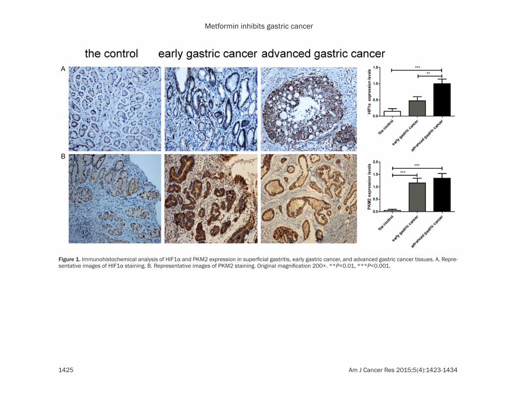
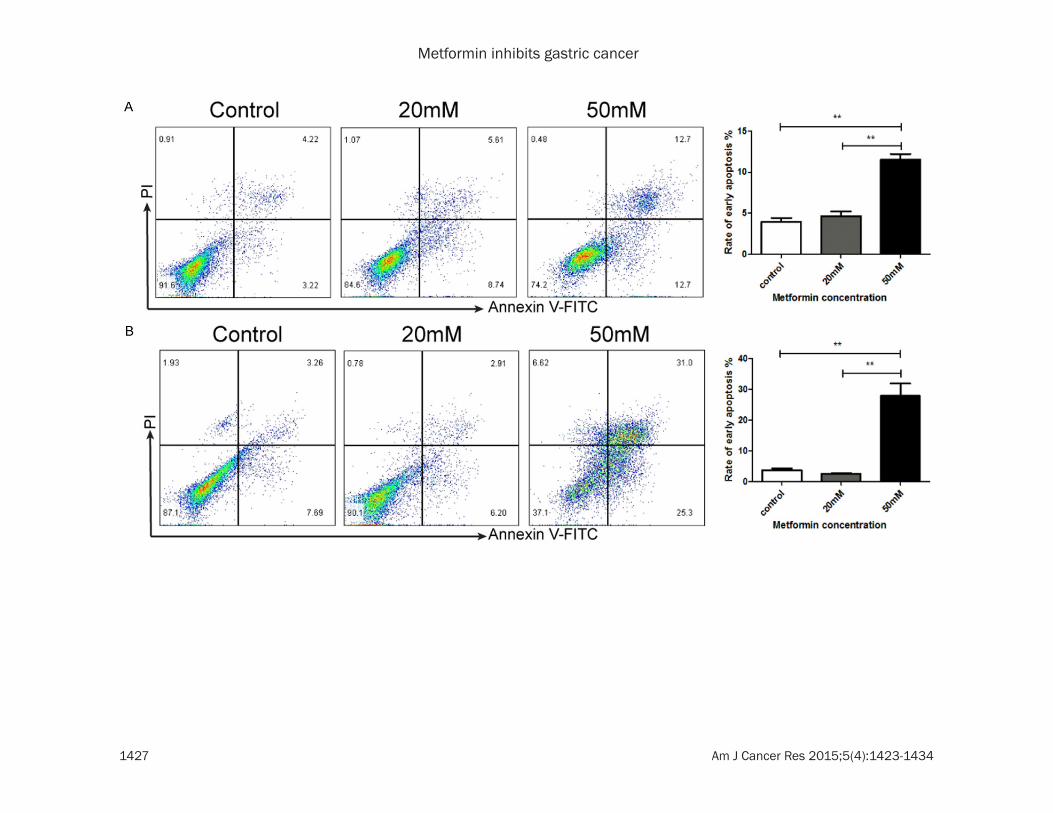
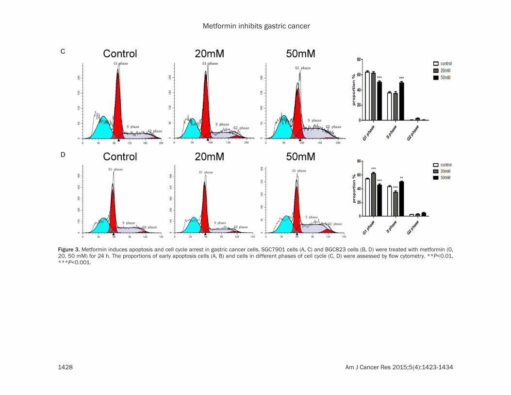
Figure 3. Metformin induces apoptosis and cell cycle arrest in gastric cancer cells. SGC7901 cells (A, C) and BGC823 cells (B, D) were treated with metformin (0, 20, 50 mM) for 24 h. The proportions of early apoptosis cells (A, B) and cells in different phases of cell cycle (C, D) were assessed by flow cytometry. **P<0.01, ***P<0.001.

Metformin inhibits gastric cancer
1429 Am J Cancer Res 2015;5(4):1423-1434
superficial gastritis and early GC tissues (Figure 1A). The expression level of PKM2 increased significantly in early and advanced GC com-pared with superficial gastritis tissue (Figure 1B).
Metformin decreases GC cell viability
We evaluated the effect of metformin on the viability of two GC cell lines: SGC7901 and BGC823. CCK-8 assay showed significant dose- and time-dependent decrease in the via-bility of SGC7901 and BGC823 cells after met-formin treatment (Figure 2).
Metformin induces apoptosis and cell cycle arrest in GC cells
To elucidate the mechanism by which metfor-min decreases the viability of GC cells, we won-dered whether metformin could induce apopto-sis and cell cycle arrest in GC cells. By Annexin V-FITC and PI staining, we observed that met-formin increased the proportion of apoptotic cells in SGC7901 and BGC823 cells in a dose dependent manner (Figure 3A, 3B). In addi-tion, by flow cytometry analysis, we found that metformin induced cell cycle arrest in SGC7901
Figure 4. Metformin reduces HIF1α, PARP and COX protein expression in gastric cancer cells. (A, B) SGC7901 cells were treated with metformin (0, 40 mM) for 24 h and then analyzed for the expression of HIF1α (A) and COX (B) by immuno-fluorescence. Original magnification 400×. (C) SGC7901 and BGC823 cells were treated with metformin (0, 40, 50 mM) for 24 h and then pro-tein expression of PI3K, Akt, HIF1α, PARP, COX, PKM2 was detected by Western blot analysis. β-actin was loading control.

Metformin inhibits gastric cancer
1430 Am J Cancer Res 2015;5(4):1423-1434
Metformin inhibits gastric cancer
1431 Am J Cancer Res 2015;5(4):1423-1434
and BGC823 cells in a dose dependent manner (Figure 3C, 3D).
Metformin reduces HIF1α, PARP, COX and PKM2 expression in GC cells
Next we evaluated the effects of metformin on PI3k, Akt, HIF1α, PARP, COX, and PKM2 protein
expression in GC cells. Immunofluorescence staining of HIF1α and COX in SGC7901 cells showed significant decrease in HIF1α and COX expression after metformin treatment (Figure 4A, 4B). Western blot analysis showed that metformin inhibited the expression level of PI3K, Akt, HIF1α, PARP, COX and PKM2 in SGC7901 and BGC823 cells (Figure 4C).
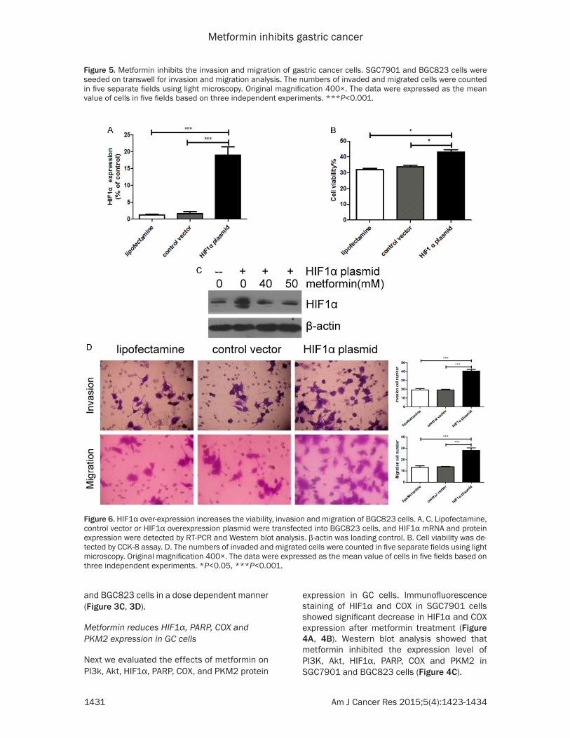
Figure 5. Metformin inhibits the invasion and migration of gastric cancer cells. SGC7901 and BGC823 cells were seeded on transwell for invasion and migration analysis. The numbers of invaded and migrated cells were counted in five separate fields using light microscopy. Original magnification 400×. The data were expressed as the mean value of cells in five fields based on three independent experiments. ***P<0.001.
Figure 6. HIF1α over-expression increases the viability, invasion and migration of BGC823 cells. A, C. Lipofectamine, control vector or HIF1α overexpression plasmid were transfected into BGC823 cells, and HIF1α mRNA and protein expression were detected by RT-PCR and Western blot analysis. β-actin was loading control. B. Cell viability was de-tected by CCK-8 assay. D. The numbers of invaded and migrated cells were counted in five separate fields using light microscopy. Original magnification 400×. The data were expressed as the mean value of cells in five fields based on three independent experiments. *P<0.05, ***P<0.001.

Metformin inhibits gastric cancer
1432 Am J Cancer Res 2015;5(4):1423-1434
Metformin inhibits the invasion and migration of GC cells
To investigate the activity of metformin against tumor metastasis, we examined the effects of metformin on the invasion and migration of GC cells. Transwell assay showed that metformin inhibited the invasion and migration of SGC- 7901 and BGC823 cells in a dose dependent manner (Figure 5).
HIF1α over-expression increases the viability, invasion and migration of GC cells
Since metformin inhibited protein expression of HIF1α in GC cells, we wondered whether HIF1α might be the key factor to mediate the effects of metformin on GC cells. We transfected HIF- 1α over-expression plasmid into BGC823 cells, and confirmed the expression of HIF1α by RT-PCR and Western blot analysis (Figure 6A, 6C). We found that HIF1 overexpression incr- eased the viability, invasion and migration of BGC823 cells (Figure 6B, 6D).
Discussion
In this study, we revealed the high expression of HIF1α and PKM2 in GC tissues, and found that metformin significantly induced apoptosis, inhibited cell invasion and migration of GC cells. The mechanism by which metformin exhibits anti-tumor activities is through the induction of apoptosis and the inhibition of HIF1α.
Recent studies showed that overexpression of HIF1α are implicated in tumorigenesis, tumor chemotherapy resistance, tumor angiogenesis, and tumor glycolysis [17-20]. Increased HIF-1α level is associated with increased risk of mor-tality in many human cancers, including gastric cancer [11]. HIF1α inhibitor inhibited tumor growth and angiogenesis [12]. In this study we found that metformin inhibited the expression of HIF1α in GC cells, suggesting that metformin may inhibit tumor cell growth and metastasis via HIF1α inhibition. However, further studies are needed to confirm our conclusion.
Targeting of tumor metabolism is emerging as a novel therapeutic strategy against cancer [21]. According to the “Warburg effect”, tumor cells exhibit an increased dependence on glycolytic pathway for ATP generation both in normoxia or hypoxia conditions [22]. PKM2 is an important executor downstream of HIF1α signaling and acts as the key enzyme of glycolysis [23]. In this
study we found high expression of PKM2 in GC tissues by immunohistochemistry, indicating the important role of glycolysis in the develop-ment of GC. Our study showed that metformin inhibited the expression of PKM2 protein, espe-cially in poorly differentiated BGC823 cells. These data suggest that metformin reduces the energy supply of GC by inhibiting HIF1α/PKM2 pathway.
The most important function of mitochondrial respiratory chain is to generate ATP by oxida-tion phosphorylation (OXPHOS). After sequen-tial electron transfer, two respiratory chains generate ATP through being catalyzed by the respiratory chain enzyme complexes IV- cyto-chrome C oxidase (COX). In energy-rich condi-tions, the mitochondria of tumor cells maintain “well-being” state and effectively shut off apop-totic machinery, resulting in the protection against cell death, even when challenged with toxic drugs. Conversely, when the mitochondria of tumor cells are in the condition of “stress”, they induce the apoptosis of tumor cells [24]. One study showed recently that metformin inhibited mitochondrial complex I of cancer cells to reduce tumorigenesis [25]. In this study we found that metformin inhibited the expres-sion level of COX in SGC7901 and BGC823 cells.
Poly(ADP)-ribose polymerase (PARP) plays a crucial role in DNA repair and the maintenance of genome stability. The proteolytic degrada-tion of PARP is caused by a variety of stimuli [26]. In the present study, the expression of PARP was decreased significantly in GC cells treated with metformin. At the same time, cell apoptosis ratio increased remarkably.
In order to confirm that HIF1α mediates the effects of metformin on GC cell proliferation, apoptosis, invasion, and migration, we trans-fected HIF1α overexpression plasmid into BGC823 cells; and found that cell viability, inva-sion and migration were obviously enhanced in the cells transfected with HIF1α plasmid. These data indicate that metformin inhibits GC cell proliferation, invasion and metastasis by inhib-iting the expression of HIF1α.
To the best of our knowledge, this is the first report demonstrating HIF1α/PKM2 signal path-way as a target of metformin in GC cells. Met- formin exhibit potent effects to inhibit malig-nant behaviors of GC cells through decreasing
Metformin inhibits gastric cancer
1433 Am J Cancer Res 2015;5(4):1423-1434
the expression of HIF1α and PKM2. However, how metformin inhibits HIF1α/PKM2 signal pathway is not clear and needs further explo- ration.
In conclusion, our study provides evidence that metformin inhibits GC growth and metastasis. The main mechanism responsible for the anti-tumor effects of metformin might be inducing intrinsic apoptosis and tumor glucose metabo-lism via the inhibition of HIF1α. These findings suggest that metformin is a promising thera-peutic agent for GC.
Acknowledgements
This study was supported by The National Natural Science Foundation of China (No. 81101814, 81272742, 81472756), Jiangsu Provincial Commission of Health and Family Planning (No. Q201413), Medical Youth Talent Reserve of Xuzhou, Xuzhou Science and Technology Plan (No. KC14SH007).
Disclosure of conflict of interest
None.
Address correspondence to: Xiaoping Zou, Depar- tment of Gastroenterology, Affiliated Drum Tower Clinical Medical School, Nanjing Medical University, Nanjing, China. Tel: 86-25-83304616; E-mail: [email protected]
References
[1] Jemal A, Bray F, Center MM, Ferlay J, Ward E, Forman D. Global cancer statistics. CA Cancer J Clin 2011; 61: 69-90.
[2] Parkin DM, Bray F, Ferlay J, Pisani P. Global cancer statistics, 2002. CA Cancer J Clin 2005; 55: 74-108.
[3] Macintyre AN, Rathmell JC. Activated lympho-cytes as a metabolic model for carcinogenesis. Cancer Metab 2013; 23: 1-5.
[4] Kumar A, Kant S, Singh SM. Antitumor and chemosensitizing action of dichloroacetate im-plicates modulation of tumor microenviron-ment: a role of reorganized glucose metabo-lism, cell survival regulation and macrophage differentiation. Toxicol Appl Pharmacol 2013; 273: 196-208.
[5] Tavares-Valente D, Baltazar F, Moreira R, Qu- eirós O. Cancer cell bioenergetics and pH regu-lation influence breast cancer cell resistance to paclitaxel and doxorubicin. J Bioenerg Bio- membr 2013; 45: 467-475.
[6] Brauer HA, Makowski L, Hoadley KA, Casbas-Hernandez P, Lang LJ, Romàn-Pèrez E, D’Arcy
M, Freemerman AJ, Perou CM, Troester MA. Impact of tumor microenvironment and epithe-lial phenotypes on metabolism in breast can-cer. Clin Cancer Res 2013; 19: 571-585.
[7] Carito V, Bonuccelli G, Martinez-Outschoorn UE, Whitaker-Menezes D, Caroleo MC, Cione E, Howell A, Pestell RG, Lisanti MP, Sotgia F. Metabolic remodeling of the tumor microenvi-ronment: migration stimulating factor (MSF) reprograms myofibroblasts toward lactate pro-duction, fueling anabolic tumor growth. Cell Cycle 2012; 11: 3403-3414.
[8] Kumar V, Gabrilovich DI. Hypoxia inducible fac-tors in regulation of immune responses in tu-mor microenvironment. Immunology 2014; 143: 512-519.
[9] Ohashi T, Akazawa T, Aoki M, Kuze B, Mizuta K, Ito Y, Inoue N. Dichloroacetate improves immu-ne dysfunction caused by tumor-secreted lac-tic acid and increases antitumor immunoreac-tivity. Int J Cancer 2013; 133: 1107-1118.
[10] Meijer TW, Kaanders JH, Span PN, Bussink J. Targeting hypoxia, HIF-1, and tumor glucose metabolism to improve radiotherapy efficacy. Clin Cancer Res 2012; 18: 5585-5594.
[11] Semenza GL. HIF1 mediates metabolic resp- onses to intratumoral hypoxia and oncogenic mutations. J Clin Invest 2013; 123: 3664-3671.
[12] Yu GT, Bu LL, Zhao YY, Liu B, Zhang WF, Zhao YF, Zhang L, Sun ZJ. Inhibition of mTOR reduce Stat3 and PAI related angiogenesis in salivary gland adenoid cystic carcinoma. Am J Cancer Res 2014; 4: 764-775.
[13] Liu Z, Jia X, Duan Y, Xiao H, Sundqvist KG, Permert J, Wang F. Excess glucose induces hy-poxia-inducible factor-1α in pancreatic cancer cells and stimulates glucose metabolism and cell migration. Cancer Biol Ther 2013; 14: 428-435.
[14] McFarland MS, Cripps R. Diabetes mellitus and increased risk of cancer: focus on metfor-min and the insulin analogs. Pharmacotherapy 2010; 30: 1159-1178.
[15] Tseng CH. Metformin reduces thyroid cancer risk in taiwanese patients with type 2 diabetes. PLoS One 2014; 9: e109852.
[16] Franciosi M, Lucisano G, Lapice E, Strippoli GF, Pellegrini F, Nicolucci A.Metformin therapy and risk of cancer in patients with type 2 diabetes: systematic review. PLoS One 2013; 2: e71583.
[17] Goscinski MA, Nesland JM, Giercksky KE, Dhakal HP. Primary tumor vascularity in eso-phagus cancer - CD34 and HIF1-α expression correlate with tumor progression. Histol His-topathol 2013; 28: 1361-1368.
[18] Tong Y, Li QG, Xing TY, Zhang M, Zhang JJ, Xia Q. HIF1 regulates WSB-1 expression to pro-mote hypoxia-induced chemoresistance in he-
Metformin inhibits gastric cancer
1434 Am J Cancer Res 2015;5(4):1423-1434
patocellular carcinoma cells. FEBS Lett 2013; 587: 2530-2535.
[19] Liu R, Li Z, Bai S, Zhang H, Tang M, Lei Y, Chen L, Liang S, Zhao YL, Wei Y, Huang C. Mechanism of cancer cell adaptation to metabolic stress: proteomics identification of a novel thyroid hormone-mediated gastric carcinogenic sig-naling pathway. Mol Cell Proteomics 2009; 8: 70-85.
[20] Semenza GL. HIF1: upstream and downstream of cancer metabolism. Curr Opin Genet Dev 2010; 20: 51-56.
[21] Kumar A, Kant S, Singh SM. Antitumor and chemosensitizing action of dichloroacetate im-plicates modulation of tumor microenviron-ment: a role of reorganized glucose metabo-lism, cell survival regulation and macrophage differentiation. Toxicol Appl Pharmacol 2013; 273: 196-208.
[22] Warburg O. On the origin of cancer cells. Science 1956; 123: 309-314.
[23] Chaneton B, Gottlieb E. Rocki ng cell metabo-lism: revised functions of the key glycolytic regulator PKM2 in cancer. Trends Biochem Sci 2012; 37: 309-316.
[24] Martinez-Outschoorn UE, Pestell RG, Howell A, Tykocinski ML, Nagajyothi F, Machado FS, Tanowitz HB, Sotgia F, Lisanti MP. Energy trans-fer in “parasitic” cancer metabolism: Mito- chondria are the powerhouse and Achilles’ heel of tumor cells. Cell Cycle 2011; 10: 4208-4216.
[25] Wheaton WW, Weinberg SE, Hamanaka RB, Soberanes S, Sullivan LB, Anso E, Glasauer A, Dufour E, Mutlu GM, Budigner GS, Chandel NS. Metformin inhibits mitochondrial complex I of cancer cells to reduce tumorigenesis. Elife 2014; 3: e02242.
[26] Ibrahim MY, Hashim NM, Mohan S, Abdulla MA, Kamalidehghan B, Ghaderian M, Dehghan F, Ali LZ, Arbab IA, Yahayu M, Lian GE, Ahm- adipour F, Ali HM. α-Mangostin from Cratoxylum arborescens demonstrates apoptogenesis in MCF-7 with regulation of NF-κB and Hsp70 protein modulation in vitro, and tumor reduc-tion in vivo. Drug Des Devel Ther 2014; 8: 1629-1647.