Embed Size (px)
Citation preview

Int J Clin Exp Med 2018;11(8):7927-7935www.ijcem.com /ISSN:1940-5901/IJCEM0063589
Original ArticleHydrogen mitigates acute lung injury through upregulation of M2 and downregulation of M1 macrophage phenotypes
Jing Zhou1, Peng Yan2, Xi-Dong Zhu3, Kai-Jiang Yu4
1Department of ICU, The Second Affiliated Hospital of Harbin Medical University, Harbin 150086, China; 2Depart-ment of Colorectal Surgery, The Second Affiliated Hospital of Harbin Medical University, Harbin 150086, China; 3Department of Anesthesiology, The Third Affiliated Hospital of Harbin Medical University, 150 Haping Street, Harbin 150081, China; 4Department of ICU, The Third Affiliated Hospital of Harbin Medical University, Harbin 150081, China
Received August 13, 2017; Accepted January 25, 2018; Epub August 15, 2018; Published August 30, 2018
Abstract: Objectives: Acute lung injury (ALI) is still a leading cause of morbidity and mortality in critically ill patients. ALI can be induced by sepsis, ventilator, hyperoxia, and ischemia-reperfusion. In the management of ALI, studies have shown that low concentration hydrogen has a therapeutic effect on acute lung injury. The present study was designed to investigate the effects and corresponding mechanism of the inhalation of hydrogen on ALI. Methods: C57 male mice underwent intraperitoneal injection of lipopolysccharide (LPS) to induce sepsis. Then, the mice were given 2% hydrogen. The survival rate, lung injury, inflammatory factors and macrophage phenotypic changes were assessed in bronchoalveolar lavage fluid (BALF). In vitro experiment, we obtained primary mouse bone mar-row-derived macrophages (BMDM) incubated by LPS to explore the anti-inflammatory effects and corresponding mechanism. Results: Treatment with 2% hydrogen showed a substantial attenuation of inflammations, adverse lung histopathological changes and decreased M1 macrophage phenotypes while increased M2 macrophage pheno-types. Importantly, the decreased M1 macrophage phenotypes while increased M2 macrophage phenotypes were observed in LPS-stimulated macrophages treated with 2% hydrogen. Further, we observed significantly low levels of TNFα in LPS-induced macrophages treated with 2% hydrogen. The effects were ascribed to an inhibition of phos-phorylation of p38 MAPK. Conclusion: 2% hydrogen may reduce sepsis-induced inflammatory responses in animals and in macrophages, and the inhibition to the activation of the MAPK/TNF-α may contribute to this protection.
Keywords: Acute lung injury, molecular hydrogen, p38 MAPK, bone marrow-derived macrophages
Introduction
Acute lung injury (ALI) is a common clinical criti-cal illness and the most common organ tissue damage induced by systemic inflammatory response syndrome, a severe systemic non-specific response to a condition (as trauma, an infection, or a burn) that provokes an acute inflammatory reaction [1]. Although the treat-ment has made great progress, the mortality rate is as high as 35%~40% [2]. Even there is good approach in the pathophysiology of ALI; there is still no effective treatment for ALI. In spite of involvement of different mechanisms in the pathogenesis of ALI, inflammation is one of the leading causes. ALI is result of the extreme inflammatory process characterized by exten-sive neutrophil influx into the lungs, the expres-
sion of pro-inflammatory mediators, and lung epithelium and endothelium damages, which results in respiratory failure. Therefore, inhibit-ing the inflammatory cascades result in good therapeutic approach for ALI. In addition, ALI is associated with the continuous macrophage-associated production of reactive oxygen/nitro-gen species (ROS/RNS), leading to lungs tissue damage. Molecular hydrogen is a good anti-inflammatory, antioxidant and antiapoptotic agent that reduces ROS level and suppresses lungs tissue damage.
LPS is a component of the cell wall of gram-negative bacteria, which can trigger a cascade of reactions by activating various effectors cells (such as neutrophils), cytokine networks and the expression of pro-inflammatory genes, lead-

Hydrogen mitigates acute lung injury
7928 Int J Clin Exp Med 2018;11(8):7927-7935
ing to the occurrence of ALI. Upon activation of defense mechanism of body toward the for-eign body (bacteria, virus), various defending cells (macrophage) release early response cyto-kines such as type I IFN, TNF-α, and IL-1β in an IRF- or NF-κB-dependent way. In ALI, TNF-alpha is considered to be the earliest and most impor-tant endogenous medium in inflammatory res- ponse. TNF-alpha can induce various inflamma-tory cells and epithelial cells in the lungs to pro-duce multiple cytokines, mediate inflammatory cells infiltrating and produce lung tissue dam-age [3]. An inducible transcription factor p38 MAPK is a driving force in the initiation and progression of systemic inflammation and sep-tic pathophysiology [4]. P38 MAPK is an impor-tant component of the MAPK family. Studies have shown that when LPS induced ALI, p38 MAPK was activated, causing the secretion of TNF-α [5]. Hence, the regulation of inflammato-ry response through the p38 MAPK signaling pathway has been a focus of studies.
Macrophages, as a group of plasticity and plu-ripotent cells, show distinct functional differ-ences in vivo and in vitro under different mic- ro environments. Macrophages also maintain airway homeostasis, and play a pivotal role in the pathogenesis of ALI [6]. According to differ-ent functions, macrophages can be divided into M1 type macrophages that classically ac- tivated macrophages, and M2 type of alterna-tively activated macrophages [7]. The M1 pro-gram is associated with release of pro-infl- ammatory mediators such as iNOS-derived NO, TNF-α, IFN-γ, and IL-12 and critically contrib-utes to pathogen elimination. In contrast, M2, which secrete anti-inflammatory cytokines like IL-1ra, IL-10, and TGF-β, downregulate IL-12, upregulate scavenger receptors, promote ang- iogenesis, and support wound healing and tis-sue remodeling. M2 macrophages prove bene-ficial in arresting the inflammatory cytokine response in sepsis [7].
Hydrogen is a kind of important physiological regulatory factors in the body and can selec-tively lower the hydroxyl free radicals and re- active oxygen species peroxynitrite anion [8], which has anti-inflammatory, antioxidant and antiapoptotic potential [9, 10]. Previous study showed that saturated hydrogen saline treat-ment improved the damage to pulmonary epi-thelial cells in rats with sepsis by increasing the expression of aquaporin-1 protein [11].
Furthermore, it showed that saturated hydro-gen saline may downregulate expression of Beclin-1 transcription and inhibits autophagy through heme oxygenase-1 and p38 MAPK sig-naling [12].
In the present study, we applied lipopolysac-charide (LPS) intraperitoneal injection to es- tablish the ALI model of mice and explored the effects of inhalation of low concentration of hydrogen on protection of ALI and the possi-ble mechanism. The study will further provide theoretical basis for the hydrogen treatment of ALI.
Materials and methods
Animal models of ALI
The experimental animal protocols were app- roved by the Institutional Animal Care and Use Committee of the Second Affiliated Hospital of Harbin Medical University. All experiments were performed according to the experimental ani-mal guidelines. Male C57 mice (20-25 g) were obtained from the Experimental Animal Center of the the Second Affiliated Hospital of Harbin Medical University. Endotoxin-induced ALI ani-mal models were established by intraperitoneal injection of LPS (50 mg/kg, Sigma Chemical, St. Louis, MO, USA). The same volume of saline was given to animals in the control group.
Inhalation of hydrogen
Animals were placed in a sealed plexi glass chamber with an inflow and outflow hose. 2% Hydrogen with mixed air was delivered to the chamber through a tube, and carbon dioxide was removed from the chamber gases with baralyme. A gas analyzer (Bruel and Kjaer, Naerum, Denmark) was used to continuously monitor the concentration of hydrogen in the outflow hose of the chamber, which was main-tained at a predetermined level during treat-ment. The animals were given two hours of inhalation of 2% hydrogen each time.
Cytokines in bronchoalveolar lavage fluid (BALF)
BALF was obtained by cannulating the trachea of mice and lavaging with 1 ml phosphate buf-fer saline (pH 7.4). Lavage samples were centri-fuged 3000 rpm at 4°C for 10 minutes, and the supernatant was stored at -80°C. The cyto-

Hydrogen mitigates acute lung injury
7929 Int J Clin Exp Med 2018;11(8):7927-7935
kines in BALF were detected using specific IL1β, TNF-α, and IL-10 ELISA kits (R&D Systems Inc., Minneapolis, Minnesota, USA), according to manufacturer’s instructions. All standards and samples were run in triplicate.
Lung myeloperoxidase (MPO) activity
MPO activity, an indicator of neutrophil infiltra-tion in lung tissues, was detected in homoge-nated lung supernatants, as previously report-ed using an MPO Assay Kit (Nanjing Jian Ch- eng Bioengineering Institute, Nanjing, China), according to manufacturer’s instructions.
Immunohistochemistry
Following PBS perfusion, the lung tissues were fixed for 1 hour by instillation of 10% PBS-buffered formalin through the trachea. Then the right lungs were taken out and fixed with 10% PBS-buffered formalin overnight at 4°C. After the paraffin embedding process, the tis-sues were sectioned in 5-μm-thick sections and the lung epithelial and endothelial bio-marker E-cadherin (1:300, Sigma-Aldrich, St. Louis, MO, USA) antibodies at 4°C overnight. Then, the sections were incubated with biotinyl-ated goat anti-mouse IgG for 1 h. The signal was detected with 3, 3’-diaminobenzidine.
Cell culture
Bone marrow derived mononuclear cells (BM- DM) were isolated from the tibias and femurs
The protein from BMDM samples was directly extracted based on manufacturer’s standard protocols (Beyotime Biotechnology, Shanghai, China). Primary rabbit antibodies for phosphor-p38 and total p38 (Cell Signaling Technology, Boston, USA) proteins were used to detect p38 protein expression. Then, the primary rab-bit antibodies forglyceraldehyde 3-phosphate dehydrogenase (GAPDH) was used as control.
Immunofluorescence analysis
For immunofluorescence assays, cultured BM- DM cells or lung tissue were rinsed once with PBS and fixed with 4% paraformaldehyde in PBS for 30 minutes, then rinsed three times with PBS, permeabilized with 0.3% Triton X-100 (Sigma) in PBS for 5 minutes, washed twice with PBS. After processing with blocking solu-tion for one hour at room temperature, cells were incubated with anti-CD68, and anti-Argi-nase 1 antibody overnight at 4°C. The slides were washed three times with PBS and incu-bated with fluorescein-conjugated secondary antibody for one hour at room temperature. DAPI staining was used for the counterstain- ing of the nucleus. Fluorescent images of the coverslips were obtained by either confocal and immunofluorescence microscope (Olymp- us, Tokyo, Japan).
Flow cytometer analysis
For flow cytometer phenotypic analysis, cells (1 × 10⁶ cells/well) were initially incubated with
Figure 1. Two hours of inhalation of 2% hydrogen improved sepsis-induced lethality. Sepsis was induced by intraperitoneal injection of LPS. A. Protective effects of 2% at 4 h after LPS administration on LPS-induced sepsis in mice. **P < 0.01 vs. Control and ##P < 0.0 vs. LPS. B. Optimum time for protective effects of 2% hydrogen on LPS-induced sepsis in mice. 2% hydrogen was treated at different time point after LPS administration. **P < 0.01 vs. time points 0, 4, 8 and 12 h.
of mice and cultured in Dulbecco’s modified Eagle’s medium along with 10% L929 cell conditioned with supple-mented 10% heat-inactivat- ed fetal calf serum (FBS)/ 1% penicillin and streptomy-cin. The incubator was set at 37°C with a humidified atmo-sphere containing 5% CO2. After 7 days of culture, the cells were stained with CD11b and analyzed by flow cytome-try analysis. The phase con-trast microscopic image was taken to show the uniform morphology.
Western blotting

Hydrogen mitigates acute lung injury
7930 Int J Clin Exp Med 2018;11(8):7927-7935
10% mouse serum for 20 minutes at 4°C. Subsequently, cells were incubated with the appropriately labeled primary antibodies for 1 hr. Anti-CD23 and anti-CD124 were used for quantifying M2 and anti-CD14 and anti-CD40 were used for quantifying M1 subtype. Then the cells were washed with washing buffer three times and incubated 20 minutes in an appropri-ate secondary antibody. All incubations were performed on ice. Appropriate isotype controls were used in all cases. Finally, the cells were washed three times with FACS buffer, resus-pended in 0.5 ml PBS, and analyzed by flow cytometer (FACSCalibur, BD Biosciences) using Cell Quest software.
Statistical analysis
The measurement data are expressed as me- an ± standard error of the mean (SEM). Intergroup differences in the levels of bioch- emical parameters, inflammatory cytokines, and the number of neutrophils were tested by one-way ANOVA, followed by the least sig- nificant difference test for multiple compari-sons. Survival studies were analyzed using K-M analysis. Survival rates were expressed in percentage. The statistical analysis was per-formed using the SPSS 17.0 software (SPSS Inc, Chicago, USA). In all tests, P < 0.05 was considered statistically significant.
Results
The inhalation of 2% hydrogen protected against sepsis-induced lethality
LPS is the major component of the outer mem-brane of Gram-negative bacteria, which are considered as one of the predominant compo-nent that causes sepsis. We found all mice in the normal control group survived. In the model group, the mice died from 12 h and the letha- lity reached maximum at 48 h. A significant improvement of the survival rate was observed in mice treated with two hours of 2% hydrogen at 4h after LPS administration (P < 0.01) (Figure 1A). It was also observed that treatment with 2% hydrogen significantly increased the surviv-al rates of animals with LPS-induced sepsis only when it was performed at 0, 4, 8 and 12 hours after LPS injection (P < 0.05) (Figure 1A). The results suggest that 2% hydrogen protects against lethality resulting from sepsis induced by LPS.
Inhalation of 2% hydrogen inhibited inflamma-tory responses
ALI, which is typically observed in individuals with sepsis, is an inflammatory disease. Dys- regulated inflammatory response in lungs, as well as the altered permeability of alveolar
Figure 2. The 2% hydrogen at 4 h after LPS administration mitigated lung injury resulting from LPS-induced sepsis in mice. A: The production of IL1β, TNF-α, and IL-10 and MPO activity in BALF at 24 h after LPS administration (n=5). B: Morphological changes of lungs were assayed by immunohistochemistry. **P < 0.01 vs. control group; ##P < 0.01 vs. model group.

Hydrogen mitigates acute lung injury
7931 Int J Clin Exp Med 2018;11(8):7927-7935
endothelial and epithelial barriers, remains as central pathophysiologic concepts in ALI and acute respiratory distress syndrome (ARDS) [13]. In this study, we found, in LPS-challenged mice, the treatment of 2% hydrogen at 4 h af- ter LPS administration significantly attenuated lung MPO activity (P < 0.01, Figure 2A), and reduced the production of TNF-α (P < 0.01, Figure 2A) and IL1β in BALF (P < 0.05, Figure 2A), but promoted the production of IL-10 (P < 0.01, Figure 2A). Furthermore, it was also observed that 2% hydrogen s at 4 h after LPS administration significantly decreased the le- vel of TNF-α, IL1β and promoted IL-10 in serum (P < 0.05, Figure 2A). Additionally, it was also found that LPS induced infiltration of inflam- matory cells in bronchial lumen, alveolar cavity and pulmonary interstitial and the treatment of 2% hydrogen at 4 h after LPS administra- tion alleviated LPS-induced lung inflammation (Figure 2B). The above results demonstrate that treatments with 2% hydrogen attenuate lung injury resulting from LPS-induced sepsis by inhibiting lung inflammation, and protecting alveolar endothelial and epithelial barriers.
2% hydrogen increased M2 macrophages in lung tissues and in BMDM after LPS stimula-tion
Immunofluorescence staining was performed to determine the immune-plasticity of the LPS-
challenged ALI mice after treatment with 2% hydrogen. Immunofluorescence staining of ma- crophage markers CD68 (pan marker, green) and Arg1 (M2 specific marker-red) showed con-sistently increased LPS-induced co-localization of M2 macrophages (yellow fluorescence in the merged image) in ALI mice (Figure 3) with 2% hydrogen treatment.
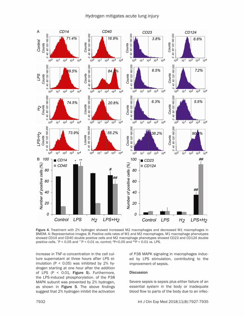
Next, BMDM were isolated from the tibias and femurs of mice and treated with LPS (1 μg/ml) for 1 hour. M1 macrophage phenotypes CD- 14 CD40 double positive cells decreased while M2 macrophage phenotypes CD23 CD124 double positive cells were observed more in BMDM that treated with 2% hydrogen than LPS-treated BMDM (Figure 4). In vivo and vitro, these data indicated that the 2% hydrogen treatment modulates the lung macrophages towards anti-inflammatory M2 subtype.
2% hydrogen inhibited the activation of the p38 pathway in BMDM after LPS stimulation
P38 MAPK is a key regulator of inducible gene expression in the immune system, and is cru-cial to the signaling networks involved in sep-sis. Previous studies have suggested that in- creasing P38 MAPK activity is associated with the development and mortality of sepsis [14]. Treatment was given on cells by 2% hydrogen starting at the addition of LPS. A significant
Figure 3. Treatment with 2% hydrogen showed increased M2 macrophages in ALI mice lung tissues.
Hydrogen mitigates acute lung injury
7932 Int J Clin Exp Med 2018;11(8):7927-7935
increase in TNF-α concentration in the cell cul-ture supernatant at three hours after LPS st- imulation (P < 0.05) was inhibited by 2% hy- drogen starting at one hour after the addition of LPS (P < 0.01, Figure 5). Furthermore, the LPS-induced phosphorylation. of the P38 MAPK subunit was prevented by 2% hydrogen, as shown in Figure 5. The above findings suggest that 2% hydrogen inhibit the activation
of P38 MAPK signaling in macrophages induc- ed by LPS stimulation, contributing to the improvement of sepsis.
Discussion
Severe sepsis is sepsis plus either failure of an essential system in the body or inadequate blood flow to parts of the body due to an infec-
Figure 4. Treatment with 2% hydrogen showed increased M2 macrophages and decreased M1 macrophages in BMDM. A: Representative images; B: Positive cells rates of M1 and M2 macrophages. M1 macrophage phenotypes showed CD14 and CD40 double positive cells and M2 macrophage phenotypes showed CD23 and CD124 double positive cells. *P < 0.05 and **P < 0.01 vs. control; #P<0.05 and ##P < 0.01 vs. LPS.

Hydrogen mitigates acute lung injury
7933 Int J Clin Exp Med 2018;11(8):7927-7935
tion, which often results in significantly poor outcomes and mortality. Sepsis occurs when toxins produced by certain bacteria cause cells in the body to release substances that trigger inflammation. Acute lung injury (ALI) secondary to sepsis is a complex syndrome associated with high morbidity and mortality. Nearly 50% of patients with severe sepsis will develop ALI, and in its more severe form, acute respiratory distress syndrome. The respiratory system is the most frequently affected organ system. Diffuse inflammation of lung parenchyma and severe lung dysfunctions are the first steps in the development of multiple organ failure and one of the leading causes of death in sepsis. In the present study, ALI occurred to animals with LPS-induced sepsis characterized by increased lung inflammation and impaired alveolar endo-thelial and epithelial barriers. These were sig-nificantly improved with the treatment of 2% hydrogen. In addition, the inhalation of 2% hydrogen improved the survival rate in animals with sepsis. The above statements further sup-port the protective action against sepsis by 2% hydrogen.
Inflammation is a beneficial response of the body for effective host defense. However, excessive inflammatory responses can cause tissue damage, fibrosis and eventual organ fail-ure. To control the excessive inflammation, nature has developed different control mecha-nism in organisms, such as anti-inflammatory cytokines and antioxidant agents. Based on lit-erature, a dysregulated, excessive proinflam-
matory cytokine expression contributes to the pathogenesis of sepsis [15]. In the present study, cytokine expression in BALF (IL-1β and TNF-α) were abnormally increased to animals with LPS-induced sepsis; which were inhibited by 2% hydrogen. In addition, 2% hydrogen pro-moted the increase of IL-10 in BALF from ani-mals with LPS-induced sepsis, which was con-sidered to be an anti-inflammatory factor [16]. These results suggest that 2% hydrogen pro-tect sepsis and its consequent injury may be through the reduction of inflammatory response in sepsis, thus, improving sepsis and sepsis-induced lung injury.
To understand the molecular changes that occur during hydrogen treatment, we evaluated the M1 and M2 macrophages in ALI animals and LPS-induced BMDMs. Increased M2 mac-rophages was noticed with treatment with 2% hydrogen in ALI mice lung tissues. Moreover, treatment with 2% hydrogen showed signifi- cant reduction in the expression of M1 macro-phage phenotypes (CD14 and CD40 double positive cells) and significant increment in the expression of M2 macrophage phenotypes (CD23 and CD124 double positive cells). These findings further support the previous observa-tions of a distinct epigenetic reprogramming being switched on in favor of an anti-inflamma-tory subtype of Macrophages.
P38 MAPK is activated by a variety of cellular stresses including osmotic shock, inflammato-ry cytokines, LPS, Ultraviolet light, and growth
Figure 5. The 2% hydrogen protected macro-phages against LPS-induced in-vitro sepsis through inhibiting activation of P38 MAPK. **P < 0.01 vs. control and ##P < 0.0 vs. LPS.

Hydrogen mitigates acute lung injury
7934 Int J Clin Exp Med 2018;11(8):7927-7935
factors. P38 MAPK is a key regulator of induc-ible gene expression in the immune system, and is crucial to the signaling networks involved in sepsis. Our results also revealed that treat-ments of 2% hydrogen protected LPS-induced in vitro sepsis by inhibiting p38 activation and expression, which plays a key role in regulating immune response to infection. Previous stud-ies have suggested that increasing P38 MAPK activity is associated with the development and mortality of sepsis [14]. The inhibition to p38 activation can reduce injury to organs resulting from sepsis, and improve survival for critically ill patients. These results indicate that the sup-pression of the hyperinflammatory phase in sepsis via the inhibition of the p38 signaling pathway contributes to the protective effects on sepsis through 2% hydrogen.
In summary, the administration of 2% hydrogen effectively protects against sepsis, demon-strating that 2% hydrogen is a safer, novel ther-apy for sepsis. Hence, our data provides a potential therapy for sepsis in clinic. We sug-gest that this protective mechanism was the inhibition of inflammation via the p38 MAPK/TNF signaling pathway. Our study contributes to a new therapeutic approach to improve sepsis and its relevant ALI, especially for the clinical treatment of sepsis.
Acknowledgements
The project was funded by National Natural Science Foundation of China (grant number: 81571871, Kai-Jiang Yu); The Second Affiliated Hospital of Harbin Medical University (grant number: KYBS2015-22, Jing Zhou).
Disclosure of conflict of interest
None.
Address correspondence to: Kai-Jiang Yu, Depart- ment of ICU, The Third Affiliated Hospital of Harbin Medical University, 150 Haping Street, Harbin 150081, China. Tel: 86-0451-86298999; E-mail: [email protected]
References
[1] Sharp C, Millar AB, Medford AR. Advances in understanding of the pathogenesis of acute respiratory distress syndrome. Respiration 2015; 89: 420-34.
[2] Villar J, Sulemanji D, Kacmarek RM. The acute respiratory distress syndrome: incidence and mortality, has it changed? Curr Opin Crit Care 2014; 20: 3-9.
[3] Togbe D, Schnyder-Candrian S, Schnyder B, Doz E, Noulin N, Janot L, Secher T, Gasse P, Lima C, Coelho FR, Vasseur V, Erard F, Ryffel B, Couillin I, Moser R. Toll-like receptor and tu-mour necrosis factor dependent endotoxin-in-duced acute lung injury. Int J Exp Pathol 2007; 88: 387-91.
[4] Qian F, Deng J, Wang G, Ye RD, Christman JW. Pivotal role of mitogen-activated protein ki-nase-activated protein kinase 2 in inflamma-tory pulmonary diseases. Curr Protein Pept Sci 2016; 17: 332-42.
[5] Le NP, Channabasappa S, Hossain M, Liu L, Singh B. Leukocyte-specific protein 1 regulates neutrophil recruitment in acute lung inflamma-tion. Am J Physiol Lung Cell Mol Physiol 2015; 309: L995-1008.
[6] Schneberger D, Aharonson-Raz K, Singh B. Pulmonary intravascular macrophages and lung health: what are we missing? Am J Physiol Lung Cell Mol Physiol 2012; 302: L498-503.
[7] Aggarwal NR, King LS, D’Alessio FR. Diverse macrophage populations mediate acute lung inflammation and resolution. Am J Physiol Lung Cell Mol Physiol 2014; 306: L709-25.
[8] Ohta S. Molecular hydrogen as a preventive and therapeutic medical gas: initiation, devel-opment and potential of hydrogen medicine. Pharmacol Ther 2014; 144: 1-11.
[9] Ishibashi T. Molecular hydrogen: new antioxi-dant and anti-inflammatory therapy for rheu-matoid arthritis and related diseases. Curr Pharm Des 2013; 19: 6375-81.
[10] Huang CS, Kawamura T, Peng X, Tochigi N, Shi-gemura N, Billiar TR, Nakao A, Toyoda Y. Hydro-gen inhalation reduced epithelial apoptosis in ventilator-induced lung injury via a mechanism involving nuclear factor-kappa B activation. Biochem Biophys Res Commun 2011; 408: 253-8.
[11] Tao B, Liu L, Wang N, Wang W, Jiang J, Zhang J. Effects of hydrogen-rich saline on aquaporin 1, 5 in septic rat lungs. J Surg Res 2016; 202: 291-8.
[12] Liu Y, Zhang J. Saturated hydrogen saline ame-liorates lipopolysaccharide-induced acute lung injury by reducing excessive autophagy. Exp Ther Med 2017; 13: 2609-2615.
[13] Sharp C, Millar AB, Medford AR. Advances in understanding of the pathogenesis of acute respiratory distress syndrome. Respiration 2015; 89: 420-34.
[14] Li L, Liu Y, Chen HZ, Li FW, Wu JF, Zhang HK, He JP, Xing YZ, Chen Y, Wang WJ, Tian XY, Li AZ, Zhang Q, Huang PQ, Han J, Lin T, Wu Q. Imped-
Hydrogen mitigates acute lung injury
7935 Int J Clin Exp Med 2018;11(8):7927-7935
ing the interaction between Nur77 and p38 reduces LPS-induced inflammation. Nat Chem Biol 2015; 11: 339-46.
[15] Hu D, Yang X, Xiang Y, Li H, Yan H, Zhou J, Cau-dle Y, Zhang X, Yin D. Inhibition of Toll-like re-ceptor 9 attenuates sepsis-induced mortality through suppressing excessive inflammatory response. Cell Immunol 2015; 295: 92-8.
[16] Peñaloza HF, Nieto PA, Muñoz-Durango N, Salazar-Echegarai FJ, Torres J, Parga MJ, Alva-rez-Lobos M, Riedel CA, Kalergis AM, Bueno SM. Interleukin-10 plays a key role in the mod-ulation of neutrophils recruitment and lung in-flammation during infection by Streptococcus pneumoniae. Immunology 2015; 146: 100-12.