Embed Size (px)
Citation preview

160
Jpn. J. Infect. Dis., 72, 160–167, 2019
Original Article
Clinical and Laboratory Findings of Middle East Respiratory Syndrome Coronavirus Infection
Se-Min Hwang1,2,3†, Baeg-Ju Na4†, Yoomi Jung5, Hyun-Suk Lim6, Jeong-Eun Seo6, Shin-Ae Park6, Young-Soo Cho6, Eun-Hyang Song6, Ji-Yeon Seo6, Sung-Ryul Kim6, Gang-Young Lee6,
Soon-Ja Kim6, Young-Suk Park6, and Haesook Seo3,6*1Korea Centers for Disease Control and Prevention, Cheongju; 2Department of Health Policy, Health & Welfare
Bureau, Sejong-si; 3Department of Preventive Medicine, Korea University College of Medicine, Seoul; 4Seoul Metropolitan Government Civil Health Bueau, Seoul; 5Korea Armed Forces Nursing Academy, Daejeon;
and 6Department of Tuberculosis, Seobuk Hospital, Seoul, Korea
INTRODUCTION
In September 2012, the Middle East respiratory syndrome coronavirus (MERS-CoV) was first reported in two patients in Saudi Arabia with severe pneumonia (1). Korean health officials reported the first imported case of MERS CoV in the country on May 20, 2015, which led to the largest transmission cluster of the disease worldwide, involving 186 human cases, 38 deaths, and 16,752 suspected cases (2). The clinical presentation and characteristics of MERS-CoV patients have been well-described (2–4). However, there is a paucity of data regarding the differences between the characteristics of suspected patients whose tests were subsequently found to be negative and those of laboratory-confirmed cases both in Saudi Arabia and South Korea (5). Moreover, the Korean outbreak was only human to human transmission (3), while the Saudi
Received May 28, 2018. Accepted December 10, 2018. J-STAGE Advance Publication December 25, 2018.DOI: 10.7883/yoken.JJID.2018.187* Corresponding author: Mailing address: Department of Preventive Medicine, Korea University College of Medicine, Seoul, Korea; and Department of Tuberculosis, Seobuk Hospital. 49, Galhyeon-ro 7-gil, Eunpyeong-gu, Seoul, Republic of Korea. Tel: +82-44-300-5724, Fax: +82-44-300-5719, E-mail: [email protected] or [email protected]
†These authors contributed equally to this work.
Arabian cases’ clinical and laboratory test results demonstrated that MERS-CoV was epidemiologically transmitted between dromedary camels (6) and humans (7). Accordingly, a comparison of data from Korean patients with suspected infection who were hospitalized and found to be either positive or negative for MERS-CoV and data from the Saudi Arabians was performed to identify meaningful indicators to detect MERS patients early and prevent further transmission in future cases in other countries.
MATERIALS AND METHODS
Subjects and Study sites: Between June 1 and July 30, 2015, seven of 62 patients who presented with acute febrile respiratory illness (fever of 37.8°C or higher, and/or a cough or other symptoms) were confirmed to be MERS-CoV positive, and their close contacts were defined as those individuals who did not wear appropriate personal protective equipment (gloves, gown, N95 mask, goggles, or face mask) and had i) stayed within 2 m of the patient, ii) stayed in the same room or ward with the patient, or iii) direct contact with the respiratory secretions of the patient (3). Patients were confirmed as infected with MERS-CoV at Seobuk Hospital, an institution with 550 beds located in Seoul, South Korea. We compared the demography, clinical symptoms, and laboratory findings between MERS-CoV-positive and MERS-CoV-negative patients. The authors retrospectively reviewed the medical charts of
SUMMARY: There is a paucity of data regarding the differentiating characteristics of patients with laboratory-confirmed and those negative for Middle East respiratory syndrome coronavirus (MERS-CoV) in South Korea. This hospital-based retrospective study compared MERS-CoV-positive and MERS-CoV-negative patients. A total of seven positive patients and 55 negative patients with a median age of 43 years (P = 0.845) were included. No statistical differences were observed with respect to their sex and the presence of comorbidities. At the time of admission, headache (28.6% vs. 3.6%; odds ratio [OR], 10.60; 95% confidence interval [CI], 1.22–92.27), myalgia (57.1% vs. 9.1%; OR, 13.33; 95% CI, 2.30–77.24), and diarrhea (57.1% vs. 14.5%; OR, 7.83; 95% CI, 1.47–41.79) were common among MERS-CoV-positive patients. MERS-CoV-positive patients were more likely to have a low platelet count (164 ± 76.57 vs. 240 ± 79.87) and eosinophil (0.27 ± 0.43 vs. 2.13 ± 2.01; P = 0.003). Chest radiography with diffuse bronchopneumonia was more frequent in MERS-CoV-positive patients than in negative patients (100% vs. 62.5%; P = 0.491). The symptoms of headache, myalgia, and diarrhea, as well as laboratory characteristics, including low platelet counts and eosinophil, and chest X-ray showing diffuse bronchopneumonia might enhance the ability to detect patients in South Korea infected with MERS-CoV.

161
Screening Results from MERS-CoV of the South Korea
Table 1. Characteristics of the participants at admission
SymptomMERS (+) : N(%) MERS (-) : N(%)
P-value* OR 95% CI Yes No Yes No
Demographics
Age, median (range) ** 43 (31-69) 43 (2-95) 0.845
Male 5 (71.4) 2 ( 28.6) 29 (52.7) 26 ( 47.3) 0.442 2.24 0.400–12.556
Symptoms
Fever 5 (71.4) 2 ( 28.6) 25 (45.5) 30 ( 54.5) 0.249 3.00 0.535–16.814
Cough 1 (14.3) 6 ( 85.7) 9 (16.4) 46 ( 83.6) 1.000 0.85 0.091– 7.956
Sputum 0 ( 0) 7 (100) 10 (18.2) 45 ( 86.5) 0.586
Chill 2 (28.6) 5 ( 71.4) 7 (12.7) 48 ( 87.3) 0.266 2.74 0.444–16.954
Rheanorrhea 1 (14.3) 6 ( 85.7) 2 ( 3.6) 53 ( 96.4) 0.306 4.42 0.347–56.259
Tonsilitis 0 ( 0) 7 (100) 3 ( 5.5) 52 ( 88.1) 1.000
Headache 2 (28.6) 5 ( 71.4) 2 ( 3.6) 53 ( 96.4) 0.059 10.60 1.218–92.268
Myalgia 4 (57.1) 3 ( 42.9) 5 ( 9.1) 50 ( 90.9) 0.006 13.33 2.302–77.243
Lowlimb pain 0 ( 0) 7 (100) 1 ( 1.8) 54 ( 98.2) 1.000
Back pain 0 ( 0) 7 (100) 1 ( 1.8) 54 ( 98.2) 1.000
Fatigue 0 ( 0) 7 (100) 1 ( 1.8) 54 ( 98.2) 1.000
Lack of appetite 1 (14.3) 6 ( 85.7) 1 ( 1.8) 54 ( 98.2) 0.215 9.00 0.497–163.125
Nausea 0 ( 0) 7 (100) 4 ( 7.3) 51 ( 87.9) 1.000
Vomiting 0 ( 0) 7 (100) 1 ( 1.8) 54 ( 98.2) 1.000
Dyspepsia 1 (14.3) 6 ( 85.7) 0 ( 0) 55 (100) 0.113
Diarrhea 4 (57.1) 3 ( 42.9) 8 (14.5) 47 ( 85.5) 0.022 7.83 1.469–41.785
Abdominal pain 0 ( 0) 7 (100) 1 ( 1.8) 54 ( 98.2) 1.000
Thirst 0 ( 0) 7 (100) 1 (1.8) 54 ( 98.2) 1.000
Chest pain 0 ( 0) 7 (100) 2 ( 3.6) 53 ( 96.4) 1.000
Dyspnea 0 ( 0) 7 (100) 2 ( 3.6) 53 ( 96.4) 1.000
Abnormal laboratory case
White blood cell 1 (14.3) 6 ( 85.7) 13 (27.7) 34 ( 72.3) 0.662 0.44 0.048–3.979
Red blood cell 0 ( 0) 7 (100) 12 (25.5) 35 ( 74.5) 0.328
Hemoglobin 0 ( 0) 7 (100) 22 (46.8) 25 ( 53.2) 0.033
Hematocrit 0 ( 0) 7 (100) 16 (34.0) 31 ( 66.0) 0.090
Platelet 4 (57.1) 3 ( 42.9) 6 (12.8) 41 ( 87.1) 0.017 9.111 1.624–51.124
Neutrophil 3 (42.9) 4 ( 57.1) 13 (28.9) 32 ( 71.1) 0.662 1.846 0.362–9.421
Eosinophil 5 (71.4) 2 ( 28.6) 10 (22.2) 35 ( 77.8) 0.016 8.750 1.470– 52.098
Basophil 0 ( 0) 7 (100) 0 ( 0) 45 (100)
Monocyte 3 (42.9) 4 ( 57.1) 7 (15.6) 38 ( 84.4) 0.120 4.071 0.744–22.292
Lymphocyte 3 (42.9) 4 ( 57.1) 21 (46.7) 24 ( 53.3) 1.000 0.857 0.172–4.277
Alkaline phosphatase 0 ( 0) 7 (100) 1 ( 2.2) 44 ( 97.8) 1.000
Aspartate aminotransferase 4 (57.1) 3 ( 42.9) 10 (21.7) 36 ( 78.3) 0.070 4.800 0.919–25.063
Alanine aminotransferase 0 ( 0) 7 (100) 8 (17.4) 38 ( 82.6) 0.577
Creatine kinase 1 (25) 3 ( 75) 5 (15.2) 28 ( 84.8) 0.524 1.867 0.160–21.742
Erythrocyte sedimentation rate 2 (28.6) 5 ( 71.4) 19 (43.2) 25 ( 56.8) 0.685 0.526 0.092–3.013
C-reactive protein 5 (71.4) 2 ( 28.6) 44 (95.7) 2 ( 4.3) 0.080 0.114 0.013–0.993
Lactate dehydrogenase 1 (20) 4 ( 80)
* :χ 2 test & Fisher’s exact test (P-value<0.05),**: Mann-Whitney test (P-value<0.05).

162
admitted patients for laboratory and radiologic findings. This study was approved by the ethics committee of Seobuk Hospital. The Institutional Review Board number is 116272-170307-HR-004.
MERS-CoV Testing: Patient sputum or tracheal-aspirate samples were collected in a sterile cap and immediately transferred to the Korea Centers for Disease Control and Prevention (KCDC). For patients from whom sputum specimens could not be acquired, specimens from nasopharyngeal or oropharyngeal swabs were obtained. Laboratory diagnosis was performed according to the World Health Organization guidelines for MERS laboratory tests (8). For the molecular detection of MERS-CoV RNA, two real-time reverse-transcription polymerase chain reaction assays targeting upstream of the MERS-CoV envelope protein gene and open reading frame 1a (ORF 1a) gene were used (9).
Review of clinical and laboratory characteristics: To analyze the clinical features, initial symptoms such as fever, cough, sputum, chill, rhinorrhea, tonsillitis, headache, myalgia, lower limb pain, back pain, fatigue, lack of appetite, nausea, vomiting, dyspepsia, diarrhea, abdominal pain, thirst, chest pain, and dyspnea were recorded at admission. The reference ranges for each laboratory measure in this study were as follows: white blood cell (WBC) count, 3.8–10×103/μL; red blood cell (RBC) count, 4.3–5.7×10⁶/μL; hemoglobin level, 13.2–17.5 g/dL; hematocrit level, 38–50%; platelets, 140–360 ×103/μL; neutrophil fraction, 36.6–73.3%; eosinophil fraction, 1–7.5%; basophil fraction, 0–1.1%; monocyte fraction, 3.0–12.5%; lymphocyte fraction, 16.7–51.0%; alkaline phosphatase (ALP) level, 30–120 U/L; aspartate aminotransferase (AST) level, 0–35 U/L; alanine aminotransferase (ALT) level, 0–45 U/L; creatine kinase (CK) level, 28–174 U/L; erythrocyte sedimentation rate (ESR), 0–20 mm/hr; C-reactive
protein (CRP) level, 0–0.05 mg/dL; and lactate dehydrogenase (LDH) level, 100–190 mg/dL. The severity of pneumonia in these patients was assessed according to the Pneumonia Severity Index (PSI) risk class and CURB scores (10–13). No other respiratory virus detective tests were available at Seobuk Hospital.
Statistical Analysis: Statistical analysis was performed using SPSS Software for Windows, version 12.0 (SPSS Inc., Chicago, IL, USA). Descriptive analyses were conducted for demographic, clinical, and laboratory data. Differences in proportions were analyzed by χ2 or Fisher's exact and Mann-Whitney tests. All reported P-values were two-sided, with values < 0.05 considered statistically significant. Odds ratios (ORs) were calculated with their 95% confidence intervals (CIs).
RESULTS
Subjects: A total of 62 patients with suspected MERS-CoV infections visited the isolation room of Seobuk Hospital for screening and quarantine. Among them, seven and 55 patients tested positive and negative, respectively. A total of 54 patients, including the seven positive patients, were hospitalized, and their medical charts were reviewed. There were no statistically significant differences in sex and age between the MERS-CoV-positive and -negative patients (Table 1). Similarly, there were no significant differences between groups in the number of days between symptom onset and admission (4.50 ± 2.59 vs. 3.25 ± 3.20; P = 0.257). The MERS-CoV-positive patients more often had close contact with confirmed cases compared to that among the negative patients (close contact cases, 100% vs. 21.4%; P = 0.001). MERS-CoV-negative patients stayed longer in the hospital than did positive patients
Fig. 1. Clinical courses and outcomes of MERS-CoV positive patients.
25. May 27. May 31. May29. May 2. June 4. June 6. June 8. June 10. June 12. June 14. June 16. June 18. June 20. June
MERS (+) No
1
2
3
4
5
6
7
• ▲ ■◆ ◇
• ▲◆
■
◇
• ◆▲
■
■• ◆▲
▲◆◇ ■
• ▲◆ ■
•◆ ▲ ■◇

163
Screening Results from MERS-CoV of the South Korea
(6.16 ± 4.51 days vs. 3.86 ± 2.73 days; P = 0.075). The latent period of MERS-CoV-positive patients was 8.14 ± 6.84 days. Contact history, clinical course, and outcome of the positive patients are shown in Fig. 1.
Patient clinical and laboratory characteristics: The clinical symptoms of both positive and negative patients are summarized in Table 1. Fever was the most common symptom in all patients. In general, headache (odds ratio [OR], 10.60; 95% CI, 1.22–92.27), myalgia (OR, 13.22; 95% CI, 2.30–77.24), and diarrhea (OR, 7.83; 95% CI, 1.47–41.79) were significantly more frequent in the MERS-CoV positive group than in the MERS-CoV negative group.
The laboratory data of the 54 patients who were admitted are presented in Table 1. One (14.3%) and 13 (27.7%) of the MERS-CoV-positive and negative patients, respectively, had leukocytosis In addition, the positive patients had a higher rate of neutrophilia (42.9% vs. 28.9%; P = 0.662), monocytosis (42.9% vs. 15.6%; P = 0.120), and lymphocytopenia (42.9% vs. 46.7%; P = 1.000). None of the patients in the positive group had abnormal ALP (0% vs. 2.2%; P = 1.000) or ALT (0% vs. 17.4%; P = 0.577) levels. However, four of the seven patients had an abnormal AST result (57.1% vs. 21.7%; P = 0.070). Of four positive patients who were tested for CK, one presented an elevated level (25% vs. 15.2%; P = 0.524). The ESR was elevated in two (28.6% vs. 43.2%; P = 0.685) of the seven MERS-CoV-
positive patients. Five of the patients in the positive group had an elevated CRP level (71.4% vs. 95.7%; P = 0.080). However, the differences between the 2 groups were not statistically significant. No abnormality was detected in the hemoglobin level, hematocrit level, and basophil count. At admission, patients with MERS-CoV were more likely to have a lower platelet count than were the negative group (57.1% vs. 12.8%; OR, 9.11; 95% CI, 1.62–51.12) and were also more likely to have eosinopenia (71.4% vs. 22.2%; OR, 8.75; 95% CI, 1.47–52.10) (Table 1). However, the average or median platelet counts were within the normal range in both MERS-CoV positive and negative groups (Table 2).
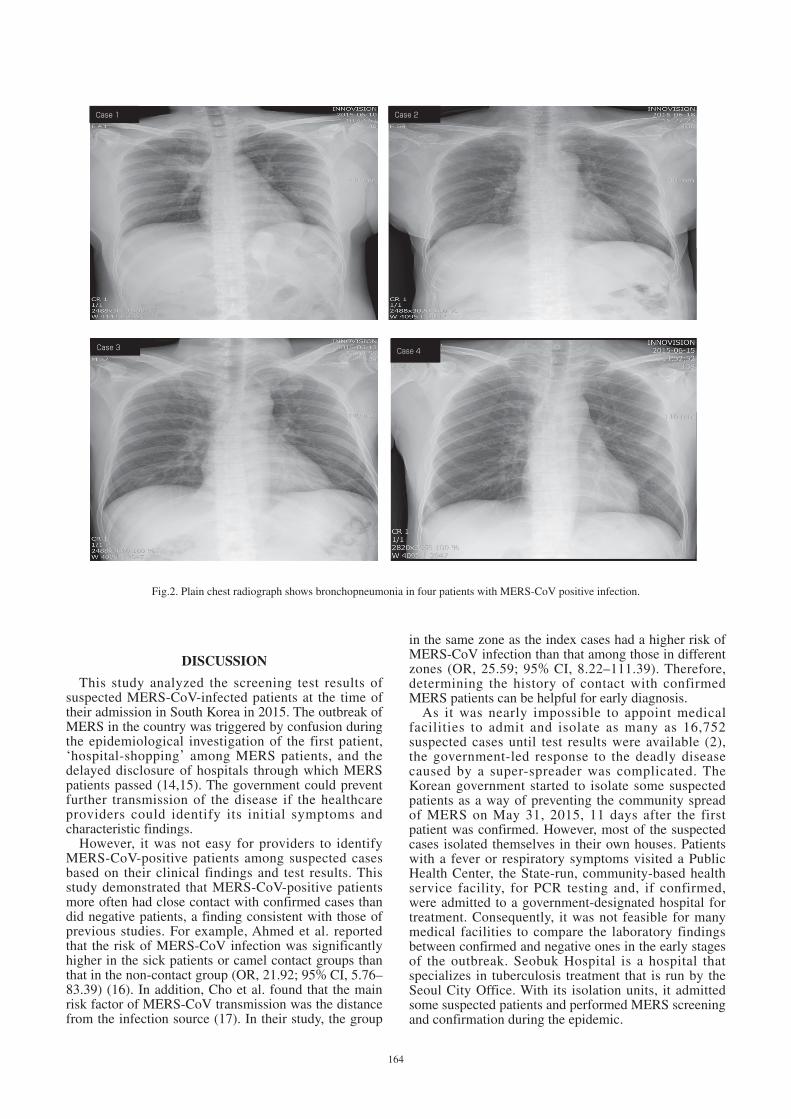
Radiologic findings and severity of patients with pneumonia: Chest X-ray images were obtained at admission from four (22.2%) of the seven MERS-CoV-positive patients. All four patients (100%) had a diffuse or bronchopneumonia type pattern (Table 3). Regarding segmental lung involvement, two (50%) patients had a lesion of left lobe origin to right lobe invasion and one (25%) had a lesion of right lobe origin (Fig. 2). In addition, the severity of pneumonia in the four patients was assessed based on PSI and CURB scores. Of the four MERS-CoV-positive patients, three (75%) were classified as PSI I and one (25%) as PSI II. All four MERS-CoV-positive patients with pneumonia (100%) had a CURB score of 0.
Table 2. Difference between laboratory results and length of hospital stay of MERS (+) and MERS (-) by median
Laboratory Index(unit)
MERS (+): N(%) MERS (-) : N(%)
P-value**Number of abnormal
patient(s) * Mean ± Std (Median, range)
Number of abnormal
patient(s) *Mean ± Std (Median, range)
White blood cell (×103/μL) 1/7 (14.3 %) 5.89±2.81 (4.90, 2.30–10.80) 13/47 (27.7 %) 7.75±3.61 (6.90, 1.80–17.90) 0.129
Red blood cell (×106/μL) 0/7 ( 0 %) 4.70±0.45 (4.75, 4.01–5.31) 12/47 (25.5 %) 4.20±0.65 (4.24, 2.07–5.44) 0.046
Hemoglobin (g/dL) 0/7 (0 %) 14.87±1.58 (15.20, 13.00–16.60) 22/47 (46.8 %) 12.90±2.00 (13, 7.30–16.90) 0.011
Hematocrit (%) 0/7 (0 %) 42.74±3.94 (43, 38.00–47.40) 16/47 (34 %) 37.75±5.76 (38, 19.20–49.70) 0.022
Platelet (×103/μL) 4/7 (57.1 %) 164±76.57 (134, 103–325) 6/47 (12.8 %) 240.30±79.87 (238, 25–449) 0.009
Neutrophil (%) 3/7 (42.9 %) 52.27±23.36 (53.30, 7.70–78) 13/45 (28.9 %) 60.86±16.42 (59.60, 6.0–90.50) 0.302
Eosinophil (%) 5/7 (71.4 %) 0.27±0.43 (0, 0–1) 10/45 (22.2 %) 2.13±2.01 (1.50, 0–8.80) 0.003
Basophil (%) 0/7 (100 %) 0.19±0.17 (2, 0–0.4) 0/45 (100 %) 0.36±0.36 (0.2, 0–1.70) 0.159
Monocyte (%) 3/7 (42.9 %) 10.40±5.94 (10.50, 0.90–17.70) 7/45 (15.6 %) 8.61±4.36 (7.70, 0.60–26.90) 0.277
Lymphocyte (%) 3/7 (42.9 %) 24.13±13.97 (24.60, 2.20–42.10) 21/45 (46.7 %) 26.00±13.07 (27.40, 1.50–49.90) 0.758
Alkaline phosphatase (U/L) 0/7 (0 %) 72.43±18.69 (71, 52–106) 1/45 (2.2 %) 73.62±64.81 (64.00, 36–483) 0.376
Aspartate aminotransferase (U/L) 4/7 (57.1 %) 32.86±5.55 (36, 25–38) 10/46 (21.7 %) 27.72±15.89 (22.50, 12–86) 0.046
Alanine aminotransferase (U/L) 0/7 (05) 25.43±5.26 (27, 17–30) 8/46 (17.4 %) 24.52±24.15 (17, 5–148) 0.072
Creatine kinase (U/L) 1/4 (25.05) 181.25±195.04 (102, 53–468) 5/33 (15.2 %) 125.24±225.05 (77, 17–1352) 0.463
Erythrocyte sedimentation rate (mm/hr) 2/7 (28.6 %) 18.29±11.74 (12, 9–42) 19/44 (43.2 %) 23.70±25.54 (13.50, 2–104) 0.658
C-reactive protein (mg/dL) 5/7 (71.4 %) 2.61±4.12 (0.63, 0.18–11.58) 44/46 (95.7 %) 3.76±6.85 (0.27, 0.02–30.46) 0.307
Lactate dehydrogenase (mg/dL) 0 1/5 (20 %) 181.80±43.83 (175, 141–256)
* : Data are number of abnormal patients/number of total patients.**: Mann-Whitney test (P-value < 0.05).
164
DISCUSSION
This study analyzed the screening test results of suspected MERS-CoV-infected patients at the time of their admission in South Korea in 2015. The outbreak of MERS in the country was triggered by confusion during the epidemiological investigation of the first patient, ‘hospital-shopping’ among MERS patients, and the delayed disclosure of hospitals through which MERS patients passed (14,15). The government could prevent further transmission of the disease if the healthcare providers could identify its initial symptoms and characteristic findings.
However, it was not easy for providers to identify MERS-CoV-positive patients among suspected cases based on their clinical findings and test results. This study demonstrated that MERS-CoV-positive patients more often had close contact with confirmed cases than did negative patients, a finding consistent with those of previous studies. For example, Ahmed et al. reported that the risk of MERS-CoV infection was significantly higher in the sick patients or camel contact groups than that in the non-contact group (OR, 21.92; 95% CI, 5.76–83.39) (16). In addition, Cho et al. found that the main risk factor of MERS-CoV transmission was the distance from the infection source (17). In their study, the group
in the same zone as the index cases had a higher risk of MERS-CoV infection than that among those in different zones (OR, 25.59; 95% CI, 8.22–111.39). Therefore, determining the history of contact with confirmed MERS patients can be helpful for early diagnosis.
As it was nearly impossible to appoint medical facilities to admit and isolate as many as 16,752 suspected cases until test results were available (2), the government-led response to the deadly disease caused by a super-spreader was complicated. The Korean government started to isolate some suspected patients as a way of preventing the community spread of MERS on May 31, 2015, 11 days after the first patient was confirmed. However, most of the suspected cases isolated themselves in their own houses. Patients with a fever or respiratory symptoms visited a Public Health Center, the State-run, community-based health service facility, for PCR testing and, if confirmed, were admitted to a government-designated hospital for treatment. Consequently, it was not feasible for many medical facilities to compare the laboratory findings between confirmed and negative ones in the early stages of the outbreak. Seobuk Hospital is a hospital that specializes in tuberculosis treatment that is run by the Seoul City Office. With its isolation units, it admitted some suspected patients and performed MERS screening and confirmation during the epidemic.
Fig.2. Plain chest radiograph shows bronchopneumonia in four patients with MERS-CoV positive infection.
Case 1 Case 2
Case 3 Case 4

165
Screening Results from MERS-CoV of the South Korea
MERS-CoV-negative patients stayed longer in the hospital than did positive patients. This is because Seobuk Hospital transferred MERS-CoV-positive patients to Seoul Medical Center for treatment as soon as their infections were confirmed by diagnostic tests. Accordingly, the positive patients’ length of stay (LOS) appeared to be shorter than that of negative patients. However, as the positive patients stayed in Seoul Medical Center until they were fully cured, it is difficult to comment that the difference in LOS between positive and negative patients is reliable. This study identified a total of seven patients who tested positive for MERS-CoV infection. In order to identify the potential predictors of the disease, the authors reviewed an additional 55 suspected patients who ultimately tested negative for the disease. The 2 groups were homogeneous in age and sex. The median age of the patients in this study was 43 years, while the previously reported median age of cases was 55 years (3,18). The male predominance (71.4% vs. 52.7% in the confirmed and negative groups, respectively) did not reach statistical significance. Other Korean studies also reported a male predominance as in the current study (3,18).
In the current study, headache, myalgia, and diarrhea were common among MERS-CoV-positive cases. Headache (20.4%), myalgia (43%), and diarrhea (19.4%)
were also frequently reported among a total of 186 MERS-CoV-positive patients in another South Korean study (18). However, a case report of five MERS-CoV patients in South Korea indicated that all patients had a myalgia on the first day of illness (19). Therefore, these symptoms have discriminating power for MERS-CoV infection in South Korea. However, these results are inconsistent with those of other studies in Saudi Arabia (5,20,21). Al-Tawfiq et al. reported that these symptoms did not differ significantly between the MERS CoV-positive and MERS-CoV-negative groups (5,20). In contrast, Garbati et al. reported diarrhea to be a common symptom in MERS-CoV-positive cases (21).
In this study, the seven confirmed cases had cough (14.3% vs. 16.4%; P = 1.000) but did not have sputum (0% vs. 18.2%; P = 0.586), although the differences between the 2 groups were not statistically significant. Another Korean study of five MERS-positive patients reported similar results, with 20% and 0% having cough and sputum, respectively, at admission (19). Saudi Arabian studies have reported consistent results. The cough rate did not differ significantly between the MERS CoV-positive and -negative groups (5, 20–22).
Despite the low rate of cough and sputum among the confirmed cases in the present study, a review of the charts of all 186 MERS-positive patients after the outbreak was resolved revealed cough and sputum rates
Table 3. X-ray images and severity of pneumonia between MERS (+)* and MERS (-)
Case no* Sex Age Comorbidity
X-ray image SeverityClinical outcomePattern Other finding PSI risk
class**CURB
score***
1 Female 43 Lumbar HNPIntestine obstruction
Diffuse or Bronchopneumonia
Right lobe origin : exacerbate Ⅰ 0 Transfer and discharge
2 Female 59 SchizophreniaDM, Dyslipidemia
Diffuse or Bronchopneumonia
Left lobe origin → Right middle lobe invasion
Ⅰ 0 Transfer and discharge
3 Male 37 Diffuse or Bronchopneumonia
Left lobe origin → Right lower lobe invasion
Ⅱ 0 Transfer and discharge
4 Male 43 Diffuse or Bronchopneumonia
Ⅰ 0 Transfer and discharge
5 Female 95 HTN, Asthma, Stroke, Dementia
Diffuse or Bronchopneumonia
Left. lower lobe Pleural effusion and cavity
Ⅴ 1 Transfer by pneumonia and arrhythmia
6 Female 46 Multiple myeloma CRF, Active Tbc Metastatic hematomas Anemia, Pneumonia
Consolidation & Ground-glass opacity
Left lobe > Rt. lobe Ⅳ 1 Death
7 Male 65 GB cancer, DM, HTNCOPD, Inactive Tbc
Ground-glass opacity Left upper lobe Ⅳ 0 Tansfer
8 Male 78 Stroke, HTN Diffuse or Bronchopneumonia
Left. lower lobe, Pleural effusion Ⅳ 0 Discharge
9 Female 72 Arrhythmia, HTN Diffuse or Bronchopneumonia
Ⅲ 0 Discharge
10 Male 69 Liver cirrhosis, DMEsophageal CA
plaque Upper thorax Ⅴ 0 Transfer
11 Male 74 B cell lymphomaNephrotic syndrome
Diffuse or Bronchopneumonia
Ⅴ 1 Transfer
12 Male 87 Rheumatic arthritis Diffuse or Bronchopneumonia
Ⅳ 0 Discharge
* : Pneumonia by MERS (+) : Case no 1–4 (bold letter), and MERS (-) : Case no 5–12.** : Mean of score of PSI risk class by MERS (+) pneumonia and MERS(-) pneumonia : 1.25 & 4.25 (Mann-Whitney test: P = 0.004).***: Mean of score of CURB by MERS (+) pneumonia and MERS(-) pneumonia : 0 & 0.38 (Mann-Whitney test: P = 0.368).
166
of 56.9% and 39.8%, respectively (18). This difference implies that the initial symptoms of MERS are less likely to include cough and sputum. Based on this observation, having suspected patients wear a mask during the entire course from onset may be effective in preventing MERS transmission from person to person in Korea (23). Healthcare providers should be aware of that a lower rate of sputum may cause a false negative result in the early stage of disease (24). The common symptoms of Korean MERS-positive and MERS-CoV-negative patients who were admitted to a hospital differed from those of Saudi Arabian patients. The difference may be attributed to the relatively small sample size in this study as well as the different infection routes between the two countries. In Korea, human-to-human transmission was the only route of infection. However, Saudi Arabia witnessed transmissions from camels to humans as well as from humans to humans (25); these different routes and their relevant factors might have caused different symptoms.
At admission, patients with MERS-CoV infection were more likely than the negative group to have a normal WBC (85.7% vs. 72.3%; P = 0.662) and less likely to have leukocytosis (14.3% vs. 27.7%). The authors found no difference in the presence of neutrophilia; monocytosis; lymphocytopenia; and elevated ALP, AST, ALT, CK, ESR, and CRP levels between the positive and negative groups. These findings were not specific to MERS-CoV-positive patients and were observed similarly in the controls (5,20). However, patients infected with MERS-CoV were more likely to have a lower platelet count and eosinopenia at admission. These results are consistent with those of other studies in Saudi Arabia and Korea (5,16,18,20–22) and seem to be related to acute viral infection and stress (26). Also, interleukin-5 (IL-5) is unique in promoting the terminal differentiation of the committed eosinophil precursor from uncommitted stem cells (26). This means that MERS-CoV may retrain IL-5 and IL-11, which are needed to develop myeloid progenitor cells into eosinophils and platelets in the early stage of infection (27). Additional studies are needed to further clarify this finding.
Chest radiography performed upon admission revealed that all four MERS-CoV-positive patients with pneumonia had a diffuse or bronchopneumonia. Among them, two and one had left and right lobe origins, respectively. A case report of five MERS-CoV-positive patients in South Korea found that four patients had left lobe infiltration (19). In addition, chest radiography with interstitial infiltrates on admission was more frequent among MERS-CoV-positive patients than that among negative patients (67% vs. 20%; OR, 8.13; P = 0.001) in Saudi Arabia (5). These results are consistent with those of previous studies in Saudi Arabia and Korea (5,19). The causative organisms of a bronchopneumonia pattern include Staphylococcus aureus, Klebsiella pneumoniae, Haemophilus influenzae, Pseudomonas aeruginosa, Escherichia coli, and Proteus species (28), while Severe Acute Respiratory Syndrome coronavirus (SARS-CoV) (29) also often entails bronchopneumonia. Therefore, the radiological finding of bronchopneumonia is not specific to MERS-CoV infection. Furthermore, chest X-ray imaging is not only normal in patients with mild
illness but also presents little change at the beginning of MERS-CoV infection (30). However, we start providing care when MERS-CoV infection is diagnosed based on X-ray imaging findings in accordance with the cause.
Also, both the PSI and CURB scores of patients with pneumonia who were assumed to be positive for MERS-CoV infection were low and no patient died during hospitalization, likely because they were younger and had fewer co-morbidities. An important observation from this study was that the pneumonia severity score was not useful for the detection of MERS-CoV patients during the early period of infection.
This study has several limitations. First, the small number of positive patients limits our ability to identify the differentiating factors for MERS-CoV patient detection (20–22). In order to overcome this limitation, this study compared the results to those of a study on all 186 confirmed cases in Korea (18). Second, patients who were admitted to and initially isolated in Seobuk Hospital to be screened for MERS-CoV were transferred to nearby hospitals for treatment after testing positive by PCR. Thus, it was not possible to follow up regarding their symptoms, laboratory results, radiology results, and prognosis after their transfer. Therefore, a longitudinal follow-up study of individual patients is warranted.
In conclusion, the symptoms of headache, myalgia, and diarrhea, as well as the laboratory characteristics of low platelet counts and eosinophil, and chest X-rays showing diffuse bronchopneumonia, might enhance the ability to detect patients in South Korea infected with MERS-CoV. However, further prospective analysis and cohort studies are needed to shed light on the possible predictors of infection.
Acknowledgments The authors thank the people and institutions that participated in the study. These data are original and have not been published previously in any journal of Korean, English, or other languages.
Conflict of interest None to declare.
REFERENCES
1. Zaki AM, van Boheemen S, Bestebroer TM, et al. Isolation of a novel coronavirus from a man with pneumonia in Saudi Arabia. N Engl J Med. 2012;367:1814-20.
2. Kim KH, Tandi TE, Choi JW, et al. Middle East respiratory syndrome coronavirus (MERS-CoV) outbreak in South Korea, 2015: epidemiology, characteristics and public health implications. J Hosp Infect. 2017;95:207-13.
3. Korea Centers for Disease Control and Prevention. Middle East respiratory syndrome coronavirus outbreak in the Republic of Korea, 2015. Osong Public Health Res Perspect. 2015;6:269-78.
4. Assiri A, Al-Tawfiq JA, Al-Rabeeah AA, et al. Epidemiological, demographic, and clinical characteristics of 47 cases of Middle East respiratory syndrome coronavirus disease from Saudi Arabia: a descriptive study. Lancet Infect Dis. 2013;13:752-61.
5. Al-Tawfiq JA, Hinedi K, Ghandour J, et al. Middle East respiratory syndrome coronavirus: a case-control study of hospitalized patients. Clin Infect Dis. 2014;59: 160-5.
6. Mohd HA, Al-Tawfiq JA, Memish ZA. Middle East respiratory syndrome coronavirus (MERS-CoV) origin and animal reservoir. Virol J. 2016;13:87.
7. Al-Tawfiq JA, Memish ZA. Middle East respiratory syndrome coronavirus: epidemiology and disease control measures. Infect Drug Resist. 2014;7:281-7.
8. World Health Organization. Laboratory testing for Middle East respiratory syndrome coronavirus. Interim recommendations. Geneva (Switzerland): WHO; 2014.

167
Screening Results from MERS-CoV of the South Korea
9. Kim YJ, Cho YJ, Kim DW, et al. Complete genome sequence of Middle East respiratory syndrome coronavirus KOR/-KNIH/002_05_2015 isolated in South Korea. Genome Announc. 2015;3:e00787-15.
10. Lim WS, van der Eerden MM, Laing R, et al. Defining community acquired pneumonia severity on presentation to hospital: an international derivation and validation study. Thorax. 2003;58:377-82.
11. Aujesky D, Auble TE, Yealy DM, et al. Prospective comparison of three validated prediction rules for prognosis in community-acquired pneumonia. Am J Med. 2005;118:384-92.
12. Buising KL, Thursky KA, Black JF, et al. A prospective comparison of severity scores for identifying patients with severe community acquired pneumonia: reconsidering what is meant by severe pneumonia. Thorax. 2006;61:419-24.
13. Spindler C, Ortqvist A. Prognostic score systems and community-acquired bacteraemic pneumococcal pneumonia. Eur Respir J. 2006;28:816-23.
14. Ki M. 2015 MERS outbreak in Korea: hospital-to-hospital transmission. Epidemiol Health. 2015;37:e2015033.
15. Khan A, Farooqui A, Guan Y, et al. Lessons to learn from MERS-CoV outbreak in South Korea. J Infect Dev Ctries. 2015;9:543-6.
16. Ahmed AE, Al-Jahdali H, Alshukairi AN, et al. Early identification of pneumonia patients at increased risk of Middle East respiratory syndrome coronavirus infection in Saudi Arabia. Int J Infect Dis. 2018;70:51-6.
17. Cho SY, Kang JM, Ha YE, et al. MERS-CoV outbreak following a single patient exposure in an emergency room in South Korea: an epidemiological outbreak study. Lancet. 2016;388:994-1001.
18. Choi WS, Kang CI, Kim Y, et al. Clinical presentation and outcomes of Middle East respiratory syndrome in the Republic of Korea. Infect Chemother. 2016;48:118-26.
19. Rhee JY, Hong G, Ryu KM. Clinical implications of 5 cases of
Middle East respiratory syndrome coronavirus infection in a South Korean outbreak. Jpn J Infect Dis. 2016;69:361-6.
20. Al-Tawfiq JA, Alfaraj SH, Altuwaijri TA, et al. A cohort-study of patients suspected for MERS-CoV in a referral hospital in Saudi Arabia. J infect. 2017;75: 378-9.
21. Garbati MA, Fagbo SF, Fang VJ, et al. A comparative study of clinical presentation and risk factors for adverse outcome in patients hospitalised with acute respiratory disease due to MERS coronavirus or other causes. PloS One. 2016;11:e0165978.
22. Mohd HA, Memish ZA, Alfaraj SH, et al. Predictors of MERS-CoV infection: a large case control study of patients presenting with ILI at a MERS-CoV referral hospital in Saudi Arabia. Travel Med Infect Dis. 2016;14:464-70.
23. Kim JY, Song JY, Yoon YK, et al. Middle East respiratory syndrome infection control and prevention guideline for healthcare facilities. Infect Chemother. 2015;47:278-302.
24. Lee JH, Lee CS, Lee HB. An appropriate lower respiratory tract specimen is essential for diagnosis of Middle East respiratory syndrome (MERS). J Korean Med Sci. 2015;30:1207-8.
25. Chen X, Chughtai AA, Dyda A, et al. Comparative epidemiology of Middle East respiratory syndrome coronavirus (MERS-CoV) in Saudi Arabia and South Korea. Emerg Microbes Infect. 2017;6:e51.
26. Wardlaw AJ. Eosinophils in the 1990s: new perspectives on their role in health and disease. Postgrad Med J. 1994;70:536-52.
27. Abbas AK, Lichtman AH, Pober JS, et al. Cellular and Molecular Immunology. 4th ed. Philadelphia: WB Saunders Co.; 2000. p. 269.
28. Husain A. editor. Thoracic Pathology. 1st ed. Amsterdam: Saunders; 2012.
29. Zhao Z, Zhang F, Xu M, et al. Description and clinical treatment of an early outbreak of severe acute respiratory syndrome (SARS) in Guangzhou, PR China. J Med Microbiol. 2003; 52:715-20.
30. Arabi YM, Balkhy HH, Hayden FG, et al. Middle East respiratory syndrome. N Engl J Med. 2017;376:584-94.