Embed Size (px)
Citation preview

Am J Cancer Res 2017;7(3):715-726www.ajcr.us /ISSN:2156-6976/ajcr0046866
Original Article Biopolymer based nanosystem for doxorubicin targeted delivery
Zsuzsanna Csikós1, Krisztina Kerekes1, Erika Fazekas1, Sándor Kun1, János Borbély1,2
1BBS Nanotechnology Ltd., Böszörményi 212., H-4032 Debrecen, Hungary; 2Department of Radiology, Faculty of Medicine, University of Debrecen, Nagyerdei krt. 94., H-4032 Debrecen, Hungary
Received December 18, 2016; Accepted January 12, 2017; Epub March 1, 2017; Published March 15, 2017
Abstract: This study describes formation of an actively and passively targeted, water-soluble drug delivery system (DDS) which contains doxorubicin (DOX). The system comprises two biocompatible and biodegradable polymers: poly-γ-glutamic acid (PGA) and chitosan (CH). Self-assembly of these biopolymers in aqueous medium results stable nanoparticles (NPs) with a hydrodynamic size of 80-150 nm and slightly negative surface charge. Folic acid (FA) was used as targeting agent bonded to the polyanion (PA) and also to the surface of the NPs. The NP’s physical stability, active targeting effect, cellular toxicity, release profile and in vivo anti-tumor efficacy were investigated. It was found that the targeted, self-assembled nanoparticles are stable at 4°C for several months, cause better in vitro toxicity effect on folate receptor (FR) positive cell lines than the doxorubicin or the non-targeted nanosystem and based on its release profile it is expected, that the nanosystem will remain stable during the circulation in the body. Pharmaco-dynamic studies demonstrated that the DOX-loaded nanoparticles can deliver greater tumor growth inhibition than the free drug molecules and the liposomal compound, with less general toxicity. It was observed that the overall survival is the main benefit of the biopolymer based drug delivery system.
Keywords: Biopolymers, self-assembled nanoparticles, doxorubicin, drug delivery, folate-targeted, in vitro release, in vivo anti-tumor efficacy
Introduction
Drug delivery systems like proteins [1], emul-sions [2, 3], liposomes [4-6], polymers [7-10] and copolymers [11] have been intensively studied in recent years. The problem to be solved for these DDSs is to decrease the non-specific effect of the chemotherapeutic agents by providing specific delivery of the active com-pound to the tumor cells, thereby reducing the dose needed, and accordingly, the adverse effects on the intact tissues. For this purpose the system should be non-leaky, stable sized and actively and/or passively targeted. Acco- rding to the conventional methods of synthesiz-ing actively targeted nanosystem, drug-loaded nanoparticles are first formed, followed by the conjugation of targeting agents to the surface of the NPs. A number of different targeting moi-eties are conjugated usually to the NPs for tar-geting to the appropriate receptors expressed in the target site, like monoclonal antibodies [12] or their fragments, polypeptides [13],
aptamers [14] and other molecules (e.g. trans-ferrin [15], ACUPA [16] or folic acid [17-21] Fo- lic acid is one of the most frequently used tar-geting agent among researchers, because com-pared with other targeting agents, folic acid offers several advantages. It is small (441 Da), stable over a broad range of temperatures and pH values, and thus amenable for site-specific chemical modification. It is inexpensive, non-immunogenic, and binds to the FR with high affinity even after conjugation to a diagnostic or therapeutic cargo [22]. In normal tissues and organs, FR expression is restricted to only a few sites, where FR is not in contact with circulating folates or intravenously administered folic acid conjugates [23]. Numerous studies have shown that FR is markedly overexpressed on the sur-face of various tumor types, including ovarian, kidney, lung, brain, endometrial, colorectal, pancreatic, gastric, prostate, testicular, blad-der, head and neck, and breast cancers, as well as non-small cell lung cancer [17, 24, 25]. Evidence also suggests that FR expression

Nanosystem for doxorubicin targeted delivery
716 Am J Cancer Res 2017;7(3):715-726
increases with advancing disease and that overexpression of FR is a negative prognostic factor for breast, colorectal, ovarian, and endo-metrial cancer [26, 27]. Thus, FR is viewed as a therapeutic target that may provide an effec-tive option for targeted personalized cancer therapy [28].
According to newer strategies pre-functional-ized polymer components with one of these tar-geting agents are first prepared followed by NP formation [29]. In this study folic acid is used as targeting agent and the two synthesizing techniques are combined, folic acid is bonded to the polymer before self-assembling, and also to the surface of the self-assembled NPs, thus an enhanced targeting is provided. Poly-γ-glutamic acid [30] is a biodegradable and bio-compatible polyanion, through its carboxyl groups PGA is capable to form ion-ion interac-tion and covalent bond with the amino function of doxorubicin [31-33]. Chitosan [34] with com-plexing agents is used as polycation (PC); it is a linear polysaccharide containing reactive amino groups. The NPs were self-assembled from these two modified biopolymers based on the ion-ion interactions of their functional groups. Nowadays, several self-assembled nanoparticles have disadvantages, they tend to dissociate [10, 35] in the body so their sizes can change, or in some cases the drug en- capsulation is not strong enough, so the drug molecules can release before they reach the tumor site.
The purpose of our project was to synthesize stable, self-assembled, targeted drug delivery system for transporting doxorubicin. The stabil-
Materials
Poly-γ-glutamic acid (PGA; >92%, Mw = 50 kDa) was purchased from Shandong Freda Bio- technology Co., Ltd., China, and was used with-out further purification. Chitosan (CH; degree of deacetylation = 88%, Mw = 320 kDa) was pur-chased from Sigma-Aldrich Co., Hungary and purified by the following method: CH was dis-solved in 2.0% aqueous acetic acid (1.0% w/w polymer concentration), filtered and dialyzed against distilled water. The solution was lyop- hilized to obtain a white chitosan powder. N- (3-Dimethylaminopropyl)-N’-ethylcarbodiimide hydrochloride (EDC*HCl), 1-hydroxybenztryaz- ole hydrate (HOBt), 1-[Bis-(dimethyl-amino)-methylene]-1H-1,2,3-triazolo-[4,5-b]-pyridinium 3-oxid hexafluorophosphate (HATU), ethylenedi-aminetetraacetic acid (EDTA), folic acid (FA), sodium bicarbonate, triethylamine (Et3N), anhy-drous dimethylformamide (DMF), trifluoroace- tic acid (TFA) and dimethyl sulfoxide (DMSO) were purchased from Sigma-Aldrich Co., Hungary, doxorubicin (DOX) hydrochloride was obtained from Carbosynth Ltd., United Kingdom and t-Boc Amine PEG Amine HCl Salt (NH2-PEG-NH-Boc, Mw = 2000 Da) was purchased from JenKem Technology USA Inc., USA, these chem-icals were used as received. DEAE Sephadex A25 anion exchange column was purchased from Sigma-Aldrich Co., Hungary. All other chemicals were analytic-grade and were used without further purification. The pH of the aque-ous polymer solutions was adjusted by the dropwise addition of NaOH (0.1 M) or HCl (0.1 M).
ity of the nanoparticles was tested by following their size changes in aqueous medium and also during the release studies. The system was char-acterized by HPLC, NMR, UV- VIS spectrophotometer and ZetaSizer instruments. The cy- totoxic and targeting effects of DOX-loaded NPs were eval-uated using folate receptor (FR)-positive cancer cell lines. The in vivo efficacy of NPs was tested on human ovarian model.
Materials and methods
Scheme 1. Schematic representation of the synthesis of folate conjugated PEG. Abb-reviations: PEG, poly (ethylene glycol).

Nanosystem for doxorubicin targeted delivery
717 Am J Cancer Res 2017;7(3):715-726
Nanoparticle preparation
Synthesis of folate-PEG conjugates (FA-NH-PEG-NH2)
To the stirred solution of folic acid (100 mg) in DMF (20 mL) HATU (86 mg in 2 mL DMF) was added dropwise at 4°C. After 10 min of stirring in the dark NH2-PEG-NH-Boc (453 mg), then 15 min later Et3N (79 ml) was added. The reaction mixture was stirred overnight at room tempera-ture in the dark. After evaporation of the DMF the residue was dissolved in TFA (20 mL) and the mixture was stirred for 3 hours at room tem-perature in the dark. TFA was removed under vacuum and the resulting yellow syrup was puri-fied by gel filtration over a DEAE Sephadex A25 column equilibrated with 0.1 M NaHCO3 to remove unconjugated folic acid. The solution of the product was desalted with dialysis and lyophilized to yield a yellow solid.
The preparation of the folate conjugated PEG is illustrated by the Scheme 1.
Folic acid-PEG-amine (FA-NH-PEG-NH2) asso-ciation with PGA
Poly-γ-glutamic acid (300 mg) was dissolved in water (300 mL) then HOBt (94 mg) was added. The solution was stirred at 4°C for 15 min then EDC*HCl (445 mg in 15 ml water) was added. The reaction mixture was stirred for 10 min, then folic FA-NH-PEG-NH2 (465 mg in 10 ml water) and Et3N (235 ml) was added and stirred at room temperature in the dark for 24 h. The PGA-PEG-FA was purified by membrane filtration.
The preparation of the targeted PGA is illustrat-ed by the Scheme 2.
Preparation of CH-EDTA conjugates
A solution was prepared from chitosan (15 mg) in water (15 mL), the pH was adjusted to 5. After the dropwise addition of aqueous EDTA
(8.2 mg, 2 mL, pH = 3.2), the mixture was stirred at room temperature for 30 min, and at 4°C for 15 min. After that, EDC*HCl (5.1 mg, in 2 mL of distilled water) was added dropwise and the reaction mixture was stirred at 4°C for 4 h, then at room temperature for 20 h. The CH-EDTA conjugate was purified by membrane filtration.
General procedure for DOX-loaded poly-γ-glutamic acid preparation (non-targeted PGA-DOX, targeted PGA-PEG-FA-DOX)
PGA or PGA-PEG-FA solution (20 mL 0.5 mg/mL) was stirred for 15 minutes at pH = 6. DOX (6.2 mg) in distilled water (1 mL) was added dropwise to the solution and the mixture was stirred for 30 min at room temperature, then for 15 min at 4°C. EDC*HCl (3.2 mg) in DMSO (1 mL) and HOBt (1.40 mg) in DMSO (1 mL) were added and the reaction was stirred at 4°C for 4 h then at room temperature for 20 h. The PGA-DOX was purified by membrane filtration
General procedure for the nanoparticle pre- paration
Solution of CH-EDTA (1 mL, 0.3 mg/mL, pH = 4.0) was added dropwise to the modified PGA solution (PGA-PEG-FA; PGA-PEG-FA-DOX or PGA-DOX, 2 mL, 0.3 mg/mL, pH = 9.5) under vigorous stirring.
Folic acid-PEG-amine (FA-NH-PEG-NH2) asso-ciation to the surface of the nanoparticles
FA-NH-PEG-NH2 (7.9 mg) in water (1 mL) was added dropwise to the solution of 15 ml doxoru-bicin loaded NP (15 mL, 0.3 mg/mL) and the mixture was stirred for 30 min at room temper-ature, then for 15 minutes at 4°C. EDC*HCl (1.4 mg) in distilled water (1 mL), HOBt (0.63 mg) in distilled water (1 mL) and Et3N (0.9 ml) were added and the reaction was stirred at 4°C for 4 h then at room temperature for 20 h. The NP-PEG-FA was purified by membrane filtration.
Scheme 2. Schematic representation of the synthesis of targeted PGA. Abbreviations: PGA, poly-γ-glutamic acid.

Nanosystem for doxorubicin targeted delivery
718 Am J Cancer Res 2017;7(3):715-726
Characterization
Characterization with nuclear magnetic reso-nance (NMR) spectroscopy
The received NH2-PEG-NH-Boc samples, and the prepared PGA-PEG-FA conjugates were characterized by 1H NMR spectroscopy. The spectra were recorded at room temperature in deuterated water (D2O) using a 400 MHz NMR spectrometer.
Determination the purity of the FA-NH-PEG-NH2 samples with HPLC
The analysis was performed on a HPLC system (Waters e2695 Separations Module) equipped with an XBridge BEH C18 column (Waters, 4.6×250 mm, 3.5 µm) and a UV/Vis detector (Waters 2489 UV/Vis detector). Briefly, 10 µL of the solution was injected to the mobile phase which was made from high purity water (Millipore RiOs-DI 3, R≥18 MΩ) and 10 mM KH2PO4 and acetonitrile. The pH of the solution was set to 2.60. The mixture was chromato-graphically separated using gradient elution. The flow rate was set to 0.80 mL/min and col-umn was maintained at 30°C.
Determination the DOX concentration with UV-Vis spectrophotometry
DOX solutions of various concentrations were prepared, and the absorptions of the solutions were recorded-from 190 to 600 nm using UV-Vis spectrophotometer (Hitachi U-1900) with a 2 nm slit width and a 1 cm path length at intervals of 1 nm, using water as the baseline reference-to obtain a calibration curve. The spectra of DOX in the absence and presence of PGA were compared.
Characterization of self-assembled, drug-load-ed nanoparticles
The hydrodynamic size and size distribution of particles was measured using a dynamic light scattering (DLS) technique with a Zetasizer Nano ZS (Malvern Instruments Ltd., Grovewood, Worcestershire, UK). This system is equipped with a 4 mW helium/neon laser with a wave-length of 633 nm and measures the particle size with noninvasive backscattering technolo-gy at a detection angle of 173°. Particle size measurements were performed using a parti-
cle-sizing cell in automatic mode. The mean hydrodynamic diameter was calculated from the autocorrelation function of the intensity of light scattered from the particles.
Electrophoretic mobility of the nanoparticles was determined using a Zetasizer Nano ZS instrument. Samples were measured in auto-matic mode with minimum runs of 10, in folded capillary cells. Each sample was measured three times, and the average data was calculated.
In vitro studies
Cell culture
The adherent SK-OV-3 (ovarian) and KB (naso-pharengeal) cancer cell lines, which overex-press folate receptors, were purchased from CLS. Cells were grown in a 5% (v/v) CO2 humidi-fied atmosphere at 37°C and passaged in DMEM medium supplemented with 10% fetal calf serum (Sigma Aldrich). The cells were main-tained in folic acid free medium (RPMI-1640, Sigma Aldrich) for two days before the in vitro tests.
In vitro cytotoxicity
1500 cells/well were plated in 96-well plate in 100 µl FA-free RPMI. The cells were incubated at 37°C for 24 h. After that the cells were treat-ed with the drug-loaded systems, and incubat-ed at 37°C for another 72 h. 10 µl MTT reagent was added to each well, and the plate was incu-bated for 4 h at 37°C, when purple precipitate was clearly visible under microscope, the super-natant was discarded and 100 µl of DMSO was added to all wells, including control wells. The absorbance of the wells was measured at 550 nm with UT-6100 Microplate Reader.
Flow cytometry
Cell surface staining: The expression of folate receptors was determined by flow cytometric analysis. 5×10⁵ cells were stained with folate-receptor specific LK-26 mAb (Abcam, 5 mg/ml), as primary and Alexa Fluor 488 labeled mouse IgG specific polyclonal antibody (Sigma-Aldrich, 25 mg/ml), as secondary antibody. The incuba-tion with antibodies was carried out for 30 min at 4°C. Cells were washed with PBS containing 1% FBS and 0.1% azide. Fluorescence of the
Nanosystem for doxorubicin targeted delivery
719 Am J Cancer Res 2017;7(3):715-726
stained cells was measured by flow cytometer (BD FACSCalibur). Flowing Software was used for data evaluation.
Test of targeting effect: The binding ability of targeted and non-targeted constructs in com-petitive flow cytometric assay was examined. FR-positive KB cells were incubated with Fo- late-RSense (FRS) molecule (fluorescent labe- lled folic acid, was purchased from Perkin-Elmer) alone or together with different samples and the binding ability of different constructs was calculated based on the inhibiting of cellu-lar uptake of FRS. Flowing Software was used for data evaluation.
Release study
The release behavior of the nanoparticles was carried out against phosphate buffer (100 mL, pH = 7.4) at 37°C. The samples were diluted ten-fold with water or RPMI-1640 (cell culture medium). 750 ml of diluted sample was intro-duced into a dialysis bag (MWCO 14 000 Da). 1400 μL of dialysis solution (DS) was collected at determined times and replaced with an equivalent volume of fresh DS. The amount of DOX released was analyzed with HPLC at 482 nm wavelength and calculated on the basis of a calibration curve using different concentrations of free DOX in PBS. Briefly, 100 μL of the solu-tion was injected to the mobile phase which
was 10 mM KH2PO4 (made from high purity water (Millipore RiOs-DI 3, R≥18 MΩ) and and acetonitrile. The pH of the buffer solution was set to 2.60. The mixture was chromato-graphically separated using gradient elution. The flow rate was set to 0.80 mL/min and col-umn was maintained at 30°C.
In vivo study
Antitumor effects in vivo
Comparative efficacy study of six i.v. injection (day 38, 41, 43, 45, 48 and 50) in SK-OV-3 s.c. xenograft SCID mouse model of ovary cancer: Tumor was induced in mice by implanting SK-OV-3 human ovary adenocarcinoma cells (5 millions/mouse) s.c. in upper region of back of SCID mice and allowing the tumors to develop to appreciable size over 38 days (60 mm3).
Results and discussion
Preparation and characterization of DOX-load-ed, self-assembled NPs
Folic acid-PEG-amine samples were synthe-sized from NH2-PEG-NH-Boc, as described in Materials and methods section and illustrated in Scheme 1. The number of ethylene glycol monomer units in the PEG chain was 53, deter-mined by NMR. The folic acid can be linked through its alpha- and gamma-carboxyl groups
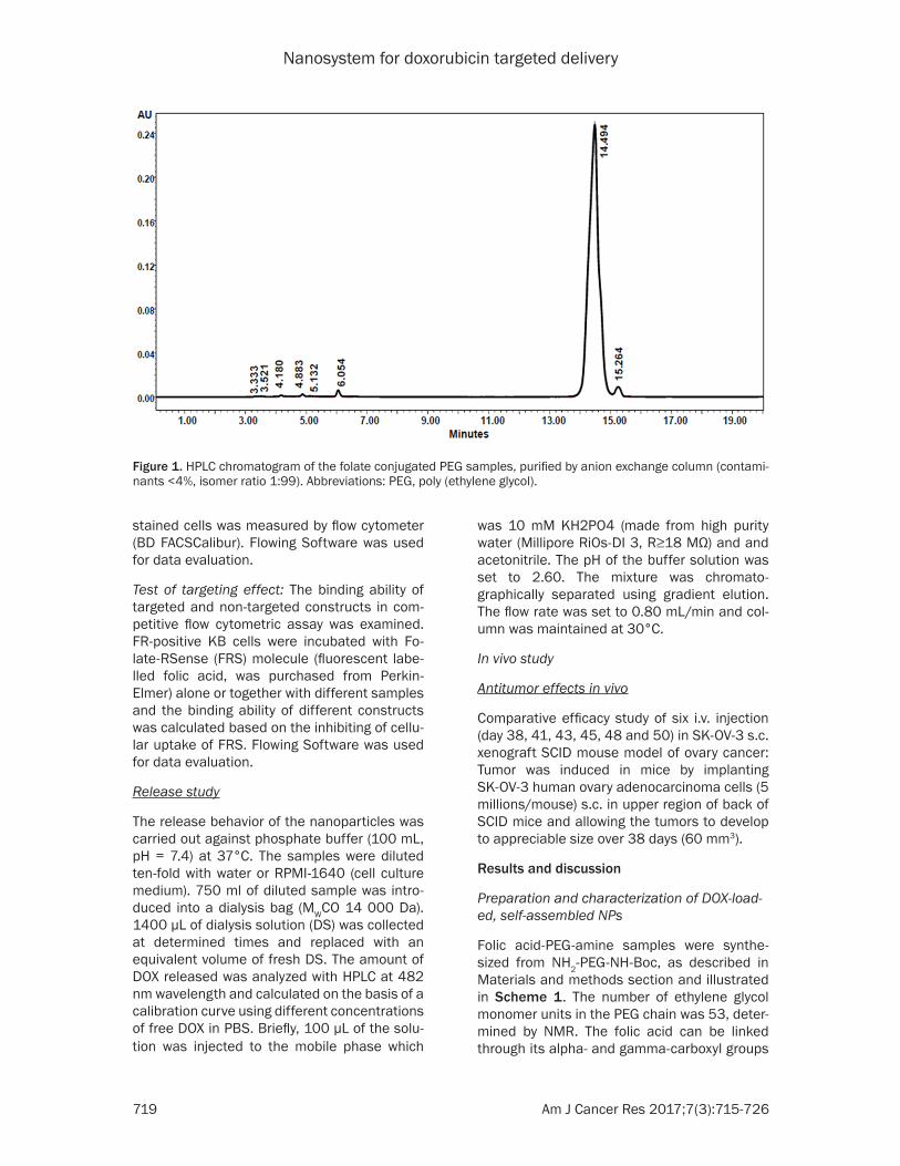
Figure 1. HPLC chromatogram of the folate conjugated PEG samples, purified by anion exchange column (contami-nants <4%, isomer ratio 1:99). Abbreviations: PEG, poly (ethylene glycol).

Nanosystem for doxorubicin targeted delivery
720 Am J Cancer Res 2017;7(3):715-726
Figure 2. 1H NMR spectra of folate targeted PGA sample, PM% = 5.8. Abbreviations: PGA, poly-γ-glutamic acid.

Nanosystem for doxorubicin targeted delivery
721 Am J Cancer Res 2017;7(3):715-726
to the amine functionalized PEG chain. Based on our HPLC results (Figure 1) it was found that after purification the ratio of the alpha- and gamma-isomer was 1:99 in the sample and the amount of contaminants was below 4%. FA-NH-PEG-NH2 (Scheme 2) and/or doxorubicin were associated with PGA via acid amide bond. The concentration of folic acid was determined by NMR and the DOX concentration by UV-Vis spectrophotometry. It was found that the naked DOX has a typical λmax at 480 nm, but in case of
A self-assembly process takes place in the mix-ture of the modified polyanion (PGA-DOX or PGA-PEG-FA-DOX) and polycation (CH-EDTA) resulting shrinkage in the hydrodynamic vol-ume of biomacromolecules and finally nanosys-tem is formed. The thus formed NPs possess negative surface charge and a narrow range of size distribution, which ensure the uniform physical and chemical characteristics. The resulting composition is a hydrophilic nanosys-tem, and forms stable colloid systems in water.
Scheme 3. Schematic illustration of self as-sembled, targeted drug delivery sytem.
Figure 3. Hydrodynamic size (A) and electrophoretic mobility (B) distribution of self-assembled NPs. Abbreviations: NPs, nanoparticles.
DOX-loaded PGA the λmax of the conjugate appeared at 495 nm, suggesting that cou-pling of DOX to γ-PGA induces red-shift of 15 nm. After puri-fication the percent of poly-mer modification (PM%) was 5-7% in case of drug molecule and 3-6% in case of targeting agent. Figure 2 shows an 1H NMR spectra of folate target-ed PGA sample, where the PM% was 5.8.

Nanosystem for doxorubicin targeted delivery
722 Am J Cancer Res 2017;7(3):715-726
Figure 4. Folate-receptor expression of KB (A) and SKOV-3 (B) cells measured by flow cytometry. Cells were stained with Folate-receptor specific LK-26 mAb and anti-mouse-IgG-A488 antibody (solid line) or only with anti-mouse-IgG-A488 (dotted line).
Figure 5. MTT results of DOX (red), non-targeted NPs (green) and targeted NPs (blue) on A2780 (A) and SK-OV-3 (B) cell lines. Abbreviations: DOX, doxorubicin; NPs, nanoparticles.
Figure 6. Competitive flow cytometric assay. Abbreviations: FA, folic acid; PA, polyanion; EDTA, ethylenediaminetetra-acetic acid; NP, nanoparticle; CH, chitosan; PEG-FA, folate conjugated poly (ethylene glycol).

Nanosystem for doxorubicin targeted delivery
723 Am J Cancer Res 2017;7(3):715-726
Modified PGA can form nanosystem with chito-san without complexing agent (without EDTA), this system is also stable in water, but during the MTT tests in some nanosystems modest aggregation was experienced in the microscop-ic images, which means that these composi-tions in vitro were failed to retain their stability to the expected extent. It was found that the chemical stability of the nanoparticles can be improved by binding a complexing agent to the polycation, thus, aggregation can be avoided. Scheme 3, shows the schematic illustration of the self-assembled nanopartilcles.
During the self-assembly of the polymers a part of the folic acid molecules may turn into the inside of the nanoparticles due to the hydro-phobic character of folic acid, thus, the target-ing effect is exerted to a smaller extent (Figure 6). Because of this fact the targeting agent is linked to the polyanion and also to the surface of the nanoparticles (Scheme 4), thus an enhanced targeting is provided (Figure 6).
The thus formed NPs possess a hydrodynamic size of 80-150 nm (Figure 3A) and its electro-phoretic mobility is between -3.0 and -1.0 mm/cm Vs at pH = 7.4 (Figure 3B). Table 1 shows some physico-chemical parameters and the stability results of DOX-loaded nanoparticles.
that there are no difference between the effect of free DOX (red) and the non-targeted DOX-loaded NPs (green) in vitro, because the pas-sive targeting may be enforced only in the body, but the actively targeted DOX-loaded NPs (blue) are more effective, they can inhibit well the cell division also in case of lower doxorubicin con-centration, because the targeting agent makes it easier for the cells to uptake the nano- particles.
Test of binding avidity of DDSs to folate-recep-tor
The active targeting effect was tested by a com-petitive flow cytometric assay using fluores-cently labeled free ligand. The inhibiting ability of samples was compared to each other and to the inhibiting ability of free, unlabelled FA. It has been found, that before self-assembly, the folate-targeted polyanion (Figure 6. Targeted PA, c = 0.3 mg/ml) can inhibit the binding of fluorescently labelled folic acid to a greater extent, than the self-assebled NPs (Figure 6, NP from targeted PA, c = 0.3 mg/ml). When the targeting agent was linked also to the surface of the nanoparticles (Figure 6, NP from target-ed PA + PEG-FA, c = 0.15 mg/ml), an enhanced targeting was provided, compared to the NP which contained folic acid only inside the NP
Scheme 4. Schematic illustration of folic acid-PEG amine association to the surface of the nanoparticle. Abbreviations: PEG, poly (ethylene glycol).
Table 1. Physico-chemical parameters and stability results of DOX-loaded NPs
Code Size(nm)
Polydispersity index
Mobility(µmcm/Vs)
Stability: size (nm)1 week 1 month 6 months
NP_1 113 0.148 -2.376 117 115 111NP_2 115 0.163 -2.261 114 115 116Abbreviations: DOX, doxorubicin; NPs, nanoparticles.
Cell surface folate-receptor expression of examined cells
The cell surface expression of folate-receptor on carcinoma cell lines used for in vitro eval-uation of folate targeted NPs was determined by flow cyto-metric analysis (Figure 4). Both KB and SKOV-3 cells showed strong FR expression, however the amount of folate-receptor on the surface of KB cells was higher, this result corresponds to the literature [36, 37].
In vitro cytotoxicity of DOX-loaded NPs
The cytotoxicity of the target-ed and non-targeted NPs were tested by MTT method on different type of FR-positive cell lines. Figure 5 shows,

Nanosystem for doxorubicin targeted delivery
724 Am J Cancer Res 2017;7(3):715-726
(Figure 6, NP from targeted PA, c = 0.15 mg/ml). The non-targeted polyanion and the CH-EDTA did not inhibit the binding of fluores-cently labelled folic acid.
Release studies
The nanoparticles were tested by release stud-ies. The amount of released drug was deter-mined by HPLC and the stability of the nano- particles was followed by measuring their size with ZetaSizer. Figure 7 shows the results of release studies. The samples were diluted ten-fold with water (Figure 7A) or cell culture medi-um (Figure 7B) and were dialyzed against phos-phate buffer at 37°C. In case of water diluted samples, 12% of doxorubicin could release from our NP within 24 hours and 69% from the
It was demonstrated in pharmacodynamic studies that doxorubicin encapsulated in NP can deliver greater tumor growth inhibition (Figure 8A) than DOX alone, and similar as doxorubicin delivered as a liposomal formula-tion, Caelyx. Our NPs caused less general toxic-ity, compared to DOX and Caelyx evidenced by acceptable body weight losses (Figure 8B) and survival data (Figure 8C).
Conclusion
Stable, folate targeted, self-assembled nano- particles were prepared, as doxorubicin deliv-ery system and an enhanced targeting was pro-vided by bonding folic acid to the polyanioan and also to the surface of the nanoparticles.
Figure 7. Release profile of DOX (red) and NP-PEG-FA (blue). The samples were diluted with water (A) and with cell culture medium (B) before dialisys. Abbreviations: DOX, doxorubicin; NP, nanoparticle; PEG-FA, folate conjugated poly (ethylene glycol).
Figure 8. Results of comparative efficacy studies in mouse model of ova-ry cancer. Abbreviations: DOX, doxorubicin; NP, nanoparticle; PEG-FA, folate conjugated poly (ethylene glycol).
free DOX. In case of cell cul-ture medium (RPMI-1640) diluted samples, 7% of doxo-rubicin could release from our NP within 24 hours and 40% from the free DOX. The size of the nanoparticles did not change during the release study. These results can pre-dict, that the nanoparticles could be stable in the body, they could retain their size and the drug molecules will not release from the NP dur-ing the circulation time.
Antitumor effects in vivo
The in vivo efficacy of NPs was tested on human ovarian model. Figure 8A shows the tumor volumes of groups dur-ing the treatment. Data repre-sent mean ± SEM of six mice per group. Figure 8B shows the body weight of groups dur-ing the treatment. Data repre-sent mean% ± STDEV of six mice per group, the measured data were plotted in propor-tion of the individual weight measured at the start of the treatment. Figure 8C shows the Kaplan-Meier survival curve. The survival data was represented based on real mortality of mice and also the body weight loss of mice (end point at 20%).

Nanosystem for doxorubicin targeted delivery
725 Am J Cancer Res 2017;7(3):715-726
The NPs had greater toxicity effect in vitro as opposed to free DOX and the release results suggested that they will probably be stable in the body, will retain their size and the DOX will not release from the NP during the circulation time. The in vivo results clearly prove that the DOX-loaded nanoparticles have significantly reduced side effects and increased therapeutic effects compared to DOX and Caelyx. These results indicate that our DOX-loaded NPs are potential candidates for cancer treatment.
Acknowledgements
This work is supported by JEREMIE-Joint Eur- opean Resources for Micro to Medium Enter- prises.
Disclosure of conflict of interest
None.
Authors’ contribution
All authors have contributed to read and approved the manuscript as submitted and are prepared to take public responsibility for the work.
Address correspondence to: Dr. János Borbély, De- partment of Radiology, Faculty Medicine, University of Debrecen, Nagyerdei krt. 94., H-4032 Debrecen, Hungary. Tel: +36 30349 6074; E-mail: [email protected]; Zsuzsanna Csikós, BBS Nanotech- nology Ltd., Böszörményi str. 212/B, H-4032 Debre- cen, Hungary. Tel: +36 52 541742; Fax: +36 52 541- 742; E-mail: [email protected]
References
[1] Ren D, Kratz F and Wang SW. Protein nanocap-sules containing doxorubicin as a pH-respon-sive delivery system. Small 2011; 7: 1051-1060.
[2] Dhanarai S, Muralidharan M, Santhi K, Hui A, Wen C and Teng H. Targeted drug delivery sys-tem-Formulation and evaluation of chitosan nanospheres containing doxorubicin hydro-chloride. International Journal of Drug Delivery 2014; 6: 186-193.
[3] Wadhwa J, Nair A and Kumria R. Emulsion forming drug delivery system for lipophilic drugs. Acta Pol Pharm 2012; 69: 179-191.
[4] Yuan MQ, Zhu F, Lou JY, Yuan WM, Fu L, Liu S, Zhang ZZ, Liu CY and He Q. The anti-tumoral efficacy of a docetaxel-loaded liposomal drug
delivery system modified with transferrin for ovarian cancer. Drug Res (Stuttg) 2014; 64: 195-202.
[5] Chen KJ, Chaung EY, Wey SP, Lin KJ, Cheng F, Lin CC, Liu HL, Tseng HW, Liu CP, Wei MC, Liu CM and Sung HW. Hyperthermia-mediated lo-cal drug delivery by a bubble-generating lipo-somal system for tumor-specific chemothera-py. Acs Nano 2014; 8: 5105-5115.
[6] Zhao W, Zhuang S and Qi XR. Comparative study of the in vitro and in vivo characteristics of cationic and neutral liposomes. Int J Nano-medicine 2011; 6: 3087-3098.
[7] Keresztessy Z, Bodnar M, Ber E, Hajdu I, Zhang M, Hartmann JF, Minko T and Borbely J. Self-assembling chitosan/poly-gamma-gluta- mic acid nanoparticles for targeted drug deliv-ery. Colloid Polym Sci 2009; 287: 759-765.
[8] Langer R and Tirrell DA. Designing materials for biology and medicine. Nature 2004; 428: 487-492.
[9] Hellmers F, Ferguson P, Koropatnick J, Krull R and Margaritis A. Characterization and in vitro cytotoxicity of doxorubicin-loaded gamma-poly-glutamic acid-chitosan composite nanoparti-cles. Biochemical Engineering Journal 2013; 75: 72-78.
[10] Zhou L, Cheng R, Tao H, Ma S, Guo W, Meng F, Liu H, Liu Z and Zhong Z. Endosomal pH-acti-vatable poly(ethylene oxide)-graft-doxorubicin prodrugs: synthesis, drug release, and biodis-tribution in tumor-bearing mice. Biomacromol-ecules 2011; 12: 1460-1467.
[11] Knop K, Pavlov GM, Rudolph T, Martin K, Pret-zel D, Jahn BO, Scharf DH, Brakhage AA, Ma-karov V, Moellmann U, Schacher FH and Schubert US. Amphiphilic star-shaped block copolymers as unimolecular drug delivery sys-tems: investigations using a novel fungicide. Soft Matter 2013; 9: 715-726.
[12] Jang M, Yoon YI, Kwon YS, Yoon TJ, Lee HJ, Hwang SI, Yun BL and Kim SM. Trastuzumab-conjugated liposome-coated fluorescent mag-netic nanoparticles to target breast cancer. Korean J Radiol 2014; 15: 411-422.
[13] Danhier F, Pourcelle V, Marchand-Brynaert J, Jerome C, Feron O and Preat V. Targeting of tu-mor endothelium by RGD-grafted PLGA-nanoparticles. Methods Enzymols 2012; 508: 157-175.
[14] Farokhzad OC, Cheng J, Teply BA, Sherifi I, Jon S, Kantoff PW, Richie JP and Langer R. Target-ed nanoparticle-aptamer bioconjugates for cancer chemotherapy in vivo. Proc Natl Acad Sci U S A 2006; 103: 6315-6320.
[15] Li H and Qian ZM. Transferrin/transferrin re-ceptor-mediated drug delivery. Med Res Rev 2002; 22: 225-250.

Nanosystem for doxorubicin targeted delivery
726 Am J Cancer Res 2017;7(3):715-726
[16] Hrkach J, Von Hoff D, Mukkaram Ali M, Andri-anova E, Auer J, Campbell T, De Witt D, Figa M, Figueiredo M, Horhota A, Low S, McDonnell K, Peeke E, Retnarajan B, Sabnis A, Schnipper E, Song JJ, Song YH, Summa J, Tompsett D, Troiano G, Van Geen Hoven T, Wright J, LoRus-so P, Kantoff PW, Bander NH, Sweeney C, Farokhzad OC, Langer R and Zale S. Preclinical development and clinical translation of a PS-MA-targeted docetaxel nanoparticle with a dif-ferentiated pharmacological profile. Sci Transl Med 2012; 4: 128ra139.
[17] Leamon CP and Low PS. Folate-mediated tar-geting: from diagnostics to drug and gene de-livery. Drug Discov Today 2001; 6: 44-51.
[18] Yoo HS and Park TG. Folate-receptor-targeted delivery of doxorubicin nano-aggregates stabi-lized by doxorubicin-PEG-folate conjugate. J Control Release 2004; 100: 247-256.
[19] Leamon CP and Reddy JA. Folate-targeted che-motherapy. Adv Drug Deliv Rev 2004; 56: 1127-1141.
[20] Lin JJ, Chen JS, Huang SJ, Ko JH, Wang YM, Chen TL and Wang LF. Folic acid-Pluronic F127 magnetic nanoparticle clusters for combined targeting, diagnosis, and therapy applications. Biomaterials 2009; 30: 5114-5124.
[21] Majoros IJ, Thomas TP, Mehta CB and Baker JR. Poly (amidoamine) dendrimer-based multi-functional engineered nanodevice for cancer therapy. J Med Chem 2005; 48: 5892-5899.
[22] Muller C and Schibli R. Folic acid conjugates for nuclear imaging of folate receptor-positive cancer. J Nucl Med 2011; 52: 1-4.
[23] Muller C. Folate based radiopharmaceuticals for imaging and therapy of cancer and inflam-mation. Curr Pharm Des 2012; 18: 1058-1083.
[24] Weitman SD, Lark RH, Coney LR, Fort DW, Fra-sca V, Zurawski VR Jr and Kamen BA. Distribu-tion of the folate receptor GP38 in normal and malignant cell lines and tissues. Cancer Res 1992; 52: 3396-3401.
[25] Parker N, Turk MJ, Westrick E, Lewis JD, Low PS and Leamon CP. Folate receptor expression in carcinomas and normal tissues determined by a quantitative radioligand binding assay. Anal Biochem 2005; 338: 284-293.
[26] Toffoli G, Russo A, Gallo A, Cernigoi C, Miotti S, Sorio R, Tumolo S and Boiocchi M. Expression of folate binding protein as a prognostic factor for response to platinum-containing chemo-therapy and survival in human ovarian cancer. Int J Cancer 1998; 79: 121-126.
[27] Hartmann LC, Keeney GL, Lingle WL, Christian-son TJH, Varghese B, Hillman D, Oberg AL and Low PS. Folate receptor overexpression is as-sociated with poor outcome in breast cancer. Int J Cancer 2007; 121: 938-942.
[28] Assaraf YG, Leamon CP and Reddy JA. The fo-late receptor as a rational therapeutic target for personalized cancer treatment. Drug Resist Updat 2014; 17: 89-95.
[29] Gu F, Zhang L, Teply BA, Mann N, Wang A, Ra-dovic-Moreno AF, Langer R and Farokhzad OC. Precise engineering of targeted nanoparticles by using self-assembled biointegrated block copolymers. Proc Natl Acad Sci U S A 2008; 105: 2586-2591.
[30] Oppermann FB, Pickartz S and Steinbuchel A. Biodegradation of polyamides. Polym Degrad Stab 1998; 59: 337-344.
[31] Manocha B and Margaritis A. Controlled re-lease of doxorubicin from doxorubicin/gamma-polyglutamic acid ionic complex. J Nanomater 2010; 780171.
[32] Bae HH, Cho MY, Hong JH, Poo H, Sung MH and Lim YT. Bio-derived poly (gamma-glutamic acid) nanogels as controlled anticancer drug delivery carriers. J Microbiol Biotechnol 2012; 22: 1782-1789.
[33] Huan M, Zhang B, Teng Z, Cui H, Wang J, Liu X, Xia H, Zhou S and Mei Q. In vitro and in vivo antitumor activity of a novel ph-activated poly-meric drug delivery system for doxorubicin. PLoS One 2012; 7: e44116.
[34] Kean T and Thanou M. Biodegradation, biodis-tribution and toxicity of chitosan. Adv Drug De-liv Rev 2010; 62: 3-11.
[35] Meng F, Zhong Y, Cheng R, Deng C and Zhong Z. pH-sensitive polymeric nanoparticles for tumor-targeting doxorubicin delivery: concept and recent advances. Nanomedicine 2014; 9: 487-499.
[36] Tomassetti A, Mangiarotti F, Mazzi M, Sforzini S, Miotti S, Galmozzi E, Elwood PC and Cane-vari S. The variant hepatocyte nuclear factor 1 activates the P1 promoter of the human alpha-Folate receptor gene in ovarian carcinoma. Cancer Res 2003; 63: 696-704.
[37] Leamon CP, Reddy JA, Vetzel M, Dorton R, Westrick E, Parker N, Wang Y and Vlahov I. Fo-late targeting enables durable and specific an-titumor responses from a therapeutically null tubulysin B analogue. Cancer Res 2008; 68: 9839-9844.