Embed Size (px)
Citation preview

ORIGINAL ARTICLE
Altered lung structure and function in mid-childhoodsurvivors of very preterm birthShannon J Simpson,1,2 Karla M Logie,1,3 Christopher A O’Dea,1,4,5
Georgia L Banton,1 Conor Murray,6 Andrew C Wilson,1,2,3,4,5 J Jane Pillow,7
Graham L Hall1,2,5
ABSTRACTRationale Survivors of preterm birth are at risk ofchronic and lifelong pulmonary disease. Follow-up datadescribing lung structure and function are scarce inchildren born preterm during the surfactant era.Objectives To obtain comprehensive data on lungstructure and function in mid-childhood from survivors ofpreterm birth. We aimed to explore relationshipsbetween lung structure, lung function and respiratorymorbidity as well as early life contributors to poorerchildhood respiratory outcomes.Methods Lung function was tested at 9–11 years inchildren born at term (controls) and at ≤32 weeksgestation. Tests included spirometry, oscillatorymechanics, multiple breath nitrogen washout anddiffusing capacity of the lung for carbon monoxide.Preterm children had CT of the chest and completed arespiratory symptoms questionnaire.Main results 58 controls and 163 preterm children(99 with bronchopulmonary dysplasia) participated.Preterm children exhibited pulmonary obstruction andhyperinflation as well as abnormal peripheral lungmechanics compared with term controls. FEV1 wasimproved by 0.10 z-scores for every additional week ofgestation (95% CI 0.028 to 0.182; p=0.008) and by0.34 z-scores per z-score increase in birth weight (0.124to 0.548; p=0.002). Structural lung changes werepresent in 92% of preterm children, with total CT scoredecreased by 0.64 (−0.99 to −0.29; p<0.001) for eachadditional week of gestation. Obstruction was associatedwith increased subpleural opacities, bronchial wallthickening and hypoattenuated lung areas on inspiratorychest CT scans (p<0.05).Conclusions Abnormal lung structure in mid-childhoodresulting from preterm birth in the contemporary era hasimportant functional consequences.
INTRODUCTIONBronchopulmonary dysplasia (BPD) is one of themost significant complications of preterm birth.BPD was described nearly 50 years ago in infantssuffering from severe respiratory distress syndromefollowing prolonged treatment with mechanicalventilation and high concentrations of inspiredoxygen (O2).
1 Infants with BPD had abnormalchest radiographs, including areas of lung collapsealternating with hyperinflation and severe emphy-sema, as well as histopathological changes
consistent with interstitial thickening, lung fibrosisand smooth muscle hypertrophy.1
Neonatal critical care has evolved considerably overthe past few decades: the implementation and stand-ardisation of postnatal surfactant therapy, increaseduse of antenatal maternal corticosteroids and thedevelopment of less aggressive ventilation strategieshave altered both the nature of and the infant popula-tion affected by BPD.2 Whereas the initial report ofBPD described an infant population with a mean ges-tational age (GA) of 34 weeks,1 babies born in theearly saccular and even the late cannalicular stages oflung development (ie, <28 weeks GA) now oftensurvive in middle-income and high-income countries.3
Thus, a ‘new’ BPD pathophysiology has emerged,characterised by fewer, larger alveoli resulting inreduced surface area for gas exchange and dysmorphicpulmonary capillary growth.4 5
Survivors of preterm birth have a high burden ofrespiratory disease in the first years of life.6 7
Key messages
What is the key question?▸ What is the impact of contemporary preterm
birth on lung structure and lung functionduring mid-childhood, and what factors predictpoorer outcomes for these children?
What is the bottom line?▸ Infants born very preterm are at significant risk
of ongoing lung disease with considerablestructural abnormalities, lung function deficitsand consequent respiratory symptoms reportedin mid-childhood.
Why read on?▸ These comprehensive data examining the lung
health of children born preterm, in thecontemporary era, raise concerns about thelong-term lung health of a subset of thispopulation. Structural lung abnormalities inchildren born preterm were common andassociated with more obstructive lung functionand more severe respiratory symptoms and mayimply active lung disease. What this means forthe health of these children as they age mustbe a priority for future investigations.
702 Simpson SJ, et al. Thorax 2017;72:702–711. doi:10.1136/thoraxjnl-2016-208985
Paediatric lung disease
To cite: Simpson SJ, Logie KM, O’Dea CA, et al. Thorax 2017;72:702–711.
► Additional material is published online only. To view please visit the journal online (http:// dx. doi. org/ 10. 1136/ thoraxjnl- 2016- 208985).
For numbered affiliations see end of article.
Correspondence toProfessor Graham L Hall, Children’s Lung Health, Telethon Kids Institute, PO Box 855, West Perth, WA 6872, Australia; graham. hall@ telethonkids. org. au
SJS and KML contributed equally.
Received 3 June 2016Revised 19 December 2016Accepted 1 January 2017Published Online First 24 January 2017
► http:// dx. doi. org/ 10. 1136/ thoraxjnl- 2016- 208524
► http:// dx. doi. org/ 10. 1136/ thoraxjnl- 2016- 209291
on Novem
ber 20, 2021 by guest. Protected by copyright.
http://thorax.bmj.com
/T
horax: first published as 10.1136/thoraxjnl-2016-208985 on 24 January 2017. Dow
nloaded from

Beyond infancy, existing follow-up data of new BPD largelydescribe small or selected populations of children with the mostsevere neonatal disease or survivors of extreme prematurity.8–10
Consequently, the lack of comprehensive and appropriatelypowered assessments of lung structure and function across thespectrum of preterm children render it difficult to determine ifthe clinical course of contemporary prematurity differs from theearly descriptions of BPD.
This study aimed to evaluate lung function, lung structureand the relationship between structure and function in a con-temporary population of children born ≤32 weeks gestation.Furthermore, we aimed to elucidate the impact of prematurityand neonatal treatments on lung function and structure findingsto provide a comprehensive snapshot of the respiratory systemduring mid-childhood following preterm birth. We hypothesisedthat worse lung structure and function at mid-childhood wouldbe associated with a more severe neonatal course. Second, wehypothesised that pulmonary function deficits would be under-pinned by structural abnormalities of the lung.
METHODSParticipantsPreterm children, with and without a neonatal classification ofBPD, and healthy term-born controls were studied at 9–11 yearsof age. Preterm and term children who had previously beenstudied by our group at 4–8 years11 were prioritised for recruit-ment to this study. The study was then supplemented with add-itional participants living in the Perth metropolitan region.Children born at King Edward Memorial Hospital (KEMH)≤32 weeks gestation between 1997 and 2003 were recruitedfrom the KEMH neonatal database. BPD was defined as at least28 days supplemental oxygen requirement as assessed at36 weeks postmenstrual age.12 Healthy term controls were atleast 37 weeks completed gestation with no lifetime history ofcardiopulmonary disease or recurrent respiratory symptoms.Written informed parental/guardian consent and child assentwere obtained prior to data collection. Approval was obtainedfrom the Princess Margaret Hospital for Children EthicsCommittee (EP1760).
Assessment of lung functionAssessment of lung function included spirometry, multiplebreath nitrogen washout, diffusing capacity of the lung forcarbon monoxide (Sensormedics Vmax, Yorba Linda,California, USA) and the forced oscillation technique (FOT)(I2M, Chess Medical, Ghent, Belgium). Lung function testswere performed according to American Thoracic Society/European Respiratory Society standards.13–16 Lung functionoutcomes were expressed as z-scores to adjust for the relevantanthropometric factors, with the exception of Lung ClearanceIndex (LCI), residual volume (RV)/total lung capacity (TLC) andfunctional residual capacity (FRC)/TLC that are independent ofanthropometrics.17–20
Assessment of lung structureCT images of the chest were acquired in children born pretermduring both inspiration and expiration. Inspiratory imagesspanned from the lung apex to diaphragm at 10 mm intervals.Three expiratory images were collected at the anatomical pointsof the tracheal carina and the midpoints between the trachealcarina and the lung apex, and tracheal carina and diaphragm(Philips Brilliance 64; Philips Medical Systems, theNetherlands). The chest CT scans were consensus scored in thesame scoring session by a specialist paediatric thoracic
radiologist (CM) and a paediatric respiratory physician (ACW)using the scoring system described by Aukland et al.21 Scorerswere blinded to severity of neonatal lung disease. Healthy con-trols did not undergo chest CT.
Other assessmentsRespiratory morbidity in the 3 months prior to the visit wasassessed via a questionnaire.22 Neonatal data extracted from theKEMH neonatal database and patient medical records includedGA, birth anthropometrics, antenatal steroid exposure, con-firmed sepsis during the birth admission, number of surfactantdoses, days of supplemental oxygen, mechanical ventilation andCPAP.
Study power and statistical analysisNormally distributed data are presented as means and SDs.Non-normally distributed data are presented as medians andIQR. Differences in lung function between groups were ana-lysed by one-way analysis of variance with post hoc compari-sons. The Mann-Whitney U test or independent samples t-testwas used for the assessment of differences between groups forall other outcomes, depending on normality of the data. Therelationships between neonatal factors and lung function orlung structure were initially assessed using univariate linearregressions. Neonatal factors with significant univariate correl-ation to structure/function outcomes were included in subse-quent stepwise multiple linear regressions. Multicollinearitiesbetween the neonatal predictors were identified and adjusted forby using residuals of independent regressions of the collinearvariables. For example, the independent impact of mechanicalventilation on lung structure/function was determined from theresidual of the regression between GA and mechanicalventilation.
A minimum sample size of 75 preterm children provides atleast 80% power for a multiple linear regression to detect a 0.2z-score difference in lung function with six predictors.Significance is considered at the 5% significance level. All datawere analysed using SPSS (V.22; IBM, Chicago, Illinois, USA).
RESULTSStudy participants at follow-up visitThere were no anthropometric differences between the termborn controls (n=58) and preterm children (n=163) at the timeof the follow-up assessment (table 1). However, children withBPD (n=99) were shorter (p<0.001) than term-born childrenand lighter (p=0.001) than their term and preterm peerswithout BPD. Parents reported respiratory symptoms such aswheeze, shortness of breath and excessive cough without coldsin approximately half of all children born preterm in the3 months prior to the visit (table 1).
The preterm cohort was born at a median (IQR) gestation of28 (25.0, 29.6) weeks (table 2). As expected from the definitionof BPD, children with BPD required a longer duration ofoxygen supplementation and increased days of respiratorysupport (p<0.001) during the neonatal period than pretermchildren without BPD. The BPD group also had a lower GA andbirth weight (p<0.001); however, there was no differencebetween birth weight z-score for preterm children with BPDand with those without BPD (p=0.121).
Generalisability of participants to the preterm populationBetween July 1998 and December 2003, 1316 children weredelivered at KEMH ≤32 weeks GA, with 371 (28.2%) of theseinfants developing BPD. Preterm infants (no BPD) not recruited
703Simpson SJ, et al. Thorax 2017;72:702–711. doi:10.1136/thoraxjnl-2016-208985
Paediatric lung disease on N
ovember 20, 2021 by guest. P
rotected by copyright.http://thorax.bm
j.com/
Thorax: first published as 10.1136/thoraxjnl-2016-208985 on 24 January 2017. D
ownloaded from

for this study were born at a median (IQR) GA of 30.4(28.3, 31.3) weeks, with a birth weight of 1440 (1165, 1680) gand required 1 (0, 3) day of supplemental O2. Infants develop-ing BPD during this time were born at 26.6 (25.1, 28.0) weeksGA, weighing 895 (705, 1055) g and required 75 (48, 98) dayssupplemental O2. There was no difference in neonatal character-istics for children recruited to this study (table 2), with theexception that recruited preterm children without BPD hadslightly lower GA (29.8; 28.3, 31.0 weeks; p=0.004) than thenon-recruited cohort.
Lung function in preterm childrenForced flows and volumes: Preterm children had significantlylower spirometry values (FEV1, FEF25–75 and FEV1/FVC) thanhealthy control children (table 3), which were further decreasedin those with BPD (see online supplementary table S1).Abnormally low lung function (defined as FEV1, FEV1/FVC orFEF25–75≤−1.96 z-scores) was not observed in any of thehealthy children, but was observed in 7 (13.5%) of the pretermgroup without BPD and 37 (46.8%) of the children with BPD.
Decreased GA and birth weight z-score were associated withlower FEV1 and FVC z-scores in mid-childhood (see figure 1and online supplementary table S2). Lower forced expiratory
flows and volumes (FEV1 and FEF25–75) were also observed inchildren with higher durations of supplemental oxygen and con-firmed septicaemia during the neonatal period. Multivariateanalysis (see online supplementary table S2) demonstrated thatFEV1 z-score was increased by 0.1 z-scores for every additionalweek of gestation (B=0.105; 95% CI 0.028 to 0.182; p=0.008)and improved by increasing birth weight z-score (B=0.336;95% CI 0.124 to 0.548; p=0.002), accounting for 16.9% ofthe variation in FEV1 in preterm children. Increasing days ofsupplemental O2 remained the only weak (R2=0.070) inde-pendent predictor of reduced FEF25–75 z-score in a multivariateanalysis such that each additional week of oxygen was associatedwith a 0.056 decrease in mid-expiratory flows after accountingfor GA and days of mechanical ventilation (B=−0.008, 95% CI−0.016 to −0.000; p=0.040).
Lung volumes by multiple breath washout: Very preterm chil-dren had increased FRC z-score and FRC/TLC, suggestive ofhyperinflation (table 3). Univariate analysis demonstrated thathyperinflation (increased FRC/TLC) was associated with lowbirth weight z-score (B=−1.428; 95% CI −2.802 to −0.054;p=0.042) and an increased duration of CPAP in the neonatalperiod independent of other respiratory support (B=0.196;95% CI 0.056 to 0.335; p=0.006) (see online supplementary
Table 2 Neonatal information about the preterm participants
Preterm
Preterm
No BPD BPD
Gestational age (PMA) 28.0 (25.0, 29.6) 29.8 (28.6, 31.0) 26.0 (24.4, 27.6)*Birth weight (g) 955 (755, 1335) 1388 (1186, 1645) 825 (700, 955)*Birth weight z-score (mean, SD) −0.14 (0.91) 0.00 (0.95) −0.22 (0.88)Birth length (cm) 35.5 (33.0, 39.5) 40.0 (38.0, 42.0) 34.0 (32.0, 36.0)*Duration of oxygen supplementation (days) 48 (2, 92) 1 (0, 3.8) 85 (59, 103)*Duration of mechanical ventilation (days) 4 (0, 28) 0 (0, 1) 20 (5, 39)*Duration of CPAP (days) 6 (1, 18) 1 (0, 3.7) 14 (4, 24)*Surfactant administered, n (%) 115 (70.6) 27 (42.7) 88 (89.8)*Antenatal steroids, n (%) 133 (81.6) 53 (82.8) 80 (81.6)
*Represents significant difference between the preterm groups with and without BPD (p<0.05).Continuous neonatal variables are presented as median (IQR), except birth weight z-score (mean, SD). Binary variables are expressed as n (%).Birth weight z-score was calculated fromFenton growth charts for preterm infants.23
BPD, bronchopulmonary dysplasia; PMA, postmenstrual age.
Table 1 Participant demographics at the time of follow-up visit
Term controls Preterm
Preterm
No BPD BPD
Participants, n 58 163 64 99Males, n (%) 26 (45) 100 (61) 42 (65) 58 (59)Age (year) 10.7 (0.9) 10.9 (0.6) 10.9 (0.6) 10.8 (0.6)Height (cm) 146.6 (9.7) 142.0 (8.0) 143.3 (9.0) 140.6 (8.0)*Weight (kg) 37.4 (8.3) 35.6 (10.6) 38.9 (13.6) 33.5 (7.4)*†Symptoms in the 3 months prior to the test visit expressed as n (% of those completing questionnaire)Asthma medication usage – 47 (32.0) 21 (38.9) 26 (28)Respiratory symptoms at rest – 26 (17.0) 16 (26.2) 10 (11.9)†Respiratory symptoms on exertion – 78 (51.0) 28 (47.5) 50 (53.2)Any respiratory symptoms past 3 months – 80 (52.6) 29 (49.2) 51 (54.8)
Participant demographics at the time of testing are presented as mean (SD) unless otherwise indicated as number (% of population).*Represents significant difference from the term controls (p<0.05).†Represents significant difference between the preterm groups with and without BPD (p<0.05).BPD, bronchopulmonary dysplasia.
704 Simpson SJ, et al. Thorax 2017;72:702–711. doi:10.1136/thoraxjnl-2016-208985
Paediatric lung disease on N
ovember 20, 2021 by guest. P
rotected by copyright.http://thorax.bm
j.com/
Thorax: first published as 10.1136/thoraxjnl-2016-208985 on 24 January 2017. D
ownloaded from

table S3). CPAP duration remained an independent predictor ofhyperinflation in a multivariate analysis (see onlinesupplementary table S3). Low birth weight z-score was also asignificant predictor of increased RV/TLC (another markerof hyperinflation) such that RV/TLC (%) was increased by1.83 units per z-score decrease in birth weight (95% CI −3.106to −0.557; p=0.005)(see figure 1 and online supplementarytable S3).
Gas exchange: None of the measures of gas exchange (diffus-ing capacity of the lung for carbon monoxide (DLCO)) were dif-ferent between healthy children and those born pretermregardless of BPD diagnosis (table 3). Higher DLCO z-score wasweakly predicted by increased duration of CPAP in the neonatalperiod independent of other respiratory support in a univariateanalysis (B=0.038; 95% CI 0.006 to 0.070; p=0.022). Noother neonatal factors predicted impairment in gas exchange(see online supplementary table S4).
Respiratory system mechanics (forced oscillation technique(FOT)): Preterm children had worse peripheral lung mechanicsthan the healthy control group, as indicated by more negativerespiratory system reactance at 8 Hz (Xrs8), increased area underthe reactance curve (AX) and increased resonant frequency (fres)(see table 3 and online supplementary table S1). Increased daysof oxygen supplementation was associated with increased fres(B=0.010; 95% CI 0.000 to 0.019; p=0.045) and AX(B=0.009; 95% CI 0.001 to 0.018; p=0.035), where FOT out-comes were increased by approximately 0.07 z-scores for eachweek of supplemental oxygen exposure (see onlinesupplementary table S5).
Respiratory symptoms and lung function: Participants withany reported respiratory symptoms in the 3 months prior to thestudy visit (n=80) (see table 1) had worse respiratory mechanicsthan those with no symptoms. Indeed, for every z-score increasein AX and z-score decrease in Xrs8, the likelihood of reportedsymptoms was increased 1.42 (95% CI 1.024 to 1.973;p=0.036) times and 1.62 times (OR 0.619; 95% CI 0.417 to0.919; p=0.017), respectively. There were no other differencesin lung function between the preterm children with recentsymptoms and those without.
Lung structure in preterm childrenStructural abnormalities were present in 92% of all childrenborn ≤32 weeks gestation (table 4). Children with BPD hadmore extensive structural lung disease on chest CT scan com-pared with children without BPD (see table 3 for median(IQR)), with total CT score ranging from 0 to 17 for thepreterm group and 0 to 26 for the BPD group out of amaximum score of 50.21
Linear and triangular subpleural opacities were the most com-monly identified abnormalities and were more severe in childrenwith BPD. Areas of collapse/consolidation and hypoattenuationon the inspiratory CT scan were also more extensive in the BPDgroup. No differences between the BPD and non-BPD groupswere observed for the other radiological findings (see figure 2for examples of radiological abnormalities).
Children with recent respiratory symptoms (67 of 127 scans)had significantly higher total CT scores (median=7; IQR=4–12)than children without recent symptoms (4; 2–7; p=0.003),
Table 3 Lung function in healthy children and preterm children during mid-childhood
Term controls (reference) Preterm Mean difference (95% CI)
Forced flows and volumes (spirometry)n successful 48 131FEV1 z-score 0.04 (0.90) −0.72 (1.13) −0.76 (−1.12 to −0.40)**FEV1/FVC z-score −0.27 (0.92) −1.25 (1.01) −0.98 (−1.33 to −0.63)**FEF25–75 z-score −0.42 (0.90) −1.46 (1.11) −1.04 (−1.42 to −0.67)**FVC z-score 0.17 (0.95) 0.13 (1.04) −0.04 (−0.40 to 0.33)
Lung volume and ventilation distribution (multiple breath washout)n successful 50 140LCI 6.14 (0.58) 6.33 (0.82) 0.19 (−0.06 to 0.44)FRC z-score −0.09 (0.96) 0.42 (1.32) 0.51 (0.10 to 0.91)*TLC z-score −0.16 (0.85) −0.05 (1.02) 0.11 (−0.21 to 0.42)RV z-score −0.45 (1.10) −0.16 (1.23) 0.28 (−0.11 to 0.68)FRC/TLC (%) 50.36 (6.65) 52.80 (7.49) 2.44 (0.07 to 4.81)*RV/TLC (%) 21.29 (6.18) 22.79 (7.02) 1.50 (−0.73 to 3.72)
Gas exchange (DLCO)n successful 38 74DLCO z-score 0.10 (0.97) 0.28 (1.27) 0.17 (−0.29 to 0.64)VA z-score −0.72 (1.16) −0.50 (1.33) 0.24 (−0.27 to 0.74)KCO z-score −0.56 (0.86) −0.57 (0.98) −0.02 (−0.39 to 0.36)
Respiratory system mechanics (forced oscillation technique)n successful 31 132Rrs8 z-score 0.00 (0.94) 0.33 (1.04) 0.32 (−0.08 to 0.72)Xrs8 z-score 0.14 (0.53) −0.43 (0.99) −0.57 (−0.94 to −0.21)*AX z-score −0.44 (0.85) 0.29 (1.15) 0.72 (0.29 to 1.16)**Fres z-score −0.18 (1.26) 0.64 (1.23) 0.82 (0.31 to 1.33)*
Data are presented as mean (SD) and unadjusted mean difference (95% CI) except for the number of successful tests.*p<0.05; **p<0.001.DLCO, diffusing capacity of the lung for carbon monoxide; FRC, functional residual capacity.
705Simpson SJ, et al. Thorax 2017;72:702–711. doi:10.1136/thoraxjnl-2016-208985
Paediatric lung disease on N
ovember 20, 2021 by guest. P
rotected by copyright.http://thorax.bm
j.com/
Thorax: first published as 10.1136/thoraxjnl-2016-208985 on 24 January 2017. D
ownloaded from
largely owing to increased scores for hypoattenuated areas oninspiration (0; 0, 2; p=0.027) and bronchial wall thickening(1; 0, 3; p=0.021).
Lower GA, lower birth weight z-score, neonatal sepsis andincreased duration of respiratory support (oxygen supplementa-tion and mechanical ventilation) were generally associated withmore extensive structural abnormalities (total CT score, linearand triangular subpleural opacities, hypoattenuation on inspir-ation and collapse/consolidation score) (see figure 3 and onlinesupplementary table S6). None of the neonatal clinical variablesexplained the extent of hypoattenuated areas of the lung duringexpiration or bronchial wall thickening.
Multivariate analysis suggested that each additional week ofgestation was associated with a 0.64-unit decrease in total CTscore (95% CI −0.99 to −0.29; p<0.001), resulting fromdecreased scores for subpleural opacities (B=−0.330; 95% CI−0.463 to −0.196; p<0.001) and hypoattenuation on
inspiration (B=−0.113; 95% CI −0.211 to −0.016; p=0.023).Additionally, after accounting for GA each week of mechanicalventilation was associated with a 0.55-unit increase in total CTscore (95% CI 0.015 to 0.142; p=0.078), largely a result ofincreased liner/triangular subpleural opacity score (B=0.029,95% CI 0.006 to 0.052; p=0.015). See online supplementarytable S6 for further details.
A random subset of CT scans (N=20) were rescored a mean(SD) time of 6.1 (1.6) years after first scoring, by the sameobservers. The intraobserver reliability was excellent with anIntraclass Correlation Coefficient (ICC) of 0.81(95% CI 0.51 to0.93) and an absolute agreement ICC of 0.76 (0.36 to 0.91).
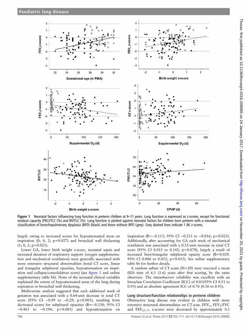
Lung structure/function relationships in preterm childrenObstructive lung disease was evident in children with moreextensive structural abnormalities on CT scan. FEV1, FEV1/FVCand FEF25–75 z-scores were decreased by approximately 0.1
Figure 1 Neonatal factors influencing lung function in preterm children at 9–11 years. Lung function is expressed as z-scores, except for functionalresidual capacity (FRC)/TLC (%) and RV/TLC (%). Lung function is plotted against neonatal factors for children born preterm with a neonatalclassification of bronchopulmonary dysplasia (BPD) (black) and those without BPD (grey). Grey dashed lines indicate 1.96 z-scores.
706 Simpson SJ, et al. Thorax 2017;72:702–711. doi:10.1136/thoraxjnl-2016-208985
Paediatric lung disease on N
ovember 20, 2021 by guest. P
rotected by copyright.http://thorax.bm
j.com/
Thorax: first published as 10.1136/thoraxjnl-2016-208985 on 24 January 2017. D
ownloaded from

z-scores for each increase in linear/triangular subpleural opaci-ties, bronchial wall thickening and hypoattenuation score duringan inspiratory chest CT (p<0.05) (see figure 4 and onlinesupplementary table S7).
Lung volumes were largely unaffected by chest CT abnormal-ities with the exception of areas of lung collapse/consolidationin preterm children that predicted reduced RV z-score(B=−1.156; 95% CI −1.892 to −0.420; p=0.002) and RV/TLC (B=−6.779; 95% CI −10.991 to −2.566; p=0.002) (seeonline supplementary table S8). Additionally, increased TLCwas apparent in children with bronchiectasis (B=0.202; 95% CI0.020 to 0.384; p=0.030).
KCO was decreased by 0.23 z-scores in preterm children foreach unit increase in hypoattenuation on an inspiratory chestCT (B=−0.233; 95% CI −0.406 to −0.061; p<0.009) (seeonline supplementary table S9). Altered respiratory systemmechanics was not independently predicted by structural lungdamage on chest CT (see online supplementary table S10).
DISCUSSIONThis study provides the most comprehensive assessment to dateof lung structure and function in school-aged children born verypreterm (≤32 weeks gestation) during the surfactant era. We
show that preterm children have a significant burden of respira-tory disease at mid-childhood with structural abnormalitiespresent in almost all preterm children and recent respiratorysymptoms reported in approximately half of the cohort. Lungfunction is also lower in children born preterm, and is asso-ciated with increased structural lung damage.
We show significant airway obstruction in children born verypreterm with reductions in FEV1, FEF25–75 and FEV1/FVC, andfurther reductions in children with BPD. Increased airwayobstruction is in line with other studies that have measured spir-ometry in preterm children with both new8 10 24 and old25 26
BPD. Findings of reduced forced expiratory flows and volumeswith stable FVC are suggestive of reduced airway calibre in thispopulation and likely results from chronic airway inflammation,airway remodelling and/or reduced parenchymal tethering.Almost half of the very preterm children in this study had evi-dence of bronchial wall thickening on chest CT that we specu-late reflects postinflammatory changes and/or ongoing airwayinflammation. Indeed, children with bronchial wall thickeninghad significantly worse obstructive lung disease and morerespiratory symptoms than their peers, which may indicate thatcloser monitoring of this group is warranted. Additionally, thesmall airways may be particularly at risk from oxygen toxicity
Table 4 The presence and extent of chest CT abnormalities in children born very preterm
PretermPreterm without BPD(n=50)
Preterm with BPD(n=83)
Hodges-Lehman mediandifference (95% CI)
Structural abnormalities on chest CTPresence, n participants (%) 123 (92) 44 (88) 79 (95)Extent (total CT score) 5 (3–9) 4 (1–7) 6 (4–11) 2 (1 to 4); p=0.008*
Linear/triangular subpleural opacitiesPresence 112 (84%) 39 (78%) 73 (88%)Extent 3 (1–5) 2 (1–3) 3 (2–6) 2 (1 to 2); p<0.001*
Decreased pulmonary attenuation—inspirationPresence 40 (30%) 9 (18%) 31 (37%)*Extent 0 (0–1) 0 (0–0) 0 (0–1) 0 (0 to 0); p=0.013*
Decreased pulmonary attenuation—expirationPresence 58 (44%) 23 (46%) 35 (42%)Extent 0 (0–2) 0 (0–2) 0 (0–2) 0 (0 to 0); p=0.926
Decreased bronchial: arterial ratioPresence 2 (2%) 1 (2%) 1 (1%)Extent 0 (0–0) 0 (0–0) 0 (0–0) 0 (0 to 0); p=0.708
BronchiectasisPresence 12 (9%) 4 (8%) 8 (10%)Extent 0 (0–0) 0 (0–0) 0 (0–0) 0 (0 to 0); p=0.801
Bronchial wall thickeningPresence 58 (44%) 20 (40%) 38 (46%)Extent 0 (0–3) 0 (0–2) 0 (0–3) 0 (0 to 0); p=0.473
BullaePresence 2 (2%) 0 (0%) 2 (2%)Extent 0 (0–0) 0 (0–0) 0 (0–0) 0 (0 to 0); p=0.271
EmphysemaPresence 4 (3%) 0 (0%) 4 (5%)Extent 0 (0–0) 0 (0–0) 0 (0–0) 0 (0 to 0); p=0.116
Collapse/consolidationPresence 15 (11%) 1 (2%) 14 (17%)*Extent 0 (0–0) 0 (0–0) 0 (0–0) 0 (0 to 0); p=0.008*
Presence indicates the number (%) of preterm children with the particular structural abnormality described. Extent scores were derived as described by Aukland et al,21 where eachparameter has a maximum score of 6 (according to the number of affected lobes) except for collapse/consolidation (maximum=2) leading to a possible total CT score of 50. Extentscores are expressed as median (IQR). Difference in presence of abnormality between groups assessed using theχ2 test and difference in extent assessed using the Mann-Whitney U test.*Represents significant difference between the preterm groups with and without BPD (p<0.05).BPD, bronchopulmonary dysplasia.
707Simpson SJ, et al. Thorax 2017;72:702–711. doi:10.1136/thoraxjnl-2016-208985
Paediatric lung disease on N
ovember 20, 2021 by guest. P
rotected by copyright.http://thorax.bm
j.com/
Thorax: first published as 10.1136/thoraxjnl-2016-208985 on 24 January 2017. D
ownloaded from

Figure 2 Chest CT abnormalities in contemporary bronchopulomary dysplasia. All images were acquired during inspiration. Images A and B arefrom an 11-year-old male (27 weeks gestation) highlighting triangular subpleural (red) and linear (blue) opacities, areas of hypoattenuation/mosaicperfusion (yellow) and emphysema (green). Image C is from an 11-year-old male (25 weeks gestation) and highlights bronchial wall thickening(orange).
Figure 3 Neonatal factors influencing lung structure in preterm children at 9–11 years. Total CT score (max 50) and extent score for linear andtriangular subpleural opacities (max 6) are plotted against gestational age, and days of mechanical ventilation for children born preterm with aneonatal classification of bronchopulmonary dysplasia (BPD) (black) and those without BPD (grey).
708 Simpson SJ, et al. Thorax 2017;72:702–711. doi:10.1136/thoraxjnl-2016-208985
Paediatric lung disease on N
ovember 20, 2021 by guest. P
rotected by copyright.http://thorax.bm
j.com/
Thorax: first published as 10.1136/thoraxjnl-2016-208985 on 24 January 2017. D
ownloaded from
during the neonatal period, with reduced midexpiratory flowsevident in those with longer exposures to oxygen.
The morphological changes in the lungs of children born verypreterm, such as larger simplified alveoli, suggest that changes inlung volume and ventilation inhomogeneity should be observedin this population. However, previous studies are inconclusive,with some reporting a degree of air trapping (elevated RV/TLC)in BPD27–29 and others failing to discriminate preterm childrenfrom healthy controls.30 31 In this study, lung volumes were gen-erally not different between preterm children with and withoutBPD, although air trapping was associated with being bornsmall for GA and the increased duration of CPAP, perhaps indi-cating that these children are more prone to airway collapse andconsequent gas trapping. Interestingly, those preterm childrenwith bronchiectasis and areas of lung collapse or consolidationon chest CT had smaller lung volumes consistent with thesepathologies. Differences between studies may reflect the
heterogeneity of lung disease in preterm children and thecohorts associated with each of these studies. By including parti-cipants from a wide spectrum of GA and neonatal lung diseaseseverity, we may have dampened the signal when comparedwith studies that highlight the extreme preterm phenotype suchas the EPICure data (≤25 weeks GA).27
Prematurity and lung injury during the neonatal periodimpair growth, branching and distribution of the pulmonaryvasculature.32 33 Surprisingly, no gas transfer measures were dif-ferent in children born preterm, likely indicating that diffusiondistances across the respiratory epithelium are similar betweenterm and preterm children. Additionally, DLCO outcomes werenot associated with neonatal factors that promote lung injury,such as mechanical ventilation or exposure to supplementaloxygen, or antenatal corticosteroid treatment which promotesthinning of alveolar walls34 and potential increased efficiency ofgas exchange. The few studies of lung function in the
Figure 4 Relationships between lung function and lung structure in preterm children. Lung function, expressed as z-scores, is plotted against chestCT outcomes for preterm children with (black) and without (grey) bronchopulmonary dysplasia.
709Simpson SJ, et al. Thorax 2017;72:702–711. doi:10.1136/thoraxjnl-2016-208985
Paediatric lung disease on N
ovember 20, 2021 by guest. P
rotected by copyright.http://thorax.bm
j.com/
Thorax: first published as 10.1136/thoraxjnl-2016-208985 on 24 January 2017. D
ownloaded from

postsurfactant era portray conflicting results for DLCO: somestudies show decreased DLCO in preterm children withBPD27 35 while others fail to detect a difference.31 Our findingsmay be influenced by the difficulty obtaining acceptable andrepeatable DLCO measures in children with BPD comparedwith the term and preterm without BPD groups. Children withBPD who were born with low birth weight z-scores were lesslikely to achieve acceptable DLCO results at school age andwere consequently under-represented in the analyses of gasexchange, biasing the results. The inability to obtain acceptableand repeatable DLCO in this population may be a manifestationof the cognitive and behavioural deficits, such as hyperactivity,inattention and dysfunction in executive functioning, known topersist in very and extremely low birth weight infants (see ref.36 for review).
In contrast to studies of young adults with old BPD,37 emphy-sema was detected in only 5% of this cohort of preterm chil-dren. However, almost half of the children in this study haveareas of decreased pulmonary attenuation on the chest CT scanthat may reflect pre-emphysematous changes to the lung (whichmay be progressive). Alternatively, hypoattenuation may berelated to disrupted pulmonary vascularisation and reducedalveolar complexity leading to decreased surface area for gasexchange seen in postmortem specimens of infants and childrenwith BPD.38 Regardless, those children who have hypoattenu-ated areas on inspiratory CT scan have more airway obstruction(lower FEV1/FVC) and reduced KCO. Consequently, this specificgroup of children may warrant follow-up to determine if theirlung disease is progressive.
We show that structural abnormalities are very common (92%)in the lungs of children born preterm. Structural damage wasmore extensive in children of lower GA who received moreoxygen and mechanical ventilation in the neonatal period, whichis consistent with the notion that more immature and fragile lungshave increased susceptibility to injury. Only two small otherstudies (of 32 and 26 subjects, respectively) report chest CT find-ings in long-term survivors of preterm birth in the surfactantera.24 39 Using the same methodology and scoring system,Aukland et al reported lower median (IQR) total CT scores (3.0(1.75–5.0)) in a cohort of preterm children born in 1991–1992than the scores reported from our cohort. While developed specif-ically for survivors of preterm birth, the extent of the chest CTfindings is likely to be limited by the insensitivity of the scoringmethod since the scoring method does not allow for multiple pre-sentations within the same lobe. Additionally, both studies per-formed only limited slice scans to minimise the radiation dose andtherefore may have underestimated the extent of the lung abnor-malities.40 We are also unable to comment on the likelihood ofobserving any of these abnormalities in the healthy control popu-lation. However, these abnormalities are, by definition, patho-logical and unlikely to be present in the control population.
We show heterogeneous impairments in lung structure andlung function at 9–11 years in children born ≤32 weeks gesta-tion. Prolonged O2 dependence in the neonatal period alonedoes not accurately predict which children will go on to haveincreased pulmonary morbidity, despite underpinning the clin-ical definition of BPD. Our data show that some infants withBPD may have normal lung function and no respiratory symp-toms. Conversely, respiratory symptoms and pulmonary func-tion deficits may appear in children born preterm who did notrequire prolonged ventilatory support during infancy.Regardless, we have shown that structural and functionalrespiratory limitations are evident in very preterm survivorsduring mid-childhood. It remains unknown whether the
involvement of the peripheral lung is related to structural abnor-malities due to preterm birth per se, such as fewer larger alveoli,or whether the changes we observe are pre-emphysematouschanges. Determining whether the structural and functionalabnormalities are progressive and what the clinical implicationof such decline might mean for the health of these children asthey age remains a priority for future investigations.
Author affiliations1Telethon Kids Institute, Perth, WA, Australia2Centre for Child Health Research, University of Western Australia, Perth, WA,Australia3School of Paediatrics and Child Health, University of Western Australia, Perth, WA,Australia4Department of Respiratory and Sleep Medicine, Princess Margaret Hospital forChildren, Perth, WA, Australia5School of Physiotherapy and Exercise Science, Faculty of Health Sciences, CurtinUniversity, Perth, WA, Australia6Diagnostic Imaging, Princess Margaret Hospital for Children, Perth, WA, Australia7Department of Anatomy, Physiology and Human Biology, University of WesternAustralia, Perth, WA, Australia
Contributors ACW, JJP and GLH: study design. SJS, KML, CAO and GLB: datacollection, lung function analysis and database entry. CM and ACW: CT scoring. SJS,JJP and GLH: statistical analysis and data interpretation. SJS, KLM and GH:manuscript preparation. All authors: revision of manuscript.
Funding NHMRC (APP634519), Princess Margret Hospital Foundation and RaineMedical Foundation. SJS (APP1073301), JJP (APP1077691) and GLH (APP1025550)are supported by NHMRC Fellowships.
Competing interests None declared.
Patient consent Obtained.
Ethics approval Princess Margaret Hospital Human Research Ethics Committee.
Provenance and peer review Commissioned; externally peer reviewed.
REFERENCES1 Northway WH Jr, Rosan RC, Porter DY. Pulmonary disease following respirator
therapy of hyaline-membrane disease. Bronchopulmonary dysplasia. N Engl J Med1967;276:357–68.
2 Kamath BD, Macguire ER, McClure EM, et al. Neonatal mortality from respiratorydistress syndrome: lessons for low-resource countries. Pediatrics2011;127:1139–46.
3 Blencowe H, Cousens S, Oestergaard MZ, et al. National, regional, and worldwideestimates of preterm birth rates in the year 2010 with time trends since 1990 forselected countries: a systematic analysis and implications. Lancet2012;379:2162–72.
4 Jobe AJ. The new BPD: an arrest of lung development. Pediatr Res 1999;46:641–3.5 Husain AN, Siddiqui NH, Stocker JT. Pathology of arrested acinar development in
postsurfactant bronchopulmonary dysplasia. Hum Pathol 1998;29:710–17.6 Elder DE, Hagan R, Evans SF, et al. Hospital admissions in the first year of life in
very preterm infants. J Paediatr Child Health 1999;35:145–50.7 Pramana IA, Latzin P, Schlapbach LJ, et al. Respiratory symptoms in preterm infants:
burden of disease in the first year of life. Eur J Med Res 2011;16:223–30.8 Fawke J, Lum S, Kirkby J, et al. Lung function and respiratory symptoms at 11 years
in children born extremely preterm: the EPICure study. Am J Respir Crit Care Med2010;182:237–45.
9 Vollsæter M, Skromme K, Satrell E, et al. Children Born Preterm at the Turn of theMillennium Had Better Lung Function Than Children Born Similarly Preterm in theEarly 1990s. PLoS ONE 2015;10:e0144243.
10 Hacking DF, Gibson AM, Robertson C, et al. Respiratory function at age 8–9 afterextremely low birthweight or preterm birth in Victoria in 1997. Pediatr Pulmonol2013;48:449–55.
11 Verheggen M, Wilson AC, Pillow JJ, et al. Respiratory function and symptoms inyoung preterm children in the contemporary era. Pediatr Pulmonol2016;51:1347–55.
12 Jobe AH, Bancalari E. Bronchopulmonary dysplasia. Am J Respir Crit Care Med2001;163:1723–9.
13 MacIntyre N, Crapo RO, Viegi G, et al. Standardisation of the single-breathdetermination of carbon monoxide uptake in the lung. Eur Respir J2005;26:720–35.
14 Miller MR, Hankinson J, Brusasco V, et al. Standardisation of spirometry. Eur RespirJ 2005;26:319–38.
710 Simpson SJ, et al. Thorax 2017;72:702–711. doi:10.1136/thoraxjnl-2016-208985
Paediatric lung disease on N
ovember 20, 2021 by guest. P
rotected by copyright.http://thorax.bm
j.com/
Thorax: first published as 10.1136/thoraxjnl-2016-208985 on 24 January 2017. D
ownloaded from

15 Oostveen E, MacLeod D, Lorino H, et al. The forced oscillation technique in clinicalpractice: methodology, recommendations and future developments. Eur Respir J2003;22:1026–41.
16 Wanger J, Clausen JL, Coates A, et al. Standardisation of the measurement of lungvolumes. Eur Respir J 2005;26:511–22.
17 Cook CD, Hamann JF. Relation of lung volumes to height in healthy personsbetween the ages of 5 and 38 years. J Pediatr 1961;59:710–14.
18 Quanjer PH, Stanojevic S, Cole TJ, et al. Multi-ethnic reference values for spirometryfor the 3–95-yr age range: the global lung function 2012 equations. Eur Respir J2012;40:1324–43.
19 Kim YJ, Hall GL, Christoph K, et al. Pulmonary diffusing capacity in healthycaucasian children. Pediatr Pulmonol 2012;47:469–75.
20 Calogero C, Simpson SJ, Lombardi E, et al. Respiratory impedance andbronchodilator responsiveness in healthy children aged 2–13 years. PediatrPulmonol 2013;48:707–15.
21 Aukland SM, Rosendahl K, Owens CM, et al. Neonatal bronchopulmonary dysplasiapredicts abnormal pulmonary HRCT scans in long-term survivors of extreme pretermbirth. Thorax 2009;64:405–10.
22 Powell CV, McNamara P, Solis A, et al. A parent completed questionnaire todescribe the patterns of wheezing and other respiratory symptoms in infants andpreschool children. Arch Dis Child 2002;87:376–9.
23 Fenton TR, Kim JH. A systematic review and meta-analysis to revise the Fentongrowth chart for preterm infants. BMC Pediatr 2013;13:59.
24 Broström EB, Thunqvist P, Adenfelt G, et al. Obstructive lung disease in childrenwith mild to severe BPD. Respir Med 2010;104:362–70.
25 Northway WH Jr, Moss RB, Carlisle KB, et al. Late pulmonary sequelae ofbronchopulmonary dysplasia. N Engl J Med 1990;323:1793–9.
26 Vrijlandt EJ, Gerritsen J, Boezen HM, et al. Lung function and exercise capacity inyoung adults born prematurely. Am J Respir Crit Care Med 2006;173:890–6.
27 Lum S, Kirkby J, Welsh L, et al. Nature and severity of lung function abnormalitiesin extremely pre-term children at 11 years of age. Eur Respir J 2011;37:1199–207.
28 Korhonen P, Laitinen J, Hyödynmaa E, et al. Respiratory outcome in school-aged,very-low-birth-weight children in the surfactant era. Acta Paediatr 2004;93:316–21.
29 Smith LJ, van Asperen PP, McKay KO, et al. Reduced exercise capacity in childrenborn very preterm. Pediatrics 2008;122:e287–93.
30 Schulzke SM, Hall GL, Nathan EA, et al. Lung volume and ventilation inhomogeneityin preterm infants at 15–18 months corrected age. J Pediatr 2010;156:542–9. e2.
31 Vom Hove M, Prenzel F, Uhlig HH, et al. Pulmonary outcome in former preterm,very low birth weight children with bronchopulmonary dysplasia: a case-controlfollow-up at school age. J Pediatr 2014;164:40–5.e4.
32 De Paepe ME, Mao Q, Powell J, et al. Growth of pulmonarymicrovasculature in ventilated preterm infants. Am J Respir Crit Care Med2006;173:204–11.
33 Coalson JJ. Pathology of new bronchopulmonary dysplasia. Semin Neonatol2003;8:73–81.
34 Willet KE, Jobe AH, Ikegami M, et al. Lung morphometry after repetitive antenatalglucocorticoid treatment in preterm sheep. Am J Respir Crit Care Med2001;163:1437–43.
35 Cazzato S, Ridolfi L, Bernardi F, et al. Lung function outcome at school age in verylow birth weight children. Pediatr Pulmonol 2013;48:830–7.
36 Saigal S, Doyle LW. An overview of mortality and sequelae of preterm birth frominfancy to adulthood. Lancet 2008;371:261–9.
37 Wong PM, Lees AN, Louw J, et al. Emphysema in young adult survivors ofmoderate-to-severe bronchopulmonary dysplasia. Eur Respir J 2008;32:321–8.
38 Sobonya RE, Logvinoff MM, Taussig LM, et al. Morphometric analysis of the lung inprolonged bronchopulmonary dysplasia. Pediatr Res 1982;16:969–72.
39 Aukland SM, Halvorsen T, Fosse KR, et al. High-resolution CT of the chest inchildren and young adults who were born prematurely: findings in apopulation-based study. AJR Am J Roentgenol 2006;187:1012–18.
40 de Jong PA, Nakano Y, Lequin MH, et al. Dose reduction for CT in children withcystic fibrosis: is it feasible to reduce the number of images per scan? Pediatr Radiol2006;36:50–3.
711Simpson SJ, et al. Thorax 2017;72:702–711. doi:10.1136/thoraxjnl-2016-208985
Paediatric lung disease on N
ovember 20, 2021 by guest. P
rotected by copyright.http://thorax.bm
j.com/
Thorax: first published as 10.1136/thoraxjnl-2016-208985 on 24 January 2017. D
ownloaded from