Embed Size (px)
Citation preview

Orgainic/inorganic pg. 14 sg
• Some cpds which contain carbon are inorganic.
Examples include carbon dioxide, carbonates, and hydrogen carbonates.
All cpds which contain no carbon are inorganic
Three types of organic cpds are found in living things, carbohydrates, proteins, lipids

Structure

• Carbon atoms can share electrons -notable being other carbon atoms, hydrogen atoms and oxygen atoms.
• The simplest organic molecules are defined as being comprised of only carbon and hydrogen = hydrocarbons

• Hydrocarbons are non-polar, hydrophobic compounds. (water hating)
• Compounds that have no charges on them will not mix with polar water.
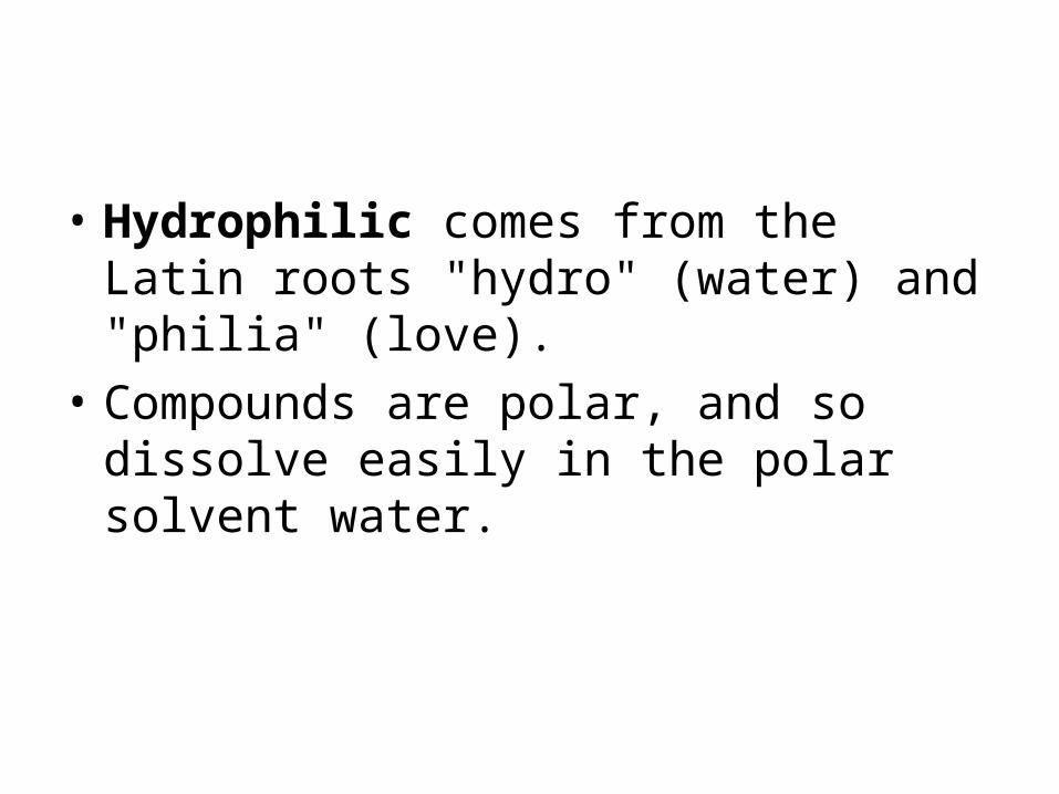
• Hydrophilic comes from the Latin roots "hydro" (water) and "philia" (love).
• Compounds are polar, and so dissolve easily in the polar solvent water.

• Structural formulas of some simple hydrocarbons.
• Methane CH4:

Hydrocarbons
• A simple chain of carbons with its full complement of hydrogens is said to be saturated.
• known as alkanes. • name ends with 'ane'.

• Hydrocarbons with double bonds in them are said to be unsaturated.
• contain at least one double bond.
• Alkenes

Branching chains
• Sometimes two hydrocarbon molecules can have the same numbers of the same atoms but have different arrangements of these atoms. We say they are isomers.

Role of hydrocarbons in fats
• A fat molecule consists of a small, non hydrocarbon component joined to three hydrocarbon tails. The tails can be broken down to provide energy. Mammalian adipose cells stockpile fat.

Functional Groups Parts of the molecules of life.
• Part of your homework tonight is to go to this web site: Building Biomolecules: The Functional Groups
• Review each of the functional groups and do the self quiz.

Functional groups

Adenosine triphosphate or ATP


Phosphate groups
• In biology this is an important group found in ATP.

• Structurally, ATP consists of the adenine nucleotide (ribose sugar, adenine base, and phosphate group, PO4-2) plus two other phosphate groups.

Exergonic / Endergonic Reactions
• Energy releasing processes, ones that "generate" energy, are termed exergonic reactions.
• Reactions that require energy to initiate the reaction are known as endergonic reactions

ATP = The primary energy transferring molecule in cells

Macromolecules – page 15sg
• Monomers make polymers. Polymers are long molecules consisting of many similar or identical building blocks linked by covalent bonds.

Condensation Reaction
• Monomers are connected by a reaction in which two molecules are covalently bonded to each other through loss of a water molecule, also known as condensation reaction (also called dehydration synthesis .
• Dehydration Synthesis-Hydrolysis

Hydrolysis
• Means - to break with water. Bonds between monomers are broken by the addition of water molecules, a hydrogen from the water attaching to one monomer and a hydroxyl group attaching to the adjacent monomer. This occurs in the digestive tract. Dehydration Synthesis-Hydrolysis
• Biology I Interactive Animations (go to biochemistry section)

Carbohydrates – fuel and building molecules pg 15 sg
• Carbohydrates have the general molecular formula CH2O
• The simplest CHO are monosaccarides.
• Disaccharides are double sugars (formed by dehydration synthesis)
• Polysaccharides are made of many sugars

Monosaccharides
• Glucose is the most common monosaccharide and is vital to life
• Three common sugars share the same molecular formula: C6H12O6. Because of their six carbon atoms, each is a hexose.

Name 2 monosaccharides p 15 sg
• Glucose
• Fructose
• Galactose
• Ribose

Glucose has the trademarks of a sugar pg 14 sg
• A hydroxyl group is attached to each carbon except one, here you find a double bond to oxygen
• Used as major nrg source for cells
• (2.2.8)

Glucose comes in different shapes
• Linear and ring forms

Draw the ring structures of glucose and ribose pg 14 sg
• Ribose ( 5 carbons) • Glucose (6 carbons)

• Most names for sugars end in “ose”
• In aqueous solutions, glucose molecules as well as most other sugars, form rings.

Disaccharide
• Consists of two monosaccharides joined together by a glycosidic linkage, a covalent bond formed during dehydration reactions Dehydration Synthesis-Hydrolysis (go to carbohydrate synthesis)

Common Disaccharides (name two disaccharides) pg 15 sg

Storage Polysaccharides
• Starch is a common PolySac of plants. Consisting of glucose molecules

Glycogen another storage molecule
• Stored mainly in liver and muscle, hydrolysis of glycogen releases glucose

Storage of starch in plants
• Plants store starch as granules within cellular structures called plastids inside the chloroplasts

Another structural poly sac is cellulose sg 15
• Found in plant cell walls- structural support

• Due to the distinctive structures of starch and cellulose, cellulose is indigestible to humans .
• While cellulose is not a nutrient for humans it remains as an important fiber.

Yet another structural poly sac
• Chitin: used by arthropods (insects, spiders, crustaceans to form exoskeleton, also found in cell wall of fungi)

• Tutorial 3.2 Macromolecules ( ann go to animations, then cho )

Lipids- Diverse hydrophobic molecules pg 15 sg
• Fat molecules are made up of four parts:
• a molecule of glycerol (on the right) and
• three molecules of fatty acids.

Structure of Fatty Acid
• Has a long carbon chain, at one end is a carboxyl group. Attached to this is a long hydrocarbon tail. The non polar C-H bond in the tails make them hydrophobic

The glycerol and fatty acid join
• One molecule of water is removed for each fatty acid joined to the glycerol.
• This results in a ester linkage.

Saturated Fatty Acid
• Contain the maximum possible amount of hydrogens, thus saturated fats. The hydrocarbon chains in these fatty acids are, fairly straight and can pack closely together, making these fats solid at room temperature.

Unsaturated Fatty Acid
• some of the carbons share double bonds, they’re not bonded to as many hydrogens as they could if they weren’t double. Therefore these oils are called unsaturated fats. They remain liquid

Look at the difference

Function of fats pag 15 sg
– energy storage molecules Fats possess more energy per molecule and less hydration compared with carbohydrates, resulting in fats possessing much more energy stored per unit mass or volume fats have 9cal/gram CHO and proteins have 4 cal/gram
– Stored as fats in animals and oils in plants – In animals such as ourselves, fats are stored in
adipose cells– Buoyancy – lipids are less dense than water allowing
animals to float
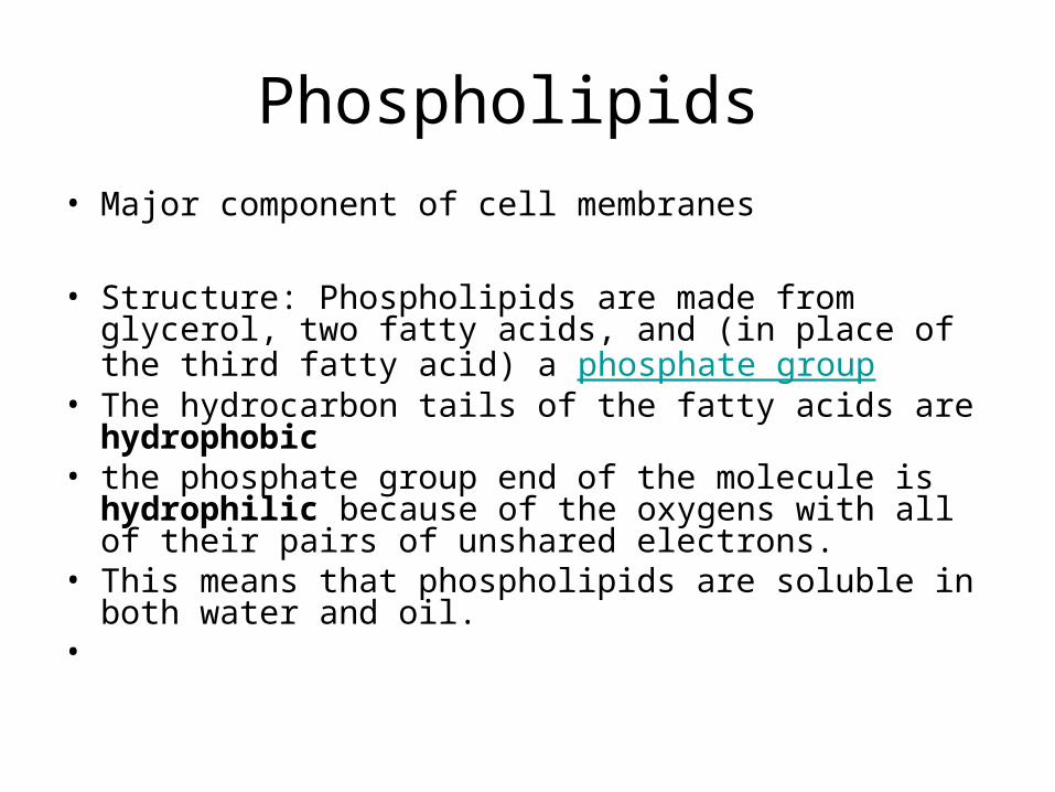
Phospholipids
• Major component of cell membranes
• Structure: Phospholipids are made from glycerol, two fatty acids, and (in place of the third fatty acid) a phosphate group
• The hydrocarbon tails of the fatty acids are hydrophobic• the phosphate group end of the molecule is hydrophilic
because of the oxygens with all of their pairs of unshared electrons.
• This means that phospholipids are soluble in both water and oil.
•

• Phospholipids have the special property of having both hydrophobic and hydrophilic parts
• A molecule that is both hydrophilic and hydrophobic is called amphipathic.

Structure of phospholipids

Function of Phospholipids
• Found in cell membranes. Act as barrier .
• When phospholipids are added to water they self assemble, with hydrophobic parts inward and hydrophilic parts outward.
• http://telstar.ote.cmu.edu/Hughes/tutorial/cellmembranes/orient2.swf

Another group of lipids are Steroids
• A lipid characterized by a carbon skeleton consisting of four fused rings. One common steroid is cholesterol.
• Many hormones are steroids, including sex hormones

• Tutorial 3.2 Macromolecules ( ann go to animations then to lipids)

Proteins
– Proteins are a major constituent of most cells (>50% dry weight)
– All proteins consist of polymers that are folded into specific conformations
– This conformation plus the chemistry of well-placed functional groups control a protein's function (another example of function follows form)
– Proteins are made up of 20 different types of amino-acid monomers

Polymers of amino acids are called peptides pg 14 sg
• Remember the structure of an amino acid. Carboxyl and amino group. A protein consists of polymers of amino acids, folded and coiled into a specific configuration.

• The side group determines characteristics, making it hydrophobic or hydrophilic, acidic or basic.

Coming together to make a peptide bond.
• amino acid basics
• Animation of Peptide Bond Formation
• Animation - Amino acid condensation
• The carboxyl group of one is adjacent to the amino group of the other, an enzyme can join the amino acids by dehydration reaction.

Several function of proteins. Pg 68 sg
• Structural proteins act as support. Examples include the silk fibers of spiders and insects, collagen in animal connective tissue, and keratin found in hair, horns, feathers.

Storage proteins
• Storage of amino acids. Examples are egg whites, casein, found in milk, and plants have storage proteins in their seeds.

Transport proteins
• Transport of other substances, Hemoglobin is an example, transporting oxygen from the lungs to other parts of the body. McGraw-Hill Online Learning Center Test<BLURT>

Hormonal Proteins
• They perform by coordinating of an organism's activities. An example is insulin.

Movement Proteins
• Used for movement. Examples are actin and myosin found in muscle tissue.

Defensive Proteins
• Protection from disease. Antibodies are an exampe.

Enzymatic Proteins
• Selective acceleration of chemical reactions. We will be discussing enzymes in detail later.

A proteins function depends on its conformation.
• A polypeptide is not the same as a protein.
• The chain of amino acids known as a polypeptide must be twisted and folded.
• Most proteins are globular (roughly spherical) others are fibrous in shape.

Shape is everything
• In order for a protein to function properly it must be able to be recognized and fit properly to another molecule. (This is important to remember when we speak of enzymes)

Polar and non polar amino acids (aa) pg 68 sg
• Polar aa have hydrophilic R groups
• Found on surface of proteins make them water soluble
• Make channels for hydrophilic substances
• Positive charged R groups allow – ions through
• Positive charged R groups allow – ions through
• Integral or transmembrane proteins

• Transmembrane protein p 68sg

Non polar aa/pg 68
• In center of water soluble proteins stabilize structure
• Remain embedded • Peripheral proteins

A proteins function depends on its conformation. Pg 66 sg
• A polypeptide is not the same as a protein.
• The chain of amino acids known as a polypeptide must be twisted and folded.
• Most proteins are globular (roughly spherical) others are fibrous in shape.

2 types you need to know pg. 66sg
• Globular - clumped into a shape of a ball. Major examples include insulin, hemoglobin, and most enzymes.
• http://student.ccbcmd.edu/~gkaiser/biotutorials/proteins/images/u4fg1b3.jpg
•

• Fibrous proteins Keratins - in wool, hair skin, fur, claws, nails, hooves, horns, scales, beaks, feathers, actin and mysin in muscle tissues and fibrinogen needed for blood clots.
• tropomycin

Four levels of protein structure all driven by chemical bonds/67 sg
• Primary structure: determined by base structure of the gene that codes fro the polypeptide.
• A chain of amino acids

Secondary Structure p 66+67
• Regular repeating structure including beta sheets, and alpha helices stabilized by hydrogen bonds between groups in the main chain of the polypeptide.

Two types of secondary structure
• Alpha - Helix: the first structure. It has a rod shape. The peptide is coiled around an imaginary cylinder and stabilized by hydrogen bonds formed between components of the peptide bonds

The second type
• Beta - pleated sheets: the amino acids adopt the conformation of a sheet of paper and the structure is stabilized by hydrogen bonds between amino acids in different polypeptide strands.

Lyzozyme

Tertiary structure pg 67
• The three dimensional conformation of a polypeptide.
• Stabilized by intramolecular bonds that form between aa in the
• Bonds form, ionic, hydrogen, hydrophobic interactions and disulfide bridges.


Quaternary Structure
• Linking together of 2 or more polypeptides to form a single protein


Hemoglobin pg 67
• Prosthetic groups – a non polypeptide structure contained in a protein
• Heme group linked to each of the four polypeptides in hemoglobin.
• Proteins with a prosthetic group are called conjugated proteins


Protein folding
• Interactive Concepts in Biochemistry - Interactive Animations
• http://www.stolaf.edu/people/giannini/flashanimat/proteins/protein structure.swf
• Proteins Structure Animation

• Tutorial 3.2 Macromolecules ( go to proteins )

Nucleic acids are informational proteins
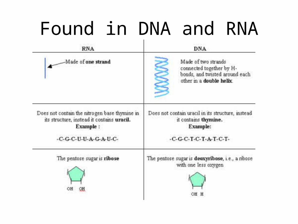
• First type of nucleic acid is deoxyribonucleic acid of DNA. The second type is ribonucleic acid or RNA.

• DNA is structurally different than RNA.
• The sugar in DNA is deoxyribose and in RNA the sugar is ribose

Nucleic acids are made of the monomer called nucleotides
• Nucleotides are made of three parts.
• 1. nitrogenous base• 2. pentose ( a five
carbon sugar)• 3. a phosphate group

Found in DNA and RNA

The Nitrogenous bases are 5 in type
• 1. Cytosine (C)
• 2. Thymine (T) found in DNA only
• 3. Uracil (U) found only in RNA
• 4. Adenine (A)
• 5. Guanine (G)

There are two families of nitrogenous bases
• The pyrimidines:• Has a six- membered
ring or carbon and nitrogen .
• These include cytosine, thymine, and uricil

• And Purines: • Larger, with the six
membered ring fused to a five membered ring.
• Includes: Adenine and guanine
Found in DNA and RNA

Building a Polynucleotide
• Polynucleotides are joined by covalent bonds between the phosphate sugar Forms phosphodiester bond
• This results in the backbone of DNA and RNA


Looks like this

DNA- you must be able to draw this
pg. 60 sg• All along the
appendages are attached the nitrogenous bases

• Tutorial 3.2 Macromolecules ( go to nucleic acids )

This all fits together
• The central dogma in molecular biology is:
• DNARNAprotein
• You must be able to draw DNA and RNA