Embed Size (px)
Citation preview

Full Terms & Conditions of access and use can be found athttp://www.tandfonline.com/action/journalInformation?journalCode=ktmp20
Download by: [Kagoshima University], [Professor Tomoyuki Kuwaki] Date: 24 November 2015, At: 02:59
Temperature
ISSN: 2332-8940 (Print) 2332-8959 (Online) Journal homepage: http://www.tandfonline.com/loi/ktmp20
Thermoregulation under pressure: a role fororexin neurons
Tomoyuki Kuwaki
To cite this article: Tomoyuki Kuwaki (2015) Thermoregulation under pressure: a role fororexin neurons, Temperature, 2:3, 379-391, DOI: 10.1080/23328940.2015.1066921
To link to this article: http://dx.doi.org/10.1080/23328940.2015.1066921
© 2015 The Author(s). Published withlicense by Taylor & Francis Group, LLC©Tomoyuki Kuwaki
Accepted author version posted online: 15Jul 2015.Published online: 15 Jul 2015.
Submit your article to this journal
Article views: 118
View related articles
View Crossmark data
Citing articles: 1 View citing articles

Thermoregulation under pressure:a role for orexin neurons
Tomoyuki Kuwaki*
Department of Physiology; Kagoshima University Graduate School of Medical and Dental Sciences; Kagoshima, Japan
Keywords: autonomic nervous system, breathing regulation, cardiovascular control, fight-or-flight, pain, stress, sleep,thermoregulation
Abbreviations: AP5, D-(¡)-2-amino-5-phosphonopentanoic acid; BAT, brown adipose tissue; CNQX, 6-cyano-7-nitroquinoxaline-2,3-dione; DHA, dorsal hypothalamic area; DMH, dorsomedial hypothalamus; GABA, g-aminobutylic acid; LHA, lateral hypotha-lamic area; ORX-AB, orexin-neuron-ablated mice; ORX-KO, orexin-knockout mice; PeF, perifornical area; PGE2, prostaglandin E2.
In the past, studies on stress responses and sleep/wake regulation were performed separately. The discovery oforexin (hypocretin) in 1998, however, dramatically changed the course of research and new findings regarding its rolein these complex processes provided a better insight into their interactions and intricacies. Orexin-containing neuronalactivity has been found to be minimal during sleep. It increases during the waking period and further increases duringthe active waking period, which includes stress responses and exploratory behaviors. Autonomic regulation of thebody, which includes body temperature, blood flow, and ventilation, is also activated along with the change invigilance states. Our recent findings suggest that orexin neurons act as a conductor of orchestration for vigilance states,behaviors, and autonomic functions. Body temperature regulation by orexin neurons seems to be mediated by one ofits cotransmitters while cardiovascular and respiratory regulation are mediated by orexin itself.
Introduction
Research on neural mechanisms of state-dependent adjust-ments of central autonomic regulation has been sparse, despitethe importance of this event from the perspective of quality oflife. In addition to calm and resting states, our daily life involvesmany perturbations that induce active conditions such as loco-motion, eating, and communication. During such active periods,cardiovascular, respiratory, and body temperature regulationneeds to be adjusted according to situational demands, which dif-fer from those during resting states, by modulating or resettinghomeostatic points.1 One of the neural substrates regulating suchadjustment, at least in the context of defensive behavior, appearsto be located in the dorsal hypothalamus because stimulation ofthis area elicits perfectly coordinated behavioral “rage” associatedwith its specific autonomic responses. This response was termedthe “defense response” and the area was termed the “defense area”of the hypothalamus.2
Several neurotransmitters have been proposed to be involved inthe modulation of the efferent pathways of defense responsesagainst stressors. For example, the activation of serotonin-1A recep-tors in the medullary raphe reduces cardiovascular changes,3 and
the inhibition of serotonin-3 receptors in the nucleus tractus soli-tarius prevents the inhibition of baroreflex bradycardia during thedefense response.4 Microinjections of adenosine into the rostralventrolateral medulla augment the increase in blood pressureinduced by electrical stimulation of the hypothalamic defensearea.5 The pros and cons of glutamate participation in the cardio-vascular component of the defense response have been a topic ofdebate.6,7 However, there is no report on the molecular basis of thedefense response and of the mechanism underlying the multiface-ted nature of simultaneous and coordinated changes in the cardio-vascular, respiratory, sensory, thermal, and behavioral parameters.Localization of orexin-containing cell bodies in the perifornicalarea (PeF) and dorsomedial hypothalamus (DMH) (Fig. 1), whichoverlap the “defense area,” prompted us to investigate the possiblerole of orexin in the defense response against stressors.
Orexin Neurons
Orexin (hypocretin)Orexins (orexin-A and orexin-B), also known as hypocretins
(hypocretin 1 and hypocretin 2, respectively), are hypothalamic
© Tomoyuki Kuwaki*Correspondence to: Tomoyuki Kuwaki; Email: [email protected]; URL: http://www.kufm.kagoshima-u.ac.jp/»physiol1/index-e.htmlSubmitted: 05/22/2015; Revised: 06/20/2015; Accepted: 06/22/2015http://dx.doi.org/10.1080/23328940.2015.1066921
This is an Open Access article distributed under the terms of the Creative Commons Attribution-Non-Commercial License (http://creativecommons.org/licenses/by-nc/3.0/), which permits unrestricted non-commercial use, distribution, and reproduction in any medium, provided the original work is properly cited. Themoral rights of the named author(s) have been asserted.
www.tandfonline.com 379Temperature
Temperature 2:3, 379--391; July/August/September 2015; Published with license by Taylor & Francis Group, LLCREVIEW
Dow
nloa
ded
by [
Kag
oshi
ma
Uni
vers
ity],
[Pr
ofes
sor
Tom
oyuk
i Kuw
aki]
at 0
2:59
24
Nov
embe
r 20
15
neuropeptides.8,9 They are cleaved from a common precursormolecule, prepro-orexin (130 residues), to form orexin-A(33 amino acids) and orexin-B (28 amino acids).9,10 Althoughorexins were first described as hypothalamic neuropeptides thatinfluenced appetite and consciousness, it was later found thatorexins also modulate reward processes,11 pain processing,12,13
and autonomic regulation of the cardiovascular,14-16 respira-tory,17,18 and neuroendocrine systems.19
Coexisting transmitter/modulatorsOrexin neurons contain not only orexin, but also other
putative neurotransmitter/modulator candidates such as gluta-mate,20-22 dynorphin,23 galanin,24 and nitric oxide.25 Dynorphinand glutamate may act synergistically with orexin to promotewakefulness.26 However, the precise role(s) of the substances thatare colocalized with orexin are largely unknown.
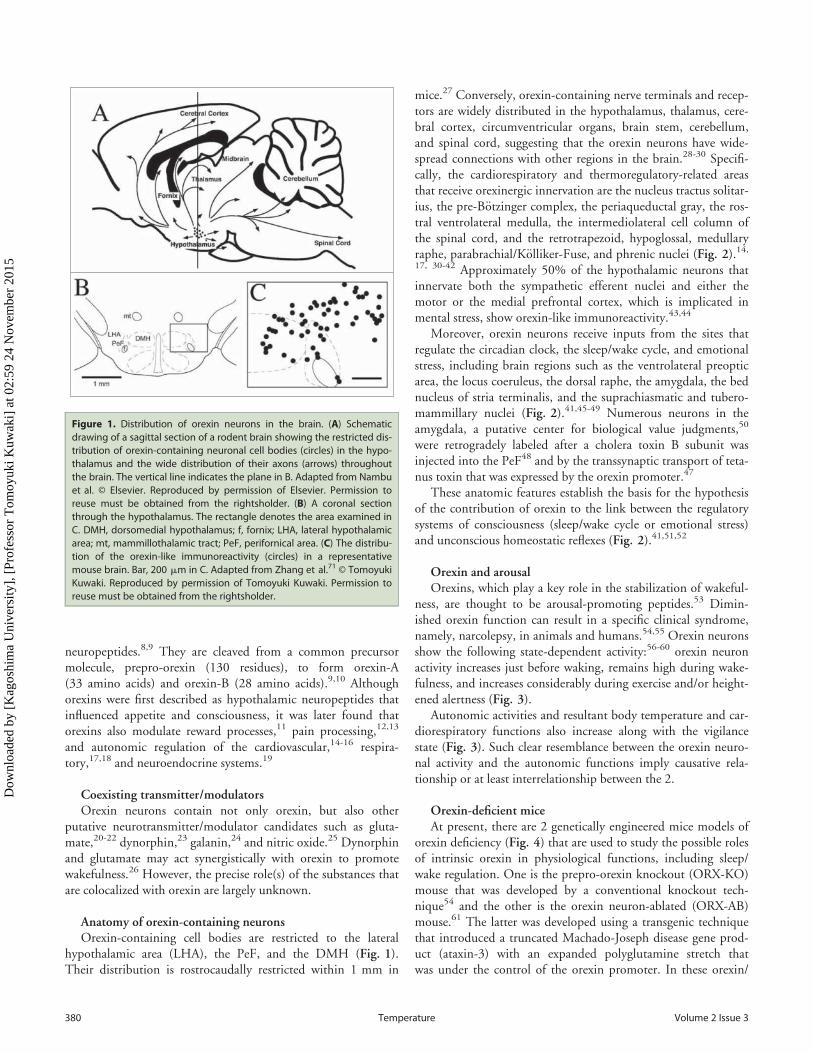
Anatomy of orexin-containing neuronsOrexin-containing cell bodies are restricted to the lateral
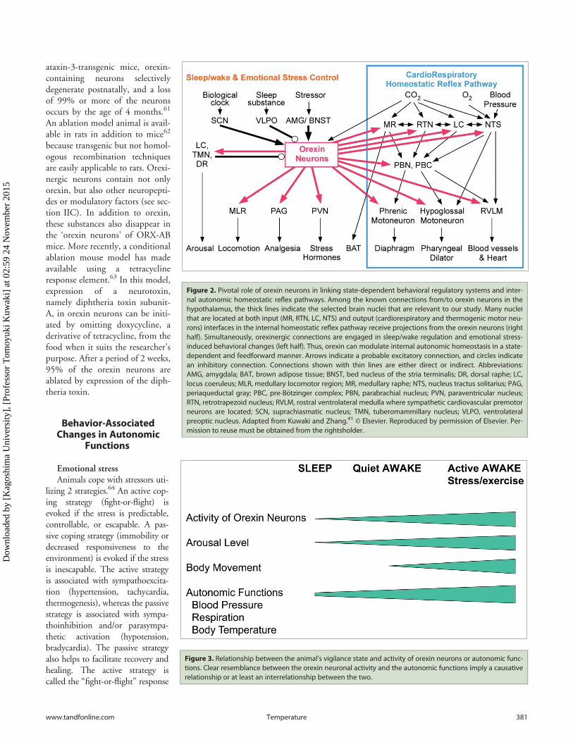
hypothalamic area (LHA), the PeF, and the DMH (Fig. 1).Their distribution is rostrocaudally restricted within 1 mm in
mice.27 Conversely, orexin-containing nerve terminals and recep-tors are widely distributed in the hypothalamus, thalamus, cere-bral cortex, circumventricular organs, brain stem, cerebellum,and spinal cord, suggesting that the orexin neurons have wide-spread connections with other regions in the brain.28-30 Specifi-cally, the cardiorespiratory and thermoregulatory-related areasthat receive orexinergic innervation are the nucleus tractus solitar-ius, the pre-B€otzinger complex, the periaqueductal gray, the ros-tral ventrolateral medulla, the intermediolateral cell column ofthe spinal cord, and the retrotrapezoid, hypoglossal, medullaryraphe, parabrachial/K€olliker-Fuse, and phrenic nuclei (Fig. 2).14,17, 30-42 Approximately 50% of the hypothalamic neurons thatinnervate both the sympathetic efferent nuclei and either themotor or the medial prefrontal cortex, which is implicated inmental stress, show orexin-like immunoreactivity.43,44
Moreover, orexin neurons receive inputs from the sites thatregulate the circadian clock, the sleep/wake cycle, and emotionalstress, including brain regions such as the ventrolateral preopticarea, the locus coeruleus, the dorsal raphe, the amygdala, the bednucleus of stria terminalis, and the suprachiasmatic and tubero-mammillary nuclei (Fig. 2).41,45-49 Numerous neurons in theamygdala, a putative center for biological value judgments,50
were retrogradely labeled after a cholera toxin B subunit wasinjected into the PeF48 and by the transsynaptic transport of teta-nus toxin that was expressed by the orexin promoter.47
These anatomic features establish the basis for the hypothesisof the contribution of orexin to the link between the regulatorysystems of consciousness (sleep/wake cycle or emotional stress)and unconscious homeostatic reflexes (Fig. 2).41,51,52
Orexin and arousalOrexins, which play a key role in the stabilization of wakeful-
ness, are thought to be arousal-promoting peptides.53 Dimin-ished orexin function can result in a specific clinical syndrome,namely, narcolepsy, in animals and humans.54,55 Orexin neuronsshow the following state-dependent activity:56-60 orexin neuronactivity increases just before waking, remains high during wake-fulness, and increases considerably during exercise and/or height-ened alertness (Fig. 3).
Autonomic activities and resultant body temperature and car-diorespiratory functions also increase along with the vigilancestate (Fig. 3). Such clear resemblance between the orexin neuro-nal activity and the autonomic functions imply causative rela-tionship or at least interrelationship between the 2.
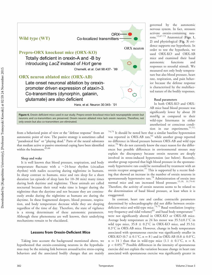
Orexin-deficient miceAt present, there are 2 genetically engineered mice models of
orexin deficiency (Fig. 4) that are used to study the possible rolesof intrinsic orexin in physiological functions, including sleep/wake regulation. One is the prepro-orexin knockout (ORX-KO)mouse that was developed by a conventional knockout tech-nique54 and the other is the orexin neuron-ablated (ORX-AB)mouse.61 The latter was developed using a transgenic techniquethat introduced a truncated Machado-Joseph disease gene prod-uct (ataxin-3) with an expanded polyglutamine stretch thatwas under the control of the orexin promoter. In these orexin/
Figure 1. Distribution of orexin neurons in the brain. (A) Schematicdrawing of a sagittal section of a rodent brain showing the restricted dis-tribution of orexin-containing neuronal cell bodies (circles) in the hypo-thalamus and the wide distribution of their axons (arrows) throughoutthe brain. The vertical line indicates the plane in B. Adapted from Nambuet al. © Elsevier. Reproduced by permission of Elsevier. Permission toreuse must be obtained from the rightsholder. (B) A coronal sectionthrough the hypothalamus. The rectangle denotes the area examined inC. DMH, dorsomedial hypothalamus; f, fornix; LHA, lateral hypothalamicarea; mt, mammillothalamic tract; PeF, perifornical area. (C) The distribu-tion of the orexin-like immunoreactivity (circles) in a representativemouse brain. Bar, 200 mm in C. Adapted from Zhang et al.71 © TomoyukiKuwaki. Reproduced by permission of Tomoyuki Kuwaki. Permission toreuse must be obtained from the rightsholder.
380 Volume 2 Issue 3Temperature
Dow
nloa
ded
by [
Kag
oshi
ma
Uni
vers
ity],
[Pr
ofes
sor
Tom
oyuk
i Kuw
aki]
at 0
2:59
24
Nov
embe
r 20
15
ataxin-3-transgenic mice, orexin-containing neurons selectivelydegenerate postnatally, and a lossof 99% or more of the neuronsoccurs by the age of 4 months.61
An ablation model animal is avail-able in rats in addition to mice62
because transgenic but not homol-ogous recombination techniquesare easily applicable to rats. Orexi-nergic neurons contain not onlyorexin, but also other neuropepti-des or modulatory factors (see sec-tion IIC). In addition to orexin,these substances also disappear inthe ‘orexin neurons’ of ORX-ABmice. More recently, a conditionalablation mouse model has madeavailable using a tetracyclineresponse element.63 In this model,expression of a neurotoxin,namely diphtheria toxin subunit-A, in orexin neurons can be initi-ated by omitting doxycycline, aderivative of tetracycline, from thefood when it suits the researcher’spurpose. After a period of 2 weeks,95% of the orexin neurons areablated by expression of the diph-theria toxin.
Behavior-AssociatedChanges in Autonomic
Functions
Emotional stressAnimals cope with stressors uti-
lizing 2 strategies.64 An active cop-ing strategy (fight-or-flight) isevoked if the stress is predictable,controllable, or escapable. A pas-sive coping strategy (immobility ordecreased responsiveness to theenvironment) is evoked if the stressis inescapable. The active strategyis associated with sympathoexcita-tion (hypertension, tachycardia,thermogenesis), whereas the passivestrategy is associated with sympa-thoinhibition and/or parasympa-thetic activation (hypotension,bradycardia). The passive strategyalso helps to facilitate recovery andhealing. The active strategy iscalled the “fight-or-flight” response
Figure 2. Pivotal role of orexin neurons in linking state-dependent behavioral regulatory systems and inter-nal autonomic homeostatic reflex pathways. Among the known connections from/to orexin neurons in thehypothalamus, the thick lines indicate the selected brain nuclei that are relevant to our study. Many nucleithat are located at both input (MR, RTN, LC, NTS) and output (cardiorespiratory and thermogenic motor neu-rons) interfaces in the internal homeostatic reflex pathway receive projections from the orexin neurons (righthalf). Simultaneously, orexinergic connections are engaged in sleep/wake regulation and emotional stress-induced behavioral changes (left half). Thus, orexin can modulate internal autonomic homeostasis in a state-dependent and feedforward manner. Arrows indicate a probable excitatory connection, and circles indicatean inhibitory connection. Connections shown with thin lines are either direct or indirect. Abbreviations:AMG, amygdala; BAT, brown adipose tissue; BNST, bed nucleus of the stria terminalis; DR, dorsal raphe; LC,locus coeruleus; MLR, medullary locomotor region; MR, medullary raphe; NTS, nucleus tractus solitarius; PAG,periaqueductal gray; PBC, pre-B€otzinger complex; PBN, parabrachial nucleus; PVN, paraventricular nucleus;RTN, retrotrapezoid nucleus; RVLM, rostral ventrolateral medulla where sympathetic cardiovascular premotorneurons are located; SCN, suprachiasmatic nucleus; TMN, tuberomammillary nucleus; VLPO, ventrolateralpreoptic nucleus. Adapted from Kuwaki and Zhang.41 © Elsevier. Reproduced by permission of Elsevier. Per-mission to reuse must be obtained from the rightsholder.
Figure 3. Relationship between the animal’s vigilance state and activity of orexin neurons or autonomic func-tions. Clear resemblance between the orexin neuronal activity and the autonomic functions imply a causativerelationship or at least an interrelationship between the two.
www.tandfonline.com 381Temperature
Dow
nloa
ded
by [
Kag
oshi
ma
Uni
vers
ity],
[Pr
ofes
sor
Tom
oyuk
i Kuw
aki]
at 0
2:59
24
Nov
embe
r 20
15
from a behavioral point of view or the “defense response” from anautonomic point of view. The passive strategy is sometimes called“paradoxical fear” or “playing dead.” Parts of the neural substratesthat mediate active vs passive emotional coping have been identifiedwithin the brainstem.65,66
Sleep and wakeIt is well known that blood pressure, respiration, and body
temperature fluctuate with a »24-hour rhythm (circadianrhythm) with nadirs occurring during nighttime in humans.In sharp contrast to humans, mice and rats sleep for a shortduration (an episode of sleep lasts for 10–30 min) many timesduring both daytime and nighttime. These animals are callednocturnal because their total wake time is longer during thenighttime than the daytime and not because they are continu-ously awake during the nighttime as humans are during thedaytime. In these fragmented sleepers, blood pressure, respira-tion, and body temperature decrease while they are sleepingregardless of the time of day. Therefore, the state of vigilanceis a strong determinant of these autonomic parameters.Although these phenomena are well known, their underlyingmechanisms remain to be elucidated.
Lessons from Orexin-Deficient Mice
Taking into account the background mentioned above, wehypothesized that orexin-containing neurons in the hypothala-mus may be the missing link between arousal/active stress-copingbehaviors and the associated bodily changes that are mainly
governed by the autonomicnervous system. In fact, stressorsactivate orexin-containing neu-rons.12,67-73 Anatomical (Figs. 1,2) and physiological (Fig. 3) evi-dence supports our hypothesis. Inorder to test the hypothesis, weused ORX-KO and ORX-ABmice and examined their basalautonomic functions andresponses to stressful stimuli. Wemeasured not only body tempera-ture but also blood pressure, heartrate, respiration, and pain behav-ior because the defense responseis characterized by the multiface-ted nature of the bodily responses.
Basal parametersIn both ORX-KO and ORX-
AB mice basal blood pressure wassignificantly lower by about 20mmHg as compared to theirwild-type littermates in eitheranesthetized or conscious condi-tion in our experiments.51,52,
74,75 It should be noted here that a similar baseline hypotensionwas reported in ORX-AB rats,62 while another group reportedno difference in blood pressure between ORX-AB and wild typemice.76 We do not currently know the exact reason for the differ-ences but possible differences in environmental stresses mayexplain the discrepancy because orexin neurons are deeplyinvolved in stress-induced hypertension (see below). Recently,another group reported that high blood pressure in the spontane-ously hypertensive rats could be normalized by treatment with anorexin receptor antagonist.77 This is supported by a recent find-ing that showed an increase in the number of orexin neurons inspontaneously hypertensive rats.78 Administration of orexin intonormal mice and rats increased blood pressure.15,18, 31,79-81
Therefore, the activity of orexin neurons seems to be related tothe determination of basal blood pressure, at least when it isexaggerated.
In contrast, heart rate and cardiac contractile parametersdetermined by echocardiography did not differ between orexin-deficient mice and wild-type mice.74,75 Basal ventilation (respira-tory frequency and tidal volume)82 and basal body temperature71
were not significantly altered in ORX-KO or ORX-AB mice.Average body temperature as 24 hrs mesor was 35.5§0.1�C inwild type mice, 35.8 § 0.2�C in ORX-KO mice, and 35.5§0.3�C in ORX-AB mice. However, change in body temperatureassociated with spontaneous exercise was significantly smaller inORX-KO (0.7 § 0.1�C, n D 8 ) and in ORX-AB (0.8 § 0.0�C,n D 14 ) than that in wild-type mice (1.1 § 0.1�C, n D 8,p < 0.05).83 Possible differences in the intensity of spontaneousexercise did not explain the variation because change in heart rateassociated with spontaneous exercise was significantly greater in
Figure 4. Orexin deficient mice used in our study. Prepro-orexin knockout mice lack neuropeptide orexin butneurons and co-transmitters are preserved. Orexin neuron ablated mice lack orexin neurons. Therefore, notonly orexin but also co-transmitters are eliminated.
382 Volume 2 Issue 3Temperature
Dow
nloa
ded
by [
Kag
oshi
ma
Uni
vers
ity],
[Pr
ofes
sor
Tom
oyuk
i Kuw
aki]
at 0
2:59
24
Nov
embe
r 20
15
ORX-KO (487 § 13 bpm) and inORX-AB (478 § 7 bpm) than inwild-type mice (437 § 9 bpm, p< 0.01).
A similar abnormality in themagnitude of state-dependentbody temperature change wasobserved when the animals fellasleep. The drop in body tempera-ture during sleep in ORX-KOmice was smaller than in wild-typemice.84 The same was true inhumans. Narcolepsy patientsshowed higher body temperatureduring sleep because their temper-ature drop was smaller than thecontrol subjects but not becausetheir body temperature was gener-ally high.85 Taken together, orexinneurons may not be involved inthe determination of basal bodytemperature when the animals areawake and at rest but do contributeto a change of body temperatureassociated with spontaneous move-ment and sleep even when there isno stimulation to the animals.
Cardiorespiratory responsesduring stress
To date, 3 lines of evidencesupport our hypothesis of the con-tribution of orexin to the defenseresponse. First, the stimulation ofthe PeF with the GABA-A receptorantagonist, bicuculline, resulted inan attenuated defense response inurethane-anesthetized ORX-KOand ORX-AB mice. Increases inarterial blood pressure, heart rate,respiratory frequency, and theß-band power of electroencephalo-gram measurements (an index ofcortical arousal) were smaller and/or shorter in ORX-KO mice thanin their wild-type littermates.74
Similarly, increased blood pres-sure, heart rate, and respiratoryminute volume and vascular dilata-tion in the skeletal muscle wereattenuated in ORX-AB mice.75
Secondly, the suppression ofthe baroreceptor reflex duringthe defense response was attenu-ated in ORX-AB mice, whereascharacteristics of the
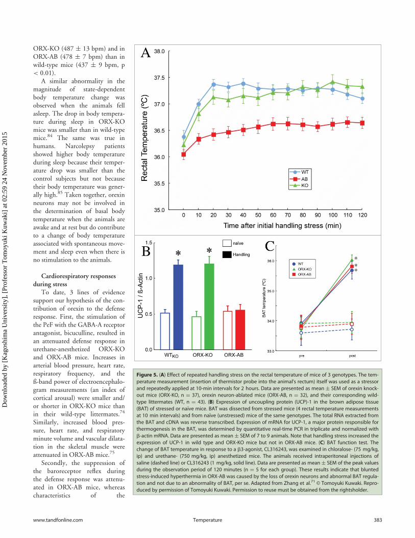
Figure 5. (A) Effect of repeated handling stress on the rectal temperature of mice of 3 genotypes. The tem-perature measurement (insertion of thermistor probe into the animal’s rectum) itself was used as a stressorand repeatedly applied at 10-min intervals for 2 hours. Data are presented as mean § SEM of orexin knock-out mice (ORX-KO, n D 37), orexin neuron-ablated mice (ORX-AB, n D 32), and their corresponding wild-type littermates (WT, n D 43). (B) Expression of uncoupling protein (UCP)-1 in the brown adipose tissue(BAT) of stressed or na€ıve mice. BAT was dissected from stressed mice (4 rectal temperature measurementsat 10 min intervals) and from na€ıve (unstressed) mice of the same genotypes. The total RNA extracted fromthe BAT and cDNA was reverse transcribed. Expression of mRNA for UCP-1, a major protein responsible forthermogenesis in the BAT, was determined by quantitative real-time PCR in triplicate and normalized withb-actin mRNA. Data are presented as mean § SEM of 7 to 9 animals. Note that handling stress increased theexpression of UCP-1 in wild type and ORX-KO mice but not in ORX-AB mice. (C) BAT function test. Thechange of BAT temperature in response to a b3-agonist, CL316243, was examined in chloralose- (75 mg/kg,ip) and urethane- (750 mg/kg, ip) anesthetized mice. The animals received intraperitoneal injections ofsaline (dashed line) or CL316243 (1 mg/kg, solid line). Data are presented as mean§ SEM of the peak valuesduring the observation period of 120 minutes (n D 5 for each group). These results indicate that bluntedstress-induced hyperthermia in ORX-AB was caused by the loss of orexin neurons and abnormal BAT regula-tion and not due to an abnormality of BAT, per se. Adapted from Zhang et al.71 © Tomoyuki Kuwaki. Repro-duced by permission of Tomoyuki Kuwaki. Permission to reuse must be obtained from the rightsholder.
www.tandfonline.com 383Temperature
Dow
nloa
ded
by [
Kag
oshi
ma
Uni
vers
ity],
[Pr
ofes
sor
Tom
oyuk
i Kuw
aki]
at 0
2:59
24
Nov
embe
r 20
15

baroreceptor reflex (gain and slope) at rest were normal inthese mice.75 During the defense response, the baroreceptor
reflex is suppressed or reset to ahigher-pressure range in orderto allow a higher blood pressurethan in resting conditions.1,86
The suppression of the barore-flex is mediated by the DMH-medullary link.4 Orexinappeared to contribute to thesuppression of the baroreflexduring defense responses, butnot to the baroreflex duringresting conditions. A pharmaco-logical study that used an orexinreceptor antagonist supports thisnotion.87
Third, an attenuation of thedefense response in the ORX-KOand ORX-AB mice was alsoobserved in the mice during natu-ral stimulation in unanesthetizedand freely moving conditions. Wetested the defense response inconscious animals using the resi-dent-intruder test or the air-jetstress paradigm in order to ruleout the possibility that theobserved differences between theorexin-deficient mice and theirwild-type littermates resultedfrom differences in anesthetic sus-ceptibilities. As expected, theemotional stressor-inducedincreases in blood pressure, heartrate, and locomotor activity weresmaller in orexin-deficient mice(ORX-KO and ORX-AB) than intheir wild-type littermates.74,75
Stress-induced analgesiaCardiorespiratory response is
not the sole characteristic of thedefense response. The defenseresponse is characterized by acoordinated change in cardiovas-cular, respiratory, sensory, andmotor functions. One of the mul-tifaceted features of the defenseresponse, stress-induced analgesia,was examined. In wild-type mice,foot shock induces long-lastinganalgesia, as evidenced byincreases in tail-flick latency fromnoxious hot water. AlthoughORX-KO mice showed moderate
analgesia, the effect was significantly smaller than that shown bytheir wild-type littermates.12
Figure 6. Effect of microinjections of PGE2 into the medial preoptic area on body temperature and the elec-tromyogram in the mice of the 4 genotypes. In chloralose (80 mg¢kg¡1)/urethane (800 mg¢kg¡1)-anaesthe-tised mice, ACSF (20 nL) and PGE2 (1 mg¢mL¡1 in ACSF) were sequentially microinjected into the medialpreoptic area (MPO) (C, D). Time-related changes in brown adipose tissue (BAT) temperature and nuchal EMG(a measure of shivering) are shown in A and B, respectively, and the changes expressed as the area under thecurve above the baseline in them for 70 min are summarized in A’ and B’, respectively. Data are presented asmean§ SEM of orexin-knockout mice (ORX-KO, n D 6), orexin neuron-ablated mice (ORX-AB, n D 6), and theircorresponding wild-type littermates (WTKO, n D 4 and WTAB, n D 5). (D) Summary for injected sites. Left-sideshows dye-distribution in WTKO and WTAB mice and right-side shows that of ORX-KO and ORX-AB mice. In theactual experiment, drugs and dye were injected into unilateral MPO. *p < 0.05 compared with baseline valuesbefore injection (Bonferroni’s post hoc test). n.s., not significant. ac, anterior commissure; MnPO, median pre-optic area. Adapted from Takahashi et al.91 © Tomoyuki Kuwaki. Reproduced by permission of TomoyukiKuwaki. Permission to reuse must be obtained from the rightsholder.
384 Volume 2 Issue 3Temperature
Dow
nloa
ded
by [
Kag
oshi
ma
Uni
vers
ity],
[Pr
ofes
sor
Tom
oyuk
i Kuw
aki]
at 0
2:59
24
Nov
embe
r 20
15
Stress-induced hyperthermiaBecause cardiorespiratory
responses during stress (see sec-tion IVB) were equally attenu-ated in ORX-KO and ORX-ABmice and stress-induced analgesiawas attenuated in ORX-KOmice (section IVC above), weconcluded at this point thatorexin was the main contributorto these responses, and colocal-ized transmitter/modulator can-didates had only a minor role, ifany. In line with this notion, wehypothesized that stress-inducedhyperthermia would also beinfluenced by orexin. On thecontrary, we found that ORX-AB mice, but not ORX-KOmice, had blunted handlingstress-induced hyperthermia(Fig. 5A).71 The brown adiposetissue (BAT), which is a majorthermogenic organ in rodents,did not respond to handlingstress (Fig. 5B), although it didrespond to direct pharmacologicstimulation (Fig. 5C). Theseabnormalities in ORX-AB micewere not observed in ORX-KOmice, in which the orexin pep-tide is deficient but the neuronsare preserved. A similar abnor-mality in stress-induced thermogenesis has recently beenreported in a rat model of orexin neuron ablation.88 Therefore,the integrity (orexin and other coexisting neurotransmitter/
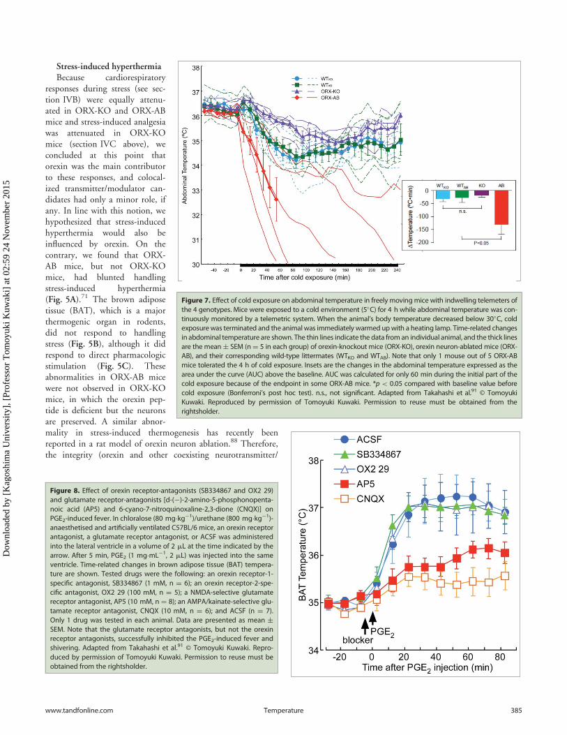
Figure 7. Effect of cold exposure on abdominal temperature in freely movingmice with indwelling telemeters ofthe 4 genotypes. Mice were exposed to a cold environment (5�C) for 4 h while abdominal temperature was con-tinuously monitored by a telemetric system. When the animal’s body temperature decreased below 30�C, coldexposurewas terminated and the animal was immediately warmed upwith a heating lamp. Time-related changesin abdominal temperature are shown. The thin lines indicate the data from an individual animal, and the thick linesare the mean§ SEM (nD 5 in each group) of orexin-knockout mice (ORX-KO), orexin neuron-ablated mice (ORX-AB), and their corresponding wild-type littermates (WTKO and WTAB). Note that only 1 mouse out of 5 ORX-ABmice tolerated the 4 h of cold exposure. Insets are the changes in the abdominal temperature expressed as thearea under the curve (AUC) above the baseline. AUC was calculated for only 60 min during the initial part of thecold exposure because of the endpoint in some ORX-AB mice. *p < 0.05 compared with baseline value beforecold exposure (Bonferroni’s post hoc test). n.s., not significant. Adapted from Takahashi et al.91 © TomoyukiKuwaki. Reproduced by permission of Tomoyuki Kuwaki. Permission to reuse must be obtained from therightsholder.
Figure 8. Effect of orexin receptor-antagonists (SB334867 and OX2 29)and glutamate receptor-antagonists [d-(¡)-2-amino-5-phosphonopenta-noic acid (AP5) and 6-cyano-7-nitroquinoxaline-2,3-dione (CNQX)] onPGE2-induced fever. In chloralose (80 mg¢kg¡1)/urethane (800 mg¢kg¡1)-anaesthetised and artificially ventilated C57BL/6 mice, an orexin receptorantagonist, a glutamate receptor antagonist, or ACSF was administeredinto the lateral ventricle in a volume of 2 mL at the time indicated by thearrow. After 5 min, PGE2 (1 mg¢mL¡1, 2 mL) was injected into the sameventricle. Time-related changes in brown adipose tissue (BAT) tempera-ture are shown. Tested drugs were the following: an orexin receptor-1-specific antagonist, SB334867 (1 mM, n D 6); an orexin receptor-2-spe-cific antagonist, OX2 29 (100 mM, n D 5); a NMDA-selective glutamatereceptor antagonist, AP5 (10 mM, n D 8); an AMPA/kainate-selective glu-tamate receptor antagonist, CNQX (10 mM, n D 6); and ACSF (n D 7).Only 1 drug was tested in each animal. Data are presented as mean §SEM. Note that the glutamate receptor antagonists, but not the orexinreceptor antagonists, successfully inhibited the PGE2-induced fever andshivering. Adapted from Takahashi et al.91 © Tomoyuki Kuwaki. Repro-duced by permission of Tomoyuki Kuwaki. Permission to reuse must beobtained from the rightsholder.
www.tandfonline.com 385Temperature
Dow
nloa
ded
by [
Kag
oshi
ma
Uni
vers
ity],
[Pr
ofes
sor
Tom
oyuk
i Kuw
aki]
at 0
2:59
24
Nov
embe
r 20
15
modulators) of the orexin neurons is indispensable for thecomplete expression of multiple facets of the fight-or-flightresponse.41,51, 52,89
Next, we asked whether theobserved abnormality in ORX-ABmice was restricted to handling stress-induced hyperthermia or whether itcould be generalized to other types ofstressors because different stressors acti-vate different brain regions.90 For thispurpose, we used 2 forms of thermo-genic perturbations including environ-mental cold exposure and brain PGE2injections that mimic inflammatoryfever.91 Again we found that ORX-ABmice, but not ORX-KO mice, exhib-ited a blunted PGE2-induced fever(Fig. 6) and intolerance to cold (5�C)exposure (Fig. 7). In addition, bodytemperature decrease in response toisoflurane anesthesia was greater inORX-AB mice than that in thewild-type mice.92 These findings weresimilar to the results previouslyobtained with handling stress-inducedthermogenesis.
As for the co-transmitter candi-dates in the orexin neurons, wefound that glutamate receptorantagonists (d-(¡)-2-amino-5-phos-phonopentanoic acid, AP-5 and 6-cyano-7-nitroquinoxaline-2,3-dione,CNQX) but not orexin receptorantagonists (SB334867 and OX229) successfully inhibited PGE2-induced fever in wild-type mice(Fig. 8).91 These results suggest thatorexin neurons are important ingeneral thermogenic processes, andtheir importance is not restricted tostress-induced thermogenesis. Inaddition, these results indicate thepossible involvement of glutamatein orexin neurons implicated inPGE2-induced fever.
Relevant Data from NormalRodents and Narcoleptic
Humans
Exogenous administration oforexin, co-transmitter candidates,and their antagonists
The exogenous administration oforexin induces both hyperthermia93,94
and hypothermia.95 Another report showed that orexin injectionsin the medullary raphe increased blood pressure and heart rate butnot BAT thermogenesis.96 Administration of an orexin-A receptor
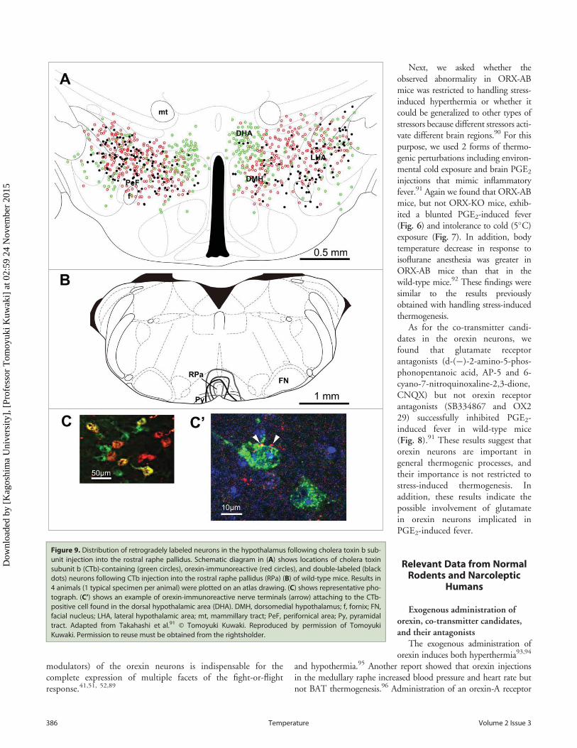
Figure 9. Distribution of retrogradely labeled neurons in the hypothalamus following cholera toxin b sub-unit injection into the rostral raphe pallidus. Schematic diagram in (A) shows locations of cholera toxinsubunit b (CTb)-containing (green circles), orexin-immunoreactive (red circles), and double-labeled (blackdots) neurons following CTb injection into the rostral raphe pallidus (RPa) (B) of wild-type mice. Results in4 animals (1 typical specimen per animal) were plotted on an atlas drawing. (C) shows representative pho-tograph. (C’) shows an example of orexin-immunoreactive nerve terminals (arrow) attaching to the CTb-positive cell found in the dorsal hypothalamic area (DHA). DMH, dorsomedial hypothalamus; f, fornix; FN,facial nucleus; LHA, lateral hypothalamic area; mt, mammillary tract; PeF, perifornical area; Py, pyramidaltract. Adapted from Takahashi et al.91 © Tomoyuki Kuwaki. Reproduced by permission of TomoyukiKuwaki. Permission to reuse must be obtained from the rightsholder.
386 Volume 2 Issue 3Temperature
Dow
nloa
ded
by [
Kag
oshi
ma
Uni
vers
ity],
[Pr
ofes
sor
Tom
oyuk
i Kuw
aki]
at 0
2:59
24
Nov
embe
r 20
15
antagonist, SB334867, inducedeither hyperthemia94,97 or had noeffect (our result, see Fig. 8). There-fore, no consensus has been reachedabout the possible role of orexinpeptides in thermoregulation. Dur-ing the course of our experimenta-tion, we found that PGE2-inducedBAT thermogenesis was normal inORX-KO mice but PGE2-inducedshivering was blunted in this mutant(Fig. 6). Therefore, we think thatorexin alone may play a role insome form(s) of the thermogenesis,but the main neurotransmitter thatis important for thermogenesis is aco-transmitter in the orexin neu-rons, which is most likely glutamate.
Microinjection of glutamatereceptor antagonists into theraphe pallidus inhibited the acti-vation of BAT sympatheticnerves that were evoked by stim-ulation of DMH/DHA,98 byPGE2,
99 and by cold expo-sure.100 Microinjection of a non-selective glutamate receptor antagonist, kynurenate, into theDMH/DHA inhibited BAT sympathetic activation evoked byPGE2 into the preoptic area.101 Conversely, dynorphin,102
nitric oxide,103 and galanin104 within the brain have beensuggested as thermolytic. Therefore, glutamate is the mostprobable transmitter for the thermogenesis associated withactivation of orexin neurons.
Functional neuroanatomyWe observed that numerous neurons with orexin-like immu-
noreactivity expressed c-Fos, a marker of neuronal activation,after foot shock stress,12,69 handling stress,71 PGE2-injection,and cold exposure.91 Moreover, disinhibition of the amygdala, aputative center for biological value judgments,50 induced signifi-cantly larger numbers of orexin-positive neurons that expressedc-Fos in the PeF/DMH (58.2 § 6.4%) than did the vehicle(18.2 § 4.4%).49
Transneuronal retrograde transport of a pseudorabies virusfrom the BAT, a key structure in nonshivering thermogenesis,identified the caudal raphe neurons as a site of orexinergic inner-vation32 and orexin-containing neurons in the hypothalamus.105
Retrograde transport of cholera toxin-B subunit from the caudalraphe nucleus identified hypothalamic neurons that containedorexin and neurons that did not contain orexin but were inner-vated by orexin (Fig. 9).91 The abovementioned neuroanatomi-cal data supports our hypothesis of the state-dependentmodulation of central thermogenic regulation by orexin neurons.
Recently, Dr. Nakamura’s group showed that activation ofDMH neurons but not of PeF/LH neurons including orexin neu-rons resulted in activation of the medullary raphe presympathetic
neurons and successive BAT thermogenesis.106 Together withour observation, we could conclude that orexin neurons in theDMH but not in PeF/LH contribute to stress-induced thermo-genesis. However, their method is unable to specifically stimulateonly orexin neurons and is activating other neurons as well. Inparticular, neurons containing melanin-concentrating hormoneintermingle in the LH with orexin neurons and activation ofthese neurons leads to hypotension, hypothermia, and sleep.107-109 Activation of both orexin neurons and melanin-concentratinghormone neurons may cancel their individual effects. Consider-ing that our conclusion was based on orexin neuron-specific loss-of-function experiments (namely knockout, ablation, and antag-onism), further studies using orexin neuron-specific gain-of-func-tion experiments such as optogenetic stimulation of geneticallydefined neurons are needed.
Human narcolepsyThere are only a few reports describing autonomic regulation
in narcoleptic patients. Sachs and Kaijser110 reported thatunmedicated narcoleptic patients showed attenuated autonomicreflexes (changes in blood pressure and heart rate) in a handgriptest and Valsalva maneuver, but not in face immersion or ortho-static tests. Because some, but not all, reflexes were disturbed,they proposed that peripheral nerves were intact and that thedefect was localized to the central nervous system. As for bodytemperature regulation, the amplitude of circadian rhythmicityof body temperature was small and this was because the nocturnaldrop was small.85 Plasma concentration of the stress hormoneadrenocorticosterone was lower in the patients than in the con-trols,111 indicating low stress sensitivity in the patients.
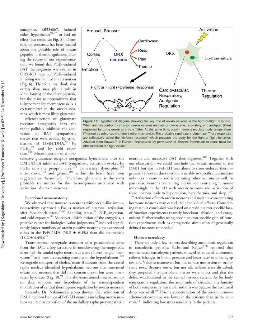
Figure 10. Hypothetical diagram showing the key role of orexin neurons in the fight-or-flight response.When animals confront a stressor, orexin neurons mediate cardiovascular, respiratory, and analgesic (Pain)responses by using orexin as a transmitter. At the same time, orexin neurons regulate body temperature(Thermo) by using cotransmitters other than orexin. The probable candidate is glutamate. These responsesare collectively called the “defense response” which prepares the body for the fight-or-flight behavior.Adapted from Kuwaki.51 © Elsevier. Reproduced by permission of Elsevier. Permission to reuse must beobtained from the rightsholder.
www.tandfonline.com 387Temperature
Dow
nloa
ded
by [
Kag
oshi
ma
Uni
vers
ity],
[Pr
ofes
sor
Tom
oyuk
i Kuw
aki]
at 0
2:59
24
Nov
embe
r 20
15

Concluding Remarks
Orexin neurons do not seem to individually regulate car-diovascular, respiratory, and body temperature systems butorchestrate them in a context-dependent manner (Fig. 10).Although vigilance state-dependent responses and emotionalstress-dependent responses may appear to be independent, weassume that the common features of these responses are state-dependent and feedforward adjustments of central ventilatoryand autonomic regulation in order to fit it to the situationaldemands that are associated with behavioral and metabolicchanges. An animal’s arousal state or alertness is minimalduring sleep, increases during quiet wakefulness, and furtherincreases during active wakefulness and involves a numberof activities such as exercise, food seeking, stress, or panic(Fig. 3). The level of this activation of arousal by orexin neu-rons is found to be greater in the dark (active period of thecircadian cycle) than in the light (inactive period) in noctur-nal mice. Ostensibly, independent autonomic regulation(associated with sleep/wake changes and with stress responses)seems to be a different facet of a single and continuous con-trol system in which orexin neurons play an important role.In line with this notion, orexin has recently been shown toplay a key role in the cardiovascular and behavioral responsesthat are associated with panic attacks in both animal modelsand humans.112
It is currently unknown why both orexin and its co-transmit-ter candidate(s) are required for the orchestration of state-depen-dent adjustment of central autonomic regulation. However,cotransmission allows for more complex communication betweenneurons. Diversity in the synaptic control of cardiovascular,
respiratory, and thermoregulatory neurons appears to be neces-sary for animals to adapt themselves to the constantly changingsituations and behavioral states (Fig. 10). The orexin and co-transmitter system is likely to function as one of the essentialmodulators for the orchestration of the circuits that control auto-nomic functions and behavior.
Disclosure of Potential Conflict of Interest
No potential conflicts of interest were disclosed.
Funding
Part of the work was supported by a Grant-in Aid for Scien-tific Research from the Ministry of Education, Culture, Sports,Science and Technology, Japan.
References
1. Kumada M, Terui N, Kuwaki T. Arterial baroreceptorreflex: its central and peripheral neural mechanisms.Prog Neurobiol 1990; 35:331-61; PMID:2263735;http://dx.doi.org/10.1016/0301-0082(90)90036-G
2. Hess WR. The Diencephalon: Autonomic and Extrapyra-midal Functions. Grune& Stratton, New York, 1954
3. Nalivaiko E, Ootsuka Y, Blessing WW. Activation of5-HT1A receptors in the medullary raphe reduces car-diovascular changes elicited by acute psychologicaland inflammatory stresses in rabbits. Am J PhysiolRegul Integr Comp Physiol 2005; 289:R596-R604;PMID:15802554; http://dx.doi.org/10.1152/ajpregu.00845.2004
4. S�evoz-Couche C, Comet MA, Hamon M, Laguzzi R.Role of nucleus tractus solitarius 5-HT3 receptors inthe defense reaction-induced inhibition of the aorticbaroreflex in rats. J Neurophysiol 2003; 90:2521-30;http://dx.doi.org/10.1152/jn.00275.2003
5. Thomas T, Spyer KM. The role of adenosine recep-tors in the rostral ventrolateral medulla in the cardio-vascular response to defence area stimulation in therat. Exp Physiol 1996; 81:67-77; PMID:8869140;http://dx.doi.org/10.1113/expphysiol.1996.sp003919
6. Sun MK, Guyenet PG. Hypothalamic glutamatergicinput to medullary sympathoexcitatory neurons inrats. Am J Physiol 1986; 251:R798-R810;PMID:3766781
7. Kiely JM, Gordon FJ. Role of rostral ventrolateralmedulla in centrally mediated pressor responses Am JPhysiol 1994; 267:H1549-56; PMID:7943401
8. de Lecea L, Kilduff T, Peyron C, Gao X, Foye P,Danielson P, Fukuhara C, Battenberg E, Gautvik V,
Bartlett Fn, et al. The hypocretins: hypothalamus-spe-cific peptides with neuroexcitatory activity. Proc NatlAcad Sci USA 1998; 95:322-7; PMID:9419374;http://dx.doi.org/10.1073/pnas.95.1.322
9. Sakurai T, Amemiya A, Ishii M, Matsuzaki I, Chem-elli RM, Tanaka H, Williams SC, Richarson JA,Kozlowski GP, Wilson S, et al. Orexins and orexinreceptors: a family of hypothalamic neuropeptidesand G protein-coupled receptors that regulate feedingbehavior. Cell 1998; 92:573-85; PMID:9491897;http://dx.doi.org/10.1016/S0092-8674(00)80949-6
10. Willie JT, Chemelli RM, Sinton CM, Yanagisawa M.To eat or to sleep? Orexin in the regulation of feedingand wakefulness. Annu Rev Neurosci 2001; 24:429-58; PMID:11283317; http://dx.doi.org/10.1146/annurev.neuro.24.1.429
11. Harris GC, Aston-Jones G. Arousal and reward: adichotomy in orexin function. Trends Neurosci 2006;29:571-7; PMID:16904760; http://dx.doi.org/10.1016/j.tins.2006.08.002
12. Watanabe S, Kuwaki T, Yanagisawa M, Fukuda Y,Shimoyama M. Persistent pain and stress activatepain-inhibitory orexin pathways. Neuroreport 2005;16:5-8; PMID:15618879; http://dx.doi.org/10.1097/00001756-200501190-00002
13. Yamamoto T, Nozaki-Taguchi N, Chiba T. Analgesiceffect of intrathecally administered orexin-A in the ratformalin test and in the rat hot plate test. Br J Phar-macol 2002; 137:170-6; PMID:12208773; http://dx.doi.org/10.1038/sj.bjp.0704851
14. Dun NJ, Le Dun S, Chen CT, Hwang LL, Kwok EH,Chang JK. Orexins: a role in medullary sympatheticoutflow. Regul Pept 2000; 96:65-70;
PMID:11102654; http://dx.doi.org/10.1016/S0167-0115(00)00202-0
15. Shirasaka T, Nakazato M, Matsukura S, Takasaki M,Kannan H. Sympathetic and cardiovascular actions oforexins in conscious rats. Am J Physiol 1999; 277:R1780-5; PMID:10600926
16. Zhang W, Shimoyama M, Fukuda Y, Kuwaki T.Multiple components of the defense response dependon orexin: Evidence from orexin knockout mice andorexin neuron-ablated mice. Autonom Neurosci:Basic Clin 2006; 126–127:139-45; PMID:16574499
17. Young JK, Wu M, Manaye KF, Kc P, Allard JS, MackSO, Haxhiu MA. Orexin stimulates breathing viamedullary and spinal pathways. J Appl Physiol 2005;98:1387-95; PMID:15557013; http://dx.doi.org/10.1152/japplphysiol.00914.2004
18. Zhang W, Fukuda Y, Kuwaki T. Respiratory and car-diovascular actions of orexin-A in mice. Neurosci Lett2005; 385:131-6; PMID:15941620; http://dx.doi.org/10.1016/j.neulet.2005.05.032
19. J�aszber�enyi M, Bujdos�o E, Pataki I, Telegdy G. Effectsof orexins on the hypothalamic-pituitary-adrenal sys-tem. J Neuroendocrinol 2000; 12:1174-8; http://dx.doi.org/10.1046/j.1365-2826.2000.00572.x
20. Abrahamson EE, Leak RK, Moore RY. The supra-chiasmatic nucleus projects to posterior hypothalamicarousal systems. Neuroreport 2001; 12:435-40;PMID:11209963; http://dx.doi.org/10.1097/00001756-200102120-00048
21. Rosin DL, Weston MC, Sevigny CP, Stornetta RL,Guyenet PG. Hypothalamic orexin (hypocretin) neu-rons express vesicular glutamate transportersVGLUT1 or VGLUT2. J Comp Neurol 2003;
About the Author
Tomoyuki Kuwaki is a Professorin Physiology at the KagoshimaUniversity Graduate School ofMedical and Dental Sciences(Japan). He is an editorial boardmember for the journals Journal ofPhysiological Sciences and Scien-tific Reports. His main researchinterest is brainmechanisms of stresscoping and relaxation of stress-induced autonomic malfunctions.
388 Volume 2 Issue 3Temperature
Dow
nloa
ded
by [
Kag
oshi
ma
Uni
vers
ity],
[Pr
ofes
sor
Tom
oyuk
i Kuw
aki]
at 0
2:59
24
Nov
embe
r 20
15

465:593-603; PMID:12975818; http://dx.doi.org/10.1002/cne.10860
22. Torrealba F, Yanagisawa M, Saper CB. Colocalizationof orexin A and glutamate immunoreactivity in axonterminals in the tuberomammillary nucleus in rats.Neurosci 2003; 119:1033-44; http://dx.doi.org/10.1016/S0306-4522(03)00238-0
23. Chou TC, Lee CE, Lu J, Elmquist JK, Hara J, WillieJT, Beuckmann CT, Chemelli RM, Sakurai T, Yana-gisawa M, et al. Orexin (hypocretin) neurons containdynorphin. J Neurosci 2001; 21:RC168(1-6);PMID:11567079
24. Hakansson M, de Lecea L, Sutcliffe JG, YanagisawaM, Meister B. Leptin receptor- and STAT3-immu-noreactivities in hypocretin/orexin neurones of the lat-eral hypothalamus. J Neuroendocrinol 1999; 11:653-63; PMID:10447804; http://dx.doi.org/10.1046/j.1365-2826.1999.00378.x
25. Cheng SB, Kuchiiwa S, Gao HZ, Kuchiiwa T, Naka-gawa S. Morphological study of orexin neurons in thehypothalamus of the Long-Evans rat, with special ref-erence to co-expression of orexin and NADPH-diaph-orase or nitric oxide synthase activities. Neurosci Res2003; 46:53-62; PMID:12725912; http://dx.doi.org/10.1016/S0168-0102(03)00026-9
26. Arrigoni E, Mochizuki T, Scammell TE. Activation ofthe basal forebrain by the orexin/hypocretin neurons.Acta Physiol 2010; 198:223-35; http://dx.doi.org/10.1111/j.1748-1716.2009.02036.x
27. Sunanaga J, Deng B-S, Zhang W, Kanmura Y,Kuwaki T. CO2 activates orexin-containing neuronsin mice. Respir Physiol Neurobiol 2009; 166:184-6;PMID:19442935; http://dx.doi.org/10.1016/j.resp.2009.03.006
28. Nambu T, Sakurai T, Mizukami K, Hosoya Y, Yana-gisawa M, Goto K. Distribution of orexin neurons inthe adult rat brain. Brain Res 1999; 827:243-60;PMID:10320718; http://dx.doi.org/10.1016/S0006-8993(99)01336-0
29. Elias CF, Saper CB, Maratos-Flier E, Tritos NA, LeeC, Kelly J, Tatro JB, Hoffman GE, Ollmann MM,Barsh GS, et al. Chemically defined projections link-ing the mediobasal hypothalamus and the lateralhypothalamic area. J Comp Neurol 1998; 402:442-59; PMID:9862320; http://dx.doi.org/10.1002/(SICI)1096-9861(19981228)402:4%3c442::AID-CNE2%3e3.0.CO;2-R
30. Marcus JN, Aschkenasi CJ, Lee CE, Chemelli RM,Saper CB, Yanagisawa M, Elmquist JK. Differentialexpression of orexin receptors 1 and 2 in the rat brain.J Comp Neurol 2001; 435:6-25; PMID:11370008;http://dx.doi.org/10.1002/cne.1190
31. Antunes VR, Brailoiu GC, Kwok EH, Scruggs P, DunNJ. Orexins/hypocretins excite rat sympathetic pre-ganglionic neurons in vivo and in vitro. Am J PhysiolRegul Integr Comp Physiol 2001; 281:R1801-7;PMID:11705764
32. Berthoud HR, Patterson LM, Sutton GM, MorrisonC, Zheng H. Orexin inputs to caudal raphe neuronsinvolved in thermal, cardiovascular, and gastrointesti-nal regulation. Histochem Cell Biol 2005; 123:147-56; PMID:15742197; http://dx.doi.org/10.1007/s00418-005-0761-x
33. Dergacheva O, Wang X, Huang ZG, Bouairi E, Ste-phens C, Gorini C, Mendelowitz D. Hypocretin-1(orexin-a) facilitates inhibitory and diminishes excit-atory synaptic pathways to cardiac vagal neurons inthe nucleus ambiguus. J Pharmacol Exp Ther 2005;314:1322-7; PMID:15947034; http://dx.doi.org/10.1124/jpet.105.086421
34. Fung SJ, Yamuy J, Sampogna S, Morales FR, ChaseMH. Hypocretin (orexin) input to trigeminal andhypoglossal motoneurons in the cat: a double-labelingimmunohistochemical study. Brain Res 2001;903:257-62; PMID:11382413; http://dx.doi.org/10.1016/S0006-8993(01)02318-6
35. Geerling JC, Mettenleiter TC, Loewy AD. Orexinneurons project to diverse sympathetic outflow
systems. Neuroscience 2003; 122:541-50;PMID:14614918; http://dx.doi.org/10.1016/j.neuroscience.2003.07.008
36. Peyron C, Tighe DK, van den Pol AN, de Lecea L,Heller HC, Sutcliffe JG, Kilduff TS. Neurons con-taining hypocretin (orexin) project to multiple neuro-nal systems. J Neurosci 1998; 18:9996-10015;PMID:9822755
37. Rosin DL, Chang DA, Guyenet PG. Afferent andefferent connections of the rat retrotrapezoid nucleus.J Comp Neurol 2006; 499:64-89; PMID:16958085;http://dx.doi.org/10.1002/cne.21105
38. Smith BN, Davis SF, Van Den Pol AN, Xu W. Selec-tive enhancement of excitatory synaptic activity in therat nucleus tractus solitarius by hypocretin 2. Neuro-science 2002; 115:707-14; PMID:12435409; http://dx.doi.org/10.1016/S0306-4522(02)00488-8
39. Volgin DV, Saghir M, Kubin L. Developmentalchanges in the orexin 2 receptor mRNA in hypoglossalmotoneurons. Neuroreport 2002; 13:433-6;PMID:11930155; http://dx.doi.org/10.1097/00001756-200203250-00014
40. Kuwaki T. Orexinergic modulation of breathingacross vigilance states. Respir Physiol Neurobiol 2008;164:204-12; PMID:18455970; http://dx.doi.org/10.1016/j.resp.2008.03.011
41. Kuwaki T, Zhang W. Orexin neurons as arousal-asso-ciated modulators of central cardiorespiratory regula-tion. Respir Physiol Neurobiol 2010; 174:43-54;PMID:20416404; http://dx.doi.org/10.1016/j.resp.2010.04.018
42. Nisimaru N, Mittal C, Shirai Y, Sooksawate T, Anan-daraj P, Hashikawa T, Nagao S, Arata A, Sakurai T,Yamamoto M, et al. Orexin-neuromodulated cerebel-lar circuit controls redistribution of arterial bloodflows for defense behavior in rabbits. Proc Natl AcadSci USA 2013; 110:14124-31; PMID:23912185;http://dx.doi.org/10.1073/pnas.1312804110
43. Krout KE, Mettenleiter TC, Loewy AD. Single CNSneurons link both central motor and cardiosympa-thetic systems: a double-virus tracing study. Neurosci-ence 2003; 118:853-66; PMID:12710992; http://dx.doi.org/10.1016/S0306-4522(02)00997-1
44. Krout KE, Mettenleiter TC, Karpitskiy V, NguyenXV, Loewy AD. CNS neurons with links to bothmood-related cortex and sympathetic nervous system.Brain Res 2005; 1050:199-202; PMID:15975562;http://dx.doi.org/10.1016/j.brainres.2005.04.090
45. Saper CB, Scammell TE, Lu J. Hypothalamic regula-tion of sleep and circadian rhythms. Nature 2005;437:1257-63; PMID:16251950; http://dx.doi.org/10.1038/nature04284
46. Sakurai T. The neural circuit of orexin (hypocretin):maintaining sleep and wakefulness. Nat Rev Neurosci2007; 8:171-81; PMID:17299454; http://dx.doi.org/10.1038/nrn2092
47. Sakurai T, Nagata R, Yamanaka A, Kawamura H,Tsujino N, Muraki Y, Kageyama H, Kunita S, Taka-hashi S, Goto K, et al. Input of orexin/hypocretinneurons revealed by a genetically encoded tracer inmice. Neuron 2005; 46:297-308; PMID:15848807;http://dx.doi.org/10.1016/j.neuron.2005.03.010
48. Yoshida K, McCormack S, Espana RA, Crocker A,Scammell TE. Afferents to the orexin neurons of therat brain. J Comp Neurol 2006; 494:845-61;PMID:16374809; http://dx.doi.org/10.1002/cne.20859
49. Zhang W, Zhang N, Sakurai T, Kuwaki T. Orexinneurons in the hypothalamus mediate cardiorespira-tory responses induced by disinhibition of the amyg-dala and bed nucleus of the stria terminalis. Brain Res2009; 1262:25-37; PMID:19368849; http://dx.doi.org/10.1016/j.brainres.2009.01.022
50. Pitkanen A, Savander V, LeDoux JE. Organization ofintra-amygdaloid circuitries in the rat: an emergingframework for understanding functions of the amyg-dala. Trends Neurosci 1997; 20:517-23;
PMID:9364666; http://dx.doi.org/10.1016/S0166-2236(97)01125-9
51. Kuwaki T. Orexin links emotional stress to autonomicfunctions. Autonom Neurosci 2011; 161:20-7;PMID:20813590; http://dx.doi.org/10.1016/j.autneu.2010.08.004
52. Kuwaki T, Zhang W. Orexin Neurons and EmotionalStress. In Sleep Hormones (Vitamins and Hormones vol89). Academic Press, New York, 2012; 135-58.PMID: 22640612
53. Carter ME, Adamantidis A, Ohtsu H, Deisseroth K,de Lecea L. Sleep homeostasis modulates hypocretin-mediated sleep-to-wake transitions. J Neurosci 2009;29:10939-49; PMID:19726652; http://dx.doi.org/10.1523/JNEUROSCI.1205-09.2009
54. Chemelli RM, Willie JT, Sinton CM, Elmquist JK,Scammell TE, Lee C, Richardson JA, Williams SC,Xiong Y, Kisanuki Y, et al. Narcolepsy in orexinknockout mice: molecular genetics of sleep regulation.Cell 1999; 98:437-51; PMID:10481909; http://dx.doi.org/10.1016/S0092-8674(00)81973-X
55. Thannickal TC, Moore RY, Nienhuis R, RamanathanL, Gulyani S, Aldrich M, Cornford M, Siegel JM.Reduced number of hypocretin neurons in humannarcolepsy. Neuron 2000; 27:469-74;PMID:11055430; http://dx.doi.org/10.1016/S0896-6273(00)00058-1
56. Lee M, Hassani O, Jones B. Discharge of identifiedorexin/hypocretin neurons across the sleep-wakingcycle. J Neurosci 2005; 25:6716-20;PMID:16014733; http://dx.doi.org/10.1523/JNEUROSCI.1887-05.2005
57. Mileykovskiy B, Kiyashchenko L, Siegel J. Behavioralcorrelates of activity in identified hypocretin/orexinneurons. Neuron 2005; 46:787-98;PMID:15924864; http://dx.doi.org/10.1016/j.neuron.2005.04.035
58. Takahashi K, Lin JS, Sakai K. Neuronal activity oforexin and non-orexin waking-active neurons duringwake–sleep states in the mouse. Neuroscience 2008;153:860-70; PMID:18424001; http://dx.doi.org/10.1016/j.neuroscience.2008.02.058
59. Sakurai T. The role of orexin in motivated behaviors.Nature Rev Neurosci 2014; 15:719-31; http://dx.doi.org/10.1038/nrn3837
60. Mahler S, Moorman D, Smith R, James M, Aston-Jones G. Motivational activation: a unifying hypothe-sis of orexin/hypocretin function. Nat Neurosci 2014;17:1298-303; PMID:25254979; http://dx.doi.org/10.1038/nn.3810
61. Hara J, Beuckmann CT, Nambu T, Willie JT, Chem-elli RM, Sinton CM, Sugiyama F, Yagami K, Goto K,Yanagisawa M, et al. Genetic ablation of orexin neu-rons in mice results in narcolepsy, hypophagia, andobesity. Neuron 2001; 30:345-54; PMID:11394998;http://dx.doi.org/10.1016/S0896-6273(01)00293-8
62. Schwimmer H, Stauss HM, Abboud F, Nishino S,Mignot E, Zeitzer JM. Effects of sleep on the cardio-vascular and thermoregulatory systems: a possible rolefor hypocretins. J Appl Physiol 2010; 109:1053-63;PMID:20705949; http://dx.doi.org/10.1152/japplphysiol.00516.2010
63. Tabuchi S, Tsunematsu T, Black S, Tominaga M, Mar-uyama M, Takagi K, Minokoshi Y, Sakurai T, KilduffT, Yamanaka A. Conditional ablation of orexin/hypo-cretin neurons: a new mouse model for the study of nar-colepsy and orexin system function. J Neurosci 2014;34:6495-509; PMID:24806676; http://dx.doi.org/10.1523/JNEUROSCI.0073-14.2014
64. Korte S, Koolhaas J, Wingfield J, McEwen B. TheDarwinian concept of stress: benefits of allostasis andcosts of allostatic load and the trade-offs in health anddisease. Neurosci Biobehav Rev 2005; 29:3-38;PMID:15652252; http://dx.doi.org/10.1016/j.neubiorev.2004.08.009
65. Nosaka S. Modifications of arterial baroreflexes: oblig-atory roles in cardiovascular regulation in stress andpoststress recovery. Jpn J Physiol 1996; 46:271-88;
www.tandfonline.com 389Temperature
Dow
nloa
ded
by [
Kag
oshi
ma
Uni
vers
ity],
[Pr
ofes
sor
Tom
oyuk
i Kuw
aki]
at 0
2:59
24
Nov
embe
r 20
15
PMID:8988438; http://dx.doi.org/10.2170/jjphysiol.46.271
66. Bandler R, Keay KA, Floyd N, Price J. Central circuitsmediating patterned autonomic activity during activevs. passive emotional coping. Brain Res Bull 2000;53:95-104; PMID:11033213; http://dx.doi.org/10.1016/S0361-9230(00)00313-0
67. Espana RA, Valentino RJ, Berridge CW. Fos immu-noreactivity in hypocretin-synthesizing and hypocre-tin-1 receptor-expressing neurons: effects of diurnaland nocturnal spontaneous waking, stress and hypo-cretin-1 administration. Neuroscience 2003;121:201-17; PMID:12946712; http://dx.doi.org/10.1016/S0306-4522(03)00334-8
68. Ida T, Nakahara K, Murakami T, Hanada R, Naka-zato M, Murakami N. Possible involvement of orexinin the stress reaction in rats. Biochem Biophys ResCommun 2000; 270:318-23; PMID:10733946;http://dx.doi.org/10.1006/bbrc.2000.2412
69. Kuwaki T, Zhang W, Nakamura A. State-dependentadjustment of the central autonomic regulation: Roleof orexin in emotional behavior and sleep/wake cycle.In Central Mechanisms of Cardiovascular Regulation.(ed. Kubo T, Kuwaki T) Research Signport, Kerala(India), 2007; 57-73
70. Winsky-Sommerer R, Yamanaka A, Diano S, BorokE, Roberts AJ, Sakurai T, Kilduff TS, Horvath TL, deLecea L. Interaction between the corticotropin-releas-ing factor system and hypocretins (orexins): a novelcircuit mediating stress response. J Neurosci 2004;24:11439-48; PMID:15601950; http://dx.doi.org/10.1523/JNEUROSCI.3459-04.2004
71. Zhang W, Sunanaga J, Takahashi Y, Mori T, SakuraiT, Kanmura Y, Kuwaki T. Orexin neurons are indis-pensable for stress-induced thermogenesis in mice. JPhysiol 2010; 588:4117-29; PMID:20807795;http://dx.doi.org/10.1113/jphysiol.2010.195099
72. Zhu L, Onaka T, Sakurai T, Yada T. Activation oforexin neurones after noxious but not conditionedfear stimuli in rats. Neuroreport 2002; 13:1351-3;PMID:12151801; http://dx.doi.org/10.1097/00001756-200207190-00027
73. Furlong TM, Vianna DML, Liu L, Carrive P. Hypo-cretin/orexin contributes to the expression of somebut not all forms of stress and arousal. Eur J Neurosci2009; 30:1603-14; PMID:19811530; http://dx.doi.org/10.1111/j.1460-9568.2009.06952.x
74. Kayaba Y, Nakamura A, Kasuya Y, Ohuchi T, Yanagi-sawa M, Komuro I, Fukuda Y, Kuwaki T. Attenuateddefense response and low basal blood pressure inorexin knockout mice. Am J Physiol Regul IntegrComp Physiol 2003; 285:R581-93;PMID:12750151; http://dx.doi.org/10.1152/ajpregu.00671.2002
75. Zhang W, Sakurai T, Fukuda Y, Kuwaki T. Orexinneuron-mediated skeletal muscle vasodilation andshift of baroreflex during defense response in mice.Am J Physiol Regul Integr Comp Physiol 2006; 290:R1654-63; PMID:16410401; http://dx.doi.org/10.1152/ajpregu.00704.2005
76. Bastianini S, Silvani A, Berteotti C, Elghozi J-L, Fran-zini C, Lenzi P, Martire VL, Zoccoli G. Sleep relatedchanges in blood pressure in hypocretin-deficient nar-coleptic mice. Sleep 2011; 34:213-8;PMID:21286242
77. Li A, Hindmarch C, Nattie E, Paton J. Antagonism oforexin receptors significantly lowers blood pressure inspontaneously hypertensive rats. J Physiol 2013;591:4237-48; PMID:23671161; http://dx.doi.org/10.1113/jphysiol.2013.256271
78. Clifford L, Dampney BW, Carrive P. Spontaneouslyhypertensive rats have more orexin neurons in theirmedial hypothalamus than normotensive rats. ExpPhysiol 2015; 100:388-98; PMID:25640802; http://dx.doi.org/10.1113/expphysiol.2014.084137
79. Chen CT, Hwang LL, Chang JK, Dun NJ. Pressoreffects of orexins injected intracisternally and to rostralventrolateral medulla of anesthetized rats. Am J
Physiol Regul Integr Comp Physiol 2000; 278:R692-7; PMID:10712290
80. Machado BH, Bonagamba LG, Dun SL, Kwok EH,Dun NJ. Pressor response to microinjection of orexin/hypocretin into rostral ventrolateral medulla of awakerats. Regul Pept 2002; 104:75-81; PMID:11830280;http://dx.doi.org/10.1016/S0167-0115(01)00351-2
81. Samson WK, Gosnell B, Chang JK, Resch ZT, Mur-phy TC. Cardiovascular regulatory actions of thehypocretins in brain. Brain Res 1999; 831:248-53;PMID:10412003; http://dx.doi.org/10.1016/S0006-8993(99)01457-2
82. Nakamura A, Zhang W, Yanagisawa M, Fukuda Y,Kuwaki T. Vigilance state-dependent attenuation ofhypercapnic chemoreflex and exaggerated sleep apneain orexin knockout mice. J Appl Physiol 2007;102:241-8; PMID:16959906; http://dx.doi.org/10.1152/japplphysiol.00679.2006
83. Miyata K, Ootsuka Y, Kuwaki T. Ultradian rhythm iscoordinated by brain system with involvement oforexin neuron system. J Physiol Sci 2012; 62:S215
84. Mochizuki T, Klerman E, Sakurai T, Scammell T.Elevated body temperature during sleep in orexinknockout mice. Am J Physiol Regul Integr CompPhysiol 2006; 291:R533-40; PMID:16556901;http://dx.doi.org/10.1152/ajpregu.00887.2005
85. Mosko S, Holowach J, Sassin J. The 24-hour rhythmof core temperature in narcolepsy. Sleep 1983; 6:137-46; PMID:6878983
86. McDowall LM, Horiuchi J, Killinger S, DampneyRAL. Modulation of the baroreceptor reflex by thedorsomedial hypothalamic nucleus and perifornicalarea. Am J Physiol Regul Integr Comp Physiol 2006;290:R1020-6; PMID:16284085; http://dx.doi.org/10.1152/ajpregu.00541.2005
87. Iigaya K, Horiuchi J, McDowall LM, Lam AC, SediqiY, Polson JW, Carrive P, Dampney RA. Blockade oforexin receptors with Almorexant reduces cardiorespi-ratory responses evoked from the hypothalamus butnot baro- or chemoreceptor reflex responses. Am JPhysiol Regul Integr Comp Physiol 2012; 303:R1011-22; PMID:23019212; http://dx.doi.org/10.1152/ajpregu.00263.2012
88. Mohammed M, Ootsuka Y, Yanagisawa M, BlessingW. Reduced brown adipose tissue thermogenesis dur-ing environmental interactions in transgenic rats withataxin-3-mediated ablation of hypothalamic orexinneurons. Am J Physiol Regul Integr Comp Physiol2014; 307:R978-89; PMID:25324552; http://dx.doi.org/10.1152/ajpregu.00260.2014
89. Kuwaki T. A key role of orexin (hypocretin) neuronsin the fight-or-flight response. Physiology News 2011;83:15-7
90. Senba E, Ueyama T. Stress-induced expression ofimmediate early genes in the brain and peripheralorgans of the rat Neurosci Res 1997; 29:183-207;PMID:9436645; http://dx.doi.org/10.1016/S0168-0102(97)00095-3
91. Takahashi Y, Zhang W, Sameshima K, Kuroki C,Matsumoto A, Sunanaga J, Kono Y, Sakurai T, Kan-mura Y, Kuwaki T. Orexin neurons are indispensablefor prostaglandin E2-induced fever and defenceagainst environmental cooling in mice. J Physiol2013; 591:5623-43; PMID:23959674; http://dx.doi.org/10.1113/jphysiol.2013.261271
92. Kuroki C, Takahashi Y, Ootsuka Y, Kanmura Y,Kuwaki T. The impact of hypothermia on emergencefrom isoflurane anesthesia in orexin neuron-ablatedmice. Anesthesia and Analgesia 2013; 116:1001-5;PMID:23477964; http://dx.doi.org/10.1213/ANE.0b013e31828842f0
93. Monda M, Viggiano A, Luca VD. Paradoxical effectof orexin A: hypophagia induced by hyperthermia.Brain Res 2003; 961:220-8; PMID:12531489; http://dx.doi.org/10.1016/S0006-8993(02)03953-7
94. Tupone D, Madden CJ, Cano G, Morrison SF. Anorexinergic projection from perifornical hypothalamusto raphe pallidus increases rat brown adipose tissue
thermogenesis. J Neurosci 2011; 31:15944-55;PMID:22049437; http://dx.doi.org/10.1523/JNEUROSCI.3909-11.2011
95. Sz�ekely M, P�eterv�ari E, Balask�o M, Hern�adi I, UzsokiB. Effects of orexins on energy balance and thermo-regulation. Regul Pept 2002; 104:47-53; http://dx.doi.org/10.1016/S0167-0115(01)00348-2
96. Luong LNL, Carrive P. Orexin microinjection in themedullary raphe increases heart rate and arterial pres-sure but does not reduce tail skin blood flow in theawake rat. Neurosci 2012; 202:209-17; http://dx.doi.org/10.1016/j.neuroscience.2011.11.073
97. Verty ANA, Allen AM, Oldfield BJ. The endogenousactions of hypothalamic peptides on brown adiposetissue thermogenesis in the rat. Endocrinology 2010;151:4236-46; PMID:20685882; http://dx.doi.org/10.1210/en.2009-1235
98. Cao W-H, Morrison SF. Glutamate receptors in theraphe pallidus mediate brown adipose tissue thermo-genesis evoked by activation of dorsomedial hypotha-lamic neurons. Neuropharmacol 2006; 51:426-37;http://dx.doi.org/10.1016/j.neuropharm.2006.03.031
99. Madden CJ, Morrison SF. Excitatory amino acidreceptor activation in the raphe pallidus area mediatesprostaglandin-evoked thermogenesis. Neurosci 2003;122:5-15; http://dx.doi.org/10.1016/S0306-4522(03)00527-X
100. Nakamura K, Morrison SF. Central efferent pathwaysmediating skin cooling-evoked sympathetic thermo-genesis in brown adipose tissue. Am J Physiol RegulIntegr Comp Physiol 2007; 292:R127-36;PMID:16931649; http://dx.doi.org/10.1152/ajpregu.00427.2006
101. Madden CJ, Morrison SF. Excitatory amino acidreceptors in the dorsomedial hypothalamus mediateprostaglandin-evoked thermogenesis in brown adiposetissue. Am J Physiol Regul Integr Comp Physiol2004; 286:R320-5; PMID:14563660; http://dx.doi.org/10.1152/ajpregu.00515.2003
102. Handler CM, Piliero TC, Geller EB, Adler MW.Effect of ambient temperature on the ability of mu-,kappa- and delta-selective opioid agonists to modulatethermoregulatory mechanisms in the rat. J PharmacolExp Ther 1994; 268:847-55; PMID:8113997
103. Steiner AA, Antunes-Rodrigues J, McCann SM,Branco LG. Antipyretic role of the NO-cGMP path-way in the anteroventral preoptic region of the ratbrain. Am J Physiol Regul Integr Comp Physiol 2002;282:R584-93; PMID:11792670; http://dx.doi.org/10.1152/ajpregu.00391.2001
104. Lyudyno V, Krasnova I, Smirnova M, Klimenko V.Antipyretic effect of neuropeptide galanin in endo-toxin-induced fever. Bull Exp Biol Med 2001; 1:60-3;http://dx.doi.org/10.1023/A:1017538814753
105. Oldfield BJ, Giles ME, Watson A, Anderson C, Col-vill LM, McKinley MJ. The neurochemical characteri-sation of hypothalamic pathways projectingpolysynaptically to brown adipose tissue in the rat.Neuroscience 2002; 110:515-26; PMID:11906790;http://dx.doi.org/10.1016/S0306-4522(01)00555-3
106. Kataoka N, Hioki H, Kaneko T, Nakamura K. Psy-chological stress activates a dorsomedial hypothala-mus-medullary raphe circuit driving brown adiposetissue thermogenesis and hyperthermia. Cell Metab2014; 20:1-13; PMID:24988453; http://dx.doi.org/10.1016/j.cmet.2014.05.018
107. Takase K, Kikuchi K, Tsuneoka Y, Oda S, Kuroda M,Funato H. Meta-analysis of melanin-concentratinghormone signaling-deficient mice on behavioral andmetabolic phenotypes. Plos one 2014; 9:e99961;PMID:24924345; http://dx.doi.org/10.1371/journal.pone.0099961
108. Glick M, Segal-Lieberman G, Cohen R, Kronfeld-Schor N. Chronic MCH infusion causes a decrease inenergy expenditure and body temperature, and anincrease in serum IGF-1 levels in mice. Endocrine2009; 36:479-85; PMID:19859841; http://dx.doi.org/10.1007/s12020-009-9252-5
390 Volume 2 Issue 3Temperature
Dow
nloa
ded
by [
Kag
oshi
ma
Uni
vers
ity],
[Pr
ofes
sor
Tom
oyuk
i Kuw
aki]
at 0
2:59
24
Nov
embe
r 20
15

109. Tsunematsu T, Ueno T, Tabuchi S, Inutsuka A,Tanaka K, Hasuwa H, Kilduff T, Terao A, Yama-naka A. Optogenetic manipulation of activity andtemporally controlled cell-specific ablation reveal arole for MCH neurons in sleep/wake regulation. JNeurosci 2014; 34:6896-909; PMID:24828644;http://dx.doi.org/tlse=10.1523/JNEUROSCI.5344-13.2014
110. Sachs C, Kaijser L. Autonomic regulation of cardio-pulmonary functions in sleep apnea syndrome andnarcolepsy. Sleep 1982; 5:227-38; PMID:6813932
111. Kok S, Roelfsema F, Overeem S, Lammers G, StrijersR, Fr€olich M, Meinders A, Pijl H. Dynamics of thepituitary-adrenal ensemble in hypocretin-deficientnarcoleptic humans: blunted basal adrenocorticotro-pin release and evidence for normal time-keeping bythe master pacemaker. J Clin Endocrinol Metab
2002; 87:5085-91; PMID:12414876; http://dx.doi.org/10.1210/jc.2002-020638
112. Johnson PL, Truitt W, Fitz SD, Minick PE, DietrichA, Sanghani S, Tr€askman-Bendz L, Goddard AW,Brundin L, Shekhar A. A key role for orexin in panicanxiety. Nature Med 2010; 16:111-5;PMID:20037593; http://dx.doi.org/10.1038/nm.2075
www.tandfonline.com 391Temperature
Dow
nloa
ded
by [
Kag
oshi
ma
Uni
vers
ity],
[Pr
ofes
sor
Tom
oyuk
i Kuw
aki]
at 0
2:59
24
Nov
embe
r 20
15
![Neonatal Thermoregulation - University of · PDF fileNeonatal Thermoregulation Julia Petty. ... A care study. Journal of Neonatal Nursing. ... 5 Thermoregulation [Compatibility Mode]](https://img.dokumen.tips/doc/110x75/5aafe83f7f8b9a6b308de3c0/neonatal-thermoregulation-university-of-thermoregulation-julia-petty-a-care.jpg)