Embed Size (px)
Citation preview

A. List neighboring anatomical regions and structures related to each wall of the orbit.
B. Describe the fascial structures of the orbit, and their relationships to the bones,eyelids, and eyeball.
C. List the layers of the eyelids; describe the conjunctiva of the eyelids & eyeball, and namethe nerves that provide sensory innervation to the eyelid skin and related conjunctiva.
D. Trace the pathways of the motor neurons coursing through the oculomotor, trochlear, and abducent nerves.
E. Trace the pathways of the sensory neurons that innervate bulbar conjunctiva as opposed to the palpebral conjunctiva and skin of the eyelids.
F. Discuss the function and describe the pathways for sympathetics and parasympathetics to smooth muscles of the orbit.
G. Describe the afferent and efferent pathways involved in the corneal and pupillary reflexes including the location of the neuronal cell bodies.
H. Diagram the arrangement of extraocular muscles within the orbit, give their innervation,and describe their anatomical actions.
Learning Objectives

• Relationships of the orbit
• Arrangement of orbital contents (fascial structures)
• Structure of eyelids & lacrimal apparatus
• Cranial nerves of the orbit (CN II-VII)
• Autonomics of the orbit (para & symp)
• Arrangement & anatomical action of extraocular muscles
• Clinical testing of extraocular muscles
Lecture Outline

Anterior cranial fossa
ORBIT
Maxillary sinus
Nasal cavity
Frontal Section through Orbit
Frontal sinusEthmoid
air cells
F
E
M
Anterior cranial fossa
N
ORBIT
Anteroposterior Radiograph
General Relationships of the Orbit

• Relationships of the orbit
• Arrangement of orbital contents (fascial structures)
• Structure of eyelids & lacrimal apparatus
• Blood supply to the orbit
• Cranial nerves of the orbit (CN II-VII)
• Autonomics of the orbit (para & symp)
• Arrangement & anatomical action of extraocular muscles
• Clinical testing of extraocular muscles
Lecture Outline

Skull
Dura mater lining the cranial cavity has two layers:
DuraPeriostealMeningeal
Cranial
Dura
Superior sagittal
sinus

Fat
Fat
Fat
Fat
Orbital septum
Tarsal plates
Periosteum
Orbital septum
Superior tarsal plate
Inferiortarsal plate
Bilaminarintracranial
dura
General Arrangement of Orbital Fascia & Fat
Optic sheath:
Periorbita (periosteum)
Optic canal
Orbital septum
DuraArachnoidPia
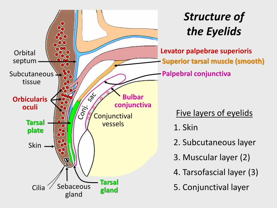
Levator palpebrae superioris
1. Skin
2. Subcutaneous layer
3. Muscular layer (2)
4. Tarsofascial layer (3)
5. Conjunctival layer
Five layers of eyelids
Skin
Subcutaneous tissue
Orbital septum
Orbicularis oculi
Tarsal plate
CiliaTarsal glandSebaceous
gland
Conjunctival vessels
Bulbar conjunctiva
Superior tarsal muscle (smooth)
Palpebral conjunctiva
Structure of the Eyelids

The pictures below show inflammations of different structures related to the eyelids. Which structure is involved in each case?
Conjunctivitis
Conjunctiva
Stye(hordeolum externum)
Sebaceous glands
Chalazion(hordeolum internum)
Tarsal glands
Clinical correlate: eye infections
(pinkeye)

Lacrimal apparatusLacrimal gland produces tears pass from lacrimal duct, across surface of eye to lacrimal canaliculi take tears from lacrimal lake to lacrimal sac down nasolacrimal duct to nasal cavity
Lacrimal lake
Lacrimal lake: pinkish shallow reservoir of tears medial canthus
Lacrimal ducts
Lacrimal canaliculus
Lacrimal gland
(CN VII)

• Relationships of the orbit
• Arrangement of orbital contents (fascial structures)
• Structure of eyelids & lacrimal apparatus
• Blood Supply to the orbit
• Cranial nerves of the orbit (CN II-VII)
• Autonomics of the orbit (para & symp)
• Arrangement & anatomical action of extraocular muscles
• Clinical testing of extraocular muscles
Lecture Outline

The blood supply to the orbit and eye is via the
ophthalmic artery:
Ophthalmic
arteries
Internal
carotid

Internal carotid artery
Central artery of the retina
Ophthalmic artery
CNII

CSF
Normal
CSF
Intracranial pressure
Papilledema with hemorrhage

Venous drainage from the orbit communicates with facial vein
anteriorly and cavernous sinus posteriorly.
Facial veinCavernous
sinus
Superior
ophthalmic
vein
These veins have no valves
Danger area
Cavernous sinus
thrombosis

• Relationships of the orbit
• Arrangement of orbital contents (fascial structures)
• Structure of eyelids & lacrimal apparatus
• Blood Supply to the orbit
• Cranial nerves of the orbit (CN II-VII)
• Autonomics of the orbit (para & symp)
• Arrangement & anatomical action of extraocular muscles
• Clinical testing of extraocular muscles
Lecture Outline

Optic (II)
Oculomotor (III)
Trochlear (IV)
Trigeminal (V)Ophthalmic (V1)Maxillary (V2)
Abducent (VI)
Cranial nerves related to the orbit

Trochlear (IV)
Oculomotor (III)
Abducent(VI)
CN II
CN V2
CN V3
3 Cranial nerves innervate extraocular muscles
CN V1

Motor axons
Abducentnucleus
Superior rectus m.
Superior oblique m.
Inferior rectus m.
Medial rectus m.
Inferior oblique m.
Levator palpebrae m.
Lateral rectus m.
Ciliary ganglion
Ciliary nerves
V3
V2
V1
Oculomotor (III)Oculomotor
nucleus
Trochlear nucleus
Trigeminal(V)
Superior orbital fissure
Internal carotid artery
Cavernous sinus
Motor Innervation of Extraocular Muscles
Long ciliary n.
Short ciliary nn.

Handy mnemonic:
LR6SO4AO3
(Lateral Rectus CN VI, Superior Oblique CN IV, All
Others CN III)
Summary of Extraocular Muscle
Innervation

CN V
Maxillary (V2)
Trigeminal ganglion
Ophthalmic (V1)
V1 sensory to:(1) skin upper eyelid
(2) Bulbar conjunctiva
(3) Palpebral conjunctiva(upper eyelid)
V2 sensory to:(1) skin lower eyelid
(2) Palpebral conjunctiva (lower eyelid)
Nasociliary
Lacrimal
Frontal
Sensory innervation to the orbit
Branches of Trigeminal n. (CN V)
mostly V1 – Ophthalmic
some V2 – Maxillary
NFL
Long ciliary n.

Bulbar conjunctiva & cornea*
Ciliary ganglion
Ciliary nerves
V3
V2
V1
CN VI
CN III
Trigeminal (V)
Sensory (skin, and bulbar & palpebral conjunctiva)
CN IV
Skin & palpepral
conjunctiva of upper
eyelid
Skin & palpepral
conjunctiva of lower
eyelid
*Corneal (blink) reflex (blinking caused by light touching of the cornea):afferent limb = CN V1 from cornea via ciliary nervesefferent limb = CN VII to orbicularis oculi
Sensory components of ophthalmic & maxillary nerves
Superior orbital fissure
Internal carotid artery
Long ciliary n.
Short ciliary nn.

• Relationships of the orbit
• Arrangement of orbital contents (fascial structures)
• Structure of eyelids & lacrimal apparatus
• Blood Supply to the orbit
• Cranial nerves of the orbit (CN II-VII)
• Autonomics of the orbit (para & symp)
• Arrangement & anatomical action of extraocular muscles
• Clinical testing of extraocular muscles
Lecture Outline

Zonular fibers attach to
lens from ciliary muscle
Smooth muscles of the eye
Lens
Ciliary muscle
(parasymp) – controls
thickness of lens
(accommodation/focus)
Sphincter pupillae m.
(parasympathetic) –
constricts pupil
Dilator pupillae m.(sympathetic) – dilates pupil

Edinger-Westphal nucleus
V3
V2
V1
Oculomotor (III)
CN V
Ciliary muscle
Dilator pupillae m.
Sphincter pupillae m.
Abducent n.
Trochlear n.
Parasympathetic components of oculomotor n.
Short ciliary nn.
Ciliary nn.
Parasympathetic pathway
Ciliary ganglion: collection of parasympathetic
post-ganglionic cell bodies.
The pupillary light reflex involves this pathway to the sphincter pupillae muscl.

Pupillary light reflex*
*Pupillary (light) reflex (constriction of pupil in response to light shone in pupil):afferent limb = CN II from stimulation of retinal ganglion cellsefferent limb = CN III (para) via ciliary nerves to pupillary sphincter

Superior cervicalsympathetic ganglion
Sympathetic trunk
Lateral gray column of upper thoracic spinal cord
C8
White ramus Ventral
root
~T1-4
TO HEAD
Symp (dilator pupillae & superior tarsal)
Levator palpebrae superioris
Superior tarsal
muscle
Dilator pupillaemuscle
Sympathetic innervation to orbit

Superior tarsal m.Levator palpebrae m.
Ciliary muscle
Dilator pupillae m.
Sphincter pupillae m.
V3
V2
V1
Oculomotor (III)
Sympathetic pathway
Superior cervicalsympathetic ganglion
Carotid plexus
CN VI
CN IV
CN V
Sympathetic innervation to orbit…
Long ciliary n.
Short ciliary nn.
Ciliary nn.

Summary: Autonomics to orbit
Parasympathetics (via cranial nerves) to:lacrimal gland
sphincter pupillae m.ciliary m.
Sympathetics (via carotid plexuses) to:blood vessels
superior tarsal m.dilator pupillae m.

1. Ptosis (droopy upper eyelid) paralysis of superior tarsal muscle
Patient with right-side Horner’s syndrome
4. Anhydrosis (dry skin due to lack of perspiration) sweat glands denervated
3. Vasodilation (flushed, warm skin) paralysis of smooth muscle in walls of vessels
2. Miosis (constricted pupil) paralysis of dilator pupillae muscle because no resistance to parasympathetically controlled pupillary sphincter
What are the signs in this patient that there is an interruption of sympathetic
innervation to the head?
Clinical correlate: Horner’s Syndrome
“Sahara dessert syndrome”

• Relationships of the orbit
• Arrangement of orbital contents (fascial structures)
• Structure of eyelids & lacrimal apparatus
• Blood Supply to the orbit
• Cranial nerves of the orbit (CN II-VII)
• Autonomics of the orbit (para & symp)
• Arrangement & anatomical action of extraocular muscles
• Clinical testing of extraocular muscles
Lecture Outline

Med
ial o
rbit
al w
allM
edial o
rbital w
all
Anatomical arrangement, innervation, & function of extraocular muscles

Movements of the Eyeball

Extraocular Muscles
The key to learning and retaining knowledge of how the 6 extraocular
muscles move the eyeball is to visualize each muscle’s anatomical position
relative to two axes (horizontal and vertical) using simple line drawings.
SR LR
MR
SO
IO
IR
IO = inferior oblique
IR = inferior rectus
LR = lateral rectus
MR = medial rectus
SO = superior oblique
SR = superior rectus
Right orbit seen from above

Horizontal axis
Vertical axis
When SR contracts relative to
horizontal axis, cornea moves
upward (ELEVATION).When SR contracts relative to
vertical axis, the cornea moves
medially (ADUCTION).
Superior Rectus Muscle: Anatomical Actions
Horizontal
axis
SR passes medial to vertical axis.
Media
l w
all

Horizontal axis
Vertical axis
Horizontal
axis
Inferior Rectus: Anatomical Actions
Med
ial w
all
When IR contracts relative to
horizontal axis, it rotates cornea
downward (DEPRESSION).
When IR rectus contracts relative
to vertical axis, it rotates cornea
medially (ADDUCTION).

Horizontal axis
Vertical axis
Media
l w
all
Inferior Oblique: Anatomical Actions
When IO contracts relative to
horizontal axis, it rotates back half
of eyeball down causing cornea to
rotate upward (ELEVATION).
Horizontal
axis
When IO contracts relative to
vertical axis, it rotates back of
eyeball medially causing cornea to
rotate laterall (ABDUCTION).

Horizontal axis
Vertical axis
Horizontal
axis
Media
l w
all
Superior Oblique: Anatomical Actions
When SO contracts relative to
horizontal axis, it rotates back of
eye forward making cornea rotate
downward (DEPRESSION).
When SO contracts relative to
vertical axis, it rotates back of the
eye medially causing cornea to
rotate outward (ABDUCTION).

Vertical axis
Media
l w
all
Learning Tip: Learning whether a muscle abducts or adducts relative to
vertical axis is made easier by realizing that the tendons for all muscles but
lateral rectus pass on medial side of the vertical axis.
.
Superior oblique
Superior rectus
Inferior oblique
Inferior rectus
Medial rectus
This is critical to drawing, or imagining, the correct positions of muscles
relative to the vertical axis.

Summary Movements Relative to Vertical Axis
ADDUCTORS:
Superior rectus
Inferior rectus
Medial rectus
SR
MR
IR
ABDUCTORS:
Superior oblique
Inferior oblique
Lateral rectus
SO
IO
LR

Summary Movements Relative to Horizontal Axis
ELEVATORS:
Superior rectus
Inferior oblique
DEPRESSORS:
Superior oblique
Inferior rectus
SR
IO
SO
IR

• Relationships of the orbit
• Arrangement of orbital contents (fascial structures)
• Structure of eyelids & lacrimal apparatus
• Blood Supply to the orbit
• Cranial nerves of the orbit (CN II-VII)
• Autonomics of the orbit (para & symp)
• Arrangement & anatomical action of extraocular muscles
• Clinical testing of extraocular muscles
Lecture Outline

Clinical Testing of Extraocular Muscles
In terms of peripheral nerve lesions that affect extraocular movements, there
are three possibilities, oculomotor (III), trochlear (IV) and abducent (VI).
Since a lesion of CNIII includes a dilated (blown) pupil, plus a ptosis, plus a
loss of many eye movements, it is typically not necessary to test each of the
muscles innervated by CNIII in order to make this diagnosis.
A lesion of CNVI (abducent) results in paralysis of the lateral rectus with the
patient having difficulty looking laterally, an easy condition to assess
clinically.
However, a lesion of CNIV (trochlear) requires testing the superior oblique
muscle in order to make a diagnosis. Therefore, the following
demonstrates how one clinically assesses the superior oblique.

Horizontal axis
Med
ial w
all
The superior oblique and the inferior rectus are the only two muscles
that can depress the eye (relative to the horizontal axis).
How can one isolate and test this action for the SO while eliminating
any contribution from the inferior rectus?
First, have the patient adduct the eye as far as possible without looking
up or down (medial rectus does this).
Horizontal axis
Notice how the horizontal
axis also rotated with the
adducted eye, making it
now nearly parallel to IR.
In this adducted position, only the SO muscle, whose line of pull is
perpendicular to the horizontal axis, can depress the eye if it is innervated
by IV; the IR cannot depress the eye since it is parallel to horizontal axis.
Superior oblique
Inferior rectus