Embed Size (px)
Citation preview
1
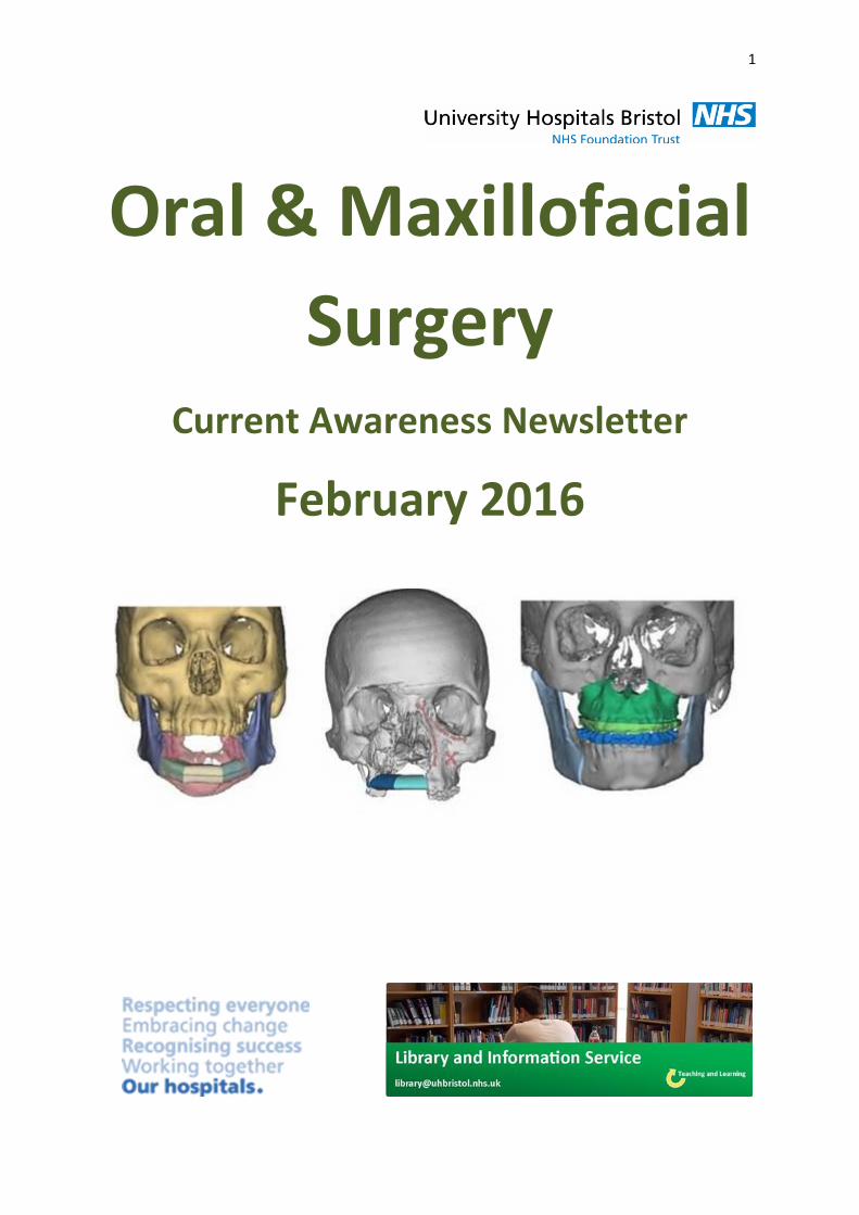
Oral & Maxillofacial
Surgery Current Awareness Newsletter
February 2016
2
Contents Your Friendly Local Librarian… ...................................................................................................... 2
Current Awareness Database Articles on Oral and Maxillofacial Surgery ........................................ 5
Oral Surgery ......................................................................................................................... 5
Bisphosphonate-related osteonecrosis of the jaw ................................................................ 10
Maxillofacial ....................................................................................................................... 12
Cleft lip and palate ............................................................................................................. 24
Journal Tables of Contents ......................................................................................................... 31
British Journal of Oral and MaxillofacialSurgery ................................................................... 31
Head and Neck ................................................................................................................... 31
Oral Surgery ....................................................................................................................... 31
Oral Surgery Oral Medicine Oral Pathology Oral Radiology .................................................. 31
The Cleft Palate-Craniofacial Journal ................................................................................... 31
Your Friendly Local Librarian… Whatever your information needs, the library is here to help. As your outreach librarian I offer
literature searching services as well as training and guidance in searching the evidence and critical
appraisal – just email me at [email protected]
Outreach: Your Outreach Librarian can help facilitate evidence-based practise for all in the oral and
maxillofacial surgery team, as well as assisting with academic study and research. We can help with
literature searching, obtaining journal articles and books, and setting up individual current
awareness alerts. We also offer one-to-one or small group training in literature searching,
accessing electronic journals, and critical appraisal. Get in touch: [email protected]
Literature searching: We provide a literature searching service for any library member. For those
embarking on their own research it is advisable to book some time with one of the librarians for a 1
to 1 session where we can guide you through the process of creating a well-focused literature
research and introduce you to the health databases access via NHS Evidence. Please email requests
3
Lunchtime Drop-in Sessions
January - June 2016
The Library and Information Service provides free specialist information skills training
for all UHBristol staff and students.
To book a place, email: [email protected]
If you’re unable to attend we also provide one-to-one or small group sessions. Contact
[email protected] to arrange a session.
Literature Searching
An in-depth guide to formulating an effective search strategy and getting the most out of searching key healthcare databases.
Understanding Articles
How to assess the strengths and weaknesses of research methods.
Examining different research designs, bias and validity, and frameworks for systematically appraising a medical paper.
Medical Statistics
A basic introduction to the key statistics in medical articles.
Giving an overview of statistics that compare risk, test confidence, analyse clinical investigations, and test difference.
Information Resources
A comprehensive overview of Library subscription resources, freely available online resources and ‘grey literature’.
January (1pm) Mon 4th Literature Searching Tues 12th Understanding articles Weds 20th Statistics Thurs 28th Information resources February (12pm) Fri 5th Literature Searching Mon 8th Understanding articles Tues 16th Statistics Weds 24th Information resources March (1pm) Thurs 3rd Literature Searching Fri 11th Understanding articles Mon 14th Statistics Tues 22nd Information resources Weds 30th Literature Searching April (12pm) Thurs 7th Understanding articles Fri 15th Statistics Mon 18th Information resources Tues 26th Literature Searching May (1pm) Weds 4th Understanding articles Thurs 12th Statistics Fri 20th Information resources Tues 31st Literature Searching June (12pm) Weds 8th Understanding articles Thurs 16th Statistics Fri 24th Information resources

4
To access electronic resources you need an
NHS Athens username and password
To register, click on the link:
https://openathens.nice.org.uk/
You need to register using an NHS PC and an NHS email address.
Registration is a quick, simple process, and will give you access to a huge range of online subscription resources,
including:
UpToDate
Dynamed
NHS Evidence
Anatomy.tv
E-journals
E-books
For more information or help with setting up your Athens account, email: [email protected]
5
Current Awareness Database Articles on Oral
and Maxillofacial Surgery
Below is a selection of articles on oral and maxillofacial surgery recently added to the healthcare
databases, grouped in the following categories:
Oral surgery
Bisphosphonate-related osteonecrosis of the jaw
Maxillofacial
Cleft lip and palate
If you would like any of the following articles in full text, or if you would like a more focused
search on your own topic, then get in touch: [email protected]
Oral Surgery Title: Professional oral health care reduces the duration of hospital stay in patients undergoing orthognathic surgery Citation: Biomedical Reports, January 2016, vol./is. 4/1(55-58), Author(s): Shigeishi H., Rahman M.Z., Ohta K., Ono S., Sugiyama M., Takechi M. Abstract: The present study reviewed the records of 58 patients who underwent orthognathic surgery [sagittal split ramus osteotomy (SSRO), Le Fort I osteotomy, genioplasty, anterior maxillary alveolar osteotomy] between 2010 and 2015. To investigate the influence of preoperative oral health care on postoperative inflammation, infection and length of hospital stay in those patients, white blood cell (WBC) count and C-reactive protein (CRP) levels were compared between patients who received and did not receive preoperative oral care. The mean CRP level throughout the postoperative term was lower in the oral care group as compared to the non-oral care group. By contrast, the oral care group had a lower occurrence of postoperative infectious complications (surgical site infection, anastomotic leak) (13.6 vs. 20.8%) and a shorter average length of hospital stay (16.2+/-3.8 vs. 21.2+/-7.4 days). These results suggest that preoperative professional oral health care decreases the duration of hospital stay following orthognathic surgery by inhibiting inflammation and infectious complications during the postoperative stage.
Title: Book Review: Oral Cancer Surgery: A Visual Guide. Citation: The Annals of otology, rhinology, and laryngology, Mar 2016, vol. 125, no. 3, p. 264-265, 0003-4894 (March 2016)
6
Author(s): McMullen, Caitlin P
Title: Oral surgery: Mandibular fracture risk. Citation: British dental journal, Jan 2016, vol. 220, no. 2, p. 44. (January 22, 2016) Author(s): Basyuni, S, Ferro, A, Cameron, M
Title: Oral anticoagulation therapy and laser surgery for benign prostatic hyperplasia: stop, replace, or continue? Citation: Current opinion in urology, Jan 2016, vol. 26, no. 1, p. 35-41 (January 2016) Author(s): Descazeaud, Aurelien, Mathieu, Romain Abstract: To update on the available literature that assessed laser surgery for benign prostatic obstruction (BPO) in patients under antithrombotic. All types of laser might be suitable to decrease the bleeding risk in patients under antithrombotic. However, there is no consensus on the appropriate perioperative management of antithrombotic. Most of the studies mixed patients with coumarin derivatives or platelet aggregation inhibitors and did not discriminate the results according to the type of antithrombotic. The continuation of low-dose aspirin is feasible and might not increase bleeding risk during the perioperative period. Conversely, the literature is still too sparse and the protocols reported are too heterogeneous to provide any firm recommendation regarding the continuation, withdrawn, or bridging of clopidogrel and coumarin derivatives during laser procedures for BPO. The approach with new oral anticoagulants is even more uncertain as no data are available in this setting. The decision to stop, continue, or replace antithrombotic should rely on both thrombotic and hemorrhagic risks. Therefore, urologist, cardiologist, and anesthesiologist should discuss altogether each case. Further studies are needed to provide a high level of evidence regarding the perioperative management of antithrombotic in the era of laser BPO procedures.
Title: The changes of oral health-related quality of life and satisfaction after surgery-first orthognathic approach: a longitudinal prospective study. Citation: Head & face medicine, Jan 2016, vol. 12, no. 1, p. 2. (2016) Author(s): Huang, Shengbin, Chen, Weiting, Ni, Zhenyu, Zhou, Yu Abstract: To our best knowledge, there was little research to assess the changes of quality of life and satisfaction after orthognathic in one trial. The aim of this study was to evaluate the changes of oral health related quality of life and satisfaction between surgery-first and orthodontic-first orthognathic surgery. (Abstract edited)
Title: Systematic review and meta-analysis of the efficacy of hilotherapy following oral and maxillofacial surgery.
7
Citation: International journal of oral and maxillofacial surgery, Jan 2016, vol. 45, no. 1, p. 110-117 (January 2016) Author(s): Bates, A S, Knepil, G J Abstract: Craniofacial surgery causes immediate postoperative pain, oedema, and functional limitations. Hilotherapy delivers cooled water to the face at 15°C and may reduce the postoperative recovery time. This work presents a meta-analysis of short-term postoperative outcomes after hilotherapy. Following a systematic literature search, comparative trials of patients undergoing surgical interventions in the maxillofacial region and receiving either hilotherapy or ice-cooling therapy were included for meta-analysis. Demographics and surgical outcomes were extracted. Data were analysed using Comprehensive Meta-Analysis software. (Abstract edited)
Title: Incidence of Venous Thromboembolism After Oral Oncologic Surgery With Simultaneous Reconstruction. Citation: Journal of oral and maxillofacial surgery : official journal of the American Association of Oral and Maxillofacial Surgeons, Jan 2016, vol. 74, no. 1, p. 212-217 Author(s): Kakei, Yasumasa, Akashi, Masaya, Hasegawa, Takumi, Minamikawa, Tsutomu, Abstract: Patients with oral cancer who undergo resection with simultaneous reconstruction are presumed to be at high risk for developing venous thromboembolism (VTE) according to current criteria. The primary purpose of this retrospective study was to report the incidence of VTE after oral cancer surgery requiring primary reconstruction and to identify the potential risk factors for VTE in this population. This retrospective study evaluated 133 consecutive patients who had undergone oral cancer resection with simultaneous reconstruction from April 2007 through December 2014. (Abstract edited)
Title: Trans-oral robotic cleft surgery (TORCS) for palate and posterior pharyngeal wall reconstruction: A feasibility study. Citation: Journal of plastic, reconstructive & aesthetic surgery : JPRAS, Jan 2016, vol. 69, no. 1, p. 97-100 Author(s): Khan, Khurram, Dobbs, Tom, Swan, Marc C, Weinstein, Gregory S, Abstract: Robot-assisted surgery has become increasingly routine, replacing open and laparoscopic techniques in certain domains, with recent extension to head and neck surgery through trans-oral access. Some advantages of the robot-assisted surgery include the ability to access confined spaces, enhanced dexterity instrumentation with intuitive movement, motion scaling, tremor elimination and three-dimensional (3D) endoscopic viewing with true depth perception. The aim of this study was to investigate the technical feasibility of trans-oral robotic cleft surgery (TORCS) to access the posterior pharyngeal wall and palate for potential use in the cleft population. All possible permutations of patient and robotic
8
instrument configurations were used with the daVinci Si Surgical System(®) (Intuitive Surgical, USA) 0° and 30° 3D endoscopes and 8-mm training instruments to determine the optimal visualization and surgical access to the palate and posterior pharynx in a paediatric airway manikin, and to simulate posterior pharyngeal wall surgery. A full robot-assisted cadaveric Hynes pharyngoplasty was performed using 5-mm training instruments. Experiments were recorded with still and video photography. TORCS is technically feasible in the paediatric cleft population. We predict a short learning curve due to the intuitive instrumentation, easier dissection and the potential to limit secondary insult compared with traditional surgery, as well as improved ergonomics for the operating surgeon. The as-yet unreported use of robotic-assisted cleft palate surgery may considerably enhance a surgeon's ability to perform difficult procedures of the palate and posterior pharynx in selected patients with limited access as well as lay the foundation for potential novel techniques.
Title: Single dose of diclofenac or meloxicam for control of pain, facial swelling, and trismus in oral surgery. Citation: Medicina oral, patología oral y cirugía bucal, Jan 2016, vol. 21, no. 1, p. e127. Author(s): Orozco-Solís, M, García-Ávalos, Y, Pichardo-Ramírez, C, Tobías-Azúa, F, Abstract: Postoperative pain associated with removal of mandibular third molars has been documented from moderate to severe during the first 24 hours after surgery, with pain peaking between 6 and 8 hours when a conventional local anesthetic is used. Dental pain is largely inflammatory, and evidence-based medicine has shown that nonsteroidal anti-inflammatory drugs are the best analgesics for dental pain. The aim of this study was to compare the analgesic, anti-inflammatory and anti-trismus effect of a single dose of diclofenac and meloxicam after mandibular third molar extraction. (Abstract edited)
Title: Effect size comparison of ketorolac nasal spray and commonly prescribed oral combination opioids for pain relief after third molar extraction surgery. Citation: Postgraduate medicine, Jan 2016, vol. 128, no. 1, p. 12-17 (January 2016) Author(s): Niebler, Gwendolyn, Dayno, Jeffrey Abstract: Opioids are frequently used for treatment of moderate to severe short-term pain, but concerns exist about this treatment approach. Ketorolac tromethamine nasal spray, a nonsteroidal anti-inflammatory, is indicated for the short-term management of moderate to moderately severe pain requiring analgesia at the opioid level. However, there are no direct comparison studies between ketorolac nasal spray and opioids. The objective of this study was to use an effect size analysis to compare the effectiveness of ketorolac nasal spray with oral combination opioid formulations in treating moderate to severe short-term pain. An effect size analysis of three randomized, double-blind, placebo-controlled studies of third molar extraction surgery compared pain relief with ketorolac nasal spray and commonly prescribed combination opioids including hydrocodone/acetaminophen (APAP), oxycodone/APAP, oxycodone/ibuprofen and tramadol HCl/APAP. Effect size comparisons
9
were made using total pain relief scores (TOTPAR6 or TOTPAR8; the weighted sum of pain relief scores through 6 or 8 h). Pain relief was measured using a five-point categorical rating scale (0 = none; 4 = complete). The effect size equivalent correlation, r, was determined using an online effect size calculator. The treatment effect size r compared with placebo was classified using established criteria (small = 0.20-0.49, moderate = 0.50-0.79 and large = ≥ 0.80). TOTPAR6 data indicated a moderate effect size for ketorolac nasal spray 31.5 mg (0.51) and oxycodone/ibuprofen 5/400 mg (0.64) and a small effect size for hydrocodone/APAP 7.5/500 mg (0.24) and oxycodone/APAP 5/325 mg (0.32). TOTPAR8 data indicated small effect sizes for ketorolac nasal spray (0.48), hydrocodone/APAP 10/650 mg (0.43), tramadol HCl/APAP 75/650 mg (0.35) and tramadol HCl/APAP 37.5/325 mg (0.17). The treatment effect sizes of ketorolac nasal spray were similar to or higher than the opioid comparators after third molar surgery, a well-accepted pain model. These results support ketorolac nasal spray as an effective treatment for moderate to moderately severe short-term pain.
Title: Association between funding, risk of bias, and outcome of randomised controlled trials in oral and maxillofacial surgery. Citation: The British journal of oral & maxillofacial surgery, Jan 2016, vol. 54, no. 1, p. 46-50 Author(s): Oomens, M A E M, Lazzari, S, Heymans, M W, Forouzanfar, T Abstract: The influence of funding on the main outcome of a random control trial (RCT) is important, as it could potentially lead to bias towards industry, and results that are too optimistic. We investigated the association between funding, the published outcome, and the risk of bias in trials in oral and maxillofacial surgery (OMFS) published from January 2000 to May 2013 listed in PubMed. (Abstract edited)
Title: Smartphone photography in oral and maxillofacial surgery. Citation: The British journal of oral & maxillofacial surgery, Jan 2016, vol. 54, no. 1, p. 104-105 Author(s): Jamil, F Abstract: An increasing number of staff in oral and maxillofacial surgery (OMFS) departments take clinical photographs with their personal phones. We report the results of a survey on the use of smartphone photography in OMFS departments in the United Kingdom, and highlight the guidelines that govern their use and the associated ethical and medicolegal implications.
Title: Oral Cancer Surgery: A Visual Guide. Citation: Annals of Otology, Rhinology & Laryngology, 2016, vol./is. 125/3(264-265),
10
Bisphosphonate-related osteonecrosis of the jaw
Title: Zoledronate induces bisphosphonate-related osteonecrosis of the jaw in osteopenic sheep. Citation: Clinical oral investigations, Jan 2016, vol. 20, no. 1, p. 31-38 (January 2016) Author(s): Voss, Pit J, Stoddart, Martin J, Bernstein, Anke, Schmelzeisen, Rainer, Abstract: Bisphosphonate-related osteonecrosis of the jaw (BP-ONJ) occurs in 1 % of patients with medication-induced osteoporosis treated with bisphosphonates. Sheep are an established large animal model for investigating osteoporotic skeletal changes. Zoledronate significantly reduces tissue mineral variability in ovariectomized sheep. The aim of this study was to analyze bone healing after tooth extraction in sheep with induced osteopenia and zoledronate administration. Eight adult ewes were randomly divided into two groups of four animals. All sheep underwent ovariectomy and a low-calcium diet. Dexamethasone was administered weekly for 16 weeks. Zoledronate was then given every third week for a further 16 weeks in four sheep; these infusions were repeated after extraction of two lower premolars. Four sheep without zoledronate administrations served as controls (Abstract edited)
Title: Managing Osteoporosis in Patients on Long-Term Bisphosphonate Treatment: Report of a Task Force of the American Society for Bone and Mineral Research. Citation: Journal of bone and mineral research : the official journal of the American Society for Bone and Mineral Research, Jan 2016, vol. 31, no. 1, p. 16-35 (January 2016) Author(s): Adler, Robert A, El-Hajj Fuleihan, Ghada, Bauer, Douglas C, Camacho, Pauline M, Abstract: Bisphosphonates (BPs) are the most commonly used medications for osteoporosis. This ASBMR report provides guidance on BP therapy duration with a risk-benefit perspective. Two trials provided evidence for long-term BP use. In the Fracture Intervention Trial Long-term Extension (FLEX), postmenopausal women receiving alendronate for 10 years had fewer clinical vertebral fractures than those switched to placebo after 5 years. (Abstract edited)
Title: Risks and Benefits of Bisphosphonate Therapies. Citation: Journal of cellular biochemistry, Jan 2016, vol. 117, no. 1, p. 20-28 (January 2016) Author(s): Reyes, Carlen, Hitz, Mette, Prieto-Alhambra, Daniel, Abrahamsen, Bo Abstract: Bisphosphonates are the mainstay of osteoporosis treatment but also play a fundamental role in treating other bone diseases such as Osteogenesis Imperfecta, Pagets' disease, and in the prevention of adverse skeletal effects in certain cancers such as prostate cancer or multiple myeloma. In the last decades, the refinement of bisphosphonates and an increase in the number of new bisphosphonates commercialized has altered the clinical
11
management of these diseases. Despite differences between randomized controlled trials and observational studies, overall all bisphosphonates licensed have proven to reduce the risk of fracture through the inhibition of bone resorption. (Abstract edited)
Title: Dose-dependent inhibitory effects of zoledronic acid on osteoblast viability and function in vitro. Citation: Molecular medicine reports, Jan 2016, vol. 13, no. 1, p. 613-622 (January 2016) Author(s): Huang, Xin, Huang, Shilong, Guo, Fengjin, Xu, Fei, Cheng, Peng, Ye, Yaping, Abstract: Zoledronic acid (ZA), which is one of the most potent and efficacious bisphosphonates, has been commonly used in clinical practice for the treatment of various bone disorders. The extensive use of ZA has been associated with increasing occurrence of jaw complications, now known as bisphosphonate‐associated osteonecrosis of the jaw (BRONJ). However, the mechanism underlying BRONJ remains to be fully elucidated. The aim of the present study was to investigate the effects of different concentrations of ZA on the MC3T3‐E1 murine preosteoblast cell line cells and examine the possible pathogenesis of BRONJ. In the present study, the effect of ZA on the viability, apoptosis, differentiation and maturation of MC3T3‐E1 cells, as well as its relevant molecular mechanism, were examined (Abstract edited)
Title: Clinical Practice. Postmenopausal Osteoporosis. Citation: New England Journal of Medicine, 2016, vol./is. 374/3(254-262), Abstract: Key Clinical Points Postmenopausal Osteoporosis Fractures and osteoporosis are common, particularly among older women, and hip fractures can be devastating. Treatment is generally recommended in postmenopausal women who have a bone mineral density T score of -2.5 or less, a history of spine or hip fracture, or a Fracture Risk Assessment Tool (FRAX) score indicating increased fracture risk. Bisphosphonates (generic) and denosumab reduce the risk of hip, nonvertebral, and vertebral fractures; bisphosphonates are commonly used as first-line treatment in women who do not have contraindications. Teriparatide reduces the risk of nonvertebral and vertebral fractures. Osteonecrosis of the jaw and atypical femur fractures have been reported with treatment but are rare. The benefit-to-risk ratio for osteoporosis treatment is strongly positive for most women with osteoporosis. Because benefits are retained after discontinuation of alendronate or zoledronic acid, drug holidays after 5 years of alendronate therapy or 3 years of zoledronic acid therapy may be considered for patients at lower risk for fracture.
Title: Histopathological Effects of Teriparatide in Medication-Related Osteonecrosis of the Jaw: An Animal Study. Citation: Journal of Oral & Maxillofacial Surgery (02782391), 2016, vol./is. 74/1(68-78), Abstract: Purpose: Osteonecrosis of the jaw after tooth extraction is a major complication in patients using bisphosphonates (BPs) for bone lesions, such as for the treatment of
12
osteoporosis. The purpose of this study was to investigate the histopathologic effects of teriparatide (a synthetic parathyroid hormone) on rats developing osteonecrosis with BP use.Materials and Methods: Rats (n = 80) that had been injected intraperitoneally with zoledronic acid for 7 weeks were used. (Abstract edited)
Maxillofacial Title: Nasolabial flap in maxillofacial gunshot trauma: A case series Citation: Archives of Trauma Research, January 2016, vol./is. 5/1(no pagination), 2251-953X;2251-9599 (23 Jan 2016) Author(s): Rahpeyma A., Khajehahmadi S. Abstract: Introduction: The nasolabial flap (NLF) has many advantages in oromaxillary reconstruction, but the majority of cases are reconstructions after pathologic resections. Its usage in trauma surgery, especially in the management of gunshot wounds, is rarely mentioned. Case Presentation: Three cases involving gunshot injuries to the face are presented: one for reconstruction of the nasal ala, another for bone graft coverage in mandibular reconstruction, and the third for the repair of premaxillary hard and soft tissue avulsive defects. Conclusions: The NLF is a thin, pliable flap and is useful for intraoral and facial reconstruction of trauma patients with small to moderate soft tissue loss.
Title: Salivary gland tumors: A 15- year report from Iran Citation: Turk Patoloji Dergisi, 2016, vol./is. 32/1(35-39), Author(s): Taghavi N., Sargolzaei S., Mashhadiabbas F., Akbarzadeh A., Kardouni P. Abstract: Objective: The aim of this study was to document the clinicopathologic characteristic of salivary gland tumors in Tehran, Iran, over a 15-year period. Material and Method: A retrospective study was conducted on salivary gland tumors diagnosed at two pathology centers of Shahid Beheshti University of Medical Sciences from March 2000 to March 2015. (Abstract edited)
Title: Computed tomography as an aid to planning intubation in the difficult airway Citation: British Journal of Oral and Maxillofacial Surgery, January 2016, vol./is. 54/1(80-82), Author(s): Grimes D., Macleod I., Taylor T., O'Connor M., Sidebottom A. Abstract: Patients having oral and maxillofacial operations often require nasal intubation, but limited mouth opening and unfavourable nasal anatomy can make it difficult. We aimed to find out whether there is an association between the prediction of difficult nasal intubation on computed tomography (CT) and actual problems. We retrospectively reviewed the imaging and anaesthetic records of 77 patients who had replacement of the
13
temporomandibular joint (TMJ) as these patients often have limited mouth opening and have had a preoperative CT. There was a positive correlation between a radiographically-assessed difficult nostril and difficulty of intubation (p<0.001). The positive predictive value was 71.4%. As a consequence, our radiologists now routinely report on the nasal cavity in these patients, and their report and the scan are then reviewed by the anaesthetist before intubation. Our results suggest that review of the CT before planned nasal intubation should be part of routine practice.
Title: New bone formation induced by surface strontium-modified ceramic bone graft substitute Citation: Oral Diseases, January 2016, vol./is. 22/1(53-61), Author(s): Park J.-W., Kang D.-G., Hanawa T. Abstract: Objectives: This study assessed the effect of surface strontium ion (Sr) modification on the osteogenic activity of an osteoconductive ceramic bone graft substitute with the hope of using the bone healing effect of Sr for potential application in periodontal and maxillofacial regenerative surgery. Materials and Methods: A simple wet chemical treatment was employed to deliver Sr to the surface of particulate porcine bone graft. The osteogenic activity of surface Sr-modified bone substitute was compared in vitro and in vivo with that of unmodified ceramic bone, other clinically available synthetic bone or osteoinductive allograft bone. Results: The resultant bone substitute showed the formation of Sr-containing microstructured surface layer along with the formation of additional nanostructures and displayed sustained Sr release. Sr modification promoted the osteogenic differentiation of bipotential ST2 stem cells. Sr-modified bone substitute increased the amount of newly formed bone at early healing period in calvarial defect of rabbits. Conclusions: These results suggest that the surface Sr modification by wet chemical treatment is a promising approach to enhance the early bone healing capacity of osteoconductive ceramic bone substitutes.
Title: Prevalence of osteoma cutis in the maxillofacial region and classification of its radiographic pattern in cone beam CT Citation: Dermatology Online Journal, 2016, vol./is. 22/1(no pagination), Author(s): Safi Y., Valizadeh S., Vasegh Z., Aghdasi M.M., Shamloo N., Azizi Z. Abstract: Background: Osteoma cutis is a rare soft tissue ossification of cutaneous tissue and may be primary or secondary. In the majority of cases it is clinically asymptomatic and may detected incidentally on radiographic examination. Cone beam computed tomography (CBCT) has can be of great assistance in the detection of this asymptomatic lesion. Objectives: In this retrospective study, the prevalence and different radiographic appearance of osteoma cutis was evaluated. (Abstract edited)
Title: Expanded Disability Status Scale-Based Disability and Dental-Periodontal Conditions in Patients with Multiple Sclerosis
14
Citation: Medical Principles and Practice, January 2016, vol./is. 25/1(49-55) Author(s): Hatipoglu H., Canbaz Kabay S., Gungor Hatipoglu M., Ozden H. Abstract: Objective: The aim of this study was to evaluate the association between different disability states in patients with multiple sclerosis (MS) as determined by the expanded disability status scale (EDSS) and dental-periodontal measures. Subjects and Methods: Eighty patients with MS (64 females and 16 males) were included in this study. Data on MS types, attack frequency, disease duration, EDSS scores and orofacial complaints prior to an MS attack were obtained from medical records. (Abstract edited)
Title: Mesenchymal stem cells derived from dental pulp: A review Citation: Stem Cells International, 2016, vol./is. 2016/(no pagination) Author(s): Ledesma-Martinez E., Mendoza-Nunez V.M., Santiago-Osorio E. Abstract: The mesenchymal stem cells of dental pulp (DPSCs) were isolated and characterized for the first time more than a decade ago as highly clonogenic cells that were able to generate densely calcified colonies. Now, DPSCs are considered to have potential as stem cell source for orthopedic and oral maxillofacial reconstruction, and it has been suggested that they may have applications beyond the scope of the stomatognathic system. To date, most studies have shown that, regardless of their origin in third molars, incisors, or exfoliated deciduous teeth, DPSCs can generate mineralized tissue, an extracellular matrix and structures type dentin, periodontal ligament, and dental pulp, as well as other structures. (Abstract edited)
Title: Head injury - A maxillofacial surgeon's perspective Citation: Journal of Clinical and Diagnostic Research, January 2016, vol./is. 10/1(ZE01-ZE06), Author(s): Choonthar M.M., Raghothaman A., Prasad R., Pradeep S., Pandya K. Abstract: Injuries and violence are one of the leading causes of mortality worldwide. A substantial portion of these injuries involve the maxillofacial region. Among the concomitant injuries, injuries to the head and cervical spine are amongst those that demand due consideration on account of their life threatening behaviour. Studies have shown that facial fractures have a strong association with traumatic brain injury. Knowledge of the types and mechanisms of traumatic brain injury is crucial for their treatment. Many a times, facial fractures tend to distract our attention from more severe and often life threatening injuries. Early diagnosis of these intracranial haemorrhage leads to prompt treatment which is essential to improve the outcome of these patients. An oral and maxillofacial surgeon should be able to suspect and diagnose head injury and also provide adequate initial management.
Title: Spontaneous fractures of the mandible concept & treatment strategy
15
Citation: Medicina Oral, Patologia Oral y Cirugia Bucal, January 2016, vol./is. 21/1(e88-e94), Author(s): Carlsen A., Marcussen M. Abstract: Background: Spontaneous fractures of the mandible dispose a surgical challenge in comparisons to fractures caused by trauma due to several complicating factors. Additionally: controversies exist concerning the terminology of the field. Material and Methods: We conducted a retrospective study of all patients with mandibular fractures, with exclusion of fractures of the coronoid process and the alveolar process, treated at the Department of Oral and Maxillofacial Surgery, Aalborg University Hospital, Denmark between February 2003 and February 2013. Data collected from the medical records included sex, age, cause of fracture, site of fracture, and treatment. Results: We identified 517 patients with 684 mandible fractures. Twenty-five of these were spontaneous fractures and 659 fractures were of traumatic origin. Condylar fractures rarely occur spontaneously, but constitute the majority of the traumatic fractures. Excluding these fractures from the analysis, we found a non-surgical approach in 14 of 24 (58%) of the spontaneous fractures and 110 of 376 (29%) of the traumatic fractures. This was statistically significant. Conclusions: We found a statistical significant difference in favor of non-surgical approach in spontaneous fractures and we discussed the treatment challenges of these fractures. We addressed the terminological controversies regarding pathological fractures, and suggested the term spontaneous fractures denoting a fracture occurring during normal jaw function being either pathological or non-pathological.
Title: Multidisciplinary management of ankyloglossia in childhood. Treatment of 101 cases. a protocol Citation: Medicina Oral, Patologia Oral y Cirugia Bucal, January 2016, vol./is. 21/1(e39-e47), Author(s): Pastor-Vera T., Ferres-Amat E., Mareque-Bueno J., Prats-Armengol J., Abstract: Background: Partial ankyloglossia is a limitation which restricts the possibility of protrusion and elevation of the tip of the tongue due to the shortness of either the lingual frenulum or the genioglossus muscles or both. The principal objective of this paper is to present our protocol of action for the treatment of ankyloglossia. The specific objectives are to study patients with ankyloglossia treated by the Service of Maxillofacial Surgery and the Service of Speech Therapy of our pediatric Hospital, describe the diagnostic procedures, the pre-surgical intervention, the surgical technique undertaken and the post-surgical rehabilitation taking into account the level of collaboration of the patients, and finally, describe the surgical complications and the referral of patients. (Abstract edited)
Title: Systematic review and meta-analysis of the efficacy of hilotherapy following oral and maxillofacial surgery Citation: International Journal of Oral and Maxillofacial Surgery, January 2016, vol./is. 45/1(110-117) Author(s): Bates A.S., Knepil G.J.
16
Abstract: Craniofacial surgery causes immediate postoperative pain, oedema, and functional limitations. Hilotherapy delivers cooled water to the face at 15 degreeC and may reduce the postoperative recovery time. This work presents a meta-analysis of short-term postoperative outcomes after hilotherapy. Following a systematic literature search, comparative trials of patients undergoing surgical interventions in the maxillofacial region and receiving either hilotherapy or ice-cooling therapy were included for meta-analysis. Demographics and surgical outcomes were extracted. Data were analysed using Comprehensive Meta-Analysis software. (Abstract edited)
Title: CT and MRI findings of primitive neuroectodermal tumor in the maxillofacial region Citation: Oral Radiology, January 2016, vol./is. 32/1(14-21), Author(s): Hou H.-J., Xu Z.-S., Xu D., Zhang H.-S., Liu J., Zhang W.-J. Abstract: Objectives: To summarize the computed tomography (CT) and magnetic resonance imaging (MRI) findings of rare primitive neuroectodermal tumors (PNETs) in the maxillofacial region. Methods: The clinical data, and CT and MRI findings of seven patients with pathologically proven PNETs in the maxillofacial region were retrospectively reviewed. The patients comprised five males and two females with a mean age of 16.4 years. The tumor location, size, margin, CT attenuation or signal intensity, contrast enhancement characteristics, and involvement of adjacent tissues were assessed. (Abstract edited)
Title: Kimura's disease of the right cheek: A case report Citation: Experimental and Therapeutic Medicine, January 2016, vol./is. 11/1(218-220), Author(s): Luo G., Gu F., Liu T., Huang Y. Abstract: Kimura's disease (KD), a chronic inflammatory disease of uncertain etiology, manifests as a painless subcutaneous swelling in the head and neck region that involves major salivary glands and regional lymph nodes. To date, the majority of cases of KD have been documented in Asian males aged 20-30 years. However, the number of reported cases of KD involving the oral and maxillofacial area is limited, and since the masses appear similar to cysts or benign tumors, the establishment of an accurate pre-operative diagnosis is challenging. The accurate diagnosis of KD is considered to require surgical excision followed by histopathological examination. In the current case, a 39-year-old man was admitted to hospital in October 2011 with a swelling evident on his right cheek. Surgical excision was performed, and histopathological observation was carried out. The formation of a lymphoid nodule accompanied by the vigorous proliferation of small blood vessels, eosinophilic infiltration and thickened cell walls were observed. No sign of recurrence of the mass has yet been observed, on the basis of the telephone follow-up interviews. These findings provide a novel insight useful in the diagnosis of KD in the oral and maxillofacial area.
17
Title: Orbit fractures: Identifying patient factors indicating high risk for ocular and periocular injury. Citation: Laryngoscope, 2016, vol./is. 16/(0-6), Abstract: Objectives/hypothesis: Maxillofacial trauma frequently involves the bony orbit that surrounds the ocular globe. Concomitant globe injury is a concern whenever orbit trauma occurs and in severe cases can occasionally result in vision loss. The mechanism of injury, physical exam findings, and radiographic imaging can provide useful information concerning the severity of the injury and concerns for vision loss. Using these three tools, it is hypothesized that the patient's history, physical exam, and radiographic findings can identify high-risk maxillofacial trauma patients with concomitant ocular injury. Identification of high risk patients who require comprehensive ophthalmologic evaluation may alter management and possibly preserve or restore vision. (Abstract edited)
Title: Preoperative and postoperative examination of occlusal and maxillofacial changes after osteochondroma extirpation. Citation: American Journal of Orthodontics & Dentofacial Orthopedics, 2016, vol./is. 149/2(259-268 Abstract: A patient came with left-side temporomandibular arthralgia, limited mandibular opening, frontal facial asymmetry, and a significant anterolateral open bite. Severe alterations in the occlusal and maxillofacial anatomy resulted from an osteochondroma associated with the mandibular condyle. We describe the changes associated with extirpation of the mandibular condylar osteochondroma and subsequent orthodontic treatment. These clinical changes resulted in improved facial symmetry and a satisfactory functional occlusion.
Title: Current Role of Carnoy's Solution in Treating Keratocystic Odontogenic Tumors. Citation: Journal of Oral & Maxillofacial Surgery (02782391), 2016, vol./is. 74/2(278-282), Abstract: Purpose: To understand the frequency of use of Carnoy's solution, as a means of chemical curettage, for treating the keratocystic odontogenic tumor (KCOT).Materials and Methods: A Web-based survey was distributed by e-mail to 6,880 members listed in the 2013 American Association of Oral and Maxillofacial Surgeons directory.Results: Eight hundred nine participants across the United States responded to the survey (12% response rate). The most common procedures performed to definitively treat a KCOT were enucleation plus mechanical curettage (curette with or without peripheral ostectomy; 66%). Of the survey participants, 198 (25%) currently use Carnoy's solution, 111 (56%) of whom are using the solution with chloroform and 83 (42%) are using it without chloroform.Conclusion: Carnoy's solution remains a common method of chemical curettage for the definitive treatment of the KCOT. Carnoy's solution with and without chloroform is being used for chemical cautery.
18
Title: Treatment of Dentofacial Deformities Secondary to Osteochondroma of the Mandibular Condyle Using Virtual Surgical Planning and 3-Dimensional Printed Surgical Templates. Citation: Journal of Oral & Maxillofacial Surgery (02782391), 2016, vol./is. 74/2(349-368), Abstract: Purpose: One-stage treatment for condylar osteochondroma and secondary facial deformities by resection and reconstruction of the mandibular condyle, orthognathic surgery, and mandibular contouring has been reported recently. This study investigated the clinical feasibility of treating osteochondroma of the mandibular condyle and secondary dento-maxillofacial deformities by virtual surgical planning and 3-dimensional (3D) printed surgical templates. (Abstract edited)
Title: Oral Surgical Procedures Performed Safely in Patients With Head and Neck Arteriovenous Malformations: A Retrospective Case Series of 12 Patients. Citation: Journal of Oral & Maxillofacial Surgery (02782391), 2016, vol./is. 74/2(0-0), Abstract: Purpose: This case series describes patients with head and neck arteriovenous malformations who underwent oral and maxillofacial surgical procedures combined with interventional radiology techniques to minimize blood loss. (Abstract edited)
Title: Special Contribution: Third Molar Clinical Trials Annotated Bibliography. Citation: Journal of Oral & Maxillofacial Surgery (02782391), 2016, vol./is. 74/1(4-12), Author(s): White, Raymond P Jr Abstract: Purpose: To provide clinicians with an annotated bibliography of published articles from research funded externally by the Oral and Maxillofacial Surgery Foundation, spanning 1996 to 2015, addressing the topic of third molar management.Materials and Methods: A brief summary for each article was generated by the respective authors.Results: The complete annotated bibliography generated by the authors is included in the Appendix.Conclusion: The annotated bibliography provides clinicians and other interested individuals with a summary of current literature emanating from clinical studies on third molar topics.
Title: Septic Arthritis of the Temporomandibular Joint-Unusual Presentations. Citation: Journal of Oral & Maxillofacial Surgery 2016, vol./is. 74/1(87-94), Abstract: This report describes 2 patients whose septic arthritis of the temporomandibular joint (SATMJ) presented atypically, resulting in treatment delay and complications. A 49-year-old man developed left-side facial allodynia, which was first treated unsuccessfully as trigeminal neuralgia. On day 21, the patient sustained facial trauma from a fall and presented to the emergency department (ED). Maxillofacial contrast-enhanced computed tomographic (CT) scan was suggestive of parotiditis, SATMJ, or hemarthrosis. His condition
19
did not improve with empiric antibiotic treatment. On day 30, contrast-enhanced magnetic resonance imaging (MRI) confirmed SATMJ. Incision and drainage yielded 6 mL of pus and produced clinical improvement. Cultures grew methicillin-resistant Staphylococcus aureus, which was treated with amoxicillin plus clavulanate and sulfamethoxazole plus trimethoprim for 30 days. On day 59, the patient still had slight preauricular pain and CT-proved TMJ osteoarthritic changes. A 56-year-old woman developed right-side facial pain after a crown procedure on her right mandibular second molar. Oral prednisone (and clindamycin) produced partial relief. Her primary physician suspected temporal arteritis, but its biopsy result on day 11 was normal. Gradually, the patient developed trismus and malocclusion refractory to various medicines. On day 49, she presented to the ED. A contrast-enhanced maxillofacial CT scan suggested SATMJ. Incision and drainage yielded 30 mL of pus and produced clinical improvement. During days 50 to 57, the patient received intravenous ampicillin plus sulbactam and metronidazole. However, preauricular tenderness and drainage from the surgical incision persisted. On day 55, CT scan showed a residual abscess. Secondary debridement yielded 5 mL of pus. Culture grew coagulase-negative S aureus. On day 141, the patient still had slight preauricular pain and TMJ osteoarthritic changes on MRI. In these cases, the SATMJ diagnosis was delayed owing to the mildness of local and systemic manifestations, the possibility of confounding conditions, and the rarity of SATMJ. Contrast-enhanced CT and MRI facilitated the diagnosis. Abscess drainage alleviated the symptoms. Postinfectious osteoarthritis developed possibly from treatment delay. SATMJ should be considered in all patients with enigmatic preauricular pain, trismus, or malocclusion.
Title: Synovial Sarcoma of the Tongue: Report of a Case. Citation: Journal of Oral & Maxillofacial Surgery (02782391), 2016, vol./is. 74/1(95-103), Abstract: This report outlines the workup and management of a 55-year-old woman with a synovial sarcoma of the lateral border of the tongue that was initially diagnosed as a glomus tumor. A review was performed of the literature on synovial sarcomas of the oral cavity and current National Comprehensive Cancer Network guidelines. Synovial sarcomas of the tongue are rare neoplasms, with variable morphologic microscopic types and immunohistochemical profiles. Fluorescence in situ hybridization analysis of the known gene translocation also can be used in diagnosis. According to the literature, resection of the tumor is the current treatment of choice; however, owing to the rarity of this entity, diagnosis and management prove challenging for the oral and maxillofacial surgeon.
Title: Decompression of Odontogenic Cystic Lesions: Past, Present, and Future. Citation: Journal of Oral & Maxillofacial Surgery (02782391), 2016, vol./is. 74/1(0-0), Abstract: Tumors and cystic lesions of the jawbones have been described since the late 1600s and it took another 200 years for classification systems to appear in the medical, surgical, and dental literatures. In the late 1800s, Carl Partsch introduced cystostomy, a method by which the cyst is converted into a pouch by suturing its lining to the mucosa of the oral cavity. The purpose of this article is to analyze the history, present, and future of cystic conditions of the jaws and decompression, a modality of treatment that during the
20
past few years has regained the attention of oral and maxillofacial surgeons and pathologists owing to its relative simplicity and effectiveness compared with other conservative options.
Title: Unusual Presentation of Guillain-Barré Syndrome After Mandibular Fracture Treatment: A Review of the Literature and a New Case. Citation: Journal of Oral & Maxillofacial Surgery (02782391), 2016, vol./is. 74/1(0-0), Abstract: Guillain-Barré syndrome (GBS) is a multifactorial and lethal inflammatory demyelinating neuronal disorder with concurrent polyradiculopathy and polyneuropathy presentations. This rare syndrome affects the peripheral nerve myelin sheath and is characterized by ascending muscle weakness and paralysis. There have been rare reports of GBS after head or brachial plexus trauma, general anesthesia, neurosurgery, orthopedic surgery, cesarean section, laparoscopy, and general surgery, and the occurrence of GBS after oral and maxillofacial surgery is not common. A review of the related literature and a new case of GBS after maxillofacial surgery are presented.
Title: A Rare Pathology: Low-Grade Fibromyxoid Sarcoma of the Maxilla. Citation: Journal of Oral & Maxillofacial Surgery (02782391), 2016, vol./is. 74/1(0-0), Abstract: Low-grade fibromyxoid sarcoma (LGFMS) is a rare tumor with a benign histologic appearance and fully malignant behavior. To date, only 5 cases of LGFMS in the maxillofacial region have been reported. This report describes the case of a 16-year-old boy who was referred to the authors' hospital with an intraosseous myxofibroblastic tumor, probably of the LGFMS type, of the right maxilla. Radical resection with wide safe margins and secondary reconstruction with a free forearm flap were performed. Six-month follow-up showed no sign of recurrence or metastasis. The authors review the scientific literature and discuss different tumor locations and treatment strategies for those in the maxillofacial region. The present case is the sixth reported case of LGFMS in the maxillofacial region (intraosseous LGFMS of the maxilla), adding another facet to the literature regarding this rare soft tissue tumor.
Title: A woman with rare blastic plasmacytoid dendritic cell neoplasm on the face. Citation: Oral Surgery, Oral Medicine, Oral Pathology & Oral Radiology, 2016, vol./is. 121/1(0-0) Abstract: Blastic plasmacytoid dendritic cell neoplasm (BPDCN) is a recently classified cutaneous lymphoma derived from the precursor of plasmacytoid dendritic cells with extremely poor prognosis. It has atypical morphologic features and is rarely found on the face. We report here a case of a 37-year-old woman with the rare BPDCN involving the maxillofacial region. She had a 2-month history of violaceous and traumatic hematoma-like skin lesions located on the left side of the face. Pathologic diagnosis of BPDCN was established by a skin biopsy, and the patient died 2 months later. Thus, a strong suspicion and early detection of BPDCN are essential in the case of violaceous or traumatic
21
hematoma-like skin lesions, especially on the face.
Title: Association between funding, risk of bias, and outcome of randomised controlled trials in oral and maxillofacial surgery. Citation: The British journal of oral & maxillofacial surgery, Jan 2016, vol. 54, no. 1, p. 46-50 (January 2016) Author(s): Oomens, M A E M, Lazzari, S, Heymans, M W, Forouzanfar, T Abstract: The influence of funding on the main outcome of a random control trial (RCT) is important, as it could potentially lead to bias towards industry, and results that are too optimistic. We investigated the association between funding, the published outcome, and the risk of bias in trials in oral and maxillofacial surgery (OMFS) published from January 2000 to May 2013 listed in PubMed. The methods used were scored using the risk of bias items given in a Delphi List. Sources of funding were recorded and categorised five ways: not funded, funded by industry, not funded by industry, supported by industry, and source of funds not clear. A total of 390 RCT met the inclusion criteria, and there was a correlation between funding and favourable main outcomes, although this was not significant. There was no correlation between the risk of bias and favourable results of the main outcome of a trial, or between the risk of bias and the reported source of funding in post-hoc analysis. We were unable to show a significant correlation between funding and a higher likelihood of a favourable result for the primary outcome in RCT in OMFS. We also failed to show a significant correlation between the risk of bias of a trial and its main outcome. In contrast, the source of funding proved to affect the risk of bias of a trial significantly, although not in post-hoc analysis. Funded trials were better organised, and so had a lower risk of bias.
Title: Systematic review and meta-analysis of the efficacy of hilotherapy following oral and maxillofacial surgery. Citation: International journal of oral and maxillofacial surgery, Jan 2016, vol. 45, no. 1, p. 110-117 Author(s): Bates, A S, Knepil, G J Abstract: Craniofacial surgery causes immediate postoperative pain, oedema, and functional limitations. Hilotherapy delivers cooled water to the face at 15°C and may reduce the postoperative recovery time. This work presents a meta-analysis of short-term postoperative outcomes after hilotherapy. Following a systematic literature search, comparative trials of patients undergoing surgical interventions in the maxillofacial region and receiving either hilotherapy or ice-cooling therapy were included for meta-analysis. Demographics and surgical outcomes were extracted. Data were analysed using Comprehensive Meta-Analysis software. (Abstract edited)
Title: Delayed Foreign Body Reaction Caused by Bioabsorbable Plates Used for Maxillofacial Fractures.
22
Citation: Archives of plastic surgery, Jan 2016, vol. 43, no. 1, p. 40-45, 2234-6163 Author(s): Jeon, Hong Bae, Kang, Dong Hee, Gu, Ja Hea, Oh, Sang Ah Abstract: Bioabsorbable plates and screws are commonly used to reduce maxillofacial bones, particularly in pediatric patients because they degrade completely without complications after bone healing. In this study, we encountered eight cases of a delayed foreign body reaction after surgical fixation with bioabsorbable plates and screws. A total of 234 patients with a maxillofacial fracture underwent surgical treatment from March 2006 to October 2013, in which rigid fixation was achieved with the Inion CPS (Inion, Tampere, Finland) plating system in 173 patients and Rapidsorb (Synthes, West Chester, PA, USA) in 61 patients. Their mean age was 35.2 years (range, 15-84 years). Most patients were stabilized with two- or three-point fixation at the frontozygomatic suture, infraorbital rim, and anterior wall of the maxilla. Complications occurred in eight (3.4%) of 234 patients, including palpable, fixed masses in six patients and focal swelling in two patients. The period from surgical fixation to the onset of symptoms was 9-23 months. Six patients with a mass underwent secondary surgery for mass removal. The masses contained fibrous tissue with a yellow, grainy, cloudy fluid and remnants of an incompletely degraded bioabsorbable plate and screws. Their histological findings demonstrated a foreign body reaction. Inadequate degradation of bioabsorbable plates caused a delayed inflammatory foreign body reaction requiring secondary surgery. Therefore, it is prudent to consider the possibility of delayed complications when using bioabsorbable plates and surgeons must conduct longer and closer follow-up observations.
Title: Biograft Block Hydroxyapatite: A Ray of Hope in the Reconstruction of Maxillofacial Defects. Citation: The Journal of craniofacial surgery, Jan 2016, vol. 27, no. 1, p. 247-252 ( Author(s): Kattimani, Vivekanand S, Chakravarthi, Pandi Srinivas, Prasad, Abstract: Improving quality of human life has been the rationale for increase in the applications of bone substitute materials for bone regeneration. High prevalence of loss of bone tissue due to disease remains a major challenge for reconstruction. Shortcomings of autografts and allografts have made the clinicians go for artificial implant materials. To prospectively study the structural and esthetic reconstruction of resected mandibular site with biograft porous block hydroxyapatite (BBHA). The study evaluated the efficacy of BBHA as a material for reconstruction of large bone defects. (Abstract edited)
Title: Dog bites and maxillofacial surgery: what can we do? Citation: British dental journal, Jan 2016, vol. 220, no. 2, p. 57. (January 22, 2016) Abstract: Children should be informed of possible dangers, and adults should be able to identify warning signs of canine aggression (The canine ladder of aggression. In: BSAVA Manual of canine and feline behavioural medicine).
23
Title: A Window View From the Orient on Trauma Involving the Inner Maxillofacial Region: From China to the Global Community With Love. Citation: The Journal of craniofacial surgery, Jan 2016, vol. 27, no. 1, p. 6. (January 2016) Author(s): Cicciù, Marco
Title: Oral and Maxillofacial Imaging. Citation: Dental clinics of North America, Jan 2016, vol. 60, no. 1, p. 1-37 (January 2016) Author(s): Mupparapu, Mel, Nadeau, Christine Abstract: This article provides the reader with the knowledge and skills of identification and diagnostic interpretative skills using planar images, tomographic images, CBCT, MDCT, pertinent MR images, as well as bone scans and PET images. The goal is to provide sufficient in-depth knowledge of the technique, anatomy, and radiographic identifiers for the diagnosis of local and systemic pathoses. The information will train the reader to be an advocate of selection criteria as well as a follower of the "Image Gently" campaign and philosophy supported by the organized dentistry in the United States, especially in Diagnostic Radiology.
Title: Inaugural Survey on Practice Patterns of Orbital Floor Fractures for American Oral and Maxillofacial Surgeons. Citation: Journal of oral and maxillofacial surgery : official journal of the American Association of Oral and Maxillofacial Surgeons, Jan 2016, vol. 74, no. 1, p. 105-122 Author(s): Christensen, Brian J, Zaid, Waleed Abstract: In recent years, several studies have reported on practitioners' preferences for the treatment of orbital floor fractures, showing widely varying practice patterns. The purpose of the present study was to identify the practice patterns among oral and maxillofacial surgeons involved in the management of orbital floor fractures in the United States and compare them with the available published data. (Abstract edited)
Title: Occupational exposure to noise in maxillofacial operating theatres: an initial prospective study. Citation: The British journal of oral & maxillofacial surgery, Jan 2016, vol. 54, no. 1, p. 94-96 Author(s): Tay, Brian Diaz, Prabhu, I S, Cousin, C H S, Cousin, G C S Abstract: Exposure to excessive noise could impair surgical performance and communication, and lead to long-term hearing loss, but it is only recently that studies on occupational exposure to noise in operating theatres have been published. The aim of this
24
prospective study was to assess mean and peak levels of noise during maxillofacial operations. We found that both were comparable to those in other surgical specialties such as orthopaedics in which power tools are used.
Title: Damage control surgery and combat-related maxillofacial and cervical injuries: a systematic review. Citation: The British journal of oral & maxillofacial surgery, Jan 2016, vol. 54, no. 1, p. 8-12 Author(s): Tong, Darryl C, Breeze, John Abstract: Damage control surgery involves rapid assessment, life-saving resuscitation, and abbreviated surgery for a patient with severe injuries. Traditionally the concept of damage control surgery has been restricted to penetrating abdominal injuries, but more recently it has been expanded to areas outside of the abdomen including the maxillofacial and neck regions. However, we know of little evidence that, when applied to injuries to the face and neck, it changes outcomes. We systematically reviewed published papers to identify those that discussed damage control in the context of combat-related trauma of the face and neck. We identified three papers that discussed the principles of managing combat-related maxillofacial injuries, all three of which were review articles that advocated the use of damage control principles in facial injuries either in isolation or as part of a multisystem approach. Anecdotal experience and opinion indicates that the concept of damage control is applicable when managing combat-related injuries of the face and neck, but no outcomes were confirmed. Further studies are required to validate the concept.
Title: Oral Propranolol With Topical Timolol Maleate Therapy for Mixed Infantile Hemangiomas in Oral and Maxillofacial Regions. Citation: The Journal of craniofacial surgery, Jan 2016, vol. 27, no. 1, p. 56-60 (January 2016) Author(s): Li, Gang, Xu, Da-Peng, Tong, Shuang, Xue, Lei, Sun, Ning-Ning, Wang, Xu-Kai Abstract: The combination treatment for mix infantile hemangiomas (IHs) using oral propranolol with topical timolol maleate was not well documented in the literature. The aim of this study was to evaluate the therapeutic effects and safety of oral propranolol along with topical timolol maleate or oral propranolol alone for treating mixed IHs in the oral and maxillofacial regions. (Abstract edited)
Cleft lip and palate
Title: Trans-oral robotic cleft surgery (TORCS) for palate and posterior pharyngeal wall reconstruction: A feasibility study
25
Citation: Journal of Plastic, Reconstructive and Aesthetic Surgery, January 2016, vol./is. 69/1(97-100) Author(s): Khan K., Dobbs T., Swan M.C., Weinstein G.S., Goodacre T.E.E. Abstract: Background/aim Robot-assisted surgery has become increasingly routine, replacing open and laparoscopic techniques in certain domains, with recent extension to head and neck surgery through trans-oral access. Some advantages of the robot-assisted surgery include the ability to access confined spaces, enhanced dexterity instrumentation with intuitive movement, motion scaling, tremor elimination and three-dimensional (3D) endoscopic viewing with true depth perception. The aim of this study was to investigate the technical feasibility of trans-oral robotic cleft surgery (TORCS) to access the posterior pharyngeal wall and palate for potential use in the cleft population. (Abstract edited)
Title: Spectral analysis of hypernasality in cleft palate children: A pre-post surgery comparison Citation: Journal of Clinical and Diagnostic Research, January 2016, vol./is. 10/1(MC01-MC03) Author(s): Dodderi T., Narra M., Varghese S.M., Deepak D.T. Abstract: Introduction: Change in resonance is the most commonly experienced speech problems in children diagnosed with cleft lip and palate. The degree of nasality during normal speech production is maintained by the changes in velopharyngeal port. These variations in speech signal are reported to be successfully captured using acoustical tools like spectral analysis. Aim: The present study investigated to note voice low tone to high tone ratio (VLHR) values for phonation samples of individuals with cleft palate before and after surgery. (Abstract edited)
Title: Dental materials for cleft palate repair. Citation: Materials science & engineering. C, Materials for biological applications, Apr 2016, vol. 61, p. 1018-1028 (April 1, 2016) Author(s): Sharif, Faiza, Ur Rehman, Ihtesham, Muhammad, Nawshad, MacNeil, Sheila Abstract: Numerous bone and soft tissue grafting techniques are followed to repair cleft of lip and palate (CLP) defects. In addition to the gold standard surgical interventions involving the use of autogenous grafts, various allogenic and xenogenic graft materials are available for bone regeneration. In an attempt to discover minimally invasive and cost effective treatments for cleft repair, an exceptional growth in synthetic biomedical graft materials have occurred. This study gives an overview of the use of dental materials to repair cleft of lip and palate (CLP). (Abstract edited)
Title: The Global Surgery Partnership: An Innovative Partnership for Education, Research, and Service.
26
Citation: Academic medicine : journal of the Association of American Medical Colleges, Jan 2016, vol. 91, no. 1, p. 75-78 Author(s): Taro, Trisa, Yao, Caroline, Ly, Stephanie, Wipfli, Heather, Magee, Kathleen, Abstract: An estimated two billion people worldwide lack access to adequate surgical care. Addressing surgical disparities requires both immediate relief efforts and long-term investments to improve access to care and surgical outcomes, train the next generation of surgical professionals, and expand the breadth of formative research in the field. While models exist for establishing short-term surgical missions in low- and middle-income countries, far less focus has been placed on models for multi-institutional partnerships that support the development of sustainable solutions. In 2011, the Global Surgery Partnership (GSP) was founded by an established children's hospital (Children's Hospital Los Angeles), an academic medical center (University of Southern California), and a nonprofit organization (Operation Smile) to build oral cleft surgical capacity in resource-poor settings through education, research, and service. Leveraging the strengths of each partner, the GSP supports three global health education programs for public health graduate students and surgical residents, including the Tsao Fellowship in Global Health; has initiated two international research projects on cleft lip and palate epidemiology; and has built upon Operation Smile's service provision. As of January 2015, Tsao fellows had operated on over 600 patients during 13 missions in countries including China, Vietnam, Mexico, and India. The GSP plans to conduct a formal evaluation and then to expand its programs. The GSP encourages other global health organizations and academic and medical institutions to engage with each other. The partnership described here provides a basic model for structuring collaborations in the global health arena.
Title: Detailed Anatomy of the Nasolabial Muscle in Human Fetuses as Determined by Micro-CT Combined With Iodine Staining. Citation: Annals of plastic surgery, Jan 2016, vol. 76, no. 1, p. 111-116 Author(s): Wu, Jiajun, Yin, Ningbei Abstract: This study aims to investigate the 3-dimensional (3D) anatomical structure of the orbicularis oris and nasalis, which are closely associated with the appearance of the upper lip and lower part of the nose. (Abstract edited)
Title: The Cleft Lip Nose: Primary and Secondary Treatment. Citation: Clinics in plastic surgery, Jan 2016, vol. 43, no. 1, p. 213-221 (January 2016) Author(s): Wolfe, Stephen Anthony, Nathan, Nirmal R, MacArthur, Ian R Abstract: This article presents an overview of the cleft lip nasal deformity and its treatment. The complex pathologic changes to normal nasal anatomy are described, and treatment
27
strategies for both unilateral and bilateral cleft lip patients are presented. The surgical technique for management of the cleft lip nasal deformity is discussed as it pertains to both primary and secondary correction.
Title: Cleft Lip Nose. Citation: Clinics in plastic surgery, Jan 2016, vol. 43, no. 1, p. 223-235 (January 2016) Author(s): Sykes, Jonathan M, Tasman, Abel-Jan, Suárez, Gustavo A Abstract: All patients with a cleft lip deformity have an associated nasal deformity that varies in degree of severity. A three-dimensional understanding of the anatomy of the cleft nose aids surgeons in selecting the proper technique for repair. Analysis and performance of orthognathic surgery should be done before nasal surgery to optimize the overall result. Goals of the secondary rhinoplasty include relief of nasal obstruction, creation of symmetry and definition of the nasal base and tip, and management of nasal scarring and webbing. Septal reconstruction in the cleft nose is a key maneuver in cleft rhinoplasty.
Title: Characterization of a group unrelated patients with arthrogryposis multiplex congenita. Citation: Jornal de pediatria, Jan 2016, vol. 92, no. 1, p. 58-64 Author(s): Valdés-Flores, Margarita, Casas-Avila, Leonora, Hernández-Zamora, Edgar, Abstract: Arthrogryposis multiplex congenita is a relatively rare neuromuscular syndrome, with a prevalence of 1:3000-5000 newborns. In this study, the authors describe the clinical features of a group of 50 unrelated Mexican patients with arthrogryposis multiplex congenita. Patients were diagnosed by physical and radiographic examination and the family history was evaluated. Of the 50 cases, nine presented other features (pectum excavatum, cleft palate, mental retardation, ulnar agenesis, etc.). Environmental factors, as well as prenatal and family history, were analyzed. The chromosomal anomalies and clinical entities associated with arthrogryposis multiplex congenita were reported. No chromosomal aberrations were present in the cases with mental retardation. Three unrelated familial cases with arthrogryposis multiplex congenita were observed in which autosomal recessive, autosomal dominant and X-linked inheritance patterns are possible. A literature review regarding arthrogryposis multiplex congenita was also conducted. It is important to establish patient-specific physical therapy and rehabilitation programs. A multidisciplinary approach is necessary, with medical, surgical, rehabilitation, social and psychological care, including genetic counseling.
Title: A multicentric association study between 39 genes and nonsyndromic cleft lip and palate in a Brazilian population. Citation: Journal of cranio-maxillo-facial surgery : official publication of the European Association for Cranio-Maxillo-Facial Surgery, Jan 2016, vol. 44, no. 1, p. 16-20
28
Author(s): Araujo, Tânia Kawasaki de, Secolin, Rodrigo, Félix, Têmis Maria, Souza, Liliane Abstract: The aim of this study was to use the TaqMan OpenArray system to evaluate associations between 39 genes and the etiology of nonsyndromic cleft lip and palate (NSCLP) in a Brazilian population. This case-control association study was designed with 80.11% statistical power according to logistic regression (GPOWER software). (Abstract edited)
Title: A new primary cleft lip repair technique tailored for Asian patients that combines three surgical concepts: Comparison with rotation-advancement and straight-line methods. Citation: Journal of cranio-maxillo-facial surgery : official publication of the European Association for Cranio-Maxillo-Facial Surgery, Jan 2016, vol. 44, no. 1, p. 27-33 Author(s): Funayama, Emi, Yamamoto, Yuhei, Furukawa, Hiroshi, Murao, Naoki, Abstract: Various techniques have been described for unilateral cleft lip repair. These may be broadly classified into three types of procedure/concept: the straight-line method (SL; Rose-Thompson effect); rotation-advancement (RA; upper-lip Z-plasty); and the triangular flap method (TA; lower-lip Z-plasty). Based on these procedures, cleft lip repair has evolved in recent decades. The cleft lip repair method in our institution has also undergone several changes. However, we have found that further modifications are needed for Asian patients who have wider philtral dimples and columns than Caucasians, while following the principles of the original techniques mentioned above. Here, we have incorporated the advantages of each procedure and propose a refined hybrid operating technique, seeking a more appropriate procedure for Asian patients. To evaluate our new technique, a comparison study was performed to evaluate RA, SL, and our technique. We have used our new technique to treat 137 consecutive cleft lip cases of all types and degrees of severity, with or without a cleft palate, since 2009. In the time since we adopted the hybrid technique, we have observed improved esthetics of the repaired lip. Our technique demonstrated higher glance impression average scores than RA/SL.
Title: Cross-specialty developments: a summary of the mutually relevant recent literature from the journal of plastic, reconstructive and aesthetic surgery. Citation: The British journal of oral & maxillofacial surgery, Jan 2016, vol. 54, no. 1, p. 13-21 Author(s): Glass, Graeme E, Mosahebi, Ash, Shakib, Kaveh Abstract: Keeping abreast of current developments is increasingly challenging when the volume of specialty articles being published is rising exponentially, and it is most acute when surgical specialties overlap, as in the case of head, neck, and facial reconstructive surgery. Here, the potential for missing key developments presents a compelling case for a summary article that highlights articles likely to be of mutual relevance. We evaluated 129 original studies and 6 reviews published in the Journal of Plastic, Reconstructive, and Aesthetic Surgery between September 2012 and August 2014, and summarised the main papers of interest and merit under the subheadings of head and neck reconstruction, cleft lip and
29
palate, craniomaxillofacial surgery, facial palsy, facial trauma, and aesthetic surgery. Most of the evidence presented (86%) is level 4.
Title: WITHDRAWN: Interventions for the management of submucous cleft palate. Citation: The Cochrane database of systematic reviews, Jan 2016, vol. 1, p. CD006703. Author(s): Nasser, Mona, Fedorowicz, Zbys, Newton, Tim, Nouri, Mahtab Abstract: Submucous cleft palate (SMCP) is a common congenital malformation of the soft palate which may present as velopharyngeal insufficiency (VPI), which can affect the quality and intelligibility of speech. Surgical techniques, which can be used to reconstruct these structural or anatomical defects and to correct velopharyngeal insufficiency, include palatal repair and procedures that rearrange the muscle attachments of the soft palate. (Abstract edited)
Title: Assessment of Labionasal Structures in Patients With Unilateral Cleft Lip. Citation: The Journal of craniofacial surgery, Jan 2016, vol. 27, no. 1, p. 78-81 Author(s): Laverde, Bruno Leonardo Bancke, da Silva Freitas, Renato, Nasser, Abstract: Unilateral cleft lip (UCL) patients have lip and nose deformities that must be addressed during lip repair. Currently, devices to achieve lip and nose improvements have been developed. The most researched presurgical molding device is the nasoalveolar molding (NAM), which has shown favorable results. However, clinical observation shows that unilateral cleft patients, even without molding devices, achieve spontaneous improvements. The aim of this study is to compare morphological and symmetry changes in nose and lip, between patients less than 30-day old and those submitted to cheiloplasty, at 6 months of age. (Abstract edited)
Title: Three-Dimensional Cartilage Graft Technique: A Different Management for Nasal Tip Surgery. Citation: The Journal of craniofacial surgery, Jan 2016, vol. 27, no. 1, p. e23. Author(s): Emsen, Ilteris Murat Abstract: Nasal tip reconstruction is an important component in aesthetic rhinoplasty. Different congenital deformities or acquired disorders (trauma, infection, operations) may affect the nasal tip structure. Additionally, the natural appearance of the nasal tip projection can be lost after rhinoplasty. The lower lateral cartilages are the main structural component of the nasal alae and tip support. Any distortion or failure on the alar cartilages may lead to both functional and aesthetic problems. In this article, the author presents a different method for nasal tip support. Between 2010 and 2014, a chart review was performed of 64 consecutive primary rhinoplasty patients (29 women, 35 men). Postoperative follow-up period was at least 12 months. (Abstract edited)
30
Title: Is alveolar cleft reconstruction still controversial? (Review of literature). Citation: The Saudi dental journal, Jan 2016, vol. 28, no. 1, p. 3-11, Author(s): Seifeldin, Sameh A Abstract: Cleft lip and palate (CL/P) is a frequent congenital malformation that manifests in several varieties including unilateral or bilateral and complete or incomplete. Alveolar cleft reconstruction remains controversial with regard to timing, graft materials, surgical techniques, and methods of evaluation. Many studies have been conducted addressing these points to develop an acceptable universal protocol for managing CL/P. The primary goal of alveolar cleft reconstruction in CL/P patients is to provide a bony bridge at the cleft site that allows maxillary arch continuity, oronasal fistula repair, eruption of the permanent dentition into the newly formed bone, enhances nasal symmetry through providing alar base support, orthodontic movement and placement of osseointegrated implants when indicated. Other goals include improving speech, improvement of periodontal conditions, establishing better oral hygiene, and limiting growth disturbances. In order to rehabilitate oral function in CL/P patients alveolar bone grafting is necessary. Secondary bone grafting is the most widely accepted method for treating alveolar clefts. Autogenous bone graft is the primary source for reconstructing alveolar cleft defects and is currently the preferred grafting material.
31
Journal Tables of Contents
The most recent issues of key journals. Click on the titles links to for the tables of contents. If you
would like any of the papers in full text then get in touch: [email protected]
British Journal of Oral and MaxillofacialSurgery February 2016 Volume 54, Issue 2
Head and Neck March 2016; Volume 38, Issue 3
Oral Surgery February 2016 Volume 9, Issue 1
Oral Surgery Oral Medicine Oral Pathology Oral
Radiology March 2016 Volume 121, Issue 3
The Cleft Palate-Craniofacial Journal January 2016Volume 53, Issue 1

32
Library Opening Times
Staffed hours: 8am-5pm, Mon-Fri
Swipe-card access: 7am-11pm 7 days a week
Level 5, Education and Research Centre
University Hospitals Bristol
Contact your outreach librarian:
Jo Hooper
UH Bristol Library Service
Ext. 20105