Embed Size (px)
Citation preview

Optimizing Recellularization of Whole DecellularizedHeart Extracellular MatrixMatthew J. Robertson1,2, Jessica L. Dries-Devlin3, Stefan M. Kren1, Jana S. Burchfield4, Doris A. Taylor1,4,5*
1 Center for Cardiovascular Repair, University of Minnesota, Minneapolis, Minnesota, United States of America, 2 Department of Molecular Cardiology, Texas Heart
Institute, Houston, Texas, United States of America, 3 Medtronic, Mounds View, Minnesota, United States of America, 4 Department of Regenerative Medicine Research,
Texas Heart Institute, Houston, Texas, United States of America, 5 Department of Integrative Biology and Physiology, University of Minnesota, Minneapolis, Minnesota,
United States of America
Abstract
Rationale: Perfusion decellularization of cadaveric hearts removes cells and generates a cell-free extracellular matrix scaffoldcontaining acellular vascular conduits, which are theoretically sufficient to perfuse and support tissue-engineered heartconstructs. However, after transplantation, these acellular vascular conduits clot, even with anti-coagulation. Here, ourobjective was to create a less thrombogenic scaffold and improve recellularized-left ventricular contractility by re-liningvascular conduits of a decellularized rat heart with rat aortic endothelial cells (RAECs).
Methods and Results: We used three strategies to recellularize perfusion-decellularized rat heart vasculature with RAECs:retrograde aortic infusion, brachiocephalic artery (BA) infusion, or a combination of inferior vena cava (IVC) plus BA infusion.The re-endothelialized scaffolds were maintained under vascular flow in vitro for 7 days, and then cell morphology, location,and viability were examined. Thrombogenicity of the scaffold was assessed in vitro and in vivo. Both BA and IVC+BA celldelivery resulted in a whole heart distribution of RAECs that proliferated, retained an endothelial phenotype, and expressedendothelial nitric oxide synthase and von Willebrand factor. Infusing RAECs via the combination IVC+BA method increasedscaffold cellularity and the number of vessels that were lined with endothelial cells; re-endothelialization by using BA orIVC+BA cell delivery significantly reduced in vitro thrombogenicity. In vivo, both acellular and re-endothelialized scaffoldsrecruited non-immune host cells into the organ parenchyma and vasculature. Finally, re-endothelialization beforerecellularization of the left ventricular wall with neonatal cardiac cells enhanced construct contractility.
Conclusions: This is the first study to re-endothelialize whole decellularized hearts throughout both arterial and venousbeds and cavities by using arterial and venous delivery. The combination (IVC+BA) delivery strategy results in enhancedscaffold vessel re-endothelialization compared to single-route strategies. Re-endothelialization reduced scaffoldthrombogencity and improved contractility of left ventricular-recellularized constructs. Thus, vessel and cavity re-endothelialization creates superior vascularized scaffolds for use in whole-organ recellularization applications.
Citation: Robertson MJ, Dries-Devlin JL, Kren SM, Burchfield JS, Taylor DA (2014) Optimizing Recellularization of Whole Decellularized Heart ExtracellularMatrix. PLoS ONE 9(2): e90406. doi:10.1371/journal.pone.0090406
Editor: Yao Liang Tang, Georgia Regents University, United States of America
Received December 18, 2013; Accepted January 31, 2014; Published February 27, 2014
Copyright: � 2014 Robertson et al. This is an open-access article distributed under the terms of the Creative Commons Attribution License, which permitsunrestricted use, distribution, and reproduction in any medium, provided the original author and source are credited.
Funding: This work was funded in part by National Institutes of Health Safety and Efficacy of Cellular Cardiomyoplasty #2R01HL063346, the National Heart Lungand Blood Institute #1U01HL100407-1, and the American Heart Association’s Jon Holden DeHaan Cardiac Myogenesis Research Center #AHA09070499Nawarded to Dr. Doris Taylor. The funders had no role in study design, data collection and analysis, decision to publish, or preparation of the manuscript.
Competing Interests: Dr. Taylor holds a financial interest in Miromatrix, Inc. and is entitled to sales royalty through the University of Minnesota for productsrelated to the research described in this paper. This relationship has been reviewed and managed by the University of Minnesota in accordance with its conflict ofinterest policies. This does not alter the authors’ adherence to PLOS ONE policies on sharing data and materials.
* E-mail: [email protected]
Introduction
Heart disease is the leading cause of death in the United States
and comprises a spectrum of disorders from congenital defects to
diseases that impair the heart’s limited potential to repair itself [1].
Cardiac tissue engineering holds promise for repairing congenital
heart defects [2], replacing diseased aortic valves [3], and restoring
scarred myocardial tissue [4]. In addition, cardiac tissue
engineering can be used to generate tissue ‘‘patches’’ that provide
support to the ventricular wall and enable delivery of reparative
stem/progenitor cells to damaged myocardium [5–8]. Eventually,
cardiac tissue engineering may even be used to create a
transplantable whole heart from a patient’s own stem/progenitor
cells.
For cardiac tissue engineering to reach its full clinical potential,
engineered tissues and organs must be structurally and functionally
similar to healthy myocardium [9]. The myocardium is a dense
highly vascular tissue that is sensitive to ischemia and has a
thickness of up to one centimeter [10]. Engineered cardiac tissues
will have to be highly vascularized like the native myocardium–
with nearly one capillary per cell–to support the high rate of
cardiomyocyte oxygen consumption and to prevent ischemia
within the construct. In addition, the engineered cardiac tissue
should integrate into the native circulation or existing heart after
transplantation. Relying on diffusion alone to support a thick
cardiac tissue–engineered construct is insufficient to compensate
for the lack of a vasculature because diffusion cannot support
PLOS ONE | www.plosone.org 1 February 2014 | Volume 9 | Issue 2 | e90406

tissues thicker than 100 microns [11]. To overcome a lack of
vascularization in engineered constructs, previous approaches
have relied on the use of porous synthetic scaffolds [12], the
ingrowth of new vessels from the recipient into the construct [13–
17], or scaffolds that have a pre-existing vasculature [18–20].
Acellular scaffolds generated from cadaveric hearts have not
only a pre-existing vasculature with a high ratio of vessel conduits
to parenchyma, but also a chemical composition, mechanical
properties, and a scaffold geometry that are similar to native heart
tissue [20]. However, acellular vessel conduits and naked
endocardium are thrombogenic and are unlikely to be usable as
perfusable tissue constructs without an endothelium. However,
endothelial cells must be delivered in a manner that appropriately
localizes them to the vascular conduit surfaces and the ventricular
cavity, not to the parenchyma of the scaffold.
We and others have shown that perfusion decellularization can
be applied to cadaveric rat, mice, and pig hearts to create acellular
scaffolds that have patent and perfusable vessel conduits [20–24].
Moreover, these scaffolds have been used to generate nascent,
beating, drug-responsive heart constructs [20,24]. Although
heterotopic transplantation of these acellular scaffolds is possible,
the scaffolds are thrombogenic even with anti-coagulation (data
not shown) [20]. In the present study, we build on our previous
work to show that perfusion-decellularized acellular scaffolds can
be re-endothelialized with functional endothelial cells, which
reduces the thrombogenicity of the scaffold. Moreover, re-
endothelialization improves contractile function of constructs that
have been re-cellularized. These re-endothelialization studies are a
first step toward generating an engineered functional arterial and
venous vasculature that can be used to create transplantable,
viable tissues and organs.
Methods
AnimalsAll experiments were performed in accordance with the US
Animal Welfare Act and were approved by the Institutional
Animal Care and Use Committee at the University of Minnesota.
Heart matrices were derived from female Sprague Dawley rats (9–
20 weeks old, Harlan Laboratories) or female Fischer 344 rats (9–
16 weeks old, Harlan Laboratories). In the transplantation
experiments, male and female athymic Hsd: RH-Foxn1rnu nude
rats (7–13 weeks old) (Harlan Laboratories) received a heart matrix
derived from Fischer 344 rats. All rats used in the generation of
heart scaffolds were anesthetized with an intraperitoneal injection
of 100 mg/kg ketamine and 10 mg/kg xylazine before systemic
heparinization and subsequent removal of the heart. In the
transplantation experiments, recipient rats were anesthetized with
sodium pentobarbital (60 mg/kg).
Decellularization of cadaveric rat heartsCadaveric rat hearts were decellularized by coronary perfusion
as previously described [20]. Briefly, rats were anesthetized and
heparinized, and a median sternotomy was performed. The
pericardium was dissected and retrosternal fat was removed to
expose the mediastinal vessels. The first three branches of the
ascending thoracic aorta were ligated and transected as were both
superior vena cavae. After transecting the inferior vena cava (IVC)
and the pulmonary vessels, we removed the heart from the
thoracic cavity and placed it in a petri dish containing phosphate-
buffered saline (PBS). Then, the heart was catheterized and
flushed with PBS. Finally, we gravity perfused the hearts with 1%
sodium dodecyl sulfate (SDS) overnight at about 80 mmHg and
washed them with deionized water, 1% Triton-X100 (Sigma), and
antibiotic-containing PBS (100 U/mL penicillin, 100 U/mL
streptomycin; Life Technologies). Immediately after decellulariza-
tion, scaffolds were transferred to a tissue culture incubator and
pre-conditioned using retrograde aortic perfusion of complete
MCDB-131 medium (Vec Technologies) overnight at 37uC.
Re-endothelialization of rat heart scaffoldsRat aortic endothelial cells (RAECs) (Vec Technologies) were
used in all re-endothelialization experiments. RAECs were
cultured on gelatin-coated T185 flasks in complete MCDB-131
medium and passaged using TrypLE Express (Life Technologies).
To determine the optimal method of re-endothelialization, we
used three different strategies to deliver RAECs into the acellular
scaffolds: 1) direct aortic infusion of cells, 2) infusion of cells into
the brachiocephalic artery (BA), or 3) a combination of venous (via
the IVC) and arterial (via the BA) cell infusions. For the aortic
infusion, we stopped retrograde aortic media perfusion of the
scaffolds, cannulated the aorta distal to the third branch of the
aorta, and perfused 2.06107cells into the decellularized scaffolds.
Cells were allowed to attach for 1 hour before constructs were re-
cannulated and perfused via the aorta with complete MCDB-131.
For BA infusions, we cannulated the BA and perfused either
2.06107 cells or 4.06107 cells. During the BA infusions, constructs
were kept under retrograde aortic perfusion of complete MCDB-
131. For the combination strategy, we stopped retrograde
perfusion of media via the aorta and cannulated the IVC. Next,
we infused 2.06107 cells, placed the scaffolds under retrograde
perfusion of medium via the aorta, and infused 2.06107 cells in the
BA as described. Scaffolds were maintained for seven days in a
tissue culture incubator. During this time, they were continuously
perfused with complete MCDB-131 via the aorta, and the flow
rate was progressively increased from 1 to 3 mL/min over three
days. For a subset of studies, we examined cell viability of RAECs
delivered by the IVC route alone; we re-endothelialized scaffolds
by stopping aortic perfusion, cannulating the IVC, and then
infusing 3.06107 RAECs. In these studies, after IVC cell delivery,
scaffolds were returned to a tissue culture incubator and
maintained under retrograde aortic perfusion without receiving
any additional cells through the aorta or BA.
Histology and cell nuclei/vessel quantificationThe re-endothelialized scaffolds were dissected into four short
axis views that were evenly spaced between the base and the apex
of the heart. The dissected scaffolds were then paraffin embedded
and sectioned (5 mm). After being rehydrated, sections were
stained with hematoxylin and eosin (H&E) or Verhoeff-Van
Gieson stain. To determine cellularity, 49,6-diamidino-2-pheny-
lindole (DAPI; Vectorlabs)-stained nuclei were quantified and
normalized to the tissue area. To quantify vessel diameter and
elastin positivity, Verhoeff-Van Gieson-stained scaffold sections
were analyzed. The diameter of re-endothelialized vessels was
obtained by measuring the short axial diameter of cell-containing
vessels with ImageJ software (NIH), and the number of elastin-
positive versus elastin-negative vessels was recorded for each
delivery strategy. We used DAPI staining of serial paraffin-
embedded sections to confirm that cell nuclei were relining the
vessels. All imaging was performed using a Nikon Eclipse TE200
inverted microscope (Fryer Co. Inc.). In the nuclei quantification,
vessel diameter, and elastin positivity studies, images were evenly
distributed between the different short axis cross-sectional views of
the re-endothelialized scaffolds to assess cell distribution across the
whole scaffold, and a total of 20 images were analyzed.
Decellularized Hearts
PLOS ONE | www.plosone.org 2 February 2014 | Volume 9 | Issue 2 | e90406

Cell labeling for tracking and viability studiesCell tracking was performed by using a montage of fluorescent
images of labeled cells. Briefly, RAECs were labeled with the
lipophilic tracers DiI or DiO on the day of re-endothelialization.
The medium (complete MCDB-131) was removed from a
confluent plate of RAECs and replaced with Dulbecco’s PBS
containing 5 mM SP-DiIC18 or SP-DiOC18 (Life Technologies).
The plates were incubated for five minutes at 37uC and then for
15 minutes at 4uC. We washed the plates once with PBS and then
added culture medium; the cells were incubated for two hours at
37uC and then trypsinized and used to re-endothelialize the
scaffolds. After one week of in vitro growth, the re-endothelialized
scaffolds were removed from the incubator and imaged on a
Stereo Discovery V20 Macro Stereo (Carl Zeiss Inc.). Then, they
were dissected, placed in Slowfade (Life Technologies), and
photographed on a 510 Meta Confocal microscope (Carl Zeiss
Inc.).
To validate cell viability, RAEC-seeded scaffolds were labeled
with the vital dye Cell Tracker Green 5-chloromethylfluorescein
diacetate (CMFDA; Life Technologies) on the last day of culture
(day 7). We removed the complete culture medium, added serum-
free CMFDA-containing DMEM (Cellgro), and circulated the
medium for 45 minutes at 37uC. Then, we replaced the CMFDA-
containing medium with complete MCDB-131and circulated the
medium for an additional 45 minutes at 37uC. The scaffolds
containing CMFDA-labeled cells were removed from the incuba-
tor, dissected, and placed in Slowfade (Invitrogen); live cells that
had converted CMFDA to a fluorescent agent were imaged on a
510 Meta Confocal microscope.
Glucose-6-phosphate dehydrogenase activity assayCell death was monitored by quantifying the release of
glucose-6-phosphate dehydrogenase (G6PDH) into the medium
by damaged and dying cells. Medium (1 mL) was harvested
daily from the perfusate of the cultured scaffolds and stored at 2
20uC. On the day of the assay, samples were thawed, and
G6PDH activity was quantified using the Vybrant Cytotoxicity
Assay Kit (Life Technologies), according to the manufacturer’s
instructions.
Terminal deoxynucleotidyl transferase dUTP nick endlabeling (TUNEL) assay
The DeadEnd Colorimetric TUNEL system (Promega) was
used to stain for nicked DNA in paraffin-embedded sections of re-
endothelialized hearts to detect dying cells. We modified the
manufacturer’s instructions as follows: after the samples were
deparaffinized and rehydrated, they were microwaved for two
minutes in a 10 mM citrate buffer solution [25], and the samples
were incubated with DyLight 594-conjugated streptavidin (Jackson
ImmunoResearch). The slides were mounted with Vectashield
mounting medium containing DAPI and imaged using a Nikon
Eclipse TE200 inverted microscope (Fryer Co. Inc.).
In vitro thrombomodulin assayTo assess scaffold thrombogenicity or the potential of the
scaffolds to clot, we examined protein C activation as a surrogate
for activation of the anticoagulation pathway. We adapted a
previously described endothelial cell thrombomodulin assay for
our studies [26,27]. Briefly, on the last day of culture, scaffolds
were washed three times by retrograde perfusion of phenol red-
free DMEM/F12 (Life Technologies) at 1 mL/min for a total of
45 minutes (15 minutes per wash) through the aorta. Then, we
continuously circulated 4 mL of phenol-red free DMEM/F12
containing human a-thrombin (0.1 NIH U/mL, Haematologic
Technologies) and human protein C (12 mg/mL, Haematologic
Technologies) retrograde through the aorta of the scaffolds for
45 min at 1 mL/min. We transferred 100 mL of the medium in
triplicate to a 96-well plate; sample-containing wells were mixed
with 50 mL of hirudin stock (12 ATU/mL, American Diagnos-
tica), and then the plate was incubated for five minutes at 37uC.
Next, the substrate S-2366 (Chromogenix) was added to a final
concentration of 0.75 mM, and the plate was incubated at room
temperature for five minutes. Finally, the absorbance at 410 nm
and 490 nm was measured using a Spectra MAX 340 (Molecular
Devices). The relative absorbance was calculated (A490-A410) and
normalized to the relative absorbance measured for acellular
scaffolds
Immunofluorescence stainingParaffin-embedded sections from re-endothelialized scaffolds
and transplanted scaffolds were rehydrated, and antigen retrieval
was performed. Briefly, the slides were boiled in 10 mM citrate
buffer with 0.05% Tween-20 at pH 6.0 for 20 minutes and then
blocked in 3% BSA in PBS for one hour. Then, we incubated the
slides with 10 ug/mL of the appropriate primary antibody in PBS
overnight at 4uC. We used antibodies to proliferating cell nuclear
antigen (PCNA) (rabbit polyclonal, Santa Cruz), CD31 (rabbit
polyclonal, Santa Cruz), endothelial nitric oxide synthase (eNOS),
calretinin, vimentin (rabbit polyclonal, Abcam), vascular endothe-
lial growth factor receptor 2 (VEGFR2; mouse monoclonal, BD
Bioscience), CD34, CD45 (mouse monoclonal, Santa Cruz), a-
smooth muscle actin (mouse monoclonal, Sigma), and von
Willebrand factor (vWF; rabbit polyclonal, Abcam; goat polyclon-
al, Santa Cruz). The slides were washed between steps with three
changes of PBS containing 0.05% Tween-20 and incubated for
one hour with the appropriate secondary antibody conjugated
with either FITC or Texas Red (Jackson Immunoresearch) at a
1:250 dilution. We mounted the slides with DAPI-containing
mounting medium and examined them on a Nikon Eclipse TE200
fluorescent microscope.
Heterotopic transplantationAfter recipient rats were anesthetized, we made a midline
incision in the abdominal wall to expose the descending aorta and
IVC. We performed an end-to-side anastomosis of the donor
heart’s ascending aorta and left pulmonary artery to the recipient
rat’s abdominal aorta and vena cava with 9-0 suture as described
[28]. Recipient rats were heparinized before transplantation and
received continued anti-coagulation therapy (sodium heparin,
100 IU/Kg twice on day of transplant, 200 IU/Kg subcutaneous
for the next two days) and daily Coumadin (0.25 mg/Kg) in the
drinking water. One week after transplantation, transplanted
scaffolds were recovered, dissected into 4 short-axis sections, and
paraffin embedded for histologic analysis.
Isolation of rat neonatal cardiac cellsWe isolated rat neonatal cardiac cells following previously
published methods [20]. Briefly, Fischer-344 rats (1 to 3 days old)
were anesthetized with 5% isoflurane (Abbott Laboratories). We
excised the hearts under sterile conditions and placed them into
50-ml conical tubes (on ice). The hearts were dissociated, and
cardiac cells were isolated using a Neonatal Cardiomyocyte
Isolation System kit (Worthington Biochemical) according to the
manufacturer’s guidelines. The neonatal cardiac cells were
suspended in a small volume of medium (approximately 1 mL)
consisting of Iscove’s Modified Dulbecco’s Medium (Life Tech-
nologies) with 10% FBS (HyClone), 2% horse serum (Life
Decellularized Hearts
PLOS ONE | www.plosone.org 3 February 2014 | Volume 9 | Issue 2 | e90406

Technologies), 100 U/ml penicillin (Life Technologies), 100 U/
ml streptomycin (Life Technologies), 2 mmol/l L-glutamine (Life
Technologies), 0.1 mmol/l 2-mercaptoethanol (Life Technolo-
gies), 1.2 mM CaCl (Fisher Scientific), and 0.8 mM MgCl
(Sigma).
Left ventricle construct recellularization and functionalevaluation
Hearts were decellularized, and 46107 RAECs were infused
into the BA. To allow for cell attachment and proliferation,
constructs were maintained for seven days in a tissue culture
incubator as described above. Next, the left atrium was
Figure 1. Cellularity and localization of labeled RAECs. (A) Number of DAPI-positive nuclei per mm2 of scaffold for each delivery method: theaorta only (Aorta), the BA only (BA), or the combined IVC+BA method (n = 3 hearts per method; mean 6SEM). The total number of cells delivered isindicated in parenthesis. (B, C) Image of whole heart in which 46107 DiI-labeled RAECs were delivered via the BA. (D, E) Image of whole heart in which26107 DiO-labeled cells were delivered via the IVC followed by an additional 26107 DiI-labeled cells administered via the BA. View of the (F) leftventricular endocardial surface, (G) right ventricular endocardial surface, and (H) the ventricle wall of a heart scaffold recellularized via the BA+IVC celldelivery technique with cells labeled as in D and E. DAPI-positive nuclei are blue (F–H), and overlapping green and red staining shows as yellow (D–H).*p,0.05. Scale bars represent 5 mm (B–E) and 50 microns (F).doi:10.1371/journal.pone.0090406.g001
Decellularized Hearts
PLOS ONE | www.plosone.org 4 February 2014 | Volume 9 | Issue 2 | e90406

cannulated, and constructs were mounted in a working heart
bioreactor based on the water-jacketed working heart system
(Radnoti Glass). We perfused the constructs with neonatal
cardiomyocyte culture medium at an atrial flow rate of 20 mL/
min. Under retrograde perfusion, rat neonatal cardiac cells
(1.36108 cells) were injected into the left ventricular wall in three
to four parallel injections, and the recellularized constructs were
maintained by using retrograde Lagendorf perfusion, as previously
described [20]. One day after injections, we sutured sterile
electrodes to the apex and base of the constructs, which were
paced continuously at a frequency of 1 Hz with 6 ms 45–60 V
pulses and a delay of 170 ms using a Grass SD-9 stimulator (Grass
Medical Instruments). The constructs were maintained for a total
of 10 days after left ventricular recellularization (9 days with
pacing); on the last day of culture, a microtip pressure catheter was
inserted into the left ventricle to assess construct function. Pressure
generation was monitored as we gradually increased the pacing
frequency from 1 Hz to 4 Hz. Measurements were recorded by
using a Powerlab 16-channel data acquisition system (AD
Instruments) and Chart 5.3 (AD Instruments). After functional
assessment, the constructs were dissected, and the sections were
stained with H&E as described. Control constructs did not receive
RAECs before injection of rat cardiac-derived cells into the left
ventricular wall.
Results
Optimizing cell perfusion strategy improves re-endothelialization of decellularized whole hearts
Cells may be delivered through the vasculature via two routes:
arterial or venous. To determine the optimal strategy for re-
endothelialization, we examined three different cell delivery
methods: arterial infusion through the aorta, arterial infusion
through the BA, or a combined venous and arterial infusion of
cells through the IVC and BA (Figure 1). At one week after cell
delivery, cells were dispersed throughout the scaffolds regardless of
delivery route. We found no statistically significant difference in
the number of endothelial cells in the matrix when 26107 cells
were delivered via the aorta or the BA (Figure 1A). The number of
cells seen in the heart after seven days increased significantly when
46107cells were delivered (Figure 1A); this finding indicates that
RAEC attachment and growth was not limited by the available
luminal space of the acellular scaffolds when 26107cells were
delivered. The greatest cellularity was observed when the
combined venous and arterial delivery route was used. Scaffolds
seeded with cells delivered using the IVC+BA route had
significantly more cells than did scaffolds re-endothelialized with
the same number of cells delivered via arterial infusion (Figure 1A),
indicating that the IVC+BA cell delivery method enabled greater
cell growth. Although no statistical difference was seen in the total
number of cells present between the aortic or BA delivery
methods, histologic study showed that cells delivered via the aorta
only were not uniformly distributed throughout the heart (data not
shown). Thus, we used the arterial BA delivery method and the
combination IVC+BA cell delivery methods as our main re-
endothelialization strategies for the remaining experiments.
Labeling RAECs with DiI and DiO before recellularization
further confirmed the uniform distribution of cells throughout the
heart matrix after BA (Figure 1B, C) or combined IVC+BA
delivery (Figure 1D, E). We examined the distribution of cells
delivered by the IVC+BA re-endothelialization approach in
greater detail to determine if cells delivered by either the venous
or arterial route preferentially recellularized different regions of
the scaffold (Figure 1F, H). The endocardial surface of the left
ventricle was predominantly recellularized with RAECs delivered
via the BA (Figure 1F), whereas the endocardial surface of the right
ventricle was populated with RAECs delivered via the IVC
(Figure 1G). Furthermore, vessels predominantly re-endothelia-
lized by cells from a single route were observed (Figure S1), and
cells delivered from both routes were seen to co-localize in some
vessels (Figure 1H) in the ventricular free walls. This finding
suggests that some vascular conduits are connected.
Scaffolds seeded with cells delivered using arterial cell perfusion
via the BA or combined venous and arterial cell perfusion via the
IVC and BA were examined to determine if there was a
correlation between cell delivery technique and the types of
vessels that were re-lined (i.e., size and elastin positivity). H&E and
Verhoeff-Van Gieson staining of re-endothelialized scaffolds
showed that vessels of varying diameters were re-lined with
RAECs (Figure 2A, C) as were both elastin-positive arterial vessels
and elastin-negative vessels (Figure 2B, D). We found no
statistically significant differences in preference for either elastin-
positive or elastin-negative vessels between arterial only (BA) and
Figure 2. Histologic assessment of decellularized rat heartscaffolds seeded with RAECs. (A,C) H&E and (B,D) Verhoeff-VanGieson staining of scaffolds recellularized by the BA only technique(A,B) and the combined IVC+BA technique (C,D); arrows indicate cell-free vessels (A), arrow heads indicate elastin-positive vessels with cellnuclei (B,D), and open arrow heads indicate elastin-negative vessels(B,D). All scaffolds were recellularized with 46107 RAECs. (E) Quantifi-cation of the number of vessels lined by DAPI-positive cell nuclei in themid-ventricular wall; the results are grouped according to vesseldiameter (n = 3 per data set; mean 6SEM). **p,0.001 for IVC+BA vs BAre-endothelialization techniques for vessels with a diameter of 11–25microns. Scale bars represent 125 microns (A–D).doi:10.1371/journal.pone.0090406.g002
Decellularized Hearts
PLOS ONE | www.plosone.org 5 February 2014 | Volume 9 | Issue 2 | e90406

venous and arterial (IVC+BA) cell delivery (data not shown).
Regardless of delivery technique, RAECs maintained a flattened
morphology and did not occlude vessel lumens (Figures 2A–D).
Quantification of vessel diameter within the ventricular wall
showed that the combined venous and arterial (IVC+BA) delivery
of cells resulted in a statistically significant increase in re-lined
small vessels (11 to 25 microns in diameter) in the mid-ventricular
wall compared to arterial (BA) delivery alone (Figure 2E). This
difference in vessel diameter distribution was not seen in apical
sections (data not shown).
Rat aortic endothelial cells survive in re-endothelializedscaffolds
Acellular scaffolds generated by detergent perfusion create a
construct that is chemically complex and structurally thick.
Endothelial cells can potentially be delivered to regions within
the scaffold that are not efficiently fed by the medium using
retrograde aortic perfusion and must rely on diffusion of nutrients.
This scenario can lead to cell death. To assess RAEC survival and
quantify cell death in recellularized scaffolds, we used three
different assays: (1) CMFDA cell labeling at the end of seven days
of in vitro culture, (2) quantification of G6PDH activity released in
the scaffold perfusate over a seven-day period, and (3) TUNEL
staining at the end of seven days of in vitro culture. CMFDA is a
non-fluorescent molecule that is cleaved by metabolically active,
viable cells to produce a green-fluorescing product. We found
fluorescent CMFDA-labeled cells lining both large and small
vessels in the ventricle wall (Figure 3A), indicating that the RAECs
that re-lined the vascular conduits were viable after a week of in
vitro culture. CMFDA-positive RAECs also lined the endocardial
wall and trabeculae (Figure S2). Cell viability measured by
CMFDA was not dependent on the delivery route, and retrograde
Figure 3. Cell survival in re-endothelialized heart scaffolds. (A) CMFDA-labeled cells (green) in the ventricle wall of a scaffold recellularizedwith 46107 RAECs via the BA technique and cultured for seven days before CMFDA labeling.(B) Quantification of G6PDH activity in the medium as anindicator of cell viability over time expressed as a percent of the initial relative fluorescence unit (RFU) measured using the Vybrant Cytotoxicity AssayKit (n = 6 for each re-endothelialization technique; results are expressed as mean 6SEM).(C–H) TUNEL staining of scaffolds re-endothelialized with46107 RAECs after seven days of culture. Images of the (C) left ventricle (LV), (D) septum, and (E) right ventricle (RV) of scaffolds seeded using the BAcell delivery technique. Images of the (F) left ventricle, (G) septum, and (H) right ventricle of scaffolds seeded using the IVC+BA cell delivery technique.(C–H) Cell nuclei are stained with DAPI (blue), and TUNEL-positive staining is red (arrows). Scale bars represent 100 microns.doi:10.1371/journal.pone.0090406.g003
Decellularized Hearts
PLOS ONE | www.plosone.org 6 February 2014 | Volume 9 | Issue 2 | e90406
aortic media perfusion was sufficient to maintain cell viability
because scaffolds re-endothelialized via the IVC-only route still
had CMFDA-positive cells after one week of culture (Figure S2).
We also quantified G6PDH activity as an indicator of ongoing cell
death. No statistically significant increases were observed in
G6PDH activity during seven days of in vitro culture; G6PDH
activity at one day after cell seeding was 10060.3% compared
with 95.961.03% at seven days post-seeding, regardless of
whether the cells were delivered using the BA only or the IVC+BA route (Figure 3B). We analyzed the sensitivity of the assay and
found that the lower limit of detection of G6PDH was 12–
30 cells/mL. Finally, TUNEL analysis demonstrated no signifi-
cant apoptosis of RAECs on day seven (Figure 3C–H) regardless of
cell location (left or right ventricle or septum) or delivery method
(BA or IVC+BA). Together, these results indicate that aortic
perfusion is sufficient to maintain RAECs throughout a re-
endothelialized scaffold for seven days after cell delivery, regardless
of delivery method and location within the scaffold.
Rat aortic endothelial cells proliferate and maintain anti-coagulant properties in re-endothelialized scaffolds
It is important that the cells in re-endothelialized scaffolds are
uniformly distributed and remain not only viable but also
functional. RAEC phenotype and function were examined by
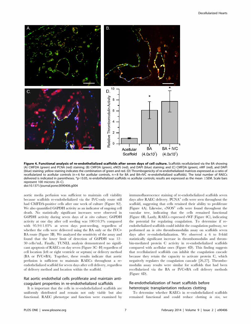
immunofluorescence staining of re-endothelialized scaffolds seven
days after RAEC delivery. PCNA+ cells were seen throughout the
scaffold, suggesting that cells retained their ability to proliferate
(Figure 4A). Likewise, eNOS+ cells were found throughout the
vascular tree, indicating that the cells remained functional
(Figure 4B). Lastly, RAECs expressed vWF (Figure 4C), indicating
the potential for regulating coagulation. To determine if re-
endothelialized scaffolds could inhibit the coagulation pathway, we
performed an in vitro thrombomodulin assay on scaffolds seven
days after re-endothelialization. We observed a 6 to 8-fold
statistically significant increase in thrombomodulin and throm-
bin-mediated protein C activity in re-endothelialized scaffolds
compared with acellular ones (Figure 4D). This finding suggests
that recellularized scaffolds can inhibit the coagulation cascade
because they retain the capacity to activate protein C, which
negatively regulates the coagulation cascade [26,27]. Thrombo-
modulin assay results were similar for scaffolds that had been
recellularized via the BA or IVC+BA cell delivery methods
(Figure 4D).
Re-endothelialization of heart scaffolds beforeheterotopic transplantation reduces clotting
To determine whether RAECs in re-endothelialized scaffolds
remained functional and could reduce clotting in vivo, we
Figure 4. Functional analysis of re-endothelialized scaffolds after seven days of cell culture. Scaffolds recellularized via the BA showing(A) CMFDA (green) and PCNA (red) staining; (B) CMFDA (green), eNOS (red), and DAPI (blue) staining; and (C) CMFDA (green), vWF (red), and DAPI(blue) staining; yellow staining indicates the combination of green and red. (D) Thrombogenicity of re-endothelialized matrices expressed as a ratio ofrecellularized to acellular controls (n = 6 for acellular controls, n = 8 for BA and BA+IVC re-endothelialized scaffolds). The total number of RAECsdelivered is indicated in parentheses. *p,0.05, re-endothelialized scaffolds vs acellular controls; results are expressed as the mean 6SEM. Scale barsrepresent 100 microns (A–C).doi:10.1371/journal.pone.0090406.g004
Decellularized Hearts
PLOS ONE | www.plosone.org 7 February 2014 | Volume 9 | Issue 2 | e90406

heterotopically transplanted acellular scaffolds or those that had
been re-endothelialized with RAECs using the BA cell delivery
method into the abdomen of recipient rats. The re-endothelialized
scaffolds were cultured in vitro for seven days before transplanta-
tion. Then, seven days after in vivo transplantation, we explanted
the scaffolds for examination (Figure 5).We found less aortic
clotting in re-endothelialized scaffolds (Figure 5F) than in acellular
scaffolds (Figure 5A). Examination of the left ventricular wall and
ventricular cavity showed greater thrombogenesis in the acellular
scaffold transplants than in the re-endothelialized scaffolds
(Figure 5B and 5G). A wider tissue distribution of blood cells
was observed in the parenchyma of the acellular scaffolds than in
re-endothelialized scaffolds (Figure 5B–E and 5G–J) as shown by
the intense red H&E staining coloration. Patent vessels with and
without blood were observed only in re-endothelialized scaffolds
(Figure 5J).
We used immunofluorescence staining to characterize the cells
present in the transplanted scaffolds. The majority of the cells
stained positive for CD31 and VEGFR2 suggesting the presence of
endothelial cells in the heterotopic transplant, even for acellular
scaffolds that were not seeded with RAECs before transplantation
(Figure 5K, L). The progenitor cell markers CD34 and CD45 were
expressed only by a small subset of the recruited cells in both the
acellular and re-endothelialized scaffolds. Heterotopic transplan-
tation of either acellular or re-endothelialized heart scaffolds did
not lead to significant staining of smooth muscle actin, vimentin,
or calretinin, which are common markers for smooth muscle cells,
fibroblasts, and mesothelium, respectively (data not shown).
Re-endothelialization before recellularization of the leftventricle wall improves contractility of the heartconstruct
To further characterize the functional benefits of using re-
endothelialized scaffolds, we examined the effects of re-endothelia-
lization on the function of beating heart constructs in which the left
ventricle was recellularized with neonatal cardiac cells. At a pacing
frequency of 2 to 4 Hz, the average maximal rate of change in
pressure was significantly higher in re-endothelialized constructs
than in constructs that had not been re-endothelialized before left
Figure 5. Characterization of heterotopic transplants. (A–E) Acellular and (F–J) re-endothelialized scaffolds seven days after heterotopictransplantation. Short axial view of an aorta with blood clot (arrow) from an acellular scaffold (A) and a non-clotted aorta (arrow) from a re-endothelialized scaffold (F) after transplantation. Short axial view of the ventricle wall of an acellular scaffold with a blood clot (B) and a re-endothelialized scaffold (G). H&E staining of a transplanted acellular scaffold (C–E) with a blood clot inside the ventricular cavity and a re-endothelialized scaffold (H–J) at increasing magnification (2X, 10X, and 20X). Arrows point to patent vessels with and without blood (J). CD31 (K) andVEGFR2 (L) (red) staining in transplanted scaffolds; DAPI-positive nuclei are blue. Scale bars represent 1 mm (A–C and F–H) and 100 microns (D, E, I, J,K, and L).doi:10.1371/journal.pone.0090406.g005
Decellularized Hearts
PLOS ONE | www.plosone.org 8 February 2014 | Volume 9 | Issue 2 | e90406
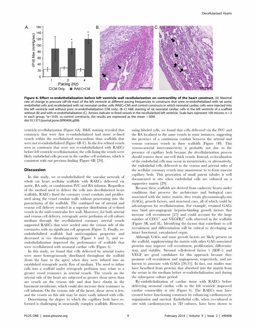
ventricle-recellularization (Figure 6A). H&E staining revealed that
constructs that were first re-endothelialized had more re-lined
vessels within the recellularized myocardium than scaffolds that
were not re-endothelialized (Figure 6B–C). In the few relined vessels
seen in constructs that were not re-endothelialized with RAECs
before left ventricle-recellularization, the cells lining the vessels were
likely endothelial cells present in the cardiac cell isolations, which is
consistent with our previous finding (Figure 6B) [20].
Discussion
In this study, we re-endothelialized the vascular network of
whole rat heart acellular scaffolds with RAECs delivered via
aortic, BA only, or combination IVC and BA infusion. Regardless
of the method used to deliver the cells into decellularized heart
scaffolds, RAECs lined the coronary vessel conduits and prolifer-
ated along the vessel conduit walls without penetrating into the
parenchyma of the scaffolds. The combined use of arterial and
venous cell delivery resulted in a superior distribution of re-lined
vessels in the mid-ventricular free wall. Moreover, for both arterial
and venous cell delivery, retrograde aortic perfusion of cell culture
medium through the recellularized coronary arterial vessels
supported RAECs that were seeded onto the venous side of the
coronaries with no significant cell apoptosis (Figure 3). Finally, re-
endothelialized scaffolds had anti-coagulant properties and
decreased in vivo thrombogenicity (Figure 4 and 5), and re-
endothelialization improved the performance of scaffolds that
were recellularized with neonatal cardiac cells (Figure 6).
In this study, we found that cells delivered by arterial routes
were more homogeneously distributed throughout the scaffold
(from the base to the apex) when they were infused into an
established retrograde aortic flow of medium. The need to infuse
cells into a scaffold under retrograde perfusion may relate to a
greater vessel resistance in arterial vessels. The vessels on the
arterial side of the heart are subjected to greater shear stress than
are vessels on the venous side and thus have elastin in the
basement membrane, which could also increase their resistance to
cell infusion. On the venous side of the heart, shear stress is less,
and the vessels on this side may be more easily infused with cells.
Determining the degree to which the capillary beds have re-
formed is challenging in structurally complex scaffolds. However,
using labeled cells, we found that cells delivered via the IVC and
the BA localized to the same vessels in some instances, suggesting
the presence of a continuous conduit between the arterial and
venous coronary vessels in these scaffolds (Figure 1H). This
venous-arterial interconnectivity is probably not due to the
presence of capillary beds because the decellularization process
should remove these one-cell thick vessels. Instead, co-localization
of the endothelial cells may occur in metarterioles, or alternatively,
the endothelial cells delivered to the venous and arterial sides of
the acellular coronary vessels may anastomose to re-form nascent
capillary beds. This generation of small patent tubules is well
documented in vitro when endothelial cells are cultured on a
supportive matrix [29].
Because these scaffolds are derived from cadaveric hearts under
conditions that preserve the architecture and biological cues
associated with the native matrix, they retain glycosaminoglycans
(GAGs), growth factors, and structural cues, all of which could be
advantageous for recellularization. For example, retained GAGs
may bind pro-angiogenic heparin-binding growth factors that
increase cell recruitment [27] and could account for the large
number of CD31+ and VEGFR2+ cells observed in the scaffolds
(Figure 5K and 5L). Identifying the factors that contribute to cell
recruitment and differentiation will be critical to developing an
intact functional, vascularized organ.
Although GAGs and some growth factors are likely present on
the scaffold, supplementing the matrix with other GAG-associated
proteins may improve cell recruitment, proliferation, differentia-
tion, and viability. Stromal cell-derived factor 1 (SDF-1) and
VEGF are good candidates for this approach because they
promote cell recruitment and angiogenesis, respectively, and are
known to associate with GAGs [30,31]. In fact, our studies may
have benefited from proteins that absorbed into the matrix from
the serum in the medium before re-endothelialization and during
the subsequent culture period.
Re-endothelialization of cardiac tissue with RAECs before
delivering neonatal cardiac cells to the left ventricle improved
cardiac contractility in vitro (Figure 6). The RAECs may have
created better functioning constructs by enhancing cardiomyocyte
organization and survival. Endothelial cells, when co-cultured in
vitro with cardiomyocytes in 2D cultures, have been shown to
Figure 6. Effect re-endothelialization before left ventricle wall recellularization on contractility of the heart construct. (A) Maximalrate of change in pressure (dP/dt max) of the left ventricle at different pacing frequencies in constructs that were re-endothelialized with rat aorticendothelial cells and recellularized with rat neonatal cardiac cells (RAEC+CM) and control constructs in which neonatal cardiac cells were injected intothe left ventricle wall without prior re-endothelialization (CM only). (B–C) H&E staining of rat neonatal cardiac cells in the left ventricle of a scaffoldwithout (B) and with re-endothelialization (C). Arrows indicate re-lined vessels in the recellularized left ventricle. Scale bars represent 100 micron; n = 3in each group; *p,0.05, vs control constructs; the results are expressed as the mean 6SEM.doi:10.1371/journal.pone.0090406.g006
Decellularized Hearts
PLOS ONE | www.plosone.org 9 February 2014 | Volume 9 | Issue 2 | e90406

promote increased cardiomyocyte organization and survival [32].
In addition, re-endothelialization of the construct before recellu-
larization of the left ventricle may have improved contractility by
enhancing nutrient transport to the neonatal cardiomyocytes,
which in turn may have also enhanced cell maturation. These
results indicate that identifying the proper combination of cell
types (vasculogenic versus myogenic) and the order of delivery will
be critical to the optimal performance of recellularized whole
organs. Moreover, our findings suggest that for the current model
re-endothelialization before left ventricle recellularization is
optimal.
In summary, we have shown that re-endothelialization of a
whole heart scaffold is optimal when cells are delivered through
both venous and arterial coronary vessels. Moreover, we found
that re-endothelialization reduces scaffold thrombogenicity and
enhances the function of a left ventricle–recellularized construct.
Our findings lay the groundwork for generating whole-heart
scaffolds or cardiac patches that have perfusable, non-thrombo-
genic vessels. Future work will involve the use of additional cells for
recellularization of constructs in order to create a fully functioning
vasculature that responds to hemodynamic changes.
Supporting Information
Figure S1 Localization of labeled RAECs in re-endothe-lialized scaffolds. Scaffolds were perfused with 26107 DiO-
labeled RAECs (green) via the IVC, followed by perfusion of
26107 DiI-labeled RAECs (red) via the BA, and were cultured for
seven days. Vessels predominantly lined with RAECs cells
delivered via the BA (A) or the IVC (B). DAPI- positive nuclei
are blue. Scale bar represents 50 microns.
(TIF)
Figure S2 CMFDA labeling of RAECs in scaffolds re-endothelialized via the IVC only. Scaffolds seeded with
36107 RAECs were labeled with CMFDA on the last day of
culture (day 7). CMFDA-positive cells in the ventricle wall (A) and
on the endocardial surface (B). Scale bar represents 100 microns.
(TIF)
Acknowledgments
We would like to thank the Lillehei Heart Institute histology and
microscopy core facilities at the University of Minnesota for sample
processing and equipment use. We would like to acknowledge Gabriel J.
Caron, MPH, for his contributions to initial cell culture expansion for the
re-endothelialization studies. Finally, we thank Rebecca A. Bartow, PhD,
of the Texas Heart Institute, for editorial assistance.
Author Contributions
Conceived and designed the experiments: MJR DAT SMK JLD.
Performed the experiments: MJR SMK JLD. Analyzed the data: MJR
JLD SMK. Wrote the paper: MJR JSB DAT.
References
1. Go AS, Mozaffarian D, Roger VL, Benjamin EJ, Berry JD, et al. (2013) Heart
disease and stroke statistics–2013 update: a report from the American Heart
Association. Circulation 127: e6–e245.2. Dean EW, Udelsman B, Breuer CK (2012) Current advances in the translation
of vascular tissue engineering to the treatment of pediatric congenital heartdisease. Yale J Biol Med 85: 229–238.
3. Butcher JT, Mahler GJ, Hockaday LA (2011) Aortic valve disease and
treatment: the need for naturally engineered solutions. Adv Drug Deliv Rev63: 242–268.
4. Karikkineth BC, Zimmermann WH (2013) Myocardial tissue engineering andheart muscle repair. Curr Pharm Biotechnol 14: 4–11.
5. Gaballa MA, Sunkomat JN, Thai H, Morkin E, Ewy G, et al. (2006) Grafting an
acellular 3-dimensional collagen scaffold onto a non-transmural infarctedmyocardium induces neo-angiogenesis and reduces cardiac remodeling. J Heart
Lung Transplant 25: 946–954.6. Kochupura PV, Azeloglu EU, Kelly DJ, Doronin SV, Badylak SF, et al. (2005)
Tissue-engineered myocardial patch derived from extracellular matrix providesregional mechanical function. Circulation 112: I144–149.
7. Kutschka I, Chen IY, Kofidis T, Arai T, von Degenfeld G, et al. (2006) Collagen
matrices enhance survival of transplanted cardiomyoblasts and contribute tofunctional improvement of ischemic rat hearts. Circulation 114: I167–173.
8. Zimmermann WH, Melnychenko I, Wasmeier G, Didie M, Naito H, et al.(2006) Engineered heart tissue grafts improve systolic and diastolic function in
infarcted rat hearts. Nat Med 12: 452–458.
9. Vunjak-Novakovic G, Tandon N, Godier A, Maidhof R, Marsano A, et al.(2009) Challenges in Cardiac Tissue Engineering. Tissue Eng Part B Rev.
10. Korecky B, Hai CM, Rakusan K (1982) Functional capillary density in normaland transplanted rat hearts. Can J Physiol Pharmacol 60: 23–32.
11. Morritt AN, Bortolotto SK, Dilley RJ, Han X, Kompa AR, et al. (2007) Cardiactissue engineering in an in vivo vascularized chamber. Circulation 115: 353–360.
12. Radisic M, Marsano A, Maidhof R, Wang Y, Vunjak-Novakovic G (2008)
Cardiac tissue engineering using perfusion bioreactor systems. Nat Protoc 3:719–738.
13. Kim MH, Hong HN, Hong JP, Park CJ, Kwon SW, et al. (2009) The effect ofVEGF on the myogenic differentiation of adipose tissue derived stem cells within
thermosensitive hydrogel matrices. Biomaterials 31: 1213–1218.
14. Nillesen ST, Geutjes PJ, Wismans R, Schalkwijk J, Daamen WF, et al. (2007)Increased angiogenesis and blood vessel maturation in acellular collagen-heparin
scaffolds containing both FGF2 and VEGF. Biomaterials 28: 1123–1131.15. Ota T, Sawa Y, Iwai S, Kitajima T, Ueda Y, et al. (2005) Fibronectin-
hepatocyte growth factor enhances reendothelialization in tissue-engineeredheart valve. Ann Thorac Surg 80: 1794–1801.
16. Zhang Z, Dong H, Liu J, Wang W, Hu B (2002) Vascular endothelial growth
factor gene transfer improves host endothelialization of xenogeneic biologicheart valve in vivo. Chin Med J (Engl) 115: 878–883.
17. Zisch AH, Schenk U, Schense JC, Sakiyama-Elbert SE, Hubbell JA (2001)
Covalently conjugated VEGF–fibrin matrices for endothelialization. J Control
Release 72: 101–113.18. Caspi O, Lesman A, Basevitch Y, Gepstein A, Arbel G, et al. (2007) Tissue
engineering of vascularized cardiac muscle from human embryonic stem cells.Circ Res 100: 263–272.
19. Suuronen EJ, Veinot JP, Wong S, Kapila V, Price J, et al. (2006) Tissue-
engineered injectable collagen-based matrices for improved cell delivery andvascularization of ischemic tissue using CD133+ progenitors expanded from the
peripheral blood. Circulation 114: I138–144.20. Ott HC, Matthiesen TS, Goh SK, Black LD, Kren SM, et al. (2008) Perfusion-
decellularized matrix: using nature’s platform to engineer a bioartificial heart.
Nat Med 14: 213–221.21. Aubin H, Kranz A, Hulsmann J, Lichtenberg A, Akhyari P (2013) Decellularized
whole heart for bioartificial heart. Methods Mol Biol 1036: 163–178.22. Park KM, Woo HM (2012) Systemic decellularization for multi-organ scaffolds
in rats. Transplant Proc 44: 1151–1154.23. Wainwright JM, Czajka CA, Patel UB, Freytes DO, Tobita K, et al. (2010)
Preparation of cardiac extracellular matrix from an intact porcine heart. Tissue
Eng Part C Methods 16: 525–532.24. Lu TY, Lin B, Kim J, Sullivan M, Tobita K, et al. (2013) Repopulation of
decellularized mouse heart with human induced pluripotent stem cell-derivedcardiovascular progenitor cells. Nat Commun 4: 2307.
25. Strater J, Gunthert AR, Bruderlein S, Moller P (1995) Microwave irradiation of
paraffin-embedded tissue sensitizes the TUNEL method for in situ detection ofapoptotic cells. Histochem Cell Biol 103: 157–160.
26. Calnek DS, Grinnell BW (1998) Thrombomodulin-Dependent AnticoagulantActivity Is Regulated by Vascular Endothelial Growth Factor. Experimental Cell
Research 238: 294–298.27. Ibrahim S, Ramamurthi A (2008) Hyaluronic acid cues for functional
endothelialization of vascular constructs. J Tissue Eng Regen Med.
28. Ono K, Lindsey ES (1969) Improved technique of heart transplantation in rats.J Thorac Cardiovasc Surg 57: 225–229.
29. Donovan D, Brown NJ, Bishop ET, Lewis CE (2001) Comparison of three invitro human ‘angiogenesis’ assays with capillaries formed in vivo. Angiogenesis 4:
113–121.
30. Robinson CJ, Mulloy B, Gallagher JT, Stringer SE (2006) VEGF165-bindingsites within heparan sulfate encompass two highly sulfated domains and can be
liberated by K5 lyase. J Biol Chem 281: 1731–1740.31. Mbemba E, Gluckman JC, Gattegno L (2000) Glycan and glycosaminoglycan
binding properties of stromal cell-derived factor (SDF)-1alpha. Glycobiology 10:21–29.
32. Narmoneva DA, Vukmirovic R, Davis ME, Kamm RD, Lee RT (2004)
Endothelial cells promote cardiac myocyte survival and spatial reorganization:implications for cardiac regeneration. Circulation 110: 962–968.
Decellularized Hearts
PLOS ONE | www.plosone.org 10 February 2014 | Volume 9 | Issue 2 | e90406