Embed Size (px)
Citation preview
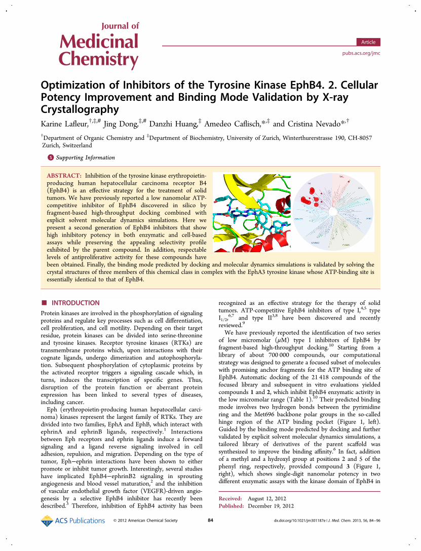
Optimization of Inhibitors of the Tyrosine Kinase EphB4. 2. CellularPotency Improvement and Binding Mode Validation by X‑rayCrystallographyKarine Lafleur,†,‡,# Jing Dong,‡,# Danzhi Huang,‡ Amedeo Caflisch,*,‡ and Cristina Nevado*,†
†Department of Organic Chemistry and ‡Department of Biochemistry, University of Zurich, Winterthurerstrasse 190, CH-8057Zurich, Switzerland
*S Supporting Information
ABSTRACT: Inhibition of the tyrosine kinase erythropoietin-producing human hepatocellular carcinoma receptor B4(EphB4) is an effective strategy for the treatment of solidtumors. We have previously reported a low nanomolar ATP-competitive inhibitor of EphB4 discovered in silico byfragment-based high-throughput docking combined withexplicit solvent molecular dynamics simulations. Here wepresent a second generation of EphB4 inhibitors that showhigh inhibitory potency in both enzymatic and cell-basedassays while preserving the appealing selectivity profileexhibited by the parent compound. In addition, respectablelevels of antiproliferative activity for these compounds havebeen obtained. Finally, the binding mode predicted by docking and molecular dynamics simulations is validated by solving thecrystal structures of three members of this chemical class in complex with the EphA3 tyrosine kinase whose ATP-binding site isessentially identical to that of EphB4.
■ INTRODUCTIONProtein kinases are involved in the phosphorylation of signalingproteins and regulate key processes such as cell differentiation,cell proliferation, and cell motility. Depending on their targetresidue, protein kinases can be divided into serine-threonineand tyrosine kinases. Receptor tyrosine kinases (RTKs) aretransmembrane proteins which, upon interactions with theircognate ligands, undergo dimerization and autophosphoryla-tion. Subsequent phosphorylation of cytoplasmic proteins bythe activated receptor triggers a signaling cascade which, inturns, induces the transcription of specific genes. Thus,disruption of the protein function or aberrant proteinexpression has been linked to several types of diseases,including cancer.Eph (erythropoietin-producing human hepatocellular carci-
noma) kinases represent the largest family of RTKs. They aredivided into two families, EphA and EphB, which interact withephrinA and ephrinB ligands, respectively.1 Interactionsbetween Eph receptors and ephrin ligands induce a forwardsignaling and a ligand reverse signaling involved in celladhesion, repulsion, and migration. Depending on the type oftumor, Eph−ephrin interactions have been shown to eitherpromote or inhibit tumor growth. Interestingly, several studieshave implicated EphB4−ephrinB2 signaling in sproutingangiogenesis and blood vessel maturation,2 and the inhibitionof vascular endothelial growth factor (VEGFR)-driven angio-genesis by a selective EphB4 inhibitor has recently beendescribed.3 Therefore, inhibition of EphB4 activity has been
recognized as an effective strategy for the therapy of solidtumors. ATP-competitive EphB4 inhibitors of type I,4,5 typeI1/2,
6,7 and type II3,8 have been discovered and recentlyreviewed.9
We have previously reported the identification of two seriesof low micromolar (μM) type I inhibitors of EphB4 byfragment-based high-throughput docking.10 Starting from alibrary of about 700 000 compounds, our computationalstrategy was designed to generate a focused subset of moleculeswith promising anchor fragments for the ATP binding site ofEphB4. Automatic docking of the 21 418 compounds of thefocused library and subsequent in vitro evaluations yieldedcompounds 1 and 2, which inhibit EphB4 enzymatic activity inthe low micromolar range (Table 1).10 Their predicted bindingmode involves two hydrogen bonds between the pyrimidinering and the Met696 backbone polar groups in the so-calledhinge region of the ATP binding pocket (Figure 1, left).Guided by the binding mode predicted by docking and furthervalidated by explicit solvent molecular dynamics simulations, atailored library of derivatives of the parent scaffold wassynthesized to improve the binding affinity.6 In fact, additionof a methyl and a hydroxyl group at positions 2 and 5 of thephenyl ring, respectively, provided compound 3 (Figure 1,right), which shows single-digit nanomolar potency in twodifferent enzymatic assays with the kinase domain of EphB4 in
Received: August 12, 2012Published: December 19, 2012
Article
pubs.acs.org/jmc
© 2012 American Chemical Society 84 dx.doi.org/10.1021/jm301187e | J. Med. Chem. 2013, 56, 84−96

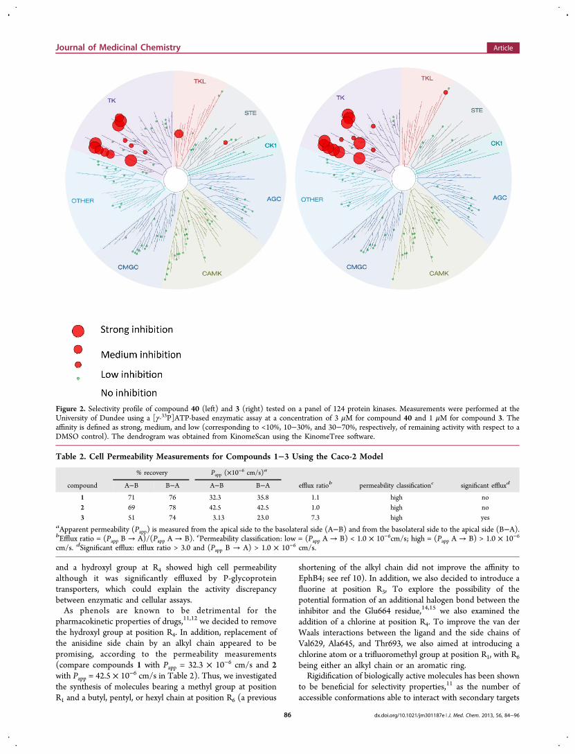
solution (Table 1). In addition, compound 3 showed apromising selectivity profile (Figure 2, right). Unfortunately, asignificant reduction of inhibitory activity on EphB4 wasobserved for this compound in a cellular assay compared to theenzymatic assays. This discrepancy could be due to reducedpermeability or increased efflux of the molecule by activetransporters. In addition, phenols can undergo glucuronidationduring phase II metabolism, thus having poor pharmacokineticproperties.11,12
With these results in hand, we embarked on a multi-disciplinary optimization campaign aimed at the developmentof a second generation of EphB4 inhibitors that combine highinhibitory potency in both enzymatic and cell-based assayswhile retaining the appealing selectivity profile exhibited by theparent compound. Here, we present the validation of thisapproach, which has led to the discovery of a lead compoundincorporating not only high potency and selectivity but also apromising pharmacological profile as well as respectable levelsof antiproliferative activity. In addition, we report herein theconfirmation of the binding mode of this chemical class to theEph receptor tyrosine kinase by means of X-ray crystallography.
■ LEAD OPTIMIZATION STRATEGY
In line with the results obtained in the enzymatic assay, theinhibition activity of compounds 1 and 2 on EphB4-transfectedChinese hamster ovary (CHO) cells has been shown to berelatively low with IC50 values in the high micromolar range.10
The high potency displayed by compound 3 in the enzymaticassay prompted us to explore its activity on a cellular setting.Thus, cellular phosphorylation assays were carried out onmurine embryonal fibroblast cells (MEF) transfected with myc-tagged human EphB4 (Proqinase). After incubation with thecorresponding inhibitor and stimulation with ephrinB2-Fc,autophosphorylation of EphB4 was quantified via sandwichELISA. While compound 3 showed single-digit nanomolaractivity in enzymatic assay, its inhibition activity in cells wasreduced by almost 2 orders of magnitude, with IC50 valueshigher than 100 nM (Table 1). There are multiple reasons for adiscrepancy between enzymatic and cellular assays, includingthe passage through the cell membrane, the natural environ-ment, and relevant ATP concentration present only in thelatter.13 To further evaluate the pharmacological potential ofcompound 3, its cell permeability was investigated first. Whilepassive transport was assessed on Caco-2 monolayers, activeefflux was tested on P-glycoprotein and BCRP transporters(Absorption Systems). Caco-2 cells express many transportproteins present on epithelial cells of the small intestine andform tight junctions between each other. Thus, measurementsof permeability through Caco-2 monolayers may also predictabsorption across intestinal tissues in vivo. Interestingly,compounds 1 and 2 bearing hydrogens at position R1 and R4
showed high cell permeability, the latter showing the highestmembrane permeation, while no significant efflux was observed(Table 2). In contrast, compound 3 with a methyl group at R1
Table 1. EphB4 Inhibition Data of Xanthine Derivatives
enzymatic assay IC50 (nM)
compound R1 R2 R3 R4 R5 R6 FRETa radiometricb cellular assay IC50 (nM)c
1 H H H H H o-methoxyphenyl 3300 4350 14% at 20 μM2 H H H H H butyl 1900 538 <10% at 20 μM3 Me H H OH H o-methoxyphenyl 5 1.6 130
aFRET-based enzymatic assay carried out using the Z′-LYTE Kinase Assay Kit−Tyr 1 Peptide (Invitrogen, Grand Island, NY) following the vendorinstructions. bMeasured at Reaction Biology Corporation using radiolabeled ATP. cCell IC50 values for compounds 1 and 2 were measured in acellular phosphorylation assay using CHO cells overexpressing EphB4. For compound 3, measurements were performed at Proqinase using MEFcells overexpressing EphB4.
Figure 1. (Left) Predicted binding mode of compounds 1 and 2.10 (Right) Binding mode of compound 3 as predicted in silico and validated by X-ray crystallography. The EphB4 residues involved in binding are shown. Thr693 is the so-called gatekeeper residue while Asp758 is the first residuein the DFG motif.
Journal of Medicinal Chemistry Article
dx.doi.org/10.1021/jm301187e | J. Med. Chem. 2013, 56, 84−9685
and a hydroxyl group at R4 showed high cell permeabilityalthough it was significantly effluxed by P-glycoproteintransporters, which could explain the activity discrepancybetween enzymatic and cellular assays.As phenols are known to be detrimental for the
pharmacokinetic properties of drugs,11,12 we decided to removethe hydroxyl group at position R4. In addition, replacement ofthe anisidine side chain by an alkyl chain appeared to bepromising, according to the permeability measurements(compare compounds 1 with Papp = 32.3 × 10⁻⁶ cm/s and 2with Papp = 42.5 × 10⁻⁶ cm/s in Table 2). Thus, we investigatedthe synthesis of molecules bearing a methyl group at positionR1 and a butyl, pentyl, or hexyl chain at position R6 (a previous
shortening of the alkyl chain did not improve the affinity toEphB4; see ref 10). In addition, we also decided to introduce afluorine at position R3, To explore the possibility of thepotential formation of an additional halogen bond between theinhibitor and the Glu664 residue,14,15 we also examined theaddition of a chlorine at position R4. To improve the van derWaals interactions between the ligand and the side chains ofVal629, Ala645, and Thr693, we also aimed at introducing achlorine atom or a trifluoromethyl group at position R1, with R6
being either an alkyl chain or an aromatic ring.Rigidification of biologically active molecules has been shown
to be beneficial for selectivity properties,11 as the number ofaccessible conformations able to interact with secondary targets
Figure 2. Selectivity profile of compound 40 (left) and 3 (right) tested on a panel of 124 protein kinases. Measurements were performed at theUniversity of Dundee using a [γ-33P]ATP-based enzymatic assay at a concentration of 3 μM for compound 40 and 1 μM for compound 3. Theaffinity is defined as strong, medium, and low (corresponding to <10%, 10−30%, and 30−70%, respectively, of remaining activity with respect to aDMSO control). The dendrogram was obtained from KinomeScan using the KinomeTree software.
Table 2. Cell Permeability Measurements for Compounds 1−3 Using the Caco-2 Model
% recovery Papp (×10−6 cm/s)a
compound A−B B−A A−B B−A efflux ratiob permeability classificationc significant effluxd
1 71 76 32.3 35.8 1.1 high no2 69 78 42.5 42.5 1.0 high no3 51 74 3.13 23.0 7.3 high yes
aApparent permeability (Papp) is measured from the apical side to the basolateral side (A−B) and from the basolateral side to the apical side (B−A).bEfflux ratio = (Papp B → A)/(Papp A → B). cPermeability classification: low = (Papp A → B) < 1.0 × 10⁻⁶cm/s; high = (Papp A → B) > 1.0 × 10⁻⁶
cm/s. dSignificant efflux: efflux ratio > 3.0 and (Papp B → A) > 1.0 × 10⁻⁶ cm/s.
Journal of Medicinal Chemistry Article
dx.doi.org/10.1021/jm301187e | J. Med. Chem. 2013, 56, 84−9686

is restricted. In addition, ortho-substitution of bicyclicmolecules with methyl or methoxy groups has been shown todecrease crystal packing and improve aqueous solubility.16 Wetherefore investigated the synthesis of a bis-ortho-methyl-substituted phenyl ring on the xanthine scaffold. Finally, poororal bioavailability of kinase inhibitors entitling an ortho-methyl/meta-hydroxyphenyl pattern11,17,18 led to the replace-ment of the phenol ring by an indazole moiety.11 Thistransformation aimed to generate a molecule with a similarhydrogen bonding pattern as compound 3 with EphB4.With these designs in mind, we set out to explore the
synthesis of a small set of customized analogues to laterevaluate their biochemical and pharmacological profile.
■ SYNTHESIS
Recently, a growing interest in the synthesis of imidazox-anthines has emerged, as a few derivatives proved to be potentserotonin,19 A3 adenosine,
20,21 or kinase receptor antagonists.6
Following our previously developed route, the synthesis of thekey intermediate 3-methyl-8-bromoxanthine 4 was achieved infive steps starting from the corresponding alkylurea (Scheme1). After alkylation with α-brominated acetophenones,cyclization usually occurs by refluxing the resulting intermediatein ethanol in the presence of a primary amine.6,19,20 However,to the best of our knowledge, the synthesis of bis-ortho-substituted imidazoxanthines has not been described yet. Inaddition, none of the mono-ortho-substituted moleculesreported so far has an alkyl chain at position R6.The synthesis of the noncommercially available acetophe-
nones used in the preparation of this new set of inhibitors is
summarized in Scheme 2. Treatment of commercially available4-fluoro-2-methylbenzaldehyde 5 with methyllithium followedby oxidation with PCC provided methyl ketone 7 in 40% yieldover two steps. The synthesis of 1-(2,6-dimethylphenyl)-ethanone started with 2-bromo-1,3-dimethylbenzene 8, whichundergoes bromo−lithium exchange in the presence of nBuli,followed by reaction with acetaldehyde to afford secondaryalcohol 9. Subsequent oxidation using PCC gave the expectedacetophenone 10,22 which was used as starting material in thesynthesis of 1,1′-(5-methyl-1H-indazole-1,4-diyl)diethanone13. Indeed, reaction with KNO3 selectively nitrated compound10 at the meta position relative to the carbonyl group.23
Reduction in the presence of iron and cyclization using isoamylnitrite afforded the protected indazole derivative 13 in good tomoderate yields.24
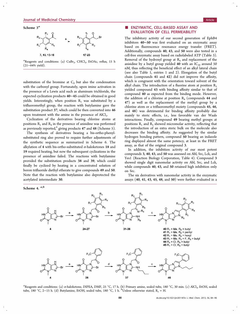
α-Bromination of the acetophenones reported in Scheme 2as well as the commercially available 2′-chloroacetophenone(14), 3′-chloroacetophenone (15), and 2′-(trifluoromethyl)-acetophenone (16), was achieved in the presence of copper(II)bromide in chloroform as shown in Scheme 3.25
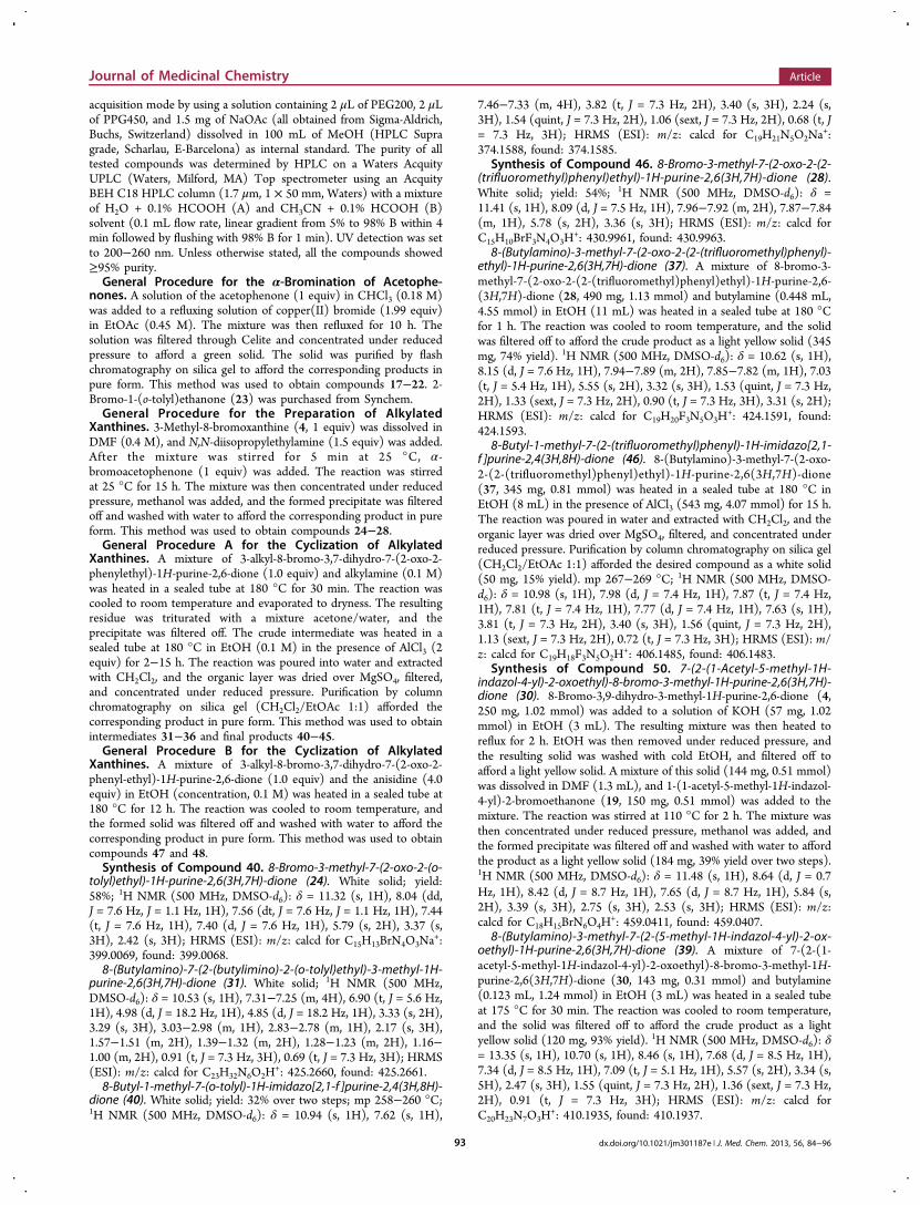
Alkylation at the N7 position of bromoxanthine 4 with meta-substituted or mono-ortho-substituted α-haloketones wasaccomplished using N,N-diisopropylethylamine in DMF togive intermediates 24−27 (Scheme 4). To our surprise, incontrast to our previous experience with anilines, thesubsequent cyclization with alkylamines under the standardconditions shown in Scheme 1 was never observed. Instead,only intermediates 31−36 could be isolated from the reactionmixtures probably due to the more electron-rich nature ofalkylamines, which are able to accomplish not only the
Scheme 1a
aReagents and conditions: (a) Primary amine, EtOH, reflux, 15 h.
Scheme 2a
aReagents and conditions: (a) MeLi, THF, 25 °C, 1 h, 55%. (b) PCC, CH2Cl2, 25 °C, 1 h, 73%. (c) BuLi, acetaldehyde, THF, −78 °C, 1.5 h. (d)PCC, CH2Cl2, 25 °C, 1 h, 56% yield over two steps. (e) KNO3, H2SO4, 0 °C, 1 h. (f) Fe, AcOH, EtOH, 90 °C, 2 h, 47% yield over two steps. (g)NaOAc, Ac2O, CHCl3, 25 °C, 30 min, then iAmONO, 60 °C, 7 h, 58%.
Journal of Medicinal Chemistry Article
dx.doi.org/10.1021/jm301187e | J. Med. Chem. 2013, 56, 84−9687
substitution of the bromine at C8 but also the condensationwith the carbonyl group. Fortunately, upon imine activation inthe presence of a Lewis acid such as aluminum trichloride, theexpected cyclization products 40−45 could be obtained in goodyields. Interestingly, when position R1 was substituted by atrifluoromethyl group, the reaction with butylamine gave thesubstitution product 37, which could be then converted into 46upon treatment with the amine in the presence of AlCl3.Cyclization of the derivatives bearing chlorine atoms at
positions R1 and R4 in the presence of anisidine was performedas previously reported,6 giving products 47 and 48 (Scheme 5).The synthesis of derivatives bearing a bis-ortho-phenyl-
substituted ring also proved to require further adjustments ofthe synthetic sequence as summarized in Scheme 6. Thealkylation of 4 with bis-ortho-substituted α-haloketones 18 and19 required heating, but now the subsequent cyclizations in thepresence of anisidine failed. The reactions with butylamineprovided the substitution products 38 and 39, which couldfinally be cyclized by heating in a concentrated solution ofboron trifluoride diethyl etherate to give compounds 49 and 50.Note that the reaction with butylamine also deprotected theacetylated intermediate 30.
■ ENZYMATIC, CELL-BASED ASSAY ANDEVALUATION OF CELL PERMEABILITY
The inhibitory activity of our second generation of EphB4inhibitors 40−50 was first evaluated on an enzymatic assaybased on fluorescence resonance energy transfer (FRET).Additionally, compounds 40, 43, and 50 were also tested in acell-free enzymatic assay based on radiolabeled ATP (Table 3).Removal of the hydroxyl group at R4 and replacement of theanisidine by a butyl group yielded 40 with an IC50 around 50nM, thus reflecting the beneficial effect of an alkyl lateral chain(see also Table 1, entries 1 and 2). Elongation of the butylchain (compounds 41 and 42) did not improve the affinity,which is congruent with the orientation toward solvent of thealkyl chain. The introduction of a fluorine atom at position R3yielded compound 43 with binding affinity similar to that ofcompound 40 as expected from the binding mode. However,the addition of a chlorine at position R4 (compounds 44 and47) as well as the replacement of the methyl group by achlorine atom or a trifluoromethyl moiety (compounds 45, 46,and 48) was detrimental for binding affinity probably duemainly to steric effects, i.e., less favorable van der Waalsinteractions. Finally, compound 49 bearing methyl groups atpositions R1 and R5 showed micromolar activity, reflecting thatthe introduction of an extra steric bulk on the molecule alsodecreases the binding affinity. As suggested by the similarhydrogen bonding pattern, compound 50 bearing an indazolering displayed almost the same potency, at least in the FRETassay, as that of the original compound 3.In addition, the inhibition activity of our most potent
compounds 3, 40, 43, and 50 was assessed on Abl, Src, Lck, andYes1 (Reaction Biology Corporation, Table 4). Compound 3showed single digit nanomolar activity on Abl, Src, and Lck,while compounds 40, 43, and 50 retained high inhibition onlyon Src.The six derivatives with nanomolar activity in the enzymatic
assays (40, 41, 43, 45, 48, and 50) were further evaluated in a
Scheme 3a
aReagents and conditions: (a) CuBr2, CHCl3, EtOAc, reflux, 15 h(21−64% yield).
Scheme 4. a,b
aReagents and conditions: (a) α-haloketone, DIPEA, DMF, 25 °C, 17 h. (b) Primary amine, sealed tube, 180 °C, 30 min. (c) AlCl3, EtOH, sealedtube, 180 °C, 2−15 h. (d) Butylamine, EtOH, sealed tube, 180 °C, 1 h. bUnless otherwise stated, Rn = H.
Journal of Medicinal Chemistry Article
dx.doi.org/10.1021/jm301187e | J. Med. Chem. 2013, 56, 84−9688

cellular phosphorylation assay on MEF cells transfected withmyc-tagged human EphB4 (Proqinase, Table 3, right column).
Except for compound 48 which showed micromolar affinity,the other five compounds displayed levels of inhibitory activity
Scheme 5. a,b
aReagents and conditions: (a) α-haloketone, DIPEA, DMF, 25 °C, 17 h. (b) Anisidine, EtOH, sealed tube, reflux, 15 h. bUnless otherwise stated, Rn= H.
Scheme 6a
aReagents and conditions: (a) 2-bromo-1-(2,6-dimethylphenyl)ethanone 18, DIPEA, DMF, 110 °C, 2 h. (b) Butylamine, EtOH, sealed tube, 180°C, 0.5−2 h. (c) BF3·OEt2, DCM, sealed tube, 180 °C, 0.5−6 h. (d) KOH, EtOH, reflux, 2 h, then 1-(1-acetyl-5-methyl-1H-indazol-4-yl)-2-bromoethanone 19, DMF, 110 °C, 2 h.
Table 3. EphB4 Inhibition Data of Xanthine Derivatives
enzymatic assay IC50 (nM)
compound R1 R2 R3 R4 R5 R6 FRETa radiometricb cellular IC50 (nM)
40 Me H H H H butyl 56 82 5041 Me H H H H pentyl 177 n.d.c 23042 Me H H H H hexyl 1300 n.d. n.d.43 Me H F H H butyl 91 139 6844 H H H Cl H butyl 22% @ 10 μM n.d. n.d.45 Cl H H H H butyl 182 n.d. 27046 CF3 H H H H butyl 15% @ 10 μM n.d. n.d.47 H H H Cl H o-methoxyphenyl 12% @ 10 μM n.d. n.d.48 Cl H H H H o-methoxyphenyl 300 n.d. 220049 Me H H H Me butyl 49% @ 10 μM n.d. n.d.50 Me H H indazole butyl 14 133 150
aFRET-based enzymatic assay carried out using the Z′-LYTE Kinase Assay Kit−Tyr 1 Peptide (Invitrogen) following the vendor instructions.bMeasured at Reaction Biology Corporation using radiolabeled ATP. cn.d.: not determined.
Journal of Medicinal Chemistry Article
dx.doi.org/10.1021/jm301187e | J. Med. Chem. 2013, 56, 84−9689

in the nanomolar range, which correlates with the potenciesmeasured in the enzymatic assays. Of particular interest is theaffinity of compounds 40 and 43, with IC50 values of 50 and 68nM, respectively.To further characterize this second generation of EphB4
inhibitors, the cell permeation of compounds 40 and 50 wasevaluated on Caco-2 monolayers (Absorption Systems). Bothcompounds showed a high cell permeability; however, onlycompound 40 was not effluxed by transport proteins (Table 5),and thus it was selected for further evaluation in terms ofselectivity (vide infra).
■ SELECTIVITY PROFILETo assess the selectivity profile of compound 40, enzymaticassays were performed on a panel of 124 kinases (NationalCentre for Protein Kinase Profiling at the University ofDundee, Table 6, Figure 2, left, and Table S1 in the SupportingInformation). In the presence of 3 μM concentration ofcompound 40, only 5 of the 124 kinases tested (Src, Lck,EphA2, EphA4, and EphB2) had a remaining activity smallerthan 10% with respect to a DMSO control. Furthermore, 5other kinases (RIPK2, Yes1, EphB1, EphB3, and EphB4)showed a remaining activity between 10% and 20%. It isimportant to note that these 10 kinases have a threonine as agatekeeper. Overall, compound 40 has a selectivity profilesimilar to that of 3, with a strong inhibition of a relatively smallfraction of the human kinome (Figure 2).
■ ANTIPROLIFERATIVE ACTIVITYOverexpression of the receptor tyrosine kinase EphB4 has beenlinked to several types of cancer, including breast,26 colon,27
and ovary.28 To assess the potential of our inhibitors on acancer model, compound 3 was submitted to the NCI-60cancer cell line panel. Compound 3 displayed remarkable levelsof cell growth inhibition against central nervous system (SNB-75, 128 nM), leukemia (K-562, 309 nM), and breast (HS 578T,562 nM) cancer cells (data shown in the SupportingInformation).
In addition, the antiproliferative activity of compounds 3, 40,41, 43, 45, and 50 was assessed on patient-derived tumor celllines using a propidium iodide-based proliferation assay anddasatinib as a reference (Oncotest, Table 7). Cell lines includedcolon, lung, kidney, pancreas, prostate, and stomach cancercells. Overall, dasatinib exhibited the highest potency, withdouble-digit nanomolar activity against RXF 393NL, LXFA983L, and PRXF DU145. Compounds 3, 40, 41, 43, 45, and 50inhibited cell proliferation in the low micromolar range, withcompound 3 exhibiting good levels of antiproliferative activityagainst RXF 393NL, PAXF 1657L, and PRXF DU145.Compounds 45 and 50 displayed a 1−2 μM activity againstthe colon cancer cell line CXF 1103L. These results confirmthe correlation between the kinase inhibitory activity of thesemolecules and their antiproliferative activity, although alsoreflect the need for further modifications to parallel theperformance of “gold standards” in this field, such as dasatinib.
■ CONFIRMATION OF THE BINDING MODE BYPROTEIN X-RAY CRYSTALLOGRAPHY
The binding mode of compounds 3, 40, and 50 wasinvestigated by X-ray crystallography using the catalytic domainof EphA3 expressed in E. coli.29 This tyrosine kinase waspreferred to EphB4 for which only expression in insect cells hasbeen reported. Note that 32 of the 36 residues in the ATPbinding site of EphA3 are identical to those in EphB4.30
Moreover, the side chains involved in binding compounds 3,40, and 50 are identical in EphA3 and EphB4. These
Table 4. Abl, Lck, Src, and Yes1 Inhibition Data of XanthineDerivativesa
compound Abl Src Lck Yes1
3 0.92 1.14 1.3 n.d.b
40 578 71 493 28443 1282 110 617 49650 203 46 264 160.4
aEnzymatic assays were carried out at Reaction Biology Corporationusing radiolabeled ATP. IC50 values are given in nanomolarconcentrations. bn.d.: not determined.
Table 5. Cell Permeability Measurements for Compounds 3, 40, and 50 Using the Caco-2 Model
% recovery Papp (×10−6 cm/s)a
compound A−B B−A A−B B−A efflux ratiob permeability classificationc significant effluxd
3 51 74 3.13 23.0 7.3 high yes40 85 89 42.7 28.1 0.7 high no50 84 96 8.02 49.0 6.1 high yes
aApparent permeability (Papp) is measured from the apical side to the basolateral side (A−B) and from the basolateral side to the apical side (B−A).bEfflux ratio = (Papp B → A)/(Papp A → B). cPermeability classification: low = (Papp A → B) < 1.0 × 10⁻⁶cm/s; high = (Papp A → B) > 1.0 × 10⁻⁶
cm/s. dSignificant efflux: efflux ratio > 3.0 and (Papp B → A) > 1.0 × 10⁻⁶ cm/s.
Table 6. Selectivity of Compound 40 Tested on a Panel of124 Protein Kinases at the University of Dundeea
kinase % activity
MKK1 56RIPK2 12Src 2Lck 5CSK 57Yes1 14Abl 43BTK 58Eph-A2 6Eph-A4 5Eph-B1 10Eph-B2 8Eph-B3 13Eph-B4 18
aThe percentage of kinase activity is measured in the presence of 3 μMof 40 compared to a 100% DMSO control (only kinases with apercentage of activity below 70% are indicated). For full data set, seeTable S1 in the Supporting Information.
Journal of Medicinal Chemistry Article
dx.doi.org/10.1021/jm301187e | J. Med. Chem. 2013, 56, 84−9690

observations explain the very similar potency on both targets ofour inhibitors and other Eph kinase inhibitors reportedpreviously.6
Crystals were obtained using the hanging drop vapordiffusion method (see Experimental Section). The finalresolution is 2.2 Å, 1.9 Å, and 2.1 Å for the complexes ofEphA3 with compounds 3, 40, and 50, respectively; furtherdetails of refinement statistics are shown in Table 8.Strikingly, the binding mode of compound 3 observed in the
crystal structure is essentially identical to that predicted bydocking and explicit solvent molecular dynamics6 (Figure 3).The pyrimidine ring is involved in two hydrogen bonds withthe hinge region, and the phenol moiety is nestled into the so-
called back pocket with its hydroxyl group accepting ahydrogen bond from the backbone NH of Asp758 (Asp764for EphA3) of the DFG loop and donating a hydrogen bond tothe side chain of Glu664 (Glu670 for EphA3) (Figure 1, right).Superposition of the crystal structures obtained for
compounds 3, 40 and 50 shows that the key features of thebinding mode are conserved, such as the hydrogen bondinteractions between the pyrimidine ring and Met702, as well asthe orientation of the phenyl ring in the back pocket (Figure 4).Interestingly, the indazole ring of compound 50 is involved intwo hydrogen bond interactions: the NH group acts as donorto the Glu670 side chain, and the imine nitrogen acts asacceptor for the backbone NH of Asp764. The methoxy groupof compound 3 matches the butyl side chains of compounds 40and 50, as both of these substituents point in the oppositedirection of the hinge region. An in-depth comparative analysisof the binding modes of compounds 3, 40, and 50, with the X-ray structures of two other series of EphB4/A3 inhibitorsdiscovered in our groups as well as with the inhibitor/Ephcomplexes available in the PDB database, will be publishedelsewhere (Jing Dong et al., in preparation).Finally, the binding mode of compound 3, predicted and
validated as belonging to type I1/2,6,7 lends itself to a
modification into type II3,8 via replacement of the hydroxylgroup by an amide linker and addition of an aromatic group,e.g., CF3-substituted benzol, to reach the so-called allosteric siteproximal to the ATP binding site. We are currently planningthe synthesis of these type II derivatives.
■ CONCLUSIONSRecently, a medicinal chemistry campaign inspired by the poseof the original hit obtained by docking resulted in the discoveryof compound 3, a single-digit nanomolar inhibitor of the EphB4tyrosine kinase. However, further evaluation of compound 3revealed a major discrepancy between enzymatic and cellularactivity (5 nM in enzymatic assay vs 130 nM in cellularphosphorylation assay). Furthermore, efficient efflux by P-glycoprotein transporters was observed on caco-2 monolayers.Aided by the docked pose of compound 3, a second
generation of EphB4 inhibitors was designed and synthesized to
Table 7. Antiproliferative Activity against Tumor Cell Linesa
aIC50 values were determined at Oncotest using a modified propidium iodide assay. Measurements were performed after 4 days of incubation withthe corresponding compound. IC50 values are given in micromolar concentrations (μM).
Table 8. X-ray Data Collection
3 40 50
space group P 1 21 1 P 1 21 1 P 1 21 1unit cell
a (Å) 52.6 53.29 53.06b (Å) 38.2 38.25 38.18c (Å) 75.7 76.04 75.84resolutionrange (Å)
38.2−2.2 34.0−1.9 38.2−2.1
uniquereflections
15076 (2090) 23747 (3297) 17532 (2402)
⟨I/σ(I)⟩ 8.8 (3.0) 8.9 (2.8) 10.6 (3.9)R merge 0.103 (0.457) 0.084 (0.405) 0.086 (0.334)completeness(%)
98.4 (94.6) 98.5 (94.8) 98.8 (93.4)
multiplicity 3.7 (3.5) 3.0 (2.9) 3.5 (3.4)refinement
resolutionrange (Å)
30.68−2.20 29.79−1.90 29.70−2.10
R factor/R free 19.15/23.25 18.45/22.34 17.37/22.53mean B factors(Å2)
30.8 24.3 28.8
RMS bonds(Å)
0.0077 0.0071 0.0071
RMS angles(deg)
1.505 1.377 1.365
Journal of Medicinal Chemistry Article
dx.doi.org/10.1021/jm301187e | J. Med. Chem. 2013, 56, 84−9691

address this dichotomy. Among this small library of carefullytailored molecules, compound 40 showed a 50 nM inhibitionactivity both in enzymatic and cell-based assays. Furtherbiological analysis suggested that compound 40 was notactively effluxed, thus improving the pharmacological propertiescompared to the previous series. In addition, strong inhibitionactivity was observed on only 5% of a 124 kinase panel whichshows that compound 40 has a selectivity profile as good as thatof compound 3.Remarkably, the binding mode of compounds 3, 40, and 50
suggested by docking followed by MD simulations with explicitsolvent has been confirmed by X-ray crystallography. Inparticular, there is a perfect overlap with the crystal structurefor the important features of the predicted binding mode ofcompounds 3, 40, and 50, such as the hydrogen bondingpattern with the hinge region and the orientation of the phenylor indazole ring in the back pocket.In summary, further synthetic efforts and additional
biochemical profiling provided new EphB4 tyrosine kinaseinhibitors with improved activity in cellular assays. Thus,compound 40 holds promise for further evaluation in in vivomodels.
■ EXPERIMENTAL SECTIONChemistry. All reactions, unless otherwise stated, were carried out
under a nitrogen atmosphere using standard Schlenk techniques. Allreagents were used as received unless otherwise noted. Solvents werepurchased in the best quality available, degassed by purging thoroughlywith nitrogen, and dried over activated molecular sieves of appropriatesize. Alternatively, they were purged with argon and passed throughalumina columns in a solvent purification system (InnovativeTechnology). Reactions were monitored by thin layer chromatography(TLC) using Merck TLC silica gel 60 F254. Flash columnchromatography was performed over silica gel (230−400 mesh).NMR spectra were recorded on AV2 400 or AV2 500 MHz Brukerspectrometers. Chemical shifts are given in ppm. The spectra arecalibrated to the residual 1H and 13C signals of the solvents.Multiplicities are abbreviated as follows: singlet (s), doublet (d),triplet (t), quartet (q), doublet−doublet (dd), quintet (quint), septet(sept), multiplet (m), and broad (br). Melting points were determinedon a Buchi Melting Point B-540 instrument. 4-Fluoro-2-methyl-benzaldehyde (5), 2-bromo-1,3-dimethylbenzene (8), 2′-chloroaceto-phenone (14), 3′-chloroacetophenone (15), and 2′-(trifluoromethyl)-acetophenone (16) were purchased from Fluka. 2-Bromo-1-(o-tolyl)ethanone (23) was purchased from Synchem. Compounds 1−4 have been previously described.6
High-resolution electrospray ionization mass spectrometry wasperformed on a Finnigan MAT 900 (Thermo Finnigan, San Jose, CA)double-focusing magnetic sector mass spectrometer. Ten spectra wereacquired. A mass accuracy ≤2 ppm was obtained in the peak-matching
Figure 3. Comparison of docking results with the X-ray structure. (a) Superposition of the X-ray structure of the complex of EphA3 with compound3 (magenta C atoms) and the pose of its anchor fragment (blue C atoms) as predicted by docking into EphB4. (b) Superposition of the X-raystructure of the complex of EphA3 with compound 3 (magenta C atoms) and the binding mode into EphB4 obtained by explicit solvent moleculardynamics (blue C atoms). The coloring scheme of the protein surface (shown only for EphA3) emphasizes donor/acceptor (blue/red) andhydrophobic (white). The residues in the ATP binding site of EphA3 and EphB4 are identical.
Figure 4. (a) X-ray structure of compound 3 (carbon atoms in cyan) in complex with EphA3. (b) Superposition of compounds 3 (carbon atoms incyan), 40 (magenta C atoms), and 50 (green C atoms).
Journal of Medicinal Chemistry Article
dx.doi.org/10.1021/jm301187e | J. Med. Chem. 2013, 56, 84−9692
acquisition mode by using a solution containing 2 μL of PEG200, 2 μLof PPG450, and 1.5 mg of NaOAc (all obtained from Sigma-Aldrich,Buchs, Switzerland) dissolved in 100 mL of MeOH (HPLC Supragrade, Scharlau, E-Barcelona) as internal standard. The purity of alltested compounds was determined by HPLC on a Waters AcquityUPLC (Waters, Milford, MA) Top spectrometer using an AcquityBEH C18 HPLC column (1.7 μm, 1 × 50 mm, Waters) with a mixtureof H2O + 0.1% HCOOH (A) and CH3CN + 0.1% HCOOH (B)solvent (0.1 mL flow rate, linear gradient from 5% to 98% B within 4min followed by flushing with 98% B for 1 min). UV detection was setto 200−260 nm. Unless otherwise stated, all the compounds showed≥95% purity.General Procedure for the α-Bromination of Acetophe-
nones. A solution of the acetophenone (1 equiv) in CHCl3 (0.18 M)was added to a refluxing solution of copper(II) bromide (1.99 equiv)in EtOAc (0.45 M). The mixture was then refluxed for 10 h. Thesolution was filtered through Celite and concentrated under reducedpressure to afford a green solid. The solid was purified by flashchromatography on silica gel to afford the corresponding products inpure form. This method was used to obtain compounds 17−22. 2-Bromo-1-(o-tolyl)ethanone (23) was purchased from Synchem.General Procedure for the Preparation of Alkylated
Xanthines. 3-Methyl-8-bromoxanthine (4, 1 equiv) was dissolved inDMF (0.4 M), and N,N-diisopropylethylamine (1.5 equiv) was added.After the mixture was stirred for 5 min at 25 °C, α-bromoacetophenone (1 equiv) was added. The reaction was stirredat 25 °C for 15 h. The mixture was then concentrated under reducedpressure, methanol was added, and the formed precipitate was filteredoff and washed with water to afford the corresponding product in pureform. This method was used to obtain compounds 24−28.General Procedure A for the Cyclization of Alkylated
Xanthines. A mixture of 3-alkyl-8-bromo-3,7-dihydro-7-(2-oxo-2-phenylethyl)-1H-purine-2,6-dione (1.0 equiv) and alkylamine (0.1 M)was heated in a sealed tube at 180 °C for 30 min. The reaction wascooled to room temperature and evaporated to dryness. The resultingresidue was triturated with a mixture acetone/water, and theprecipitate was filtered off. The crude intermediate was heated in asealed tube at 180 °C in EtOH (0.1 M) in the presence of AlCl3 (2equiv) for 2−15 h. The reaction was poured into water and extractedwith CH2Cl2, and the organic layer was dried over MgSO4, filtered,and concentrated under reduced pressure. Purification by columnchromatography on silica gel (CH2Cl2/EtOAc 1:1) afforded thecorresponding product in pure form. This method was used to obtainintermediates 31−36 and final products 40−45.General Procedure B for the Cyclization of Alkylated
Xanthines. A mixture of 3-alkyl-8-bromo-3,7-dihydro-7-(2-oxo-2-phenyl-ethyl)-1H-purine-2,6-dione (1.0 equiv) and the anisidine (4.0equiv) in EtOH (concentration, 0.1 M) was heated in a sealed tube at180 °C for 12 h. The reaction was cooled to room temperature, andthe formed solid was filtered off and washed with water to afford thecorresponding product in pure form. This method was used to obtaincompounds 47 and 48.Synthesis of Compound 40. 8-Bromo-3-methyl-7-(2-oxo-2-(o-
tolyl)ethyl)-1H-purine-2,6(3H,7H)-dione (24). White solid; yield:58%; 1H NMR (500 MHz, DMSO-d6): δ = 11.32 (s, 1H), 8.04 (dd,J = 7.6 Hz, J = 1.1 Hz, 1H), 7.56 (dt, J = 7.6 Hz, J = 1.1 Hz, 1H), 7.44(t, J = 7.6 Hz, 1H), 7.40 (d, J = 7.6 Hz, 1H), 5.79 (s, 2H), 3.37 (s,3H), 2.42 (s, 3H); HRMS (ESI): m/z: calcd for C15H13BrN4O3Na
+:399.0069, found: 399.0068.8-(Butylamino)-7-(2-(butylimino)-2-(o-tolyl)ethyl)-3-methyl-1H-
purine-2,6(3H,7H)-dione (31). White solid; 1H NMR (500 MHz,DMSO-d6): δ = 10.53 (s, 1H), 7.31−7.25 (m, 4H), 6.90 (t, J = 5.6 Hz,1H), 4.98 (d, J = 18.2 Hz, 1H), 4.85 (d, J = 18.2 Hz, 1H), 3.33 (s, 2H),3.29 (s, 3H), 3.03−2.98 (m, 1H), 2.83−2.78 (m, 1H), 2.17 (s, 3H),1.57−1.51 (m, 2H), 1.39−1.32 (m, 2H), 1.28−1.23 (m, 2H), 1.16−1.00 (m, 2H), 0.91 (t, J = 7.3 Hz, 3H), 0.69 (t, J = 7.3 Hz, 3H); HRMS(ESI): m/z: calcd for C23H32N6O2H
+: 425.2660, found: 425.2661.8-Butyl-1-methyl-7-(o-tolyl)-1H-imidazo[2,1-f ]purine-2,4(3H,8H)-
dione (40). White solid; yield: 32% over two steps; mp 258−260 °C;1H NMR (500 MHz, DMSO-d6): δ = 10.94 (s, 1H), 7.62 (s, 1H),
7.46−7.33 (m, 4H), 3.82 (t, J = 7.3 Hz, 2H), 3.40 (s, 3H), 2.24 (s,3H), 1.54 (quint, J = 7.3 Hz, 2H), 1.06 (sext, J = 7.3 Hz, 2H), 0.68 (t, J= 7.3 Hz, 3H); HRMS (ESI): m/z: calcd for C19H21N5O2Na
+:374.1588, found: 374.1585.
Synthesis of Compound 46. 8-Bromo-3-methyl-7-(2-oxo-2-(2-(trifluoromethyl)phenyl)ethyl)-1H-purine-2,6(3H,7H)-dione (28).White solid; yield: 54%; 1H NMR (500 MHz, DMSO-d6): δ =11.41 (s, 1H), 8.09 (d, J = 7.5 Hz, 1H), 7.96−7.92 (m, 2H), 7.87−7.84(m, 1H), 5.78 (s, 2H), 3.36 (s, 3H); HRMS (ESI): m/z: calcd forC15H10BrF3N4O3H
+: 430.9961, found: 430.9963.8-(Butylamino)-3-methyl-7-(2-oxo-2-(2-(trifluoromethyl)phenyl)-
ethyl)-1H-purine-2,6(3H,7H)-dione (37). A mixture of 8-bromo-3-methyl-7-(2-oxo-2-(2-(trifluoromethyl)phenyl)ethyl)-1H-purine-2,6-(3H,7H)-dione (28, 490 mg, 1.13 mmol) and butylamine (0.448 mL,4.55 mmol) in EtOH (11 mL) was heated in a sealed tube at 180 °Cfor 1 h. The reaction was cooled to room temperature, and the solidwas filtered off to afford the crude product as a light yellow solid (345mg, 74% yield). 1H NMR (500 MHz, DMSO-d6): δ = 10.62 (s, 1H),8.15 (d, J = 7.6 Hz, 1H), 7.94−7.89 (m, 2H), 7.85−7.82 (m, 1H), 7.03(t, J = 5.4 Hz, 1H), 5.55 (s, 2H), 3.32 (s, 3H), 1.53 (quint, J = 7.3 Hz,2H), 1.33 (sext, J = 7.3 Hz, 2H), 0.90 (t, J = 7.3 Hz, 3H), 3.31 (s, 2H);HRMS (ESI): m/z: calcd for C19H20F3N5O3H
+: 424.1591, found:424.1593.
8-Butyl-1-methyl-7-(2-(trifluoromethyl)phenyl)-1H-imidazo[2,1-f ]purine-2,4(3H,8H)-dione (46). 8-(Butylamino)-3-methyl-7-(2-oxo-2-(2-(trifluoromethyl)phenyl)ethyl)-1H-purine-2,6(3H,7H)-dione(37, 345 mg, 0.81 mmol) was heated in a sealed tube at 180 °C inEtOH (8 mL) in the presence of AlCl3 (543 mg, 4.07 mmol) for 15 h.The reaction was poured in water and extracted with CH2Cl2, and theorganic layer was dried over MgSO4, filtered, and concentrated underreduced pressure. Purification by column chromatography on silica gel(CH2Cl2/EtOAc 1:1) afforded the desired compound as a white solid(50 mg, 15% yield). mp 267−269 °C; 1H NMR (500 MHz, DMSO-d6): δ = 10.98 (s, 1H), 7.98 (d, J = 7.4 Hz, 1H), 7.87 (t, J = 7.4 Hz,1H), 7.81 (t, J = 7.4 Hz, 1H), 7.77 (d, J = 7.4 Hz, 1H), 7.63 (s, 1H),3.81 (t, J = 7.3 Hz, 2H), 3.40 (s, 3H), 1.56 (quint, J = 7.3 Hz, 2H),1.13 (sext, J = 7.3 Hz, 2H), 0.72 (t, J = 7.3 Hz, 3H); HRMS (ESI): m/z: calcd for C19H18F3N5O2H
+: 406.1485, found: 406.1483.Synthesis of Compound 50. 7-(2-(1-Acetyl-5-methyl-1H-
indazol-4-yl)-2-oxoethyl)-8-bromo-3-methyl-1H-purine-2,6(3H,7H)-dione (30). 8-Bromo-3,9-dihydro-3-methyl-1H-purine-2,6-dione (4,250 mg, 1.02 mmol) was added to a solution of KOH (57 mg, 1.02mmol) in EtOH (3 mL). The resulting mixture was then heated toreflux for 2 h. EtOH was then removed under reduced pressure, andthe resulting solid was washed with cold EtOH, and filtered off toafford a light yellow solid. A mixture of this solid (144 mg, 0.51 mmol)was dissolved in DMF (1.3 mL), and 1-(1-acetyl-5-methyl-1H-indazol-4-yl)-2-bromoethanone (19, 150 mg, 0.51 mmol) was added to themixture. The reaction was stirred at 110 °C for 2 h. The mixture wasthen concentrated under reduced pressure, methanol was added, andthe formed precipitate was filtered off and washed with water to affordthe product as a light yellow solid (184 mg, 39% yield over two steps).1H NMR (500 MHz, DMSO-d6): δ = 11.48 (s, 1H), 8.64 (d, J = 0.7Hz, 1H), 8.42 (d, J = 8.7 Hz, 1H), 7.65 (d, J = 8.7 Hz, 1H), 5.84 (s,2H), 3.39 (s, 3H), 2.75 (s, 3H), 2.53 (s, 3H); HRMS (ESI): m/z:calcd for C18H15BrN6O4H
+: 459.0411, found: 459.0407.8-(Butylamino)-3-methyl-7-(2-(5-methyl-1H-indazol-4-yl)-2-ox-
oethyl)-1H-purine-2,6(3H,7H)-dione (39). A mixture of 7-(2-(1-acetyl-5-methyl-1H-indazol-4-yl)-2-oxoethyl)-8-bromo-3-methyl-1H-purine-2,6(3H,7H)-dione (30, 143 mg, 0.31 mmol) and butylamine(0.123 mL, 1.24 mmol) in EtOH (3 mL) was heated in a sealed tubeat 175 °C for 30 min. The reaction was cooled to room temperature,and the solid was filtered off to afford the crude product as a lightyellow solid (120 mg, 93% yield). 1H NMR (500 MHz, DMSO-d6): δ= 13.35 (s, 1H), 10.70 (s, 1H), 8.46 (s, 1H), 7.68 (d, J = 8.5 Hz, 1H),7.34 (d, J = 8.5 Hz, 1H), 7.09 (t, J = 5.1 Hz, 1H), 5.57 (s, 2H), 3.34 (s,5H), 2.47 (s, 3H), 1.55 (quint, J = 7.3 Hz, 2H), 1.36 (sext, J = 7.3 Hz,2H), 0.91 (t, J = 7.3 Hz, 3H); HRMS (ESI): m/z: calcd forC20H23N7O3H
+: 410.1935, found: 410.1937.
Journal of Medicinal Chemistry Article
dx.doi.org/10.1021/jm301187e | J. Med. Chem. 2013, 56, 84−9693

8-Butyl-1-methyl-7-(5-methyl-1H-indazol-4-yl)-1H-imidazo[2,1-f ]purine-2,4(3H,8H)-dione (50). 8-(Butylamino)-3-methyl-7-(2-(5-methyl-1H-indazol-4-yl)-2-oxoethyl)-1H-purine-2,6(3H,7H)-dione(39, 40 mg, 0.09 mmol) was heated in a sealed tube at 175 °C inCH2Cl2 (4 mL) in the presence of BF3·OEt2 (0.4 mL, 3.24 mmol) for30 min. The reaction was poured in water and extracted with CH2Cl2,and the organic layer was dried over MgSO4, filtered, and concentratedunder reduced pressure. Purification by column chromatography onsilica gel (gradient EtOAc:MeOH 99:1 to 98:2) afforded the desiredcompound as a white solid (1.1 mg, 3% yield). 1H NMR (400 MHz,DMSO-d6): δ = 13.21 (s, 1H), 10.93 (s, 1H), 7.83 (s, 1H), 7.69 (s,1H), 7.63 (d, J = 8.5 Hz, 1H), 7.39 (d, J = 8.5 Hz, 1H), 3.77 (t, J = 7.3Hz, 2H), 3.43 (s, 3H), 2.31 (s, 3H), 1.48 (quint, J = 7.3 Hz, 2H), 0.98(sext, J = 7.3 Hz, 2H), 0.55 (t, J = 7.3 Hz, 3H); HRMS (ESI): m/z:calcd for C20H21N7O2Na
+: 414.1649, found: 414.1646.FRET-Based Enzymatic Assay. Compounds were tested in the
Z′-LYTE Kinase Assay Kit−Tyr 1 Peptide (Invitrogen, USA) in aCorning 384 well microtiter plate. Fluorescence progress curves weremeasured upon excitation at 400 nm and emission at 445 and 520 nm.The assay contained a final concentration of EphB4 and ATP of 25ng/μL and 125 μM (which is near its Km), respectively, and was run atroom temperature for 2 h. IC50 values (inhibitor concentration atwhich enzyme activity is reduced by 50%) are determined aftercarrying out assays at 10 different concentrations between 20 μM and10 pM.[γ-33P]ATP-Based Enzymatic Assay. The enzymatic assays for
the selectivity profile were performed at the National Centre forProtein Kinase Profiling at the University of Dundee. All assays (25.5μL volume) were carried out robotically at room temperature andwere linear with respect to time and enzyme concentration under theconditions used. Assays were performed for 30 min using MultidropMicro reagent dispensers (Thermo Electron Corporation, Waltham,MA) in a 96-well format. The concentration of magnesium acetate inthe assays was 10 mM and [γ-33P]ATP (800 cpm/pmol) was used at 5,20, or 50 μM to be at or below the Km for ATP for each kinase. Theassays were initiated with MgATP, stopped by the addition of 5 μL of0.5 M orthophosphoric acid, and spotted onto P81 filter plates using aunifilter harvester (PerkinElmer, Boston, MA). The data is presentedas mean percentage activity of duplicate assays at single concentrationcompared to DMSO controls. A similar protocol was used at ReactionBiology Corporation to measure the inhibitory activity of compounds3, 40, 43, and 50 against Abl, Src, Lck, and Yes1.Cellular Phosphorylation Assays. The following experiments
were performed at ProQinase GmbH. Mouse embryonal fibrablastcells, stably transfected to overexpress full-length human EphB4, wereplated at 4 × 10⁴ cells/well in DMEM supplemented with 10% FCS in48-well culture dishes. Medium was replaced by DMEM without FCSbefore test compounds prediluted in 100% DMSO were added (finalDMSO concentration of 1%). After incubation for 90 min at 37 °C,cells were stimulated for 2 h at 4 °C using murine ephrinB2-Fc at afinal concentration of 2 μg/mL. Quantification of EphB4 phosphor-ylation was assessed in a 96-well plate via sandwich ELISA using a myccapture antibody and an antiphosphotyrosine detection antibody.Cellular Permeability Assays. The following experiments were
performed at Absorption Systems. Caco-2 monolayers were grown toconfluence on collagen-coated, microporous, polycarbonate mem-branes in 12-well Costar Transwell plates. The permeability assaybuffer for the donor chambers was Hanks Balanced Salt Solutioncontaining 10 mM HEPES and 15 mM glucose at a pH of 7.4. Thebuffer in the receiver chambers also contained 1% bovine serumalbumin. Cells were dosed on the apical side (A-to-B) or basolateralside (B-to-A) and incubated at 37 °C with 5% CO2 in a humidifiedincubator. After 2 h, aliquots were taken from the donor and receiverchambers. Each determination was performed in duplicate. The flux oflucifer yellow was also measured for each monolayer after beingsubjected to the test compounds to ensure that no damage wasinflicted to the cell monolayers during the flux period. All sampleswere assayed by LC-MS/MS using electrospray ionization. Theapparent permeability, Papp, and percent recovery were calculated asfollows:
= × ×P C t V A C(d /d ) /( )app r r A (1)
= × × + ×
×
V C V C
V C
percent recovery 100 (( ) ( ))/
( )r r
finald d
final
d 0 (2)
where, dCr /dt is the slope of the cumulative concentration in thereceiver compartment versus time in μM·s−1, Vr is the volume of thereceiver compartment in cm3, Vd is the volume of the donorcompartment in cm3, A is the area of the cell monolayer (1.13 cm2 for12-well Transwell), C0 is the nominal concentration of the dosingsolution in micromolar, CA is the average of the nominal concentrationof the dosing solution and the measured donor concentration at 120min in μM, Cr
final is the cumulative receiver concentration inmicromolar at the end of the incubation period, and Cd
final is theconcentration of the donor in micromolar at the end of the incubationperiod.
Monolayer Assay. Cell lines were routinely passaged once ortwice weekly and maintained in culture for up to 20 passages. All cellswere cultured in RPMI 1640 medium supplemented with 10% (v/v)fetal calf serum and 0.1 mg/mL gentamicin (medium and allcomponents from PAA, Colbe, Germany) at 37 °C in a humidifiedatmosphere with 5% CO2.
A modified propidium iodide (PI) assay was used to assess theanticancer activity of the compounds. Briefly, cells were harvested fromexponential phase cultures, counted, and plated in 96-well flat-bottommicrotiter plates at a cell density of 4000−20 000 cells per well. After a24 h recovery period, allowing the cells to resume exponential growth,10 μL of culture medium (four control wells/plate) and culturemedium with the test compound were added by the liquid handlingrobotic system and treatment was continued for four days. Thecompounds were applied in half log increments at 10 concentrations induplicate. Next, cells were washed with 200 μL of PBS to remove deadcells. Subsequently, 200 μL of a solution containing 7 μg/mLpropidium iodide (PI) and 0.1% (v/v) Triton X-100 was added. Afteran incubation period of 1−2 h at room temperature, fluorescence(FU) was measured using the Cytofluor 4000 microplate reader(excitation λ = 530 nm, emission λ = 620 nm) to quantify the amountof attached viable cells. For calculations, the mean value of duplicate/quadruplicate (untreated control) data was used. Quality criteria for asuccessful assay included fluorescence intensity signal of >500 unitsfrom the untreated control wells.
X-ray Crystallography. The atomic coordinates and structurefactors of EphA3 in complex with the inhibitors 3, 40, and 50 havebeen deposited with the Protein Data Bank as entries 4GK2, 4GK3,and 4GK4, respectively.
Protein Expression and Purification. A clone of the EphA3 kinasedomain (residues: 606−947) was obtained from Prof. Sirano Dhe-Paganon’s group29 and expressed in E. coli strain BL21 (DE3). Cellsexpressing EphA3 were induced with a 1 mM solution of isopropyl β-D-thiogalactopyranoside (IPTG) for 12 h at 15 °C. Cell pellets wereresuspended in buffer A (50 mM Tris, pH 8.0, and 100 mM NaCl,supplemented with protease inhibitors) and lysed by sonication. Aftercentrifugation at 15 000 rpm for 1 h, the soluble fraction of EphA3 waspurified using HisTrap FF crude and HiTrap Q HP columns (GEHealthcare), followed by gel filtration chromatography (Superdex75;GE Healthcare). The appropriate fractions were combined andconcentrated to ∼10 mg/mL using Amicon filter devices (10 kDa ascutoff) in a storage solution (100 mM sodium chloride and 10 mMTris-HCl pH 8.0, 5% glycerol). The resulting solution was aliquotedand stored at −80 °C for further usage.
Crystallization, Data Collection, and Structure Determination.Crystals of the EphA3 kinase domain were grown at 20 °C using thehanging drop vapor diffusion method. Equal volumes of protein andreservoir solutions (0.1 M sodium cacodylate pH 6.5, 0.15 Mammonium sulfate, 22.5% PEG 3350) were mixed, and crystalsappeared after 1 to 2 days. A 5 mM solution of inhibitor (in 100%DMSO) was added to the hanging drop to reach a final DMSOconcentration of 10% (v/v). The crystals were soaked for 1 to 24 hand flash-frozen in liquid nitrogen without extra cryoprotectant.
Journal of Medicinal Chemistry Article
dx.doi.org/10.1021/jm301187e | J. Med. Chem. 2013, 56, 84−9694

Data sets were collected on a MarCCD detector and indexed,integrated, and scaled with the XDS31 and CCP4 programs.32 Thestructures were solved by molecular replacement with PHASER33
using the apo EphA3 kinase domain structure (PDB entry 2GSF) as asearch model and refined with PHENIX.34
The Simulated Annealing Composite OMIT map (SupportingInformation), in which compound 3 was omitted from structure factorcalculation, was generated in a region within 1.6 Å of compound 3using PHENIX and Pymol (http://www.pymol.org).
■ ASSOCIATED CONTENT*S Supporting InformationSelectivity data of compound 40, antiproliferative activity ofcompound 3, 1H NMR and 13C NMR of selected compounds,and HPLC traces (for purity) of tested compounds andsimulated annealing composite OMIT map. This material isavailable free of charge via the Internet at http://pubs.acs.org.
■ AUTHOR INFORMATIONCorresponding Author*Phone: (41) 446353945. Fax: (41) 446353948. E-mail:[email protected], [email protected] Contributions#These two authors contributed equally to this work.NotesThe authors declare no competing financial interest.
■ ACKNOWLEDGMENTSWe are grateful to Maja Bollhalder, the late Sara Savaresi,Sabrina Sonda, and Changchuan Xie for technical help in apreliminary phase of this work. We also thank Xiao-Dan Li andChitra Rajendran for interesting discussions and help with datacollection. Prof. Sirano Dhe-Paganon is kindly acknowledgedfor providing the plasmid for EphA3 expression. This work wassupported financially in part by a grant of the Sino-Swissprogram to A.C. and by the Forschungskredit of the Universityof Zurich to K.L.
■ ABBREVIATIONS: USEDAbl, abelson murine leukemia viral oncogene homologue; ATP,adenosine triphosphate; DCM, dichloromethane; DFG,aspartate-phenylalanine-glycine; DIPEA, diisopropylethyl-amine; DMEM, Dulbecco’s Modified Eagle Medium; DMF,dimethylformamide; DMSO, dimethyl sulfoxide; E. coli,Escherichia coli; ELISA, enzyme-linked immunosorbent assay;Eph, erythropoietin-producing human hepatocellular carcinomareceptor; FCS, fetal calf serum; FRET, fluorescence-resonanceenergy transfer; Lck, lymphocyte-specific kinase; MD, molec-ular dynamics; Papp, apparent permeability; PBS, phosphate-buffered saline; PEG, polyethylene glycol; PCC, pyridiniumchlorochromate; SAR, structure−activity relationship; RIPK2,receptor interacting serine-threonine protein kinase 2; THF,tetrahydrofuran
■ REFERENCES(1) Pasquale, E. B. Eph receptor signalling casts a wide net on cellbehaviour. Nat. Rev. Mol. Cell. Biol. 2005, 6, 462−475.(2) Adams, R. H. Vascular patterning by Eph receptor tyrosinekinases and ephrins. Semin. Cell. Dev. Biol. 2002, 13, 55−60.(3) Martiny-Baron, G.; Holzer, P.; Billy, E.; Schnell, C.; Brueggen, J.;Ferretti, M.; Schmiedeberg, N.; Wood, J. M.; Furet, P.; Imbach, P. Thesmall molecule specific EphB4 kinase inhibitor NVP-BHG712 inhibitsVEGF driven angiogenesis. Angiogenesis 2010, 13, 259−267.
(4) Bardelle, C.; Cross, D.; Davenport, S.; Kettle, J. G.; Ko, E. J.;Leach, A. G.; Mortlock, A.; Read, J.; Roberts, N. J.; Robins, P.;Williams, E. J. Inhibitors of the tyrosine kinase EphB4. Part 1:Structure-based design and optimization of a series of 2,4-bis-anilinopyrimidines. Bioorg. Med. Chem. Lett. 2008, 18, 2776−2780.(5) Miyazaki, Y.; Nakano, M.; Sato, H.; Truesdale, A. T.; Stuart, J. D.;Nartey, E. N.; Hightower, K. E.; Kane-Carson, L. Design and effectivesynthesis of novel templates, 3,7-diphenyl-4-amino-thieno and furo-[3,2-c]pyridines as protein kinase inhibitors and in vitro evaluationtargeting angiogenetic kinases. Bioorg. Med. Chem. Lett. 2007, 17, 250−254.(6) Lafleur, K.; Huang, D.; Zhou, T.; Caflisch, A.; Nevado, C.Structure-based optimization of potent and selective inhibitors of thetyrosine kinase erythropoietin producing human hepatocellularcarcinoma receptor B4 (EphB4). J. Med. Chem. 2009, 52, 6433−6446.(7) Zuccotto, F.; Ardini, E.; Casale, E.; Angiolini, M. Through the“gatekeeper door”: exploiting the active kinase conformation. J. Med.Chem. 2010, 53, 2681−2694.(8) Mitchell, S. A.; Danca, M. D.; Blomgren, P. A.; Darrow, J. W.;Currie, K. S.; Kropf, J. E.; Lee, S. H.; Gallion, S. L.; Xiong, J. M.;Pippin, D. A.; DeSimone, R. W.; Brittelli, D. R.; Eustice, D. C.;Bourret, A.; Hill-Drzewi, M.; Maciejewski, P. M.; Elkin, L. L.Imidazo[1,2-a]pyrazine diaryl ureas: inhibitors of the receptor tyrosinekinase EphB4. Bioorg. Med. Chem. Lett. 2009, 19, 6991−6995.(9) Noberini, R.; Lamberto, I.; Pasquale, E. B. Targeting Ephreceptors with peptides and small molecules: progress and challenges.Semin. Cell. Dev. Biol. 2012, 23, 51−57.(10) Kolb, P.; Kipouros, C. B.; Huang, D.; Caflisch, A. Structure-based tailoring of compound libraries for high-throughput screening:discovery of novel EphB4 kinase inhibitors. Proteins 2008, 73, 11−18.(11) Bamborough, P.; Angell, R. M.; Bhamra, I.; Brown, D.; Bull, J.;Christopher, J. A.; Cooper, A. W.; Fazal, L. H.; Giordano, I.; Hind, L.;Patel, V. K.; Ranshaw, L. E.; Sims, M. J.; Skone, P. A.; Smith, K. J.;Vickerstaff, E.; Washington, M. N-4-Pyrimidinyl-1H-indazol-4-amineinhibitors of Lck: indazoles as phenol isosteres with improvedpharmacokinetics. Bioorg. Med. Chem. Lett. 2007, 17, 4363−4368.(12) Jeong, E. J.; Liu, X.; Jia, X.; Chen, J.; Hu, M. Coupling ofconjugating enzymes and efflux transporters: impact on bioavailabilityand drug interactions. Curr. Drug Metab. 2005, 6, 455−468.(13) Manley, P. W.; Drueckes, P.; Fendrich, G.; Furet, P.; Liebetanz,J.; Martiny-Baron, G.; Mestan, J.; Trappe, J.; Wartmann, M.; Fabbro,D. Extended kinase profile and properties of the protein kinaseinhibitor nilotinib. Biochim. Biophys. 2010, 1804, 445−453.(14) Auffinger, P.; Hays, F. A.; Westhof, E.; Ho, P. S. Halogen bondsin biological molecules. Proc. Natl. Acad. Sci. U.S.A. 2004, 101, 16789−16794.(15) Huber, K.; Brault, L.; Fedorov, O.; Gasser, C.; Filippakopoulos,P.; Bullock, A. N.; Fabbro, D.; Trappe, J.; Schwaller, J.; Knapp, S.;Bracher, F. 7,8-dichloro-1-oxo-β-carbolines as a versatile scaffold forthe development of potent and selective kinase inhibitors with unusualbinding modes. J. Med. Chem. 2012, 55, 403−413.(16) Ishikawa, M.; Hashimoto, Y. Improvement in aqueous solubilityin small molecule drug discovery programs by disruption of molecularplanarity and symmetry. J. Med. Chem. 2011, 54, 1539−1554.(17) Maier, J. A.; Brugel, T. A.; Sabat, M.; Golebiowski, A.;Laufersweiler, M. J.; VanRens, J. C.; Hopkins, C. R.; De, B.; Hsieh, L.C.; Brown, K. K.; Easwaran, V.; Janusz, M. J. Development of N-4,6-pyrimidine-N-alkyl-N′-phenyl ureas as orally active inhibitors oflymphocyte specific tyrosine kinase. Bioorg. Med. Chem. Lett. 2006,16, 3646−3650.(18) Sabat, M.; VanRens, J. C.; Laufersweiler, M. J.; Brugel, T. A.;Maier, J.; Golebiowski, A.; De, B.; Easwaran, V.; Hsieh, L. C.; Walter,R. L.; Mekel, M. J.; Evdokimov, A.; Janusz, M. J. The development of2-benzimidazole substituted pyrimidine based inhibitors of lymphocytespecific kinase (Lck). Bioorg. Med. Chem. Lett. 2006, 16, 5973−5977.(19) Zagorska, A.; Jurczyk, S.; Pawlowski, M.; Dybala, M.; Nowak,G.; Tatarczynska, E.; Nikiforuk, A.; Chojnacka-Wojcik, E. Synthesisand preliminary pharmacological evaluation of imidazo[2,1-f]purine-2,4-dione derivatives. Eur. J. Med. Chem. 2009, 44, 4288−4296.
Journal of Medicinal Chemistry Article
dx.doi.org/10.1021/jm301187e | J. Med. Chem. 2013, 56, 84−9695

(20) Baraldi, P. G.; Preti, D.; Tabrizi, M. A.; Fruttarolo, F.;Romagnoli, R.; Zaid, N. A.; Moorman, A. R.; Merighi, S.; Varani, K.;Borea, P. A. New pyrrolo[2,1-f]purine-2,4-dione and imidazo[2,1-f]purine-2,4-dione derivatives as potent and selective human A3adenosine receptor antagonists. J. Med. Chem. 2005, 48, 4697−4701.(21) Baraldi, P. G.; Preti, D.; Tabrizi, M. A.; Romagnoli, R.;Saponaro, G.; Baraldi, S.; Botta, M.; Bernardini, C.; Tafi, A.;Tuccinardi, T.; Martinelli, A.; Varani, K.; Borea, P. A. Structure-activity relationship studies of a new series of imidazo[2,1-f]purinonesas potent and selective A(3) adenosine receptor antagonists. Bioorg.Med. Chem. 2008, 16, 10281−10294.(22) Messaoudi, S.; Treguier, B.; Hamze, A.; Provot, O.; Peyrat, J. F.;De Losada, J. R.; Liu, J. M.; Bignon, J.; Wdzieczak-Bakala, J.; Thoret,S.; Dubois, J.; Brion, J. D.; Alami, M. Isocombretastatins A versuscombretastatins A: The forgotten isoCA-4 isomer as a highlypromising cytotoxic and antitubulin agent. J. Med. Chem. 2009, 52,4538−4542.(23) Alcalde, E.; Mesquida, N.; Frigola, J.; Lopez-Perez, S.; Merce, R.Indene-based scaffolds. Design and synthesis of novel serotonin 5-HT6 receptor ligands. Org. Biomol. Chem. 2008, 6, 3795−3810.(24) Takami, A.; Iwakubo, M.; Okada, Y.; Kawata, T.; Odai, H.;Takahashi, N.; Shindo, K.; Kimura, K.; Tagami, Y.; Miyake, M.;Fukushima, K.; Inagaki, M.; Amano, M.; Kaibuchi, K.; Iijima, H.Design and synthesis of Rho kinase inhibitors (I). Bioorg. Med. Chem.2004, 12, 2115−2137.(25) Bakke, B. A.; McIntosh, M. C.; Turnbull, K. D. Improvedalkylation and product stability in phosphotriester formation throughquinone methide reactions with dialkyl phosphates. J. Org. Chem.2005, 70, 4338−4345.(26) Kumar, S. R.; Singh, J.; Xia, G.; Krasnoperov, V.; Hassanieh, L.;Ley, E. J.; Scehnet, J.; Kumar, N. G.; Hawes, D.; Press, M. F.; Weaver,F. A.; Gill, P. S. Receptor tyrosine kinase EphB4 is a survival factor inbreast cancer. Am. J. Pathol. 2006, 169, 279−293.(27) Stephenson, S. A.; Slomka, S.; Douglas, E. L.; Hewett, P. J.;Hardingham, J. E. Receptor protein tyrosine kinase EphB4 is up-regulated in colon cancer. BMC Mol. Biol. 2001, 2, 15.(28) Castellano, G.; Reid, J. F.; Alberti, P.; Carcangiu, M. L.;Tomassetti, A.; Canevari, S. New potential ligand-receptor signalingloops in ovarian cancer identified in multiple gene expression studies.Cancer Res. 2006, 66, 10709−10719.(29) Choi, Y.; Syeda, F.; Walker, J. R.; Finerty, P. J., Jr.; Cuerrier, D.;Wojciechowski, A.; Liu, Q.; Dhe-Paganon, S.; Gray, N. S. Discoveryand structural analysis of Eph receptor tyrosine kinase inhibitors.Bioorg. Med. Chem. Lett. 2009, 19, 4467−4470.(30) Huang, D.; Zhou, T.; Lafleur, K.; Nevado, C.; Caflisch, A.Kinases selectivity potential for inhibitors targeting the ATP bindingsite: a network analysis. Bioinformatics 2010, 26, 198−204.(31) Kabsch, W. Automatic processing of rotation diffraction datafrom crystals of initially unknown symmetry and cell constants. J. Appl.Crystallogr. 1993, 26, 795−800.(32) The CCP4 suite: programs for protein crystallography. ActaCrystallogr., Sect. D: Biol. Crystallogr. 1994, 50, 760-763.(33) McCoy, A. J.; Grosse-Kunstleve, R. W.; Adams, P. D.; Winn, M.D.; Storoni, L. C.; Read, R. J. Phaser crystallographic software. J. Appl.Crystallogr. 2007, 40, 658−674.(34) Adams, P. D.; Grosse-Kunstleve, R. W.; Hung, L. W.; Ioerger, T.R.; McCoy, A. J.; Moriarty, N. W.; Read, R. J.; Sacchettini, J. C.;Sauter, N. K.; Terwilliger, T. C. PHENIX: building new software forautomated crystallographic structure determination. Acta Crystallogr.,Sect. D: Biol. Crystallogr. 2002, 58, 1948−1954.
Journal of Medicinal Chemistry Article
dx.doi.org/10.1021/jm301187e | J. Med. Chem. 2013, 56, 84−9696