Embed Size (px)
Citation preview

Louisiana State UniversityLSU Digital Commons
LSU Doctoral Dissertations Graduate School
2010
Optimization and toxocologic effects of cancerimmuno-electrogene therapy using a tumor-targeted interleukin-12 gene constructScott Douglas ReedLouisiana State University and Agricultural and Mechanical College, [email protected]
Follow this and additional works at: https://digitalcommons.lsu.edu/gradschool_dissertations
Part of the Medicine and Health Sciences Commons
This Dissertation is brought to you for free and open access by the Graduate School at LSU Digital Commons. It has been accepted for inclusion inLSU Doctoral Dissertations by an authorized graduate school editor of LSU Digital Commons. For more information, please [email protected].
Recommended CitationReed, Scott Douglas, "Optimization and toxocologic effects of cancer immuno-electrogene therapy using a tumor-targetedinterleukin-12 gene construct" (2010). LSU Doctoral Dissertations. 657.https://digitalcommons.lsu.edu/gradschool_dissertations/657

OPTIMIZATION AND TOXICOLOGIC EFFECTS OF CANCER IMMUNO-ELECTROGENE THERAPY USING A TUMOR-TARGETED INTERLEUKIN-12
GENE CONSTRUCT
A Dissertation
Submitted to the Graduate Faculty of the Louisiana State University and
Agricultural and Mechanical College in partial fulfillment of the
requirements for the degree of Doctor of Philosophy
in
The Interdepartmental Program in Veterinary Medical Science
through The Department of Comparative Biomedical Sciences
by Scott Douglas Reed
B.S., Virginia Tech, 1984 D.V.M., University of Florida, 1990
December 2010

ii
DEDICATION
This dissertation is dedicated to my parents. Their academic prowess was far greater than
mine, but their sacrifices to family and the advancement of their children prevented them from
pursuing the academic path that I have been fortunate enough to follow. My mom, a nuclear
chemist who was at the top of her high school class and one of the few women in science in her
college classes, gave up her career to assure the successes of her sons and daughter. My dad,
who was valedictorian of his high school class, dedicated himself to a career in government
where he served as a chemist working for both the Food and Drug Administration and the
Environmental Protection Agency. Despite a job that was often full of frustrations, he always
was there to provide for our family and encouraged us to pursue careers in science and to be
critical thinkers. Both parents have never stopped being there to support us and have continued
to support my sister Ginny, who has Down syndrome, long into their retirement years.
Although this may seem trivial, to those who have known the unconditional love and
companionship that only a dog can provide, my additional dedication to my dog Chi who has
helped me through the worst times of my life will come as no surprise. Since he has no use for
such a dedication, I will let him roll in something stinky in honor of our travels together.
iii
ACKNOWLEDGEMENTS
Obviously this dissertation would not have been possible without the financial support
and mentorship provided by Doctor Shulin Li. I am grateful for his help and guidance.
Additional funding of this investigation was supported by the National Institutes of Health under
a Ruth L. Kirschstein T-32 National Research Service Award; I am indebted to Doctor Thomas
Klei, Associate Dean for Research and Academic Affairs, and Doctor Andrew Lackner, Director
of the Tulane Regional Primate Center, for securing this funding. Additionally, there have been
a number of people who have facilitated my research and who I owe great appreciation, starting
with my committee members. My pathology mentors, Doctors Timothy Morgan and Nobuko
Wakamatsu have taught me a great deal about pathology and have served as a great source of
expertise and advice. Doctor Inder Sehgal has been nothing but pleasant, cooperative, and
helpful with everything I needed from him, including serving as co-chair on my committee on
short notice after Doctor Li accepted a position at the MD Anderson Cancer Center. The dean’s
representative, Doctor Prosanta Chakrabarty has also been very flexible and pleasant throughout
my interactions with him.
From the Li laboratory, Doctors Denada Dibra and Jeff Cutrera provided tumor cells and
their valuable time demonstrating a variety of techniques; Summer Xia was also helpful in
locating items and providing therapeutic plasmids. Histopathology technical expertise was
provided by Cheryl Crowder, Hal Holloway, Kendra Schultz, and Sherry Ring – without their
help, the bulk of my work could not have been completed. Del Phillips also deserves thanks for
processing immunohistochemistry slides. Marylin Dietrick was very helpful in processing and
analyzing flow cytometry data; she is a tremendous asset to LSU.

iv
Last, but not least, the dedicated animal husbandry staff in the Department of Laboratory
Animal Medicine always went above and beyond in caring for our experimental mice. I owe
special thanks to Ms Cecelia Koon and Doctor Rhett Stout for their help and facilitating animal
orders and assuring regulatory compliance. More importantly, the hundreds of mice that were
sacrificed in order to help other species affected by cancer were essential for this work. Without
the ability to do therapeutic testing using laboratory animals, many of the great advances in
human and veterinary medicine would not be possible.

v
TABLE OF CONTENTS DEDICATION ………………………………………….……………………………………….ii ACKNOWLEDGEMENTS.........................................................................................................iv ABSTRACT…..………………………………………………..……………………………….vii CHAPTER 1. INTRODUCTION…………..………………………………………………….1 REFERENCES…………………………………………………………………………....3 CHAPTER 2. ELECTROPORATION ADVANCES IN LARGE ANIMALS………….......5 INTRODUCTION ………………………………………………………………………..6 THE MECHANISM FOR EP-MEDIATED DNA ENTRY INTO CELLS ……………..7 ACHIEVING HIGH-LEVEL AND LONG-TERM GENE EXPRESSION …………...9
ELECTRON AVALANCHE TRANSFECTION AND ELECTROSONOPORATION..…………………………………………………….…….9
EP FORMULATIONS AND NOVEL EP PARAMETERS ………………………...….11 APPLICATION OF EP IN LARGE ANIMALS ………………………………………15 APPLICATION OF EP FOR DNA VACCINES – OPTIMIZATION…………………18 APPLICATION OF EP FOR DNA VACCINES – MUSCLE VERSUS SKIN………..19 APPLICATION OF EP FOR DNA VACCINES – ROLE OF ADJUVANTS ………..21 APPLICATION OF EP FOR “CELL VACCINES”..…………………………………..21
APPLICATION OF EP FOR DNA VACCINES – LARGE ANIMAL AND PRIMATE STUDIES ………………………………………………………………………………..22
INNOVATIVE APPLICATIONS OF EP GENE THERAPY …………………………22 UNSOLVED CONCERNS PERTAINING TO EP ……………………………………25 CONCLUSION …………………………………………………………………………27 REFERENCES ………………………………………………………………………….27 CHAPTER 3. BLEOMYCIN/INTERLEUKIN-12 ELECTROCHEMOGENE THERAPY FOR TREATING NATURALLY OCCURRING SPONTANEOUS NEOPLASMS IN DOGS ………………………………………………………………………………………...…40 INTRODUCTION ………………………………………………………..……………..41
MATERIALS AND METHODS…………………..……………………………………42 Selection of Naturally Occurring Tumor-Bearing Dogs…………………...….…42 In Vitro IL12-Augmented Cytotoxicity Determination…………………………43 Imaging…………………………………………………………………………..43 Other Diagnostics………………………………………………………………..44 Treatment………...………………………………………………………………44 RESULTS………………………………………………………………………………..45 DISCUSSION …………………………………………………………………………..53 CONCLUSIONS …………………………………………………………………….….55 REFERENCES ………………………………………..………………………………...56

vi
CHAPTER 4. PRE-CLINICAL TOXICITY ASSESSMENT OF TUMOR-TARGETED INTERLEUKIN-12 LOW-INTENSITY ELECTROGENETHERAPY ………………..…59
INTRODUCTION.............................................................................................................60 MATERIALS AND METHODS.......................................................................................62 GLP and GLP-Like Protocols……………………………………………...….…62 Mice ……………………………………………………………………….…….62 Cell Lines and Propagation …………………………………………….………..62 Tumor Inoculation and Monitoring …………………………………………..…63 Anesthesia………………………………………………………………………..63 Tumor Resection Surgery………………………………………………………..63 Plasmid……………………………………………………………….…………..64 Electroporation……………………………………………………………..…….64 Toxicity Study……………………………………………………………..……..64 Surgery Augmented with ttIL-12 EP Study…………………………………..….65 Physical Exam/Behavior Monitoring………………………………………….…65 CBC and Chemistry Analysis ……………………………………………..….…65 Euthanasia and Gross Necropsy……………………………………………….…66 Histopathology…………………………………………………………………...66 Statistical Analysis ……………………………………………………………....66 RESULTS......................................................................................................................... 66 Optimization of Medpulser™ Parameters..……………………………………...66 Cytokine Expression after ttIL-12 EGT …………..……………………..………67 Background Lesions in C3h/Hej Mice Over Time ……………...………….…...68 Local Effects of Electroporation…………………………………………….…..68 Liver Toxicity from ttIL-12……………………………………………….….….69 Systemic Immune Effects of ttIL-12 EGT…………………………………….....71 Dystrophic Cardiac Calcification …………………………………...…………...71 Effect of ttIL-12 EGT on Wound Healing …………………………………..….74 DISCUSSION...................................................................................................................76 ADDITIONAL OBSERVATIONS……………………………………………………...80 REFERENCES..................................................................................................................80
CHAPTER 5. CONCLUDING REMARKS..............................................................................96 APPENDIX 1: TABLES OF CLINICAL OBSERVATIONS…………………….................98 APPENDIX 2: LETTERS OF PERMISSION........................................................................101 VITA...........................................................................................................................................106

vii
ABSTRACT
This dissertation includes a comprehensive current review of reversible electroporation
(EP) and other related physical gene transfection techniques; an overview of results of
electrochemogene therapy (ECGT) used to treat naturally occurring spontaneous neoplasms in
dogs; and the results of comprehensive, pre-clinical toxicology testing of electrogene therapy
(EGT) of a tumor-targeted version of interleukin-12 (IL-12) in mice.
Intralesional bleomycin (BLM) and feline interleukin-12 (fIL-12) DNA injection
combined with trans-lesional EP resulted in complete cure of two recurrent oral squamous cell
carcinomas and an acanthomatous ameloblastoma in a series of six cases of spontaneous
neoplasia in pet dogs. The three remaining dogs, which had no other treatment options, had
partial responses to ECGT. One of these dogs had mandibular melanoma with pulmonary and
lymph node metastases; one dog had cubital histiocytic sarcoma with spleen metastases; and one
had soft palate fibrosarcoma. Treatment of all six dogs was associated with minimal side effects,
was easy to perform, was associated with repair of bone lysis in cured dogs; improved the quality
of life for dogs with partial responses; and extended overall survival time.
For the purpose of meeting pre-clinical safety requirements for an Investigational New
Drug filing, we assessed the safety of tumor-targeted interleukin-12 (ttIL-12) when administered
by EGT in C3H/HeJ mice by identifying an initial safe dose for human dose escalation schemes,
toxicity target organs, markers of toxicity, and toxicity reversibility. Dystrophic cardiac
calcification in older, 5 µg ttIL-12-treated mice was the only serious toxicity. Based on these
results and the lack of any effect on wound healing when combined with surgery, low-intensity
EGT with ttIL-12 appears to be safe and well tolerated as both a single treatment modality and
when combined with surgical tumor resection.

1
CHAPTER 1 INTRODUCTION
2
Although there have been tremendous advances in cancer treatment over the past few
decades, it still remains a major killer; in 2007, the cancer mortality rate was second only to heart
disease. In other words, cancer represented roughly a quarter of all deaths in the United States or
562,875 people1. The majority of these deaths continue to be a result of recurrent or metastatic
disease; therefore treatments addressing prevention or treatment of cancer recurrence and
metastasis are essential for making a significant impact in the war against cancer.
Surgery remains the primary treatment modality for many solid tumors and is often
combined with or supplanted by radiation and/or chemotherapy. In some cases surgery alone
may result in a clinical cure, but many times either the location of the tumor or pre-existing
micrometastases preclude complete cure. Despite lack of clinical cure, one of the benefits of
surgical removal of the primary tumor is that the majority of metastatic tumor cells enter into a
growth phase either because of lack of growth factor inhibition by the primary tumor or because
of inflammatory effects of the surgery. Entry into the growth phase is essential for efficacy of
radiation or chemotherapy. Other small molecule inhibitors and monoclonal antibodies also
perform best in the setting of cell proliferation. Cancer immunotherapy is somewhat unique in
that it usually does not require actively proliferating cells, instead relying on tumor specific
antigens or lack of MHC expression to recognize and kill tumor cells through the
immunosurveillance activities of the immune system. This becomes even more important with
the knowledge that many tumors have a small quiescent population of tumor stem cells which
drive tumor recurrence and may be important in tumor metastasis. Targeting this population of
tumor cells may be the key to curing many cancers.
Cancer immunotherapy has been used for a variety of tumors for several years now.
Immunotherapeutic strategies include recombinant cytokine therapy, dendritic cell manipulation

3
through either ex vivo manipulation and re-introduction to other in vivo modifications, and use
of a variety of non-specific immunostimulants. Although recombinant cytokine therapy has had
a number of safety issues, cytokine gene therapy avoids these problems by targeting tumors
allowing safe sustained (for a limited period of time) systemic levels of circulating cytokines.
Our laboratory has been actively developing a number of these gene therapies, the most
promising of which is Interleukin-12 (IL-12) electrogene therapy. We have also looked at co-
stimulatory molecule gene therapy and a number of other cytokine genes, but at this point IL-12
appears to offer the most promise. We have found this therapy to be efficacious in mice and to
be both safe and effective when combined with bleomycin electrochemotherapy or surgery in
both mice and dogs.
Given that our chosen means of gene transfection is by electroporation, chapter two
covers recent advances in large animal electroporation therapy.2 In chapter three, examples of
clinical use of combination chemotherapy and cytokine gene therapy mediated by
electroporation is discussed with demonstration of safety and efficacy in dogs with a variety of
neoplasms.3 In chapter four, controlled experimental pre-clinical safety studies provide firm
evidence of the safety of this treatment prior to escalation to human clinical trials.4 Finally,
chapter five summarizes our findings and suggests further directions for this work along with
potential applications.
REFERENCES
1. Cancer facts and figures. In: American Cancer Society 2010.
2. Reed SD, Li S. Electroporation Advances in Large Animals. Curr Gene Ther 2009.
3. Reed SD FA, Buckholz J, Zhang B, Cutrera J, Shiomitsu K, Li S. Bleomycin/interleukin-
12 electrochemogenetherapy for treating naturally occurring spontaneous neoplasms in dogs. Cancer Gene Therapy 2010.

4
4. Reed SD LS. Pre-clinical toxicity assessment of tumor-targeted interleukin-12 low-
intensity electrogenetherapy. Cancer Gene Therapy 2010; (In press).

5
CHAPTER 2 ELECTROPORATION ADVANCES IN LARGE ANIMALS*
*Reprinted with permission from Bentham Science Publishers Ltd , 2009©

6
INTRODUCTION
Gene therapy has become an important potential treatment modality for a variety of
disorders and has shown great promise for treating genetic deficiencies and mutations, as well as
for providing secreted therapeutic proteins and immune modulators. Gene therapy has also
become important in preventing disease by use of DNA vaccines. Furthermore, in production
animals, gene therapy has been used to enhance reproductive efficiency and production gains.
A variety of means for introducing genes into mammalian cells have been used that can
be broadly categorized as viral, chemical, and physical methods. There is a general consensus
that viral vectors are the most efficient means of gene transfection, but significant safety
concerns, such as the potential to illicit an immune response and/or cause cellular transformation
limit virus use in a variety of settings. Chemical methods can be effective in vitro, but their use
in vivo needs further improvement because their transfection efficiency is lower than for viral
and physical methods in most of cases. Physical transfection avoids many of the undesirable
effects of chemical and viral methods, can be used repetitively, is relatively simple and cost-
effective, and has essential no limitation on the coding length of the gene to be introduced. Of
the physical methods of gene transfection, electroporation (EP) is the most commonly used
method in a variety of animal and human trials.
Historically, gene therapy in large animals (defined as species of animals other than rats,
mice, and small rodents) has been pursued using viral and non-viral vectors carrying genes to be
transfected into host cells. Since in vivo EP has emerged as one of the few powerful non-viral
vector delivery methods for efficiently and effectively delivering plasmid DNA and nucleic acids
in vivo, this review will be limited to EP-based gene therapy. For the purpose of this review, in
vivo EP refers to reversible EP for the purpose of gene delivery, not irreversible EP where the

7
therapeutic intent is ablation of cells using EP technique alone. Several recent reviews
examining the use of irreversible 5-11 and reversible 12-15 EP are published elsewhere. The
focuses of this review are novel aspects of EP as applied to large animals, improvement of EP
delivery technique, and development of EP-based vaccines.
THE MECHANISM FOR EP-MEDIATED DNA ENTRY INTO CELLS
During EP, a series of square-wave electric pulses are used to drive naked DNA into
cells. A stable, non-dividing population of muscle cells is transfected when long-term
expression of a gene is desired, for example in supplementing clotting factors to treat hemophilia
(though immunogenicity often neutralizes the circulated clotting factor).16, 17 Alternatively, a
tumor’s population of neoplastic cells, stromal cells, and attending inflammatory cells are
transfected in tumor gene therapy, for example introducing interleukin 12 to trigger anti-tumor
immunity.18 Regardless of the target cells transfected, EP exposes tissue to a brief electric field
which induces temporary and reversible breakdown of cell membranes and formation of pores.
The electric field also takes advantage of the tendency of negatively-charged nucleic acid to
migrate toward the positive pole in an electric field (an electrophoretic effect).
Within the cell membrane, pores form within 10 ns 19 and initially are less than 10 nm in
diameter. 20, 21 While the duration of pore formation may be short, reconstitution of the cell
membrane may be prolonged by decreasing temperature. However with longer duration electric
field application, pore number increases and pores begin to coalesce. When large enough pores
form, the damage becomes irreversible and cells die (irreversible EP). Fortunately, the size of
pores normally is not a limiting factor, both small oligonucleotides and nucleotides larger than
150 kb, which is larger than the pores, have been shown to readily enter the cell during EP. This
fact suggests that the mechanism of gene transfer into cells may simply be based on diffusion;22
or, as other researchers have suggested, that electric-pulse-induced membrane instability causes

8
membrane bound vesicles containing DNA to form which are carried into the cell by
endocytosis.23 Despite numerous theories, the mechanism of nucleic acid entry into cells remains
open to conjecture.24 All models explaining nucleotide migration through the cell membrane
must be based on several physical postulates including: the existence of long-lived
electropores,25-27 a preliminary binding step at the cell surface due to membrane plasmid DNA
interaction and then DNA diffusion through electropores,26 electrophoretic forces generated by
the external field which push the plasmid DNA through the membrane.28, 29 Interactions have
been observed between DNA and model lipid bilayers which suggest that other mechanisms,
including endocytosis, may also play a role in membrane-DNA interaction. In fact, DNA-
induced endocytosis has been observed in a number of studies, in the absence of any electric
field.30, 31
It is generally accepted that when the cell membrane is not permeabilized, electric field
lines or vectors follow the outer profile of the cell, and DNA flows in the direction of the field
around the cell to the anode. When the membrane is permeabilized, electric field lines enter the
cell membrane and DNA is trapped in the region of the cell membrane opposite the cathode
where it is effectively pushed up against the membrane by electrophoretic force.24 Interaction
with the permeabilized membrane prevents DNA from flowing around the cell. Thus, Favard, et
al., conclude that electrotransfection is a multistep process where negatively charged DNA
migrates by electrophoresis towards the cell plasma membrane on the cathode side where it
accumulates.24 When electric fields exceed a certain threshold, the plasma membrane is
permeabilized allowing accumulated plasmid DNA to enter. This translocation of plasmid DNA
from the plasma membrane to the cytosol and subsequent passage to the nuclear envelope takes
minutes to hours. Intracellular movement also occurs by an as yet undetermined mechanism

9
which may involve simple diffusion, endocytosis, or electrophoretic movement. Upon entering
the nucleus, gene transcription from plasmid DNA can take place.24
ACHIEVING HIGH-LEVEL AND LONG-TERM GENE EXPRESSION
In most cases, the DNA that enters the nucleus is transiently transcribed and rarely
integrated into the host cell genome. Although this means that the frequency of genome
disruption is lower than techniques involving integration of the gene in the host genome,32 the
optimal conditions for long-term levels of transgene expression without adverse side effects,
remains a primary objective for researchers.33 Despite the fact that transient expression of the
transgene remains one of the major shortcomings of nonviral DNA delivery, this shortcoming
can partially be alleviated by using post-mitotic, stable cells, such as myocytes. EP-mediated
gene therapy in continually dividing cells often yields declining transgene expression, probably
due to degradation, since extrachromosomal DNA is known to persist in post-mitotic tissues.
Several recent studies, however, have shown that the integrase from bacteriophage φC31 confers
genomic integration of plasmid DNA and long-term expression in mammalian cells in a variety
of contexts. Used together, EP-mediated transfection and φC31 integrase could be a powerful
combination for long-term, nonviral gene therapy.34, 35
ELECTRON AVALANCHE TRANSFECTION AND ELECTROSONOPORATION
Improvements in EP methods, equipment, and protocols are inevitable and there have
been numerous alterations in EP technique and attempts to understand the mechanisms
underlying EP in recent years. An interesting recent example of a technique based on EP that
has been used for gene therapy in rabbit eyes, is EAT. In electrosonoporation, an electric field
with high voltage amplitude is produced from microelectrodes adjacent to plasma bubbles or
blebs (between the retina and choroid in this study). This forms a transient vapor cavity in the

10
plasma space which is ionized, allowing conductance from the electrode through the vapor cavity
to the tissue. At the same time, the cavitation bubble generates a propagating acoustic wave that
exposes the tissue to mechanical stress synchronized with an electric field. In initial studies on
chorioallantoic membrane, electron avalanche transfection was >10,000-fold more efficient and
produced less tissue damage than conventional EP. Efficient plasmid DNA transfer to the rabbit
retina after subretinal DNA injection and trans-scleral EAT was also demonstrated in this study.
Electroretinograms and histology showed no evidence of damage from the procedure.36
EAT differs from conventional EP by using microelectrodes instead of large electrodes;
by relying on ionization of the vapor cavity to deliver the electric field and mechanical stress;
and by using short, biphasic electric pulses. Since arc production is considered detrimental in
conventional EP, EAT delivers the electrical charge via an ionized vapor cavity which prevents
arc generation, allowing use of much higher electric fields. Furthermore, short, biphasic pulses
cause little or no muscle movement which is desirable for precision and patient comfort.
Increased EP efficiency under the tensile stress created during EAT may occur because of
increased lipid bilayer instability and resulting increased susceptibility to permeabilization.36
Another modification of EP, electrosonoporation (ES) is similar to EAT in that EP is
combined with a physical method of inducing pore formation through interaction with cavitation
bubbles. Sonoporation (SP) uses ultrasound to temporarily permeabilize cell membranes
allowing uptake of compounds from the extracellular environment; since membrane alteration is
transient, the compound is left trapped inside the cell after ultrasound exposure. Ultrasound
produces microscopic cavitation bubbles within the extracellular milieu; the cavitation bubbles
implode producing a shockwave while on or near the surface of a cell membrane; and the tiny
shockwave produces pores in the cell membrane allowing compounds to diffuse into the cell.37

11
Steps in ES include preparation of a DNA-microbubble preparation, injection, and tissue
ultrasound exposure.37
Ultrasound offers good penetration through soft tissue, minimal damage to cells/tissues, and
does not damage DNA; however, it is limited by breakdown of cell cytoskeleton which among
other perturbations, alters DNA trafficking within cells.38
Combining EP with SP has been used in multiple studies for gene transfer and has been
found to be effective.39, 40 When used to transfect the luciferase reporter gene into muscle, ES
was found to be twofold more effective than EP alone.40 The following table (table 1) has a brief
comparison of some of the benefits and shortcomings associated with these techniques.
Regardless of how poration occurs, persistent, high-level gene expression remains a
challenge. This is particularly true for secreted proteins because of their immunogenicity. Our
results suggest that the secreted alkaline phosphatase (SEAP) reporter gene maintains long-term
expression at a level of 5 ng/mL in blood, but expression is shut down rapidly after exceeding
this level following IM delivery of SEAP gene via EP (Figure 1). This observation is most likely
due to development of an immune response, since a titer of anti-SEAP antibody was detected in
the animals expressing a high level of SEAP. Other mechanisms such as death of the cells with
high reporter gene expression may also contribute.
EP FORMULATIONS AND NOVEL EP PARAMETERS
Although EP is the determining factor that dictates gene transduction in vivo, the
carrier solution in which genes are delivered by EP can affect the efficacy and damaging effects
of EP. Physiologic saline is most commonly used, and when combined with EP, expression of
luciferase is enhanced by 10,000 fold over direct injection in muscle.41 In tumors, expression
after EP of luciferase gene in saline was increased 1200 fold.42 Alternatively, concentrated

12
Table 1. Summary Comparing Different Poration Gene Transfection Techniques Method Advantages Disadvantages ReferencesEP � Equipment is readily
available & currently in clinical use � Short-term gene expression may be an advantage in some settings (cancer therapy) � Large amount of literature pertaining to optimization & implementation � Proven in a variety of species with numerous transfection products � Local tissue damage may offer advantages in terms of cytokine release � Inhibition of angiogenesis & local tumor destruction
� Mild transient discomfort at EP site & mild local tissue damage � Long-term gene expression remains a challenge
[8-11, 20, 28-29]
SP � Simple � Equipment readily available & currently in clinical use � Minimal damage to tissues and DNA
� Breakdown of cell cytoskeleton alters DNA trafficking � Long-term gene expression remains a challenge � Limited species and genes tested � Lack of tissue damage may limit release of beneficial cytokines
[33, 34]
EAT � Minimal tissue damage (applicable to sensitive neural tissue) � High efficiency of transfection (greater than 10,000 fold more effective than EP) � Minimal patient discomfort � High precision
� Specialized equipment � Cumbersome for clinical use & requires specialized training to implement � Long-term gene expression remains a challenge � Limited species and genes tested � Lack of tissue damage may limit release of beneficial cytokines
[32]
ES � More effective transfection than EP alone in one study
� Combination equipment is not currently in clinical use � Long-term gene expression remains a challenge � Limited species and genes tested
[35, 36]

Figure. (encodingexpressiolow levelof SEAPunder a lEach cur
DNA for
levels of
altering t
al., demo
expressio
was dimi
by low co
mM for E
Io
acids; a v
formulati
increase
aspartate
formulati
(1). (A) (Lefg DNA coordon declines sl was express
ow electric frve represent
rmulation in
expression i
the ionic atm
onstrated a g
on with decr
inished by ti
onductivity.
EP in skeleta
onic strength
variety of po
ions. Addit
efficacy and
, dextran sul
ions have be
ft panel). Aftdinated withsharply in se
sed after IMfield (250 V/ts the SEAP
phosphate-b
in skeletal m
mosphere, ion
general trend
easing vehic
ssue damage
Ultimately
al muscle.44
h is not the o
olymers and
ion of a vari
d decrease to
lfate, pectin,
een examined
ter IM adminh electric pulerum on day
administrat/cm), SEAP activity from
buffered salin
muscle.43 Sal
nic strength,
d toward incr
cle cationic s
e because of
these author
only consider
adjuvants (in
iety of polym
oxicity of EP
, poloxamer
d and found
13
nistration of ses (n=5) in 20 after the
ion of a lowexpression
m an individ
ne (10 mg p
lt concentrat
and conduc
reasing effici
strength. Ho
f hypo-osmo
rs found that
ration when
n the case of
mers to DNA
P. Poly-L-glu
188, polyvin
effective.17,
f a large dosean electric ftreatment. (B
w dose (10 µg(ng/mL seru
dual mouse.
plasmid DNA
tions influen
ctivity of the
iency of luci
owever, over
otic stress and
at the optima
formulating
f vaccines) h
A formulation
utamate, pol
nylpyrrolido
, 45-52 Additi
e (100 µg) ofield of 350VB) (Right pa
g) of SEAP-um) persisted
A per mL) yi
nce transfect
DNA formu
iferase repor
rall transfect
d electrical i
al saline conc
g a medium f
have also bee
ns have been
lyacrylic aci
one, and cati
ion of 15-50
f SEAP-V/cm, SEAPanel). When
encoding DNd for 7 week
ielded high
ion efficienc
ulation. Lee
rter gene
tion efficien
injury induc
centration w
for EP of nu
en added to
n shown to
id, poly-L-
onic liposom
kDa poly-L
P a
NA ks.
cy by
e, et
cy
ed
was 71
ucleic
EP
mal
L-
14
glutamate is one example of the increased efficacy, without toxic effect, that may be provided by
adding polymers to the DNA formulation; 6 mg/ml of ploy-L-glutamate has consistently
improved EP efficiency by 4-12 fold.45, 49 In the case of poly-L-glutamate, these effects are
thought to be a result of its ability to decrease DNA clearance and increase DNA stability in
muscle.45, 49 Poloxamer 188 provides an example of a polymer which has been added to decrease
EP damage.50 Perhaps most promising, are the cationic liposomal formulations that have been
shown to increase transfection in a variety of mouse tumor systems.52 Addition of adjuvants to
the formulation can increase vaccine efficacy and will be discussed in the vaccine application
section of this review.
The actual EP parameters and conditions are as important as or more important than the
nucleic acid formulation for effective electrotransfection. Aside from the desirability of having
adaptive constant-current EP discussed previously,53 longer duration electric pulses with lower
voltage have been shown to give the same EP effect as high voltage shorter duration pulses.
Specifically, a pulse of 100 V/cm lasting 100 ms yields expression equivalent to 25 V/cm for 160
ms.54 Thus, to minimize tissue injury, lower voltages can be used to decrease heat build-up and
resultant necrosis. In other studies, Satukauskas, et al., have shown that a train of long identical
pulses, or combinations of pore-creating high-voltage, short-duration electric pulses and
electrophoretic low-voltage, long-duration electric pulses are necessary for efficient gene
transfection.55, 56 More recently, Andre’, et al., demonstrated high level gene expression in
muscle by delivering a single 800 V/cm, 100 microsecond pulse, followed by four 80 V/cm, 100
millisecond pulses.57, 58 Furthermore, when these investigators examined the effect of fast
versus slow injection of transfection medium, they demonstrated that very fast injection of
transfection medium into tissue (20 ul/2 sec) increases gene expression by 500-fold compared to
the classic slow injection (20 ul/25 sec) technique.59

15
APPLICATION OF EP IN LARGE ANIMALS
Until now, in vivo EP has been primarily conducted in murine models, but attempts and
applications to large animals have gained momentum in recent years. Most applications in large
animals use muscle as the target tissue. As alluded to previously, skeletal muscle is an ideal
tissue for EP-mediated gene transfer. Muscle fibers are long-lived post-mitotic cells, and muscle
is well vascularized, allowing efficient transport of gene products into the systemic circulation.
Access to numerous muscle groups is also relatively easy in most species. Furthermore, gene
expression in muscle after EP-mediated gene transfer has been reported to be as long as 9-19
months.42, 60, 61 Thus, skeletal muscle-targeted EP has been used for introduction of numerous
genes to supplement production of critical secretory molecules in deficient hosts or augment
levels of gene product already present.
Perhaps the largest amount of work using EP-mediated gene therapy in large animals has
been conducted optimizing EP parameters in pigs. Bureau, et al. demonstrated efficient EP-
mediated transduction of growth hormone releasing hormone (GHRH) gene using electric pulses
of low field intensity. They also found that internal needle electrodes give a 25-fold increase in
expression levels compared with caliper electrodes in skeletal muscle in swine, and demonstrated
that by optimizing the EP method, favorable physiological changes, such as enhanced weight
gain and improved body composition, could be obtained at extremely low plasmid doses in a
large mammal. Furthermore, they found that the degree of permeabilization of the muscle cells
is dependent on the electric field intensity, length of pulses, shape and type of electrodes.62
Somiari, et al. found that cell size was also an important parameter in determining degree of
permeabilization.63 Use of needle electrodes in large mammals, such as pigs or humans, is
necessary because of the increased resistance of the skin, the thickness of the subcutaneous fat

16
tissue, and the concern for tissue damage if the intensity of the electric field were to be
proportionally increased using caliper or plate-type external electrodes.43 Brown, et al. further
optimized muscle EP-mediated gene transfer by determining that using constant current pulses,
between 0.4 and 0.6 A applied 80 seconds after injection of 0.5 mg plasmid DNA expressing
secreted embryonic alkaline phosphatase reporter gene in a total volume of 2 mL produced the
highest level of expression in semimembranosis muscle in pigs. Increased injection volumes and
increasing lag time between injection and EP did not improve transfection efficiency.64
Numerous other studies have applied EP-mediated gene transfer in pigs with excellent
results. The bulk of applications thus far have been directed at regulating fat and muscle mass.
Draghia-Akli, et al. note that EP-mediated gene transfer is particularly appropriate for
modulating the intrinsic properties and mass of muscle and fat. Treatment conditions such as
cachexia associated with chronic diseases, autoimmune diseases (e.g., myasthenia gravis),
stimulation or suppression of appetite, and in vivo manipulation of glucose metabolism and fat
deposition in patients with diabetes are some of the applications of EP-mediated gene therapy in
muscle. Basic studies of muscle-specific transcription factors and their impact on development,
also benefit from use of EP-mediated gene therapy. Additionally, it has recently been suggested
that administration of the gene for leptin, a hormone predominantly produced by adipocytes and,
functionally, a key regulator of body weight, may ameliorate obesity from a variety of causes.65
Young pigs that underwent muscle EP with GHRH plasmid had significantly greater
weight gain, significantly increased lean body mass, and decreased fat mass when compared with
controls. Additionally, pigs undergoing EP with GHRH plasmid were leaner at end of study
than controls, and had a proportional increase in all internal organs and higher bone density.43
Similarly, pregnant sows treated with GHRH gene IM EP had offspring with optimal health and
growth characteristics and significantly reduced morbidity and mortality. Treated pigs also

17
expressed GHRH for at least one year, and beneficial effects on offspring occurred for three
consecutive pregnancies.66
Similarly, a study of GHRH gene IM EP in thirty-two Holstein heifers yielded cows with
improved immune function, health status, significantly increased body weights at 100 days of
milk production, and improved body condition scores.67
Myogenic plasmid containing GHRH has also been delivered by muscle EP in severely
debilitated dogs with naturally occurring tumors, and yielded significantly increased
concentrations of IGF-1 and increased muscle mass.68 Similar to the previously mentioned work
by Andre’, et al., work in dogs demonstrated that a combination of 1 high voltage pulse (600
V/cm, 100 μs), followed by 4 low voltage pulses (80 V/cm, 100 ms, 1 Hz) yielded the same
transfection efficiency as the standard trains of low voltage pulses, and was able to yield
detectable systemic expression of human interleukin-12. Only mild and transitory local side
effects, without clinically detectable systemic side effects, were seen, indicating that
electrotransfection is a feasible, effective, and safe method for muscle targeted gene therapy in
dogs, which could have potential for clinical applications in small animal veterinary practice.69
In other applications of muscle EP in dogs, Fewell et al. were able to produce measurable
levels of factor IX in treatment of hemophilia B and described a method for producing high
transfection efficiency with high levels of systemic factor IX following a single administration;16,
17 Draghia-Akli, et al. were able to demonstrate effects of GHRH in young, healthy Beagles;70
and Tone, et al. were able to demonstrate long-term gene expression in muscle EP in dogs.61
Electrotransfer of plasmid DNA into skeletal muscle has been successfully achieved in
many different experimental animals including mice, rats and rabbits,41, 71 cattle,67, 72 goats,72
sheep,73 pigs,74, 75 dogs,17, 68 and monkeys.76 As in the dog studies previously mentioned, it has
been shown that a better transfection efficiency can be achieved using combination of one high

18
voltage electric pulse followed by different numbers of low voltage electric pulses.56 It has been
hypothesized that the high voltage pulse first causes permeabilization of cell membrane,
followed by electrophoresis of DNA across destabilized cell membrane during the low voltage
pulses.56, 62, 77 Large animal models and production use of EP-mediated gene therapy is growing
significantly; applications of EP in general seem almost limitless, including the use of EP-
mediated vaccine applications.
APPLICATION OF EP FOR DNA VACCINES – OPTIMIZATION
Vaccines (biological agents capable of triggering specific immunity against infectious
diseases or cancer) can be delivered in a variety of ways, including EP. They can be categorized
as inactivated/killed, attenuated/live, toxoid, component, and gene-based (DNA, RNA,
oligonucleotides) vaccines that can be administered for a variety of purposes. These purposes
were historically limited to infectious disease prevention, but now include tumor vaccine
development and use in a variety of immune-mediated degenerative diseases. Of the different
types of vaccine and their different targets, perhaps use of genetic vaccines as applied to both
tumor vaccines and vaccination for infectious diseases shows the most promise when combined
with EP.
Gene vaccines evolved from revolutions in molecular engineering and gene delivery, and
their usage has become commonplace over the last few years. Gene vaccines use DNA to
express immunogen and induce an immune response. Various gene delivery approaches are
available to administer gene vaccines which, similar to other gene therapies, can be categorized
as viral and non-viral. As stated previously, the use of viral vectors can be very effective in
transfecting cells and inducing an immune response, but is limited by safety issues. Injection of
naked DNA vaccine is safe, and in muscle yields long-term gene expression, but very little
antigen response is produced.78 DNA injection followed by EP is much more effective, and

19
induces a similar level of immune response as protein immunization. 79 Despite some early
experimental successes, developing safe and effective DNA vaccines requires optimization of
several variables before widespread EP-mediated gene vaccine administration becomes
commonplace. Ultimately, the simplicity and effectiveness of genetic vaccination using EP may
allow widespread use of gene vaccination in large animals and humans in the near future.
Optimization of gene construction can markedly enhance transfection efficiency and
resulting immune responses when applied to DNA vaccines. To be an effective vector, plasmid
DNA should contain a strong viral promoter and a strong polyadenylation transcription
termination signal. Additionally, most vaccination vectors also contain an intron to increase
expression. When the whole antigen is toxic or immunosuppressive, epitopes from the antigen
may be utilized and can be expressed as mini-genes, which are inserted into unrelated but highly
immunogenic sequences that successfully induce both cellular and humoral responses.
APPLICATION OF EP FOR DNA VACCINES – MUSCLE VERSUS SKIN
Optimization of EP parameters can also improve the outcome of genetic vaccine. As
noted previously, to select optimal parameters for EP-mediated DNA delivery in vivo, specific
needs for different tissues, vaccine formulations, and DNA dosages must be considered
simultaneously. Also noted previously, muscle is the most commonly targeted tissue for EP-
mediated gene delivery because of, among other reasons, the large quantity of tissue and its rich
blood supply which allows systemic circulation of secreted proteins. For vaccination, EP-
induced muscle cell damage may be beneficial because of the release of a variety of cytokines
which may help initiate immune response by attracting antigen presenting cells (APCs) to the
injection site.73 Muscle selection is also important; aside from accessibility, muscle should be
chosen based on EP efficiency difference in different muscles. For example, in mice the anterior

20
tibialis muscle has been demonstrated to have the highest expression of muscles tested for
secreted alkaline phosphatase.53 Unfortunately, even when all the aforementioned factors are
optimized, IM administration of gene vaccine may produce a less than optimal immune response
in some circumstances.53
Skin is a more traditional target tissue for vaccination because it is readily accessible and
has a large population of unique antigen presenting cells. Keratinocytes are primarily
responsible for transgene expression after intradermal (ID) administration.80 Expression of
immunogen by keratinocytes can induce an immune response through interaction with bone
marrow-derived dermal Langerhan’s cells and dermal dendritic cells.81 Similar to the findings of
enhanced IM expression of genes when delivered by EP in muscle, 100 to 1000-fold higher gene
expression was induced after ID delivery of plasmid DNA when introduced by EP. Specifically,
higher levels of prostate-specific antigen (PSA)-stimulated CD8+ T cells were induced after
intradermal EP delivery of low-dose PSA DNA vaccine in a mouse model.82 Thus, skin
continues to be a common target of EP-mediated vaccine use now and in the future.
Conversely, there is some evidence that in certain settings, IM EP does produce better
immunization than ID EP of gene vaccines.71 The low levels of EP-mediated, gene vaccine-
induced immunity in muscle alluded to before, may be a result of the lack of cytokines released
by professional APCs. When expressed immunogen is secreted and taken up by large numbers
of professional APCs, the APCs present antigen and cross-prime large numbers of cells.83 In
contrast, ID administration exposes a much smaller number of APCs to transfected cells and
development of immunity.

21
APPLICATION OF EP FOR DNA VACCINES – ROLE OF ADJUVANTS
As is the case for traditional vaccines, addition of adjuvant can significantly increase the
magnitude and duration of vaccine-induced immune response in gene vaccines.
Lipopolysaccharide (LPS), a component of gram negative bacterial cell walls and potent
endotoxin, has been used to augment immune responses through toll-like receptor 4 (TLR4).84
Because granulocyte macrophage-colony stimulating factor (GM-CSF) has a potent effect on DC
differentiation and maturation, and also on expression of MHC and co-stimulatory molecules, it
has been utilized as immune adjuvant for vaccine against numerous infectious diseases and
cancer.85-89 Oligonucleotides are also being investigated as adjuvant with promising initial
results.90-94 Incorporating adjuvant into the gene construct has been demonstrated in an elegant
example of enhanced anti-tumor vaccine efficacy using dendritic cells electrotransfected with
mRNA containing the gene for tumor associated antigens (TAA) linked to mRNA encoding
ubiquitin. The resulting ubiquitinated TAA product was effectively targeted to the proteasome,
enhancing degradation of TAA which resulted in more efficient priming of TAA-specific CD8+
T-cells.88, 95
APPLICATION OF EP FOR “CELL VACCINES”
EP has also been used in the development of “cell vaccines”. For example, dendritic
cells have been EP transfected with the gene for tumor-associated antigens ex vivo and
reintroduced to patients to enhance their anti-tumor immune response. EP transfection of DCs
with mRNA results in higher protein expression in DCs than DNA, and carries no risk of
integration into host genome. RNA instability can be minimized by modifying the mRNA with a
3’-poly(A) tail and a 5’ 7-methylguanosine cap. A number of studies have been reported using
this approach to various antigens such as melanoma, carcinoembryonic antigen,86 human

22
telomerase reverse transcriptase, and HER-2/neu antigen.96, 97 Using this approach for infectious
disease vaccines has been much less common; nevertheless one recent publication reported
improvement in hepatitis C prevention using mRNA-transfected DC-mediated vaccine.98
APPLICATION OF EP FOR DNA VACCINES – LARGE ANIMAL AND PRIMATE STUDIES
Although active research using EP-mediated gene vaccines has grown exponentially over
the past few years, most of the research to date has been in small animals. In general, studies in
large animals have demonstrated less efficacy than in small animals, but the number of studies
using large animals pales in comparison to those in mice. Effective EP-mediated gene
vaccination in primates has been demonstrated however; Zhao and Xu looked at numerous
combinations of EP parameters for vaccination against hepatitis B virus (HBV) and found a great
variation in efficiency depending on the EP parameters selected.99 These authors also provided
another example of using immune-modulating fusion genes (interleukin-2 and gamma interferon)
as an adjuvant enhancing immune responses in EP-mediated gene vaccination.99 Thus EP-
mediated gene vaccination shows great promise for achieving high gene expression, efficient
humoral and cellular responses, and specific protection against antigens, including in more
clinically relevant species such as the Rhesus macaques used in this study. Furthermore, EP-
mediated gene vaccination has proven safe, stable, easy to manipulate, and relatively
inexpensive.
INNOVATIVE APPLICATIONS OF EP GENE THERAPY
Investigations into treatment of type I diabetes mellitus (T1D) have also used muscle-
targeted gene via EP delivery. T1D is due to a loss of immune tolerance to islet antigen and
thus, there is intense interest in developing therapies that can re-establish tolerance. Tolerance is
maintained by complex mechanisms that include inhibitory molecules and several types of

23
regulatory T cells (Treg). A major historical question is whether gene therapy can be employed to
generate Treg cells. Recent studies indicate that gene transfer of immunoregulatory molecules can
prevent T1D and other autoimmune diseases. In studies by Prud’homme, et al., in vivo EP-
mediated gene transfer was thought to have the potential to be used to perform DNA vaccination
against islet cell antigens. When combined with appropriate immune ligands, this would result
in the generation of Treg cells and protection against T1D. In vivo, EP can also be applied for
non-immune therapy of diabetes. It can be used to deliver protein drugs such as glucagon-like
peptide 1 (GLP-1), leptin, or transforming growth factor beta (TGF-beta). These act in T1D or
type II diabetes (T2D) by restoring glucose homeostasis, promoting islet cell survival and growth
or improving wound healing and other complications of T1D.100
Bone marrow cells, splenocyte and T cells generally are difficult to achieve a high level
of gene delivery, regardless of gene delivery methods employed. Studies by Tervo, et al. found
that both EP and nucleofection resulted in high-level transgene expression (up to 60% transgene-
positive T cells) from both small and large green fluorescent protein reporter constructs in
activated rabbit T cells with moderate cytotoxicity. Both non-viral gene delivery methods were
vastly superior to retroviral, lentiviral, or adenoviral transduction approaches. These studies also
established conventional EP as an efficient and inexpensive procedure to render primary rabbit T
cells accessible to rapid functional ex vivo analyses. Furthermore, the viability of electroporated
rabbit T cells was remarkably high (47±7%); compared to analogous studies conducted in
primary T cells from rats and mice.101
In an interesting variant of in vivo EP using transplantation of autologous hepatocytes
that underwent EP in vitro, hepatocytes were isolated from a surgically resected liver wedge,
electroporated with an insulin expression plasmid ex vivo and reimplanted intraparenchymally

24
under ultrasonic guidance into the liver in each of 10 streptozotocin-induced diabetic Yorkshire
pigs. Based on positive results, authors concluded that autologous hepatocytes could be
efficiently, simply and safely modified by EP of a plasmid DNA to express, process and secrete
insulin. This strategy achieved significant and sustained therapeutic efficacy, and may have
broader future applications for the treatment of other acquired and inherited diseases for which
systemic reconstitution of a specific protein deficiency is desirable. Combining autologous
hepatocytes with ex vivo gene transfer has several advantages. Using this technique, hepatocytes
are likely to be of higher quality and can be used fresh (instead of preserved). This also allows
use of high voltage EP for transfecting primary somatic cells which otherwise might cause tissue
necrosis in vivo. Using autologous cells also overcomes the problem of donor scarcity and
avoids the need for chronic immunosuppressive therapy.102
Small double strand RNAs, involved in gene silencing or RNA interference, or closely
related micro RNAs derived from endogenous hairpin precursors can bind to RNA-induced
silencing complexes and either degrade messenger RNA, block translation, or otherwise suppress
gene expression. EP may help overcome the fact that routine therapy or studies with siRNAs is
complicated by the fact that these highly charged molecules do not easily enter cells.103
In nonhuman primates, gene targeting can produce animal models for translational
studies of human diseases. Gene targeting in fibroblasts followed by somatic cell nuclear transfer
(SCNT) has been successful in numerous large animal species, including primates. In rhesus
macaques gene targeting in a primary culture of adult rhesus macaque fibroblasts was
accomplished by culture of adult male fibroblasts transfected by EP of S-phase synchronized
cells with a construct containing a SV40 enhancer with human telomerase reverse transcriptase to
overcome senescence and allow long term in vitro manipulations.104 It is thought that these cell
lines can be used for the production of null mutant rhesus macaque models of human genetic

25
disease using SCNT technology.105 Null mutant sheep, goats, pigs and cattle have been
produced using an alternative approach: gene targeting in somatic cells followed by nuclear
transfer to enucleated oocytes (SCNT; reproductive cloning) whose gene targeting efficiency
could also potentially be improved using EP.106-115
UNSOLVED CONCERNS PERTAINING TO EP
One concern in EP-mediated gene transfer in vivo is the amount of tissue damage
produced secondary to heat generated. Draghia-Akli, et al. and others have suggested that
constant current EP (instead of constant voltage) may reduce tissue damage and contribute to
overall success.46, 67, 74 Unfortunately, exclusively focusing on using lowered voltage pulses in
order to decrease cell death through necrosis/oncosis, may not prevent death through cell
apoptosis which has been shown to take place even with low voltage EP.116 Although most of
the adverse effects of EP have been characterized in muscle, mild damage has also been reported
with ID EP, but this damage was resolved within one week of EP.81 Additional means of
decreasing tissue damage include addition of polymers to the injected DNA formulation,
alterations in ionic strength and composition, and augmentation of EP with other transfection
techniques such as sonoporation.
Pain is another concern in cases of in vivo EP – especially if the technique is to be applied
clinically to non-anesthetized patients. In humans, patients describe muscle contractions as
being surprising, sometimes unpleasant, but not painful.117 Pain from EP is proportional to the
absolute applied voltage,118 and one way of lowering the total voltage is by decreasing the gap
between electrodes to 0.4 cm.117 During EP of cutaneous masses, muscle contractions can also
be palliated by elevating or tenting the skin to be electroporated well above the underlying
musculature.119 In certain settings, ex vivo EP may be practical, which would allow for the EP

26
procedure to be conducted on cells harvested from the patient for EP, and subsequently
reintroduced in situ, completely eliminating the chance of EP-induced pain.102
Vascular effects of EP have also been a concern for many investigators, but recent studies
suggest that changes in afferent and efferent vessels during EP may be beneficial, particularly
when applied to tumor gene therapy. High voltage pulses cause a brief reflex constriction of
afferent arterioles in normal tissue, and in tumor tissue (which have more fragile and tortuous
blood vessels) long-term hypoperfusion can occur after EP.120 These vascular effects may be
beneficial in electrochemotherapy because higher concentrations of drug may remain trapped in
the tumor due to lack of “wash out” at the time of EP. Similarly, in gene therapy, transient
hypoperfusion has been shown to enhance gene expression.121-123
One of the advantages of using EP and other non-viral vectors is that they are not
hampered by vector immunogenicity if properly designed (by removal of CpG islands and
residual bacterial sequences). If not properly designed, CpG-mediated nonspecific
inflammatory effects (e.g. mediated through binding to TLR-9) can injure tissues and/or confuse
the interpretation of immunological studies. Additionally, many viral promoters are turned off
by inflammatory cytokines.124-126 Another approach to minimize immunogenicity is to delete
most vector elements producing “minicircles” containing the expression cassette.127
Furthermore, if the gene being transfected is to be secreted, signal peptide sequences may also
play an important role in functional expression. For example, studies involving nonhuman
primates that received an erythropoietin encoding plasmid showed that changing the transgene
leader sequence and optimizing the gene codon usage yielded higher levels of circulating
transgene product and a more significant biological effect than the wild-type gene.76 Thus, by
altering the plasmid or transfected DNA design and sequence, one can minimize the dose

27
necessary to attain physiological levels of the target hormone, enzyme, or peptide, and
manipulate the expression of the newly produced transgene product.
CONCLUSION
In summary, in vivo EP-mediated gene therapy is gaining ground as one of the most
important means for non-viral gene therapy. Further understanding of the mechanisms of target
cell DNA entry, intracellular DNA transport, and nuclear processing will allow further
optimization of the technique through optimization of gene formulations and electrical pulse
parameters. Furthermore, the ability to augment EP with tension forces (in EAT and ES), use it
in an ex vivo setting, and incorporate integrase enzyme to allow genomic integration of
transfected genes may broaden the appeal of this technique. At this point, there appear to be few
limitations, and potential uses continue to grow as these limitations are overcome.
Because of the multitude of advantages in using EGT to attain gene expression, we have
chosen this as our gene transfection method of choice for cancer immunogene therapy. In the
next chapter, we discuss the effectiveness of using EP-mediated gene transfection of interleukin-
12 when combined with EP-mediated chemotherapy for treatment of a variety of neoplasms in
dogs.
REFERENCES 1. Onik G, Mikus P, Rubinsky B. Irreversible electroporation: implications for prostate
ablation. Technol Cancer Res Treat 2007; 6(4): 295-300.
2. Esser AT, Smith KC, Gowrishankar TR, Weaver JC. Towards solid tumor treatment by
irreversible electroporation: intrinsic redistribution of fields and currents in tissue. Technol Cancer Res Treat 2007; 6(4): 261-74.
3. Rubinsky B. Irreversible electroporation in medicine. Technol Cancer Res Treat 2007;
6(4): 255-60.

28
4. Rubinsky B, Onik G, Mikus P. Irreversible electroporation: a new ablation modality--clinical implications. Technol Cancer Res Treat 2007; 6(1): 37-48.
5. Miller L, Leor J, Rubinsky B. Cancer cells ablation with irreversible electroporation.
Technol Cancer Res Treat 2005; 4(6): 699-705.
6. Davalos RV, Mir IL, Rubinsky B. Tissue ablation with irreversible electroporation. Ann
Biomed Eng 2005; 33(2): 223-31.
7. Al-Sakere B, Andre F, Bernat C, Connault E, Opolon P, Davalos RV et al. Tumor
ablation with irreversible electroporation. PLoS ONE 2007; 2(11): e1135.
8. Mir LM. Application of electroporation gene therapy: past, current, and future. Methods
Mol Biol 2008; 423: 3-17.
9. Miyazaki M, Obata Y, Abe K, Furusu A, Koji T, Tabata Y et al. Gene Transfer Using
Nonviral Delivery Systems. Perit Dial Int 2006; 26(6): 633-640.
10. Teissie J, Golzio M, Rols MP. Mechanisms of cell membrane electropermeabilization: a
minireview of our present (lack of ?) knowledge. Biochim Biophys Acta 2005; 1724(3): 270-80.
11. Gehl J. Electroporation: theory and methods, perspectives for drug delivery, gene therapy
and research. Acta Physiol Scand 2003; 177(4): 437-47.
12. Fewell JG. Factor IX gene therapy for hemophilia. Methods Mol Biol 2008; 423: 375-82.
13. Fewell JG, MacLaughlin F, Mehta V, Gondo M, Nicol F, Wilson E et al. Gene therapy
for the treatment of hemophilia B using PINC-formulated plasmid delivered to muscle with electroporation. Mol Ther 2001; 3(4): 574-83.
14. Daud AI, DeConti RC, Andrews S, Urbas P, Riker AI, Sondak VK et al. Phase I trial of
interleukin-12 plasmid electroporation in patients with metastatic melanoma. J Clin Oncol 2008; 26(36): 5896-903.
15. Benz R, Zimmermann U. Pulse-length dependence of the electrical breakdown in lipid
bilayer membranes. Biochim Biophys Acta 1980; 597(3): 637-42.

29
16. Benz R, Zimmermann U. The resealing process of lipid bilayers after reversible electrical breakdown. Biochim Biophys Acta 1981; 640(1): 169-78.
17. Chang DC, Reese TS. Changes in membrane structure induced by electroporation as
revealed by rapid-freezing electron microscopy. Biophys J 1990; 58(1): 1-12.
18. Knutson JC, Yee D. Electroporation: parameters affecting transfer of DNA into
mammalian cells. Anal Biochem 1987; 164(1): 44-52.
19. Xie TD, Sun L, Tsong TY. Study of mechanisms of electric field-induced DNA
transfection. I. DNA entry by surface binding and diffusion through membrane pores. Biophys J 1990; 58(1): 13-9.
20. Favard C, Dean DS, Rols MP. Electrotransfer as a non viral method of gene delivery.
Curr Gene Ther 2007; 7(1): 67-77.
21. Neumann E, Kakorin S, Toensing K. Fundamentals of electroporative delivery of drugs
and genes. Bioelectrochem Bioenerg 1999; 48(1): 3-16.
22. Xie TD, Tsong TY. Study of mechanisms of electric field-induced DNA transfection. V.
Effects of DNA topology on surface binding, cell uptake, expression, and integration into host chromosomes of DNA in the mammalian cell. Biophys J 1993; 65(4): 1684-9.
23. de Gennes PG. Passive entry of a DNA molecule into a small pore. Proc Natl Acad Sci U
S A 1999; 96(13): 7262-4.
24. Klenchin VA, Sukharev SI, Serov SM, Chernomordik LV, Chizmadzhev Yu A.
Electrically induced DNA uptake by cells is a fast process involving DNA electrophoresis. Biophys J 1991; 60(4): 804-11.
25. Sukharev SI, Klenchin VA, Serov SM, Chernomordik LV, Chizmadzhev Yu A.
Electroporation and electrophoretic DNA transfer into cells. The effect of DNA interaction with electropores. Biophys J 1992; 63(5): 1320-7.
26. Angelova MI, Hristova N, Tsoneva I. DNA-induced endocytosis upon local
microinjection to giant unilamellar cationic vesicles. Eur Biophys J 1999; 28(2): 142-50.
27. Angelova MI, Tsoneva I. Interactions of DNA with giant liposomes. Chem Phys Lipids
1999; 101(1): 123-37.

30
28. Drinkwater NR, Klinedinst DK. Chemically induced mutagenesis in a shuttle vector with
a low-background mutant frequency. Proc Natl Acad Sci U S A 1986; 83(10): 3402-6.
29. Isaka Y, Imai E. Electroporation-mediated gene therapy. Expert Opin Drug Deliv 2007;
4(5): 561-71.
30. Thyagarajan B, Olivares EC, Hollis RP, Ginsburg DS, Calos MP. Site-specific genomic
integration in mammalian cells mediated by phage phiC31 integrase. Mol Cell Biol 2001; 21(12): 3926-34.
31. Hollis RP, Nightingale SJ, Wang X, Pepper KA, Yu XJ, Barsky L et al. Stable gene
transfer to human CD34(+) hematopoietic cells using the Sleeping Beauty transposon. Exp Hematol 2006; 34(10): 1333-43.
32. Chalberg TW, Vankov A, Molnar FE, Butterwick AF, Huie P, Calos MP et al. Gene
transfer to rabbit retina with electron avalanche transfection. Invest Ophthalmol Vis Sci 2006; 47(9): 4083-90.
33. Ohta S, Suzuki K, Ogino Y, Miyagawa S, Murashima A, Matsumaru D et al. Gene
transduction by sonoporation. Dev Growth Differ 2008; 50(6): 517-20.
34. Skorpikova J, Dolnikova M, Hrazdira I, Janisch R. Changes in microtubules and
microfilaments due to a combined effect of ultrasound and cytostatics in HeLa cells. Folia Biol (Praha) 2001; 47(4): 143-7.
35. Yamashita Y, Shimada M, Minagawa R, Tsujita E, Harimoto N, Tanaka S et al. Muscle-
targeted interleukin-12 gene therapy of orthotopic hepatocellular carcinoma in mice using in vivo electrosonoporation. Mol Cancer Ther 2004; 3(9): 1177-82.
36. Yamashita Y, Shimada M, Tachibana K, Harimoto N, Tsujita E, Shirabe K et al. In vivo
gene transfer into muscle via electro-sonoporation. Hum Gene Ther 2002; 13(17): 2079-84.
37. Mir LM, Bureau MF, Gehl J, Rangara R, Rouy D, Caillaud JM et al. High-efficiency
gene transfer into skeletal muscle mediated by electric pulses. Proc Natl Acad Sci U S A 1999; 96(8): 4262-7.
38. Bettan M, Emmanuel F, Darteil R, Caillaud JM, Soubrier F, Delaere P et al. High-level
protein secretion into blood circulation after electric pulse-mediated gene transfer into skeletal muscle. Mol Ther 2000; 2(3): 204-10.

31
39. Draghia-Akli R, Ellis KM, Hill LA, Malone PB, Fiorotto ML. High-efficiency growth
hormone-releasing hormone plasmid vector administration into skeletal muscle mediated by electroporation in pigs. FASEB J 2003; 17(3): 526-8.
40. Lee MJ, Cho SS, Jang HS, Lim YS, You JR, Park J et al. Optimal salt concentration of
vehicle for plasmid DNA enhances gene transfer mediated by electroporation. Exp Mol Med 2002; 34(4): 265-72.
41. Nicol F, Wong M, MacLaughlin FC, Perrard J, Wilson E, Nordstrom JL et al. Poly-L-
glutamate, an anionic polymer, enhances transgene expression for plasmids delivered by intramuscular injection with in vivo electroporation. Gene Ther 2002; 9(20): 1351-8.
42. Draghia-Akli R, Khan AS, Cummings KK, Parghi D, Carpenter RH, Brown PA.
Electrical enhancement of formulated plasmid delivery in animals. Technol Cancer Res Treat 2002; 1(5): 365-72.
43. Quaglino E, Iezzi M, Mastini C, Amici A, Pericle F, Di Carlo E et al. Electroporated
DNA vaccine clears away multifocal mammary carcinomas in her-2/neu transgenic mice. Cancer Res 2004; 64(8): 2858-64.
44. Spadaro M, Ambrosino E, Iezzi M, Di Carlo E, Sacchetti P, Curcio C et al. Cure of
mammary carcinomas in Her-2 transgenic mice through sequential stimulation of innate (neoadjuvant interleukin-12) and adaptive (DNA vaccine electroporation) immunity. Clin Cancer Res 2005; 11(5): 1941-52.
45. Maurer PH. Antigenicity of polypeptides (poly-alpha-amino acids). XVII. Immunologic
studies in humans with polymers containing L or D and L-alpha-amino acids. J Immunol 1965; 95(6): 1095-9.
46. Hartikka J, Sukhu L, Buchner C, Hazard D, Bozoukova V, Margalith M et al.
Electroporation-facilitated delivery of plasmid DNA in skeletal muscle: plasmid dependence of muscle damage and effect of poloxamer 188. Mol Ther 2001; 4(5): 407-15.
47. Mendiratta SK, Thai G, Eslahi NK, Thull NM, Matar M, Bronte V et al. Therapeutic
tumor immunity induced by polyimmunization with melanoma antigens gp100 and TRP-2. Cancer Res 2001; 61(3): 859-63.
48. Cemazar M, Sersa G, Wilson J, Tozer GM, Hart SL, Grosel A et al. Effective gene
transfer to solid tumors using different nonviral gene delivery techniques:

32
electroporation, liposomes, and integrin-targeted vector. Cancer Gene Ther 2002; 9(4): 399-406.
49. Draghia-Akli R, Khan AS, Brown PA, Pope MA, Wu L, Hirao L et al. Parameters for
DNA vaccination using adaptive constant-current electroporation in mouse and pig models. Vaccine 2008; 26(40): 5230-7.
50. Muramatsu T, Nakamura A, Park HM. In vivo electroporation: a powerful and
convenient means of nonviral gene transfer to tissues of living animals (Review). Int J Mol Med 1998; 1(1): 55-62.
51. Satkauskas S, Andre F, Bureau MF, Scherman D, Miklavcic D, Mir LM. Electrophoretic
component of electric pulses determines the efficacy of in vivo DNA electrotransfer. Hum Gene Ther 2005; 16(10): 1194-201.
52. Satkauskas S, Bureau MF, Puc M, Mahfoudi A, Scherman D, Miklavcic D et al.
Mechanisms of in vivo DNA electrotransfer: respective contributions of cell electropermeabilization and DNA electrophoresis. Mol Ther 2002; 5(2): 133-40.
53. Hojman P, Gissel H, Andre F, Cournil-Henrionnet C, Eriksen J, Gehl J et al.
Physiological Effects of High and Low Voltage Pulse Combinations for Gene Electrotransfer in Muscle. Hum Gene Ther 2008; (November 2008): 1249-1260.
54. Andre F, Gehl J, Sersa G, Preat V, Hojman P, Eriksen J et al. Efficiency of High and
Low Voltage Pulse Combinations for Gene Electrotransfer in Muscle, Liver, Tumor and Skin. Hum Gene Ther 2008; (November 2008): 1261-1272.
55. Andre FM, Cournil-Henrionnet C, Vernerey D, Opolon P, Mir LM. Variability of naked
DNA expression after direct local injection: the influence of the injection speed. Gene Ther 2006; 13(23): 1619-27.
56. Matsumoto T, Komori K, Shoji T, Kuma S, Kume M, Yamaoka T et al. Successful and
optimized in vivo gene transfer to rabbit carotid artery mediated by electronic pulse. Gene Ther 2001; 8(15): 1174-9.
57. Tone CM, Cardoza DM, Carpenter RH, Draghia-Akli R. Long-term effects of plasmid-
mediated growth hormone releasing hormone in dogs. Cancer Gene Ther 2004; 11(5): 389-96.

33
58. Bureau MF, Gehl J, Deleuze V, Mir LM, Scherman D. Importance of association between permeabilization and electrophoretic forces for intramuscular DNA electrotransfer. Biochim Biophys Acta 2000; 1474(3): 353-9.
59. Somiari S, Glasspool-Malone J, Drabick JJ, Gilbert RA, Heller R, Jaroszeski MJ et al.
Theory and in vivo application of electroporative gene delivery. Mol Ther 2000; 2(3): 178-87.
60. Brown PA, Khan AS, Draghia-Akli R. Delivery of DNA into skeletal muscle in large
animals. Methods Mol Biol 2008; 423: 215-24.
61. Draghia-Akli R, Khan AS. Muscle and fat mass modulation in different clinical models.
Methods Mol Biol 2008; 423: 449-60.
62. Draghia-Akli R, Fiorotto ML. A new plasmid-mediated approach to supplement
somatotropin production in pigs. J Anim Sci 2004; 82 E-Suppl: E264-269.
63. Brown PA, Davis WC, Draghia-Akli R. Immune-enhancing effects of growth hormone-
releasing hormone delivered by plasmid injection and electroporation. Mol Ther 2004; 10(4): 644-51.
64. Draghia-Akli R, Malone PB, Hill LA, Ellis KM, Schwartz RJ, Nordstrom JL. Enhanced
animal growth via ligand-regulated GHRH myogenic-injectable vectors. FASEB J 2002; 16(3): 426-8.
65. Pavlin D, Tozon N, Sersa G, Pogacnik A, Cemazar M. Efficient electrotransfection into
canine muscle. Technol Cancer Res Treat 2008; 7(1): 45-54.
66. Draghia-Akli R, Cummings KK, Khan AS, Brown PA, Carpenter RH. Effects of
plasmid-mediated growth hormone releasing hormone supplementation in young, healthy Beagle dogs. J Anim Sci 2003; 81(9): 2301-10.
67. Aihara H, Miyazaki J. Gene transfer into muscle by electroporation in vivo. Nat
Biotechnol 1998; 16(9): 867-70.
68. Tollefsen S, Vordermeier M, Olsen I, Storset AK, Reitan LJ, Clifford D et al. DNA
injection in combination with electroporation: a novel method for vaccination of farmed ruminants. Scand J Immunol 2003; 57(3): 229-38.

34
69. Scheerlinck JP, Karlis J, Tjelle TE, Presidente PJ, Mathiesen I, Newton SE. In vivo electroporation improves immune responses to DNA vaccination in sheep. Vaccine 2004; 22(13-14): 1820-5.
70. Khan AS, Smith LC, Abruzzese RV, Cummings KK, Pope MA, Brown PA et al.
Optimization of electroporation parameters for the intramuscular delivery of plasmids in pigs. DNA Cell Biol 2003; 22(12): 807-14.
71. Babiuk S, Baca-Estrada ME, Foldvari M, Middleton DM, Rabussay D, Widera G et al.
Increased gene expression and inflammatory cell infiltration caused by electroporation are both important for improving the efficacy of DNA vaccines. J Biotechnol 2004; 110(1): 1-10.
72. Fattori E, Cappelletti M, Zampaglione I, Mennuni C, Calvaruso F, Arcuri M et al. Gene
electro-transfer of an improved erythropoietin plasmid in mice and non-human primates. J Gene Med 2005; 7(2): 228-36.
73. Andre F, Mir LM. DNA electrotransfer: its principles and an updated review of its
therapeutic applications. Gene Ther 2004; 11 Suppl 1: S33-42.
74. Wolff JA, Malone RW, Williams P, Chong W, Acsadi G, Jani A et al. Direct gene
transfer into mouse muscle in vivo. Science 1990; 247(4949 Pt 1): 1465-8.
75. Wu CJ, Lee SC, Huang HW, Tao MH. In vivo electroporation of skeletal muscles
increases the efficacy of Japanese encephalitis virus DNA vaccine. Vaccine 2004; 22(11-12): 1457-64.
76. Porgador A, Irvine KR, Iwasaki A, Barber BH, Restifo NP, Germain RN. Predominant
role for directly transfected dendritic cells in antigen presentation to CD8+ T cells after gene gun immunization. J Exp Med 1998; 188(6): 1075-82.
77. Medi BM, Singh J. Skin targeted DNA vaccine delivery using electroporation in rabbits
II. Safety. Int J Pharm 2006; 308(1-2): 61-8.
78. Roos AK, Moreno S, Leder C, Pavlenko M, King A, Pisa P. Enhancement of cellular
immune response to a prostate cancer DNA vaccine by intradermal electroporation. Mol Ther 2006; 13(2): 320-7.
79. Condon C, Watkins SC, Celluzzi CM, Thompson K, Falo LD, Jr. DNA-based
immunization by in vivo transfection of dendritic cells. Nat Med 1996; 2(10): 1122-8.

35
80. Ueda Y, Itoh T, Fuji N, Harada S, Fujiki H, Shimizu K et al. Successful induction of
clinically competent dendritic cells from granulocyte colony-stimulating factor-mobilized monocytes for cancer vaccine therapy. Cancer Immunol Immunother 2007; 56(3): 381-9.
81. Yen HH, Scheerlinck JP. Co-delivery of plasmid-encoded cytokines modulates the
immune response to a DNA vaccine delivered by in vivo electroporation. Vaccine 2007; 25(14): 2575-82.
82. Park MY, Kim HS, Woo SJ, Kim CH, Park JS, Sohn HJ et al. Efficient antitumor
immunity in a murine colorectal cancer model induced by CEA RNA-electroporated B cells. Eur J Immunol 2008; 38(8): 2106-17.
83. Onodera S, Ohshima S, Tohyama H, Yasuda K, Nishihira J, Iwakura Y et al. A novel
DNA vaccine targeting macrophage migration inhibitory factor protects joints from inflammation and destruction in murine models of arthritis. Arthritis Rheum 2007; 56(2): 521-30.
84. Hosoi A, Takeda Y, Sakuta K, Ueha S, Kurachi M, Kimura K et al. Dendritic cell
vaccine with mRNA targeted to the proteasome by polyubiquitination. Biochem Biophys Res Commun 2008; 371(2): 242-6.
85. Zhang X, Divangahi M, Ngai P, Santosuosso M, Millar J, Zganiacz A et al. Intramuscular
immunization with a monogenic plasmid DNA tuberculosis vaccine: Enhanced immunogenicity by electroporation and co-expression of GM-CSF transgene. Vaccine 2007; 25(7): 1342-52.
86. Smooker PM, Rainczuk A, Kennedy N, Spithill TW. DNA vaccines and their application
against parasites--promise, limitations and potential solutions. Biotechnol Annu Rev 2004; 10: 189-236.
87. Roman M, Martin-Orozco E, Goodman JS, Nguyen MD, Sato Y, Ronaghy A et al.
Immunostimulatory DNA sequences function as T helper-1-promoting adjuvants. Nat Med 1997; 3(8): 849-54.
88. Chu RS, Targoni OS, Krieg AM, Lehmann PV, Harding CV. CpG oligodeoxynucleotides
act as adjuvants that switch on T helper 1 (Th1) immunity. J Exp Med 1997; 186(10): 1623-31.
89. Erhardt M, Gorschluter M, Sager J, Ziske C, Strehl J, Lilienfeld-Toal MV et al.
Transfection of human monocyte-derived dendritic cells with CpG oligonucleotides. Immunol Cell Biol 2005; 83(3): 278-85.

36
90. Kim CH, Yoon JS, Sohn HJ, Kim CK, Paik SY, Hong YK et al. Direct vaccination with
pseudotype baculovirus expressing murine telomerase induces anti-tumor immunity comparable with RNA-electroporated dendritic cells in a murine glioma model. Cancer Lett 2007; 250(2): 276-83.
91. Sasawatari S, Tadaki T, Isogai M, Takahara M, Nieda M, Kakimi K. Efficient priming
and expansion of antigen-specific CD8+ T cells by a novel cell-based artificial APC. Immunol Cell Biol 2006; 84(6): 512-21.
92. Gilboa E, Vieweg J. Cancer immunotherapy with mRNA-transfected dendritic cells.
Immunol Rev 2004; 199: 251-63.
93. Grunebach F, Muller MR, Brossart P. New developments in dendritic cell-based
vaccinations: RNA translated into clinics. Cancer Immunol Immunother 2005; 54(6): 517-25.
94. Yu H, Babiuk LA, van Drunen Littel-van den Hurk S. Immunity and protection by
adoptive transfer of dendritic cells transfected with hepatitis C NS3/4A mRNA. Vaccine 2007; 25(10): 1701-11.
95. Zhao YG, Xu Y. Electroporation-mediated HBV DNA vaccination in primate models.
Methods Mol Biol 2008; 423: 487-95.
96. Prud'homme GJ, Draghia-Akli R, Wang Q. Plasmid-based gene therapy of diabetes
mellitus. Gene Ther 2007; 14(7): 553-64.
97. Tervo HM, Allespach I, Keppler OT. High-level transfection of primary rabbit T
lymphocytes. J Immunol Methods 2008; 336(1): 85-9.
98. Chen NK, Wong JS, Kee IH, Lai SH, Thng CH, Ng WH et al. Nonvirally modified
autologous primary hepatocytes correct diabetes and prevent target organ injury in a large preclinical model. PLoS ONE 2008; 3(3): e1734.
99. Prud'homme GJ, Glinka Y, Khan AS, Draghia-Akli R. Electroporation-enhanced nonviral
gene transfer for the prevention or treatment of immunological, endocrine and neoplastic diseases. Curr Gene Ther 2006; 6(2): 243-73.
100. Meehan DT, Zink MA, Mahlen M, Nelson M, Sanger WG, Mitalipov SM et al. Gene
targeting in adult rhesus macaque fibroblasts. BMC Biotechnol 2008; 8: 31.

37
101. Norgren RB, Jr. Creation of non-human primate neurogenetic disease models by gene
targeting and nuclear transfer. Reprod Biol Endocrinol 2004; 2: 40.
102. Phelps CJ, Koike C, Vaught TD, Boone J, Wells KD, Chen SH et al. Production of alpha
1,3-galactosyltransferase-deficient pigs. Science 2003; 299(5605): 411-4.
103. Lai L, Kolber-Simonds D, Park KW, Cheong HT, Greenstein JL, Im GS et al. Production
of alpha-1,3-galactosyltransferase knockout pigs by nuclear transfer cloning. Science 2002; 295(5557): 1089-92.
104. Dai Y, Vaught TD, Boone J, Chen SH, Phelps CJ, Ball S et al. Targeted disruption of the
alpha1,3-galactosyltransferase gene in cloned pigs. Nat Biotechnol 2002; 20(3): 251-5.
105. Denning C, Burl S, Ainslie A, Bracken J, Dinnyes A, Fletcher J et al. Deletion of the
alpha(1,3)galactosyl transferase (GGTA1) gene and the prion protein (PrP) gene in sheep. Nat Biotechnol 2001; 19(6): 559-62.
106. Piedrahita JA. Targeted modification of the domestic animal genome. Theriogenology
2000; 53(1): 105-16.
107. McCreath KJ, Howcroft J, Campbell KH, Colman A, Schnieke AE, Kind AJ. Production
of gene-targeted sheep by nuclear transfer from cultured somatic cells. Nature 2000; 405(6790): 1066-9.
108. Yu G, Chen J, Yu H, Liu S, Xu X, Sha H et al. Functional disruption of the prion protein
gene in cloned goats. J Gen Virol 2006; 87(Pt 4): 1019-27.
109. Shen SN, Xu Z, Qian XP, Ding YT, Yu LX, Liu BR. RNA-electroporated CD40-
activated B cells induce functional T-cell responses against HepG2 cells. Eur J Cancer Care (Engl) 2008; 17(4): 404-11.
110. Kuroiwa Y, Kasinathan P, Matsushita H, Sathiyaselan J, Sullivan EJ, Kakitani M et al.
Sequential targeting of the genes encoding immunoglobulin-mu and prion protein in cattle. Nat Genet 2004; 36(7): 775-80.
111. Richt JA, Kasinathan P, Hamir AN, Castilla J, Sathiyaseelan T, Vargas F et al.
Production of cattle lacking prion protein. Nat Biotechnol 2007; 25(1): 132-8.

38
112. Matsuki N, Ishikawa T, Imai Y, Yamaguchi T. Low voltage pulses can induce apoptosis. Cancer Lett 2008; 269(1): 93-100.
113. Gehl J, Geertsen PF. Efficient palliation of haemorrhaging malignant melanoma skin
metastases by electrochemotherapy. Melanoma Res 2000; 10(6): 585-9.
114. Heller R, Jaroszeski M, Atkin A, Moradpour D, Gilbert R, Wands J et al. In vivo gene
electroinjection and expression in rat liver. FEBS Lett 1996; 389(3): 225-8.
115. Gehl J. Electroporation for drug and gene delivery in the clinic: doctors go electric.
Methods Mol Biol 2008; 423: 351-9.
116. Sersa G, Cemazar M, Parkins CS, Chaplin DJ. Tumour blood flow changes induced by
application of electric pulses. Eur J Cancer 1999; 35(4): 672-7.
117. Takeshita S, Isshiki T, Sato T. Increased expression of direct gene transfer into skeletal
muscles observed after acute ischemic injury in rats. Lab Invest 1996; 74(6): 1061-5.
118. Tsurumi Y, Takeshita S, Chen D, Kearney M, Rossow ST, Passeri J et al. Direct
intramuscular gene transfer of naked DNA encoding vascular endothelial growth factor augments collateral development and tissue perfusion. Circulation 1996; 94(12): 3281-90.
119. Gehl J, Skovsgaard T, Mir LM. Vascular reactions to in vivo electroporation:
characterization and consequences for drug and gene delivery. Biochim Biophys Acta 2002; 1569(1-3): 51-8.
120. Bromberg JS, Debruyne LA, Qin L. Interactions between the immune system and gene
therapy vectors: bidirectional regulation of response and expression. Adv Immunol 1998; 69: 353-409.
121. Chen D, Ding Y, Zhang N, Schroppel B, Fu S, Zang W et al. Viral IL-10 gene transfer
inhibits the expression of multiple chemokine and chemokine receptor genes induced by inflammatory or adaptive immune stimuli. Am J Transplant 2003; 3(12): 1538-49.
122. Qin J, Peng Z, McLeod MV. In vitro mutagenesis to define functional domains. Methods
Mol Biol 2004; 241: 189-94.

39
123. Darquet AM, Rangara R, Kreiss P, Schwartz B, Naimi S, Delaere P et al. Minicircle: an improved DNA molecule for in vitro and in vivo gene transfer. Gene Ther 1999; 6(2): 209-18.

40
CHAPTER 3 BLEOMYCIN/INTERLEUKIN-12 ELECTROCHEMOGENE THERAPY FOR
TREATING NATURALLY OCCURRING SPONTANEOUS NEOPLASMS IN DOGS*
*Reprinted by permission from Macmillan Publishers Ltd, 2010©

41
INTRODUCTION
As noted in chapter 2, electroporation (EP) is the application of a series of square-wave
electrical pulses to increase cell permeability and allow movement of molecules into cells.
Several recent reviews describe application of this technique to gene 2, 12-15 and drug therapy15,
128-131. Over the past fifteen years, EP has become a common technique for introducing large
molecules into cells, both in vitro and in vivo.
IL-12 treatment can have a dramatic anti-tumor effect on a variety of neoplasms 132-137.
Both innate resistance and adaptive immunity are affected by IL-12 mediated anti-tumor
resistance. IFN-gamma and other cytokines induced by IL-12 either directly, or indirectly
through other cytokine signaling, have both direct toxic effects on the tumor cells, and potent
anti-angiogenic activity on tumor neovasculature. IL-12’s action in adaptive immunity relies on
its ability to trigger or augment Th1 and CTL responses. Thus IL-12 has a potent adjuvant
activity in cancer therapy and in vivo electroporation delivery of genes or electrogenetherapy
(EGT) has been described in numerous tumor-bearing murine models and in early human trials
18, 138. In these studies, IL-12 EGT eradicates tumors and induces long-term anti-tumor memory,
but is unable to eradicate large tumors 138.
BLM, an antitumor antibiotic, causes breaks in double-stranded DNA and releases free
radicals which cause cellular oxidant damage 139. Delivery of BLM by local electrochemotherapy
(ECT) is an ideal way to increase efficacy while reducing potential toxic side effects. Once
BLM enters tumor cells during ECT, it becomes sequestered because of its large size and lack of
transmembrane transporters. This sequestered site prevents non-target organ toxicity and allows
specific targeting to the local EP site. In experimental animal models, delivery of BLM via
intratumoral ECT consistently eradicates large tumors but often fails to extend survival time over
standard therapy. EP delivery of chemotherapeutics has been described in animals for the

42
treatment of SCC in cats 140 and a variety of cutaneous and subcutaneous tumors in cats, dogs,
and horses 131. Bleomycin ECT has also been well tolerated in humans with between 9 and
100% complete response rates depending on the technique used.141 In fact, the European
medical community has embraced ECT for many years, as evidenced by the results of the
European Standard Operating Procedures of Electrochemotherapy (ESOPE) trials completed in
2003. These trials demonstrated that ECT was not only safe and effective, but also efficacious
for local control of a variety of tumors.142 Two of the limitations of ECT are that it is currently
only suitable for local treatment of superficial tumors, and it only targets the primary tumor.142
Thus the combination of BLM, which is able to eradicate large primary tumors, and the
gene for IL-12, which can induce anti-tumor immunity, seems ideally suited for treatment of
most neoplasia which is accessible to local treatment. In fact, we and others have previously
demonstrated that this combination can successfully eradicate and prevent recurrence of
mammary cancer and melanoma in mouse tumor models 138, 143. In addition to being efficacious,
this treatment appears to be well-tolerated, has minimal side effects, preserves tissue
architecture, and could be easily adapted to clinical use. However, this combination has not been
tested in large animals bearing naturally occurring tumors, nor has it been used in humans at this
point. In order to determine whether experimental results translate into positive clinical results,
we used ECGT to treat six client-owned dogs with a variety of tumors.
MATERIALS AND METHODS
Selection of Naturally Occurring Tumor-Bearing Dogs. Privately owned dogs that
were referred to the Cancer Treatment Unit of the Louisiana State University’s (LSU) Veterinary
Teaching Hospital and Clinics were selected for the study from 2004 to 2008. Aside from
having histologically confirmed neoplastic disease, inclusion criteria included the absence of
overt heart, renal, or other life-threatening illness. Dogs were staged by obtaining a thorough

43
anamnesis, physical examination, complete blood cell count, serum biochemistry profile,
urinalysis, electrocardiogram, thoracic radiographs (three view metastasis check), and abdominal
ultrasound if indicated. Additionally, two of the dogs had computerized tomography (CT)
scanning of the skull to gauge the degree of local tumor invasion and bone lysis.
In Vitro IL-12-Augmented Cytotoxicity Determination. A SCC cell line (SCCVII)
was transfected with either two micrograms fIL-12 DNA or control DNA by in vitro EP, grown
for 24 hours, and conditioned medium was collected. DNA was prepared using endotoxin-free
preparation kits (Qiagen, Germantown, Maryland) using the standard protocol provided by the
manufacture. Leukocytes were harvested from pre-treatment blood samples obtained from the
second SCC-bearing dog. Leukocytes (5 x 10⁵) were then added to 1.5 mL of the conditioned
medium and incubated for 18 hours at 37oC . Conditioned leukocytes were then mixed in a 50:1
ratio with fluorescent-labeled canine osteosarcoma target cells (D17) and incubated for 24 hours
at 37oC. The number of remaining fluorescent cells (live cells) per microliter were then counted
and compared to numbers remaining with control DNA.
Imaging. Except for abdominal ultrasound on the dog with histiocytic sarcoma, dogs
were anesthetized for CT scans. An intravenous catheter was placed in the cephalic vein and
maintenance fluids were delivered during anesthesia. Dogs were premedicated with various
combinations of anxietolytic, analgesic, and parasympatholytic drugs, induced with propofol,
intubated, and maintained on isoflurane in oxygen. Hemoglobin oxygen saturation, blood
pressure, and electrocardiography were monitored during anesthesia.
For staging via thoracic radiographs, ventrodorsal, left lateral, and right lateral
radiographs were obtained using digital radiography equipment (Siemens Medical Solutions
USA, Incorporated, Malvern, Pennsylvania) and were evaluated by boarded veterinary
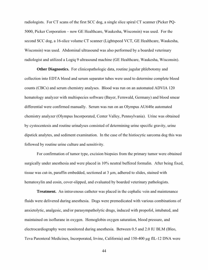
44
radiologists. For CT scans of the first SCC dog, a single slice spiral CT scanner (Picker PQ-
5000, Picker Corporation – now GE Healthcare, Waukesha, Wisconsin) was used. For the
second SCC dog, a 16-slice volume CT scanner (Lightspeed VCT, GE Healthcare, Waukesha,
Wisconsin) was used. Abdominal ultrasound was also performed by a boarded veterinary
radiologist and utilized a Logiq 9 ultrasound machine (GE Healthcare, Waukesha, Wisconsin).
Other Diagnostics. For clinicopathologic data, routine jugular phlebotomy and
collection into EDTA blood and serum separator tubes were used to determine complete blood
counts (CBCs) and serum chemistry analyses. Blood was run on an automated ADVIA 120
hematology analyzer with multispecies software (Bayer, Fernwald, Germany) and blood smear
differential were confirmed manually. Serum was run on an Olympus AU640e automated
chemistry analyzer (Olympus Incorporated, Center Valley, Pennsylvania). Urine was obtained
by cystocentesis and routine urinalyses consisted of determining urine specific gravity, urine
dipstick analytes, and sediment examination. In the case of the histiocytic sarcoma dog this was
followed by routine urine culture and sensitivity.
For confirmation of tumor type, excision biopsies from the primary tumor were obtained
surgically under anesthesia and were placed in 10% neutral buffered formalin. After being fixed,
tissue was cut-in, paraffin embedded, sectioned at 3 µm, adhered to slides, stained with
hematoxylin and eosin, cover-slipped, and evaluated by boarded veterinary pathologists.
Treatment. An intravenous catheter was placed in the cephalic vein and maintenance
fluids were delivered during anesthesia. Dogs were premedicated with various combinations of
anxietolytic, analgesic, and/or parasympatholytic drugs, induced with propofol, intubated, and
maintained on isoflurane in oxygen. Hemoglobin oxygen saturation, blood pressure, and
electrocardiography were monitored during anesthesia. Between 0.5 and 2.0 IU BLM (Bleo,
Teva Parenteral Medicines, Incorporated, Irvine, California) and 150-400 µg fIL-12 DNA were

45
administered in 1-2 mL of 0.45% NaCl injected parenterally into the center of the mass,
immediately followed by insertion of a hexagonal array of six needle electrodes (Inovio
Biomedical Corporation, Blue Bell, Pennsylvania) surrounding the injection point (trans-
lesionally) and electroporation. The only exception was the first case of SCC where simple
caliper electrodes were used to deliver the pulses. Two 20 msec 400 V/cm electrical pulses were
administered 100 msec apart using an ECM 830 electroporator (BTX, Genetronics – now Inovio
Biomedical Corporation, Blue Bell, Pennsylvania). Additional treatments occurred ten or more
days after the first. The number of treatments varied from one to three, and the study was
approved by the Institutional Review Board of the LSU School of Veterinary Medicine
Response to treatment was evaluated through recheck examinations, tumor measurement
and gross characterization, communication with the pet’s owner, follow-up CBCs and serum
biochemistry profiles, and in two cases, with follow-up CT scans. Complete response is defined
as disappearance of all measurable tumors for 21 days and partial response was defined as more
than 50% reduction in measurable tumor for 21 days. Lack of measurable tumor for greater than
eight months was characterized as a cure for this study.
RESULTS
fIL-12 depicted in figure 2a provided by Dr Stephen Dunham (University of Nottingham,
United Kingdom) was chosen due to the lack of canine IL-12 availability. There is 91%
homology between the canine and feline IL-12 144 suggesting that the fIL12 should be functional
on canine cells. To confirm this assumption, the biologic ability of fIL-12 to activate canine
mononuclear cells was determined. Canine leukocytes activated by fIL-12 induced significantly
more (p<0.05) canine target tumor cell death when compared to control DNA (figure 2b).

a.
Figure 2encodingcytolytic (p<0.05) pfIL12 reencoding A
from thos
of the tum
CT or ca
A
of lesion
samples a
carcinom
fact that t
Both the
tissue sar
high mito
2: Analysis og gene in the
activity of ctarget cell d
epresent cong plasmid DN
All dogs were
se used in m
mor receivin
liper measur
All tumors w
s and/or met
are depicted
mas and sarco
tumor embo
histiocytic s
rcomas and c
otic rate, poo
of fIL12 gen pCI-neo veccanine leukodeath occurrenditioned meNA, respecti
e treated wit
mice 138. The
ng EP. For e
rements), 0.5
ere examine
tastatic poten
d in figure 3.
omas, and al
oli were note
sarcoma and
carried a poo
or differentia
ne function actor (Promeg
ocytes activaed when canedium obtainively.
th combinati
e dose of BL
each cm2 of t
5 IU of BLM
ed histologic
ntial. Repre
As noted pr
ll were diagn
d within lym
d the fibrosar
or prognosis
ation, and di
46
b.
against dogga Corporati
ated by fIL12nine leukocytned by transf
ons of BLM
M and Il-12
tumor at its m
M and 150 µg
ally to confi
sentative ph
reviously, tum
nosed by boa
mphatics in th
rcoma were c
s 145. The me
iagnosis of ly
g target tumion, Madison2 conditionetes were treafecting cells
M and fIL-12
2 DNA was o
maximum cr
g of fIL-12 D
irm histopath
hotomicrogra
mors treated
arded pathol
he biopsy fr
considered g
elanoma was
ymph node a
mor cells. a. n, Wisconsind medium. ated with fILwith control
with a dosa
often divided
ross sectiona
DNA were u
hology diagn
aphs of avail
d represented
logists. Note
rom the first
grade 3 or hi
s also a high
and pulmona
The fIL-12 n). b. AnalySignificant
L12. pCtrl al DNA and f
ages extrapol
d between ar
al area (base
used.
noses and ex
lable biopsy
d a range of
eworthy is th
dog with SC
igh-grade so
h-grade given
ary metastas
ysis of
and fIL12
lated
reas
ed on
xtent
he
CC.
oft-
n its
ses.

Figure 3papillarymelanomsarcoma.µm. All biopsies. R
ECGT. F
significan
dog had n
it difficul
unchange
metastati
histiocyti
: Histologicy squamous cma (amelanot
Bar = 100 sections are
Regardless o
Furthermore
nt side effec
numerous ot
lt to directly
ed after treat
ic melanom
ic sarcoma,
c analysis ocell carcinomtic). Bar = 1µm d. Sect of hematox
of tumor typ
e, treatment
ct was 48 ho
ther medical
y link the dia
tment for m
ma had clini
its bacterial
f dog tumorma. Bar = 50100 µm c. Stion from do
xylin and eos
pe and anato
in all cases
ours of diarr
l problems a
arrhea to the
most dogs (Ta
icopathologi
l cystitis res
47
r biopsies. a00 µm b. SeSection fromog with acantsin stained fo
omic locatio
was associat
rhea for the
and demonstr
ECGT. Cli
able 2). On
cally signif
olved during
a. Section frection from t
m the cubital jthomatous aormalin-fixe
on, all tumo
ted with min
dog with m
rated marked
inicopatholo
ly the dogs
ficant abnor
g treatment
rom the first the dog withjoint of dog
ameloblastomed, paraffin-e
ors had clin
nimal side e
metastatic me
d stress (agg
ogic data rem
with histioc
rmalities. I
and there w
dog treated h malignant
with histiocma. Bar = 20embedded
nical respon
effects. The
elanoma, bu
gression), ma
mained essen
cytic sarcom
In the dog
was no signif
with
cytic 00
nse to
most
ut this
aking
ntially
a and
with
ficant

48
progression of its cholestasis or hypercholesterolemia based on serum chemistries. In the dog
with melanoma, alkaline phosphatase increased and was thought to represent increasing primary
tumor bone involvement or cholestasis; this dog’s neutrophilic leukocytosis also progresses
slightly and was thought to be related to increased necrosis and ulceration of the primary tumor.
It is worthwhile noting that both these dogs had significant changes prior to treatment; therefore
it is not thought that ECGT played any role in progression of pre-existing disease.
Table 2: Summary of clinicopathologic abnormalities and side effects of treatment:
Signalment 6.5 kg, 8-month-
old, M Shih Tzu
with maxillary
SCC
4.5 kg, 6-month-old,
F Jack Russell
Terrier with
mandibular SCC
40 kg, 9-year-old,
FS Rottweiler with
histiocytic sarcoma
13 kg, 19-year-old,
F Chow Chow cross
w/ metastatic
melanoma
37 kg, 9.5-year-old,
FS Golden Retriever
w/ acanthomatous
ameloblastoma
Pre-treatment
clinicopathologic
findings (followed
by reference
interval)
NSF NSF ALP 488 (0-100)
TB 1.1 (0-0.4)
Chol 333 (150-240)
UC: E.Coli
ALP 269 (0-100)
Alb 2.4 (2.6-4.2)
TP 5.6 (5.8-7.5)
Plat 1003 (220-600)
WBC 16.2 (8-14.5)
Neut 13.4 (3-11.5)
NSF
Post-treatment
clinicopathologic
abnormalities
NSF NSF ALP 535 (0-100)
TB 1.0 (0-0.4)
Chol 335 (150-240)
ALP 399 (0-100)
Plat 1008 (220-600)
WBC 18.2 (8-14.5)
Neut 15.6 (3-11.5)
NSF
Treatment Side
Effects
Lethargy &
decreased appetite
None Swollen limb distal
to tumor
Diarrhea 2 days None
Alb = Albumin ALP = Alkaline Phosphatase Chol = Cholesterol
F = Female FS = Female Spayed M = Male
Neut = Neutrophils (x103/dl) NSF = No significant findings Plat = Platelets x103/dl
TB = Total Bilirubin TP = Total Protein UC = Urine culture
WBC = White blood count (x103/dl)
Of the six dogs treated, three had complete responses and three had partial responses.
The first dog (with maxillary SCC) treated, had a complete response. This dog was the only dog
to receive BLM/IL-12 ECGT using caliper electrodes as performed in tumor-bearing mice (17).
Morphologically and on CT scan, this dog had clear bone lysis and a large tumor initially (figure
4a), whereas after the second treatment, visible tumor was eradicated. After six months, the

primary l
Compare
easily acc
later (tab
The seco
via a six
dog with
bone (fig
treatment
Compare
ECGT. A
This dog
Figure 4treatmenmonths asix monthal. 2008)All rightstransmittotherwise
lesion had he
ed to surgery
complished
ble 3).
ond dog treat
needle array
mandibular
gure 5a). By
t, most of th
ed to surgery
Aside for a s
remains tum
4: Photograpnt. a. Pre-treafter treatmenhs after treat with kind ps reserved. Nted in any foe without wr
ealed and m
y and radiatio
within a sho
ted also had
y electrode (a
r SCC demon
y the time the
he bony lysis
y and radiatio
single day of
mor free alm
phic and CTeatment phont. c. Pre-trtment. (Figu
permission ofNo part of thrm or by anyritten permis
much of the as
on therapy, t
ort period of
a complete r
as did all sub
nstrate mark
e second CT
s and much o
on treatment
f lethargy an
most three yea
T scan analyotograph in threatment CT
ure 3 is from f Springer Shese figures y means, elession from th
49
ssociated bo
this dog’s tre
time. This d
response. Th
bsequent dog
ked malocclu
T images wer
of the occlus
t, this dog ex
nd decreased
ars later (tab
ysis of tumohe dog with
T scan in the figure 24.1 cience and Bmay be repr
ectronic, phohe Publisher
ony lysis had
eatment was
dog remains
his dog was
gs). CT ima
usion and tum
re taken 1.5 m
sal changes h
xperienced m
d appetite, no
ble 3).
or volume chmaxillary Sdog with maand 24.2, pa
Business Meroduced, storotocopying, mr).
d been repair
s without sid
s tumor free
the first to r
ages taken in
mor invasion
months after
had resolved
minimal side
o side effects
hange pre- CC. b. Photaxillary SCCage 324 (Cutedia. ©2008red in a retrimicrofilming
red (figure 4
de effects and
almost five
receive ECG
nitially for th
n into adjace
r the last
d (figure 5b).
e effects from
s were noted
and post-tograph six C. d. CT sctrera, Torrer
8 Humana Prieval systemg, recording
b).
d
years
GT
his
ent
.
m
d.
can ro et ress. , or or

Table 3:
Signalment
Primary Tum
Metastases
WHO Stage
Imaging
Treatments
Adjunct trea
Follow-up
Figure 5lysis/repmandibutransverssections ttreatment
Clinical Su
6.5 kg
old, M
mor 2 cm di
of
maxill
Tumo
biopsy
T
CT c
w/r
2
10 da
atment Previo
Res
month
5. Pre- and air for the dlar SCC & d
se sections arthrough the t CT scan. T
ummary (as
g, 8-month-
M Shih Tzu
4
iameter SCC
L rostral
lary gingiva
2
m
or noted in
y lymphatics
T2bN0M0
consistent
esolution
ECGT
ay interval
ous surgery
solved, 56
s tumor-free
R
post-treatmdog with madisplacementre labelled stumor site (pThe previous
s of 7/2009)
.5 kg, 6-month-old
F Jack Russell
Terrier
2 cm diameter SCC
of the L rostral
mandibular gingiva
None detected
T2bN0M0
CT consistent
w/resolution
2 ECGT
10 day interval
Previous surgery
Resolved, 27 month
tumor-free
ment CT scaandibular St of incisor aequentially fpre- and poss tumor site
50
:
d, 40 kg, 9-ye
FS Rottw
C
a
4-5 cm dia
histiocytic s
of L cubita
Splenic m
T3N0M
U/S shows
metasta
1 ECG
Tramad
hs Primary tu
resolved, eut
due to met
diseas
ns delineatiSCC. a. Pre-and canine tefrom rostral t-treatment iis circled.
ear-old,
weiler
13 k
F Ch
ameter
sarcoma
al joint
6 cm
malig
of
masses Subm
node
M1
splenic
asis
Rad
pulmo
GT
10
dol Pirox
tumor
thanized
tastatic
se
50%
reduc
due to
ing tumor a-treatment Ceeth with bonto caudal (1
images cove
kg, 19-year-old,
how Chow cross
x 4 cm diameter
gnant melanoma
R mandibular
gingiva
mandibular lymph
es & pulmonary
T3N2bM1
diographs show
onary metastases
2 ECGT
0 day interval
xicam, tramadol
% primary tumor
ction, euthanized
o poor quality of
life
and degree oCT demonstr
ny lysis (circ1-6) and refeer the same a
37 kg, 9.5-yea
FS Golden Re
1.6 cm x 1
diameter mand
acanthomat
ameloblasto
None detec
T2N0M0
Radiographs w
normal lim
3 ECGT
10 day inter
Previous sur
Resolved, 9 m
tumor-fre
of bone ating invasivcled). CT
er to transverarea). b. Pos
ar-old,
etriever
cm
dibular
tous
oma
cted
0
within
mits
T
rvals
rgery
months
ee
ve
rse st-

T
the initia
radiation
associate
significan
desirable
numerou
the photo
eradicate
radiation
complete
tumor fre
Figure 6ameloblaAfter twoerosion s
T
treatment
partial re
The dog with
l diagnosis,
n therapy. W
ed with signi
nt orthopedi
e cosmetic an
us periods of
ograph taken
ed with ECG
n therapy. Th
ely leaving n
ee (table 3).
6. Pre- and astoma. a. Po more ECGsubsequently
The remainin
t options (w
esponses to E
h acanthomat
this dog’s ow
While both of
ificant side e
c pain, the c
nd functiona
f anesthesia,
n at the time
GT, avoiding
he area of gi
no residual ti
post-treatmPhotographs GT treatmenty resolved).
ng dogs, whic
ere not cand
ECGT. The
tous amelobl
wners were
f these option
effects. In th
chance of sur
al results. Ra
and the pote
of the last tr
many of the
ingival erosi
issue damage
ment photogafter the firss (three total
ch were pres
didates for su
dog with his
51
lastoma is ou
offered eithe
ns offer a hig
he case of su
rgical compl
adiation wou
ential for rad
reatment (fig
e complicatio
on depicted
e. Nine mo
graphs of cost ECGT treal), tumor wa
sented becau
urgery, chem
stiocytic sarc
ur most rece
er segmental
gh probabili
urgery, the do
lications, and
uld have requ
diation side e
gure 6b), the
ons associate
in figure 5b
onths later, th
ompletely eratment (arroas completely
use they wer
motherapy, or
coma had m
ent success (
l mandibulec
ity of cure, th
og would ha
d would hav
uired frequen
effects. As c
e tumor was
ed with surg
b subsequent
his ten-year-
radicated acows point to ty eradicated
re thought to
r radiation th
marked enlarg
figure 6a). A
ctomy or
hey are
ave had
ve had less
nt treatments
can be seen f
completely
gery and
ly resolved
-old dog rem
canthomatothe tumor).
d (the gingiva
o have no oth
herapy), had
gement of th
After
s,
from
mains
ous b.
al
her
d
he left

cubital jo
After trea
7b).
Figure 7prior to trECGT tre Post-trea
and the d
size of th
resolution
increased
metastase
B
reduction
partial re
progressi
euthanas
owner’s d
oint associate
atment, the p
7. Pre- and reatment of eatment (pre
atment nodul
diameter of th
he joint in co
n of the prim
d her quality
es.
Both the dog
n in tumor si
esponders. In
ive neoplasti
ia. The dog
declined furt
a
ed with the p
primary tum
post-treatmthe left cubi
evious tumor
les present w
he joint was
omparison to
mary tumor.
y of life, she
with metast
ize by greate
n the case of
ic disease an
with the fib
ther treatme
primary tum
mor was eradi
ment photogitus (circled)r site circled
were thought
reduced by
o the contrala
Although th
ultimately w
atic melanom
er than 50%
f the dog wit
nd numerous
brosarcoma a
nt.
b
52
mor and poorl
icated and la
raphs of the). b. Photogd).
to represent
1.5 cm. W
ateral cubital
he improvem
was euthaniz
ma and the d
after ECGT
th melanoma
s other medic
also respond
b
ly-weight-be
ameness had
e histiocyticgraph sixty-n
t scar tissue
While not con
l joint was th
ment in this d
ed due to co
dog with fibr
T and therefo
a, this sixtee
cal problems
ed initially,
earing lamen
d improved m
c sarcoma. nine days aft
from the bio
nfirmed histo
he same sug
dog’s ambul
omplications
rosarcoma h
ore were also
en-year-old d
s which nece
but had rapi
ness (figure 7
markedly (fig
a. Photograter a single
opsy procedu
ologically, th
ggesting com
latory ability
from spleni
had an initial
o considered
dog had
essitated
id recurrence
7a).
gure
aph
ure,
he
mplete
y
ic
d
e and

53
The effect of treatment on metastatic disease was not determined. Both dogs with
metastatic disease prior to treatment, ultimately were euthanized because of complications either
associated with their metastases or with recurrence of the primary tumor. Generalizations
regarding the type of tumors amenable to ECGT were also difficult given the small number of
dogs treated. As is true with most neoplasia therapies, dogs with advanced metastatic neoplasia
appear to be poor candidates for ECGT. At least two types of invasive oral neoplasms, SCC and
acanthomatous ameloblastoma, responded well to treatment.
Table 3 summarizes the clinical findings and outcomes for five of the dogs treated. The
sixth dog (not included in this table), a dog with fibrosarcoma of the soft palate, had initial tumor
regression with rapid recrudescence after a single treatment and owners terminated their
participation in this study.
DISCUSSION
As noted by several authors 146, using client-owned dogs for clinical investigation of novel
cancer therapies offers several advantages over mice and humans. Compared with humans, dogs
have similar anatomy and physiology; dogs often share the same environment as humans; dogs
naturally develop cancers sharing many characteristics with human cancers, the canine genome
has been sequenced; and cancers in dogs grow over long periods of time yet their growth occurs
over a short enough time period to be useful in evaluating new therapies. Dogs also share many
of the intra-individual variations that humans do, including varied genetic constitution, varied
immune responses, intra-tumoral heterogeneity, varied development of recurrent or resistant
disease, and a varied degree of metastatic disease. Pet dogs also have the advantage of being
cared for until ages commonly associated with the highest risk for cancer. For many of the
naturally occurring cancers in dogs, histological appearance, tumor genetics, biological behavior,

54
and response to therapy are similar to their human counterparts. Furthermore, the lack of
established “gold standard” veterinary cancer treatment protocols, allows dogs to be treated
humanely with less proven treatment alternatives not possible in humans 146.
From this series of treatments in dogs with spontaneous neoplasms and varied genetic
makeup, environmental conditions, nutrition, and husbandry, we can conclude that the ECGT
was well tolerated and had minimal side effects. Additionally, the treatments themselves can be
easily accomplished with less than fifteen minutes of anesthesia. While not particularly painful,
humans receiving EP describe uncomfortable muscle twitching; this discomfort combined with
the need to access poorly accessible oral sites necessitates general anesthesia.
ECGT resulted in disease free intervals of greater than eight months in three tumors
(considered a cure in this study) ; two of which had recurred after attempts at surgical resection
prior to receiving this ECGT. Two of these tumors were SCCs which have previously been
shown to respond to BLM ECT without gene therapy 131; however, the period of follow-up was
much less than the five cancer-free years that we report here. We were unable to evaluate
whether these dogs would have been cured with BLM ECT alone, but theoretically, the
augmentation with IL-12 gene therapy should help stimulate immune surveillance/memory and
prevent recurrence as we found in mice (17). Acanthomatous ameloblastoma has not previously
been reported to be ECT-sensitive; however, the lack of control subjects did not allow us to
compare ECT to ECGT for this tumor either.
The dog with fibrosarcoma did not receive a full series of treatments. This dog’s partial
response to therapy suggests that fibrosarcomas may be partially responsive to this treatment
modality. In the two dogs with advanced metastatic neoplasia, ECGT enhanced the dogs’ quality
of life prior to euthanasia; the effect of ECGT on metastatic disease was not determined. Both of

55
these dogs were not deemed to be candidates for surgery, radiation, or chemotherapy because of
their advanced disease; thus ECGT offered a safe, relatively non-invasive, method of local tumor
control without the side effects associated with conventional cancer therapies.
In humans, the rates of complete response after once-only treatments of single-agent,
BLM ECT is reported to be between 9 and 100% depending on the technique used. 141
Electrogene therapy of IL-12 has also reached phase one clinical trials in humans and has had
similar positive results. In the first trial of gene transfer using electroporation in humans, the
therapy was deemed, “safe, effective, reproducible, and titratable.”18 Moreover, IL-12 EGT
without any other therapy gave complete regression of non-electroporated distant metastases in
10% of the subjects and produced disease stabilization or partial response in an additional 42%.18
Although impressive, if these therapies were combined, as in our study, the responses should
improve significantly based on our findings in dogs and mice.138
CONCLUSIONS
Given the previous results achieved with experimentally induced tumors in mice 138 and
the encouraging responses achieved in three of the dogs in this study, ECGT offers a promising
new approach to simple, relatively non-invasive tumor therapy with minimal side effects in dogs.
Furthermore, ECGT may also be applicable to other species with readily available species-
specific IL-12 DNA. These early applications support further research and treatment of a variety
of neoplasms in dogs, and offer great promise for other species, including humans. For humans,
additional pre-clinical studies assuring the safety of both EP and gene transfection with IL-12 are
needed before applying for Investigational New Drug status. In the next chapter, safety studies
looking at the toxicity of a tumor targeted version of the interleukin 12 gene were conducted to
determine potential toxicity.

56
REFERENCES
1. Mir LM. Application of electroporation gene therapy: past, current, and future. Methods Mol Biol 2008; 423: 3-17.
2. Miyazaki M, Obata Y, Abe K, Furusu A, Koji T, Tabata Y et al. Gene Transfer Using
Nonviral Delivery Systems. Perit Dial Int 2006; 26(6): 633-640.
3. Teissie J, Golzio M, Rols MP. Mechanisms of cell membrane electropermeabilization: a
minireview of our present (lack of ?) knowledge. Biochim Biophys Acta 2005; 1724(3): 270-80.
4. Gehl J. Electroporation: theory and methods, perspectives for drug delivery, gene therapy
and research. Acta Physiol Scand 2003; 177(4): 437-47.
5. Reed SD, Li S. Electroporation Advances in Large Animals. Curr Gene Ther 2009.
6. Sammeta SM, Vaka SR, Narasimha Murthy S. Transdermal drug delivery enhanced by
low voltage electropulsation (LVE). Pharm Dev Technol 2009; 14(2): 159-64.
7. Barbul A, Antov Y, Rosenberg Y, Korenstein R. Enhanced delivery of macromolecules
into cells by electroendocytosis. Methods Mol Biol 2009; 480: 141-50.
8. Campana LG, Mocellin S, Basso M, Puccetti O, De Salvo GL, Chiarion-Sileni V et al.
Bleomycin-Based Electrochemotherapy: Clinical Outcome from a Single Institution's Experience with 52 Patients. Ann Surg Oncol 2009; 16(1): 191-199.
9. Cemazar M, Tamzali Y, Sersa G, Tozon N, Mir LM, Miklavcic D et al.
Electrochemotherapy in veterinary oncology. J Vet Intern Med 2008; 22(4): 826-31.
10. Qian C, Liu XY, Prieto J. Therapy of cancer by cytokines mediated by gene therapy
approach. Cell Res 2006; 16(2): 182-8.
11. Lollini PL, De Giovanni C, Pannellini T, Cavallo F, Forni G, Nanni P. Cancer
immunoprevention. Future Oncol 2005; 1(1): 57-66.
12. Mortara L, Giuliani L, De Lerma Barbaro A, Accolla RS, Noonan DM. Experimental
therapeutic approaches to adenocarcinoma: the potential of tumor cells engineered to

57
express MHC class II molecules combined with naked DNA interleukin-12 gene transfer. Surg Oncol 2007; 16 Suppl 1: S33-6.
13. Weiss JM, Subleski JJ, Wigginton JM, Wiltrout RH. Immunotherapy of cancer by IL-12-
based cytokine combinations. Expert Opin Biol Ther 2007; 7(11): 1705-21.
14. Mazzolini G, Prieto J, Melero I. Gene therapy of cancer with interleukin-12. Curr Pharm
Des 2003; 9(24): 1981-91.
15. Persano L, Crescenzi M, Indraccolo S. Anti-angiogenic gene therapy of cancer: current
status and future prospects. Mol Aspects Med 2007; 28(1): 87-114.
16. Daud AI, DeConti RC, Andrews S, Urbas P, Riker AI, Sondak VK et al. Phase I trial of
interleukin-12 plasmid electroporation in patients with metastatic melanoma. J Clin Oncol 2008; 26(36): 5896-903.
17. Torrero MN, Henk WG, Li S. Regression of high-grade malignancy in mice by
bleomycin and interleukin-12 electrochemogenetherapy. Clin Cancer Res 2006; 12(1): 257-63.
18. Takimoto CH CE. Principles of Oncologic Pharmacotherapy. In: Pazdur R WL,
Camphausen KA, Hoskins WJ (ed) Cancer Management: A Multidisciplinary Approach, 11 edn, 2008.
19. Spugnini EP, Vincenzi B, Citro G, Tonini G, Dotsinsky I, Mudrov N et al.
Electrochemotherapy for the treatment of squamous cell carcinoma in cats: a preliminary report. Vet J 2009; 179(1): 117-20.
20. Kishida T, Asada H, Itokawa Y, Yasutomi K, Shin-Ya M, Gojo S et al. Electrochemo-
gene therapy of cancer: intratumoral delivery of interleukin-12 gene and bleomycin synergistically induced therapeutic immunity and suppressed subcutaneous and metastatic melanomas in mice. Mol Ther 2003; 8(5): 738-45.
21. Buttner M, Belke-Louis G, Rziha HJ, McInnes C, Kaaden OR. Detection, cDNA cloning
and sequencing of canine interleukin 12. Cytokine 1998; 10(4): 241-8.
22. Ehrhart N. Soft-tissue sarcomas in dogs: a review. J Am Anim Hosp Assoc 2005; 41(4):
241-6.

58
23. Khanna C, Lindblad-Toh K, Vail D, London C, Bergman P, Barber L et al. The dog as a cancer model. Nat Biotechnol 2006; 24(9): 1065-6.

59
CHAPTER 4 PRE-CLINICAL TOXICITY ASSESSMENT OF TUMOR-TARGETED INTERLEUKIN-
12 LOW-INTENSITY ELECTROGENETHERAPY*
*Reprinted by permission from Macmillan Publishers Ltd, 2010©

60
INTRODUCTION
One of the major challenges associated with traditional surgical oncology, radiation, and
chemotherapy is tumor recurrence and metastasis. One strategy to prevent tumor recurrence and
metastasis is to induce or augment anti-tumor immunity. Interleukin 12 (IL-12) induces and
maintains a powerful Th1-mediated anti-tumor immune response and has been used
therapeutically in humans to treat neoplasia. The major effects of IL-12 are the stimulation of
IFN-γ production by natural killer (NK) and T cells, which in turn stimulates additional IL-12
production147, and induction of anti-angiogenesis genes 148. Another important role of IL-12 is to
exhibit immunoregulatory functions in the generation of T helper 1 (Th1) and cytotoxic T
lymphocytes (CTL)149. For these reasons, daily systemic administration of IL-12 recombinant
protein (rIL-12) has been shown to generate a significant inhibitory effect on the metastatic
tumor growth of B16F10 melanoma, established murine renal carcinoma (RENCA), and CT26
tumors148, 150, 151. Unfortunately, rIL-12 has also been associated with significant adverse effects,
including death152, 153. Investigation of rIL-12 toxicity suggests that cytokine-induced shock
causing early deaths was mediated by NK cell production of IFNγ 154. Much of this toxicity was
attenuated by giving low, “desensitizing” doses of rIL-12 prior to treatment with the normal dose
regimen; nevertheless, use of rIL-12 has now been supplanted by use of IL-12 gene therapy for
cancer treatment.
Systemic administration of cytokines at pharmacologic doses results in high
concentrations of cytokines in the circulation and often in suboptimal levels in tissues at the site
of tumors, therefore therapy is more likely to cause systemic toxicity and less likely to be
efficacious in treating the primary tumor. In contrast, EP-mediated cytokine gene transfection
allows localized expression of cytokine at targeted sites, avoiding deleterious side effects and

61
resembling the normal paracrine effect of cytokines. This localized effect can be enhanced more,
if the gene is somehow targeted to specific tissue or the tumor itself.
Our laboratory has previously demonstrated and published the efficacy of intratumoral
EP of the gene for IL-12155. Others have also found that therapy using the gene for IL-12 is safe
and effective in a variety of pre-clinical models along with in human phase 1 trials treating
melanoma 18. However, in these safety trials B16 tumor-bearing mice were used, making it
difficult to extrapolate to IL-12 cancer gene therapy in general (and making it difficult to rule out
variables associated with tumor burden). Moreover, the electroporation field in those studies
was a much higher voltage (1300 V/cm) and shorter duration (95µsec) than the parameters used
in this study156. Our laboratory and others have found that lower voltage, longer duration pulses
are associated with comparable transfection efficiency in a variety of mouse tumor models using
standard reporter gene assays2, 54, 157. Additionally, currently available commercial
electroporators deliver low voltage over longer intervals.
Safety of EP has also been examined in numerous studies and numerous EP protocols
have been found to be safe and effective for most commonly used tissues158-160. There is some
concern regarding use of EP in heart muscle, and patients with defibrillators or arrhythmias may
not be good candidates for EP 160. To our knowledge, nobody has examined the toxic effects of
EP over time when applied under clinical conditions in a systematic way, nor has anyone
reported the safety and toxicity of a relatively low voltage, long duration electroporation
protocol; therefore we sought to characterize EP changes at numerous acute and a chronic end-
points using a clinical electroporator to determine if there was severe or lasting toxicity.
To increase the efficacy of anti-tumor immune induction and further address the concern of
safety, we have developed a plasmid DNA vector encoding a tumor-targeted peptide linked to
the IL-12 gene, allowing the gene product to preferentially accumulate in tumors regardless of

62
the site of injection. Pre-clinical therapeutic studies using murine ttIL-12 encoding plasmid have
been completed in four independent tumor models; we have found that accumulation of IL12
protein in tumors is safe and triggers a more aggressive anti-tumor immune response than
systemic rIL-12 therapy (Cutrera, submitted manuscript). In this study, the safety and toxicity
of ttIL-12 was thoroughly analyzed (according to FDA guidelines for Investigational New Drug
(IND) approval) in order to determine whether this novel fusion gene can be safely used in
humans under clinical circumstances.
MATERIALS & METHODS
GLP or GLP-like Protocols. Serum chemistries and complete blood counts (CBCs)
were performed by Antech Diagnostics (Memphis TN), which is fully compliant with Good
Laboratory Practice (GLP) Regulations as set forth in 21 CFR Part 58 as well as other regulatory
requirements. All other procedures, including tissue archival, quality control, and data keeping
procedures were conducted in GLP-like conditions (as stipulated by GLP regulations, but not
certified by the Food and Drug Administration (FDA)).
Mice. All animal use studies had approval from the Louisiana State University
Institutional Animal Care and Use Committee (Baton Rouge, LA). This committee follows
USPHS Policy on Humane Care and Use of Laboratory Animals. Young adult male and female
C3H/HeJ mice were obtained from a commercial vendor (Harlan Laboratories, Inc.,
Indianapolis, Indiana), were fed ad libitum a standard diet (Harlan Teklad irradiated mouse diet
7912, Madison, WI), and were housed in a temperature-controlled animal facility with a 12/12-
hour light/dark cycle. Each group for the toxicity studies consisted of six male and six female
mice; the age of mice at the time of the first treatment in this study was 8.5 weeks.
Cell Lines and Propagation. For tumor resection and wound healing studies, SCCVII
murine squamous cell carcinoma cells which are syngeneic in C3H mice, originally obtained

63
from Dr Bert O’Malley (Baylor College of Medicine) were maintained as monolayers in
Dulbecco’s Modified Eagle Medium. Tumors from study mice were harvested and confirmed to
be spindyloid squamous cell carcinoma cells based on routine histopathology, cytokeratin
immunohistochemistry, and transmission electron microscopy. Cells were harvested from flasks
using trypsin-EDTA (0.05% trypsin-0.53 mmol/L EDTA; Mediatech), collected by
centrifugation, and washed. Cells were counted and their concentration was adjusted to 2 x 10⁵
cells/ml using sterile phosphate- buffered saline (PBS) prior to inoculation into mice.
Tumor Inoculation and Monitoring. Tumor cells were inoculated in syngeneic C3H
mice by intradermal injection of SCCVII cells in the caudodorsal skin. 30 µl of approximately 2
x 105 SCCVII tumor cells in sterile PBS was delivered to each mouse. Tumors were monitored
using a digital vernier caliper and volumes were calculated using the formula 4/3π(a2 b) where
“a” is the tumor long dimension (diameter) and “b” is the shortest measurement perpendicular to
“a”.
Anesthesia. For all procedures requiring anesthesia, mice were anesthetized by chamber
induction with 4% isoflurane in oxygen and were subsequently maintained by mask
administration of isoflurane in oxygen to effect. During longer procedures, an infrared heat lamp
was used for thermal support. Respiratory effort and rate along with anal tone and withdrawal
reflexes were monitored throughout anesthesia.
Tumor Resection Surgery. When tumor areas averaged 30,000 mm3, mice were
anesthetized, the area surrounding the tumor was clipped to remove hair, and the surgical site
was prepared with three alternating 70% ethanol/2% chlorhexidine scrubs. The site was draped
with a sterile drape and an elliptical incision was made with minimal tumor margins
(approximately 1 mm – no effort was made to completely excise tumors beyond a minimal

64
margin) and the tumor was removed. The skin defect was closed with 2-3 cruciate sutures of 5-0
silk.
Plasmid. DNA encoding the tumor-targeting peptide (CHP) was inserted into the murine
IL-12 gene construct immediately upstream to the p40 termination codon (Cutrera, submitted
manuscript). This gene construct expressed CHP-IL-12, herein referred to ttIL-12, and was
inserted into a pCLneo mammalian plasmid expression vector (Promega Corporation, Madison,
Wisconsin).
Electroporation. Either skin overlying the dorsal lumbar epaxial musculature or
quadriceps femoris muscle were electroporated using a fixed-voltage, four-needle array
disposable electrode with a 4 mm gap delivering two 60 ms pulses of 46 V (200 V/cm) spaced
approximately 190 ms apart, attached to a Medpulser™ (Inovio Incorporated, San Diego,
California) clinical electroporator. For SC application (the first treatment), either 30 µL of
0.45% saline or ttIL-12 in 0.45% saline were injected subcutaneously; within 15-20 seconds the
electrode was inserted transcutaneously straddling the injection site; and the pulses were
delivered. For IM use, the same volume was injected bilaterally in the quadriceps muscles (the
total volume was therefore 60 µL, and the total DNA was divided between the two limbs),
followed by insertion of the electroporator needles deep into the musculature surrounding the
injection site.
Toxicity Study. Groups of six female mice and six male mice divided equally between
no treatment controls, 0.45% NaCl EP, 1 µg ttIL-12 DNA in 0.45% NaCl, and 5 µg ttIL-12 DNA
in 0.45% NaCl were treated. The volumes injected were 30 µL in the case of SC treatment and
60 µL divided bilaterally into two 30 µL aliquots for IM treatment. Treatment consisted of
initial SC EP followed ten days later by IM EP. Control and treatment groups were then
sacrificed at acute time points (1, 3, and 7 days) and a 30 day chronic time point.

65
Dosages of ttIL-12 were a presumably toxic mega-dose of 5 ug IL12 DNA and a therapeutic
dose of 1 µg IL-12 DNA. The presumed toxic dose was chosen for determining the toxicity limit
but will not be translated to humans because it would equal an impractical 22.5 mg in a 90 kg
human (requiring a minimum injection of approximately 25 mL). The therapeutic dose chosen as
most likely to be translated into human trials is 1 µg for multiple administrations, which is equal
to 4.5 mg in a 90 kg human.
Surgery Augmented with ttIL-12 EP Study. To examine the effect of ttIL-12 EGT on
recovery from tumor resection surgery, groups of six male mice were treated with the
aforementioned protocol, but a massive dose of 20 µg of ttIL-12 was administered during each
episode of EGT.
Physical Exam/Behavior Monitoring. A brief physical exam including body weight,
respiratory rate and effort, mucus membrane character, cutaneous lesions, musculoskeletal
changes, and localized tissue damage was performed on each mouse the day after EP and every
seven days thereafter. Mice were also monitored for gait abnormalities and grooming behavior
along with any other detectable behavioral anomalies.
CBC & Chemistry Analysis. Mice were anesthetized and the brachial artery was
accessed through an incision created in the ventral axilla; the artery was severed and pooled
blood was collected with a sterile glass pipette. Approximately 400-600 µl of blood was
collected into a miniature EDTA tube (BD Microtainer™ Tubes, Becton, Dickenson and
Company) and mixed well. The remaining blood – usually approximately 800 µL – was
collected into a miniature plasma separator tube (BD Microtainer™, Becton, Dickenson and
Company) and thoroughly mixed. A blood smear was made from the EDTA blood sample and
the remaining blood and plasma were sent to a private GLP lab (Antech Diagnostics, Memphis,
Tennessee) for a CBC and select chemistry analysis.

66
Euthanasia and Gross Necropsy. Under isoflurane general anesthesia, mice were
exsanguinated and death was assured through cervical dislocation. All mice received a complete
necropsy. Any tissue which had gross lesions (in addition to those routinely collected) was
saved and processed for histopathologic examination.
Histopathology. All major perfused organs as well as any tissues with lesions (2
sections of skin) were fixed in 10% neutral-buffered formalin. Routine tissues examined
histologically on all mice were liver, spleen, kidneys, lungs, heart, thymus, tracheobronchial
lymph nodes, brain, bone marrow (lumbar spine & femurs), and quadriceps musculature. After
fixation the tissue was cut-in, embedded in paraffin, sectioned on a microtome, mounted on glass
slides, and stained with hematoxylin and eosin. Cover-slipped slides were examined by a
pathologist and lesions were recorded. All slides and paraffin blocks were subsequently
archived.
Statistical Analysis. Groups in each phase of evaluation were compared to control mice
using a two-tailed paired T-test with a confidence interval of 95%. Charts of group means
include standard error bars unless indicated otherwise.
RESULTS
Optimization of Medpulser™ Parameters. Our ultimate goal is to effectively deliver
ttIL-12 gene to tumors, tissues at the margins of tumors, or systemically via musculature using
EP in order to prevent tumor recurrence after primary tumor ablation. To achieve this goal, it is
important to have a set of EP parameters enhancing gene delivery in multiple tissues. To
determine such a set of parameters, we compared each of the clinically used applicators provided
by the manufacturer (Inovio, San Diego, California) of the electroporator used in this study. We
found that using the Medpulser™ fixed-voltage applicator delivering 107 V/cm for two
consecutive 60 msec pulses separated by a pause of approximately 190 msec allowed statistically

67
significant (p = 0.002 and p < 0.001) high levels of reporter gene and cytokine expression when
delivered both SC and IM (Fig. 8A). This applicator delivers 46 V across a 4 mm gap, four-
needle electrode array that is inserted transcutaneously.
Cytokine Expression after ttIL-12 EGT. In previous use of rIL-12, toxicity of IL-12
protein was seen with a large systemic acute dose of rIL-12. It is generally agreed that low
constitutive IL-12 expression kinetics are produced by IL-12 EGT and one of the advantages of
EGT is local expression without significant, potentially toxic systemic levels of cytokine. To
determine whether clinical application of the optimized EP parameters with ttIL-12 yielded the
safe expression kinetics, we determined plasma cytokine levels in all of the sacrificed mice. The
previously determined parameters yielded efficient transfection of the gene for ttIL-12 in vivo
using the Medpulser™ as indicated by detection of significant plasma cytokine levels only in IL-
12 DNA-treated mice (Fig. 8B & 8C), yet we found no statistically significant elevation in
systemic plasma cytokine levels. Mean levels of IL-12 p70 protein peaked at about 600 pg/mL
in the 5 µg dose group on day three, and at approximately 60 µg/mL in the 1 µg therapeutic dose
group on day seven. Levels of IFNγ also increased after treatment, with the mean peak value
reaching approximately 120 pg/mL on day seven in the 5 µg treatment group, and approximately
360 pg/mL in the 1 µg treatment group.
Thus, EP parameters using the Medpulser™ electroporator and four-needle electrode
array were effective in numerous reporter genes and for therapeutic ttIL-12. As expected,
systemic levels of IL-12 were not significant enough to potentially cause systemic toxicity.
Additionally, levels of IL-12 were greater in the higher dose group – particularly on day three.
The results for IFNγ were similar, but higher levels were seen in the low dose IL-12 group
compared to the high dose group at acute time points. It is possible that IFNγ production was
higher in the lower dose ttIL-12 group because of greater attenuation of IFNγ response by the

high dose
fatality in
conclusio
Figure 8levels aftusing difmurine ILmurine IFhigher letreatment
B
were eva
EGT ther
or incide
tissue hy
of enviro
and overg
spleen w
extramed
and over
L
electropo
microsco
e of ttIL-12,
n rIL-12 ther
ons, a greate
8. Optimizatter ttIL-12 Efferent voltagL-12p70 cytFNγ also wevels over timt. Error bars
Background
aluated histol
rapy. Two m
nce in any p
perplasia inc
onmental inh
growth incre
ere noted in
dullary hema
time.
Local Effects
oration, musc
opically. Foc
similar to th
rapy153. Unf
er number of
tion of MedpEGT. A, Comge applicatortokine levelsere not statistme with returs represent s
Lesions in
logically, we
minor chang
particular trea
creased over
halant antigen
eased as mic
all mice (bo
atopoiesis, a
s of Electrop
cle and skin
cal to multif
he desensitiz
fortunately, t
f mice would
pulser ™ elemparison of rs. B, There s between grotically differrn to normaltandard erro
C3H/HeJ M
e also noted
es were note
atment group
r time and w
n exposure).
ce aged. Lym
oth treatment
common fre
poration. In
were harves
focal acute rh
68
zing dose of
to achieve si
d be required
ctroporationluciferase geis no statistioups, but trerent betweenl. X-axis is tor of the mea
Mice over Ti
lesions that
ed over time
p). The inci
was unassocia
. Additional
mphoid hype
t and control
equent findin
n order to de
sted from ele
habdomyocy
rIL-12 used
ignificance a
d.
n settings andene expressiical differencends are illusn groups (p >the number an.
ime. Since a
appeared to
in all group
idence of bro
ated with tre
lly, the incid
erplasia of ly
ls) througho
ng in mice, w
etermine the
ectroporation
yte necrosis
d to prevent I
and make an
d Plasma ILion after elecce (p > 0.05strated. C, M> 0.05), but dof days after
all major per
o have no rel
ps (without in
onchial-asso
eatment (pres
dence of inci
ymph nodes,
out the study
was constant
e local effect
n sites and e
with subseq
IFNγ shock a
ny definitive
-12 and IFNctroporation ) in mean pl
Mean plasmadid trend towr the IM EG
rfused organ
ation to ttIL
ncreased sev
ociated lymp
sumably bec
isor malocclu
, thymus and
y; and splenic
t between gr
ts of
examined
quent
and
e
Nγ
lasma a ward T
ns
-12
verity
phoid
cause
usion
d
c
roups

69
pyogranulomatous to granulomatous myositis followed by myoregeneration and resolution were
seen at site of needle electrode insertion in numerous mice in all of the groups that received IM
EP (Fig. 9). The incidence of detectable muscle damage peaked at seven days for the ttIL-12
treated mice, and was maximal on day 1 for the 0.45% saline treated mice. These trends may
reflect the more widespread tissue inflammation over time and chances of detecting a change,
more than reflecting increasing severity; in fact, by day 30, no muscle lesions were detected
except for a single small focus of muscle degeneration, myofiber loss, and mineralization in a
mouse of the 5 µg ttIL-12 treatment group (in a mouse that also had marked dystrophic cardiac
calcification (DCC)).
We also monitored behavior and performed periodic physical examinations of the mice
after EP and found that several mice were transiently lame in one or both pelvic limbs after IM
EP. Mice that were lame were rechecked periodically and clinically detectable lameness
resolved completely in all mice by seven days after IM EP. There was not relationship between
the incidence or severity of lameness and the substance injected.
Liver Toxicity from ttIL-12. The biggest concern regarding adverse effects from IL-12
was hepatotoxicity; therefore several lobes of liver were examined histologically for ttIL-12 EGT
lesions and a scoring system was used to document severity and incidence of lesions in different
treatment groups (Fig. 10). There was a mild increase in the detection of liver lesions over time
in the two ttIL-12 treatment groups, but none seen in either the control mice or the 0.45% saline
treated mice (Fig. 10). Although none of the toxicity appeared serious, the most severe lesions,
and the highest incidence of lesions, were seen on day seven (Fig. 10). Lesion incidence and
severity were also greater in the 5 µg ttIL-12 treatment group than in the 1 µg ttIL-12 treatment
group, but had essentially resolved by day 30.

T
analytes
(ALT) an
(Fig. 10)
treatment
increase
day 30. L
and level
ttIL-12 tr
individua
Figure 9of muscleillustratinafter intrathird panhypereosinfiltrate post-EP daggregateillustratinfixed tisswith dete
To determine
correlated w
nd alkaline p
. For alkalin
t groups or o
in treatment
Levels never
ls of ALT in
reatment gro
al animal bas
9. Muscle dae damage deng the normaamuscular E
nel is from a sinophuilia, aof histiocyte
demonstratines of histiocyng normal msue. 200x mectable quadr
e if there wer
with histologi
phosphatase
ne phosphata
over time. F
t group devia
r reached cli
n saline treatm
oups. Nevert
sis and is tho
amage assocetected over al muscle ap
EP demonstramouse threeand hyalinizes and neutrng resolvingytes. The la
musculature. magnification
ricpes lesion
re any serolo
ic lesions, w
(ALP) activ
ase and albu
For ALT, the
ation from co
inically relev
ment groups
theless, ALT
ought to be a
iated with Etime. On th
ppearance. Tating separate days post-Eation of myoophils. The
g muscle damast panel is fr
Hematoxylin, bar = 100 µns.
70
ogic indicato
we compared
ity in each g
umin, there w
ere was a mil
ontrols up to
vant increase
s were essent
T levels did c
a good moni
EP. A, Photohe left is quadThe next pantion of the enEP demonstrofibers with fourth pane
mage with eafrom a mousein & eosin stµm. B, Cha
ors of liver to
d mean levels
group as wel
was no signif
ld, but statis
o day seven,
es (2-3 fold h
tially as high
correlate wit
itoring param
omicrographdriceps from
nel is from a ndomysium rating fragma moderate
el is from a marly regenerae sacrificed otaining of paart illustratin
oxicity and t
s of alanine
ll as levels o
ficant differe
stically insign
which retur
higher than
h as or highe
th liver toxic
meter for acu
hs illustratinm an untreate
mouse sacriby fibrin anentation, roumultifocal to
mouse sacrifiation and muon day 30 poaraffin embeng the percen
to see if thes
transaminas
f albumin (A
ence between
nificant (p.0
rned to norm
normal rang
er than in the
city on an
ute liver toxi
g the spectrued mouse ificed one dand edema. Tunding, o coalescing
ficed seven dultifocal ost-EP edded formalntage of mic
se
e
ALB)
n
0.05),
mal by
ge),
e
icity.
um
ay The
g days
lin-ce

71
Systemic Immune Effects of ttIL-12 EGT. In order to determine if there were any significant
systemic effects of ttIL-12 EGT on the hematopoietic system and systemic immunity, CBCs and
bone marrow histology were evaluated. In the case of WBC, there was a trend to decreasing
total counts up to day seven which returned to normal by day 30 (Fig. 11). The trend was not
significantly different from control mice in the 1 µg treatment group (p = 0.053 on day 3 and p =
0.109 on day 7), but was in the 5 µg treatment group (p = 0.017 on day three and p = 0.023 on
day seven). The incidence of infectious disease was essentially non-existent (except possibly for
mild BALT hyperplasia) in all groups except for the 5 µg ttIL-12 treatment group, which had
two mice on day 30 that had severe, potentially life-threatening, infectious diseases. One mouse
had a moderate to severe, multifocal to coalescing, pyogranulomatous cholangiohepatitis (Fig.
11B), and the other mouse had severe, bilateral, subacute to chronic, diffuse, pyogranulomatous
pyelonephritis (Fig.11C). Of note is the fact that the second mouse also was one of the mice
with DCC. No effects on bone marrow or other hematopoietic parameters were noted.
Dystrophic Cardiac Calcification. In histologically evaluating all other organs for
potential ttIL-12 EGT toxicity, the only other major change seen in any of the mice was seen in
the day 30, 5 µg ttIL-12 treatment group, which had 3 mice (25%) with moderate to severe DCC
(Fig. 12). Of these mice, one died, and one had evidence of biventricular congestive heart failure
(pulmonary edema with splenic and hepatic congestion) at the time of sacrifice.
As noted above, the only fatality in the 192 mice of this study was a female in the mega-
dose 5µg chronic 30 day group that died on day 22 after the IM EGT treatment. The cause of
death in this mouse was thought to be biventricular congestive heart failure secondary to DCC.
Aside from severe myocardial mineralization (similar to Fig. 5B, but more severe), this mouse
had hepatosplenomegaly due to congestion and mild hemosiderosis in the liver, spleen, and
lungs. In the lungs, numerous alveolar macrophages contained either erythrocytes or

Figure 1histopathwas mildwith a grlivers wit100x fielhyperplaparaffin-liver lesiotreated w(large boparametesignifican(p>0.05)
0. Incidenchology for thd. The panelrade 1 score th 1-3 lesionld. Lesions csia with sporembedded foons by lesion
with 1 µg ttILold number iners by group nce for ALT.
e of mild livhe liver toxicl on the left icharacterize
ns per 100x fconsisted of radic neutroormalin-fixen score and t
L-12. C, Incn the center)and day. Th
T on day 7 (p
ver toxicity incity scoring sillustrates noed by ≤ 1 lesifield. The fof small foci ophils & lym
ed tissue. 10total group sidence of liv) for mice trehe 0.45% sap=0.51), but
72
n ttIL-12 tresystem used on-affected lion per 100 xourth panel iof hepatocell
mphocytes. H00x magnificscore (numbver lesions beated with 5 line treatmenall other cha
eated mice. Aindicating th
liver. The sex field. Theis a grade 3 llular necrosiHematoxylincation, bar =ber at the topby lesion sco
µg ttIL-12. nt group appanges were n
A, Examplehat in all casecond panel e third panel liver with 3-is with Kupfn and eosin st= 200 µm. Bp of the pie core and total
D, Plasma proached stanot statistica
es of liver ses the toxicis consistentis a grade 2
-8 lesions peffer cell taining of , Incidence ohart) for micgroup score levels of live
atistical ally significa
city t
er
of ce
er
ant
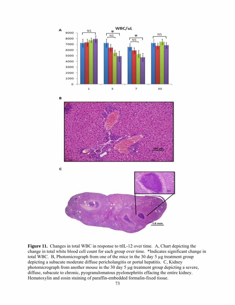
Figure 1change intotal WBdepictingphotomicdiffuse, sHematox
1. Changes n total white
BC. B, Photog a subacute crograph frosubacute to cxylin and eos
in total WBe blood cell comicrographmoderate dim another m
chronic, pyosin staining o
BC in responcount for each from one ofiffuse perich
mouse in the granulomatoof paraffin-e
73
se to ttIL-12ch group ovef the mice in
holangitis or 30 day 5 µg
ous pyelonepembedded fo
2 over time. er time. *Indn the 30 day portal hepat
g treatment gphritis effaciormalin-fixed
A, Chart dedicates signi5 µg treatm
titis. C, Kidngroup depictiing the entired tissue.
epicting the ificant chang
ment group ney ing a severe,e kidney.
ge in
,

Figure 1cardiac cdemonstrdemonstrthroughoHematox
hemoside
expanded
between
mouse w
sacrifice
E
EGT with
was critic
2. Photomiccalcification rating no evirating moder
out the ventrixylin and eos
erin (“heart f
d. Definitive
death and po
with the heart
as evidence
Effect of ttIL
h surgical re
cal for assur
crographs co(DCC). A, Nidence of myrate, multifoicular free wsin staining o
failure cells”
e evidence o
ost-mortem
t depicted (F
d by pulmon
L-12 EGT o
esection of p
ring that the t
omparing anNormal hearyocardial miocal myocardwall and interof paraffin-e
”), and the in
of pulmonary
examination
Fig. 12B) wa
nary edema a
n Wound H
rimary tumo
therapy did n
74
n untreated mrt section wiineralizationdial mineralirventricular embedded fo
nterstitial spa
y alveolar ed
n was estima
s also in con
along with h
Healing. Bec
ors, analysis
not interfere
mouse heart tith inset of thn. B, Sectionization (intenseptum char
ormalin-fixed
aces surroun
dema was no
ated to be 12
ngestive hear
hepatic and s
cause we ant
of wound he
e with surgic
to a heart wihe myocardin and inset frnsely basophracteristic ofd tissue.
nding major
ot seen, but t
-24 hours.
rt failure at t
splenic cong
ticipate com
ealing when
cal protocols
ith dystrophiium rom a mousehilic foci) f DCC.
vessels were
the interval
The female
the time of
estion.
mbining ttIL-
n ttIL-12 EG
s. Therefore
ic
e
e
12
T
, we

grossly e
tumor-be
figure de
treatment
evaluated
can be se
with mas
wound re
Figure 1healing sThere waand ttIL-
evaluated wo
earing C3H m
epicts healed
ts of 20 µg t
d by observin
een from the
ssive doses o
e-epithelializ
3. Therapy wseven days afas no signific12 EGT gro
ound healing
mice and com
d tumor resec
ttIL-12 prior
ng the time t
plot compar
of ttIL-12 pro
zation.
with tumor-tfter surgery cant differenups (p=0.54
g after EGT a
mpared with
ction incision
r to surgery (
to complete
ring wound
oduced no s
targeted intein the untrea
nce in the tim)
75
and tumor re
h wound heal
ns seven day
(Fig. 13A &
wound epith
epithelializa
statistical dif
rleukin 12 dated control me to comple
esection in sq
ling in non-t
ys after surge
13B). Wou
helialization
ation times (F
fference (p=
does not delagroup (A) aete epithelia
quamous cel
treatment co
ery in mice t
und healing w
n (n =5 mice
Fig. 13C), E
0.5415) in p
ay wound heand the ttIL-1lization betw
ll carcinoma
ontrols. The
treated with
was subjecti
per group).
EGT treatmen
primary inten
ealing. Incisi12 group (B)ween the con
a
last
two
vely
As
nt
ntion
on ). ntrol

76
DISCUSSION
Several clinical trials are beginning or underway in humans using EP to deliver DNA in
the form of vaccines or therapeutic genes; however, only one toxicology study was found in the
literature using tumor-bearing mice to examine the safety of IL-12 gene EGT 156. Furthermore,
the aforementioned study used high voltage (1,300 V/cm), short duration (0.1 msec) EP pulses in
contrast to the low voltage (200 V/cm), and long-duration (60 msec) pulses used in the current
study. The electrode array used was also different than the one used in this study (a six-needle
array versus the current four-needle array).
Comparing the different EP parameters, we did find effective transfection using the
current parameters (Fig. 8), but it was important to also determine if these EP parameters caused
any serious adverse effects. In addition to establishing the safety of the current set of EP
parameters, it was also important to establish the safety of ttIL-12. Theoretically, a tumor-
targeted gene should be both safer and more efficacious than a systemically-administered, non-
targeted gene. As expected, therapeutic use of ttIL-12 EGT was found to be safe and well
tolerated under conditions likely to be used for EGT clinically (Figs. 9, 10, & 11).
To our knowledge, this is also the first large scale study of IL-12 toxicity in non-tumor
bearing mice, and the first examining the effects of relatively low voltage, long duration EP
pulses. Tumor-bearing mice are not normal immunologically161-163, so extrapolation from
studies in tumor-bearing mice is fraught with a number of variables which may affect toxicity
and efficacy of immune therapy. Additionally, both transfection efficiencies and local EP
effects vary widely depending on the EP system used.
Using these parameters, local muscle changes in response to EP were completely
resolved by day 30. Interestingly, damage was more frequently detected on days one and three

77
in the group treated with carrier only – perhaps because of the hypotonic osmolarity of the half-
strength saline compared to DNA solutions. A protective effect of ttIL-12, other than adding
colloidal pressure, seems unlikely at these acute time points but cannot be ruled out. Although
the damage was detectable, albeit transient, these changes should be viewed with the perspective
that inserting the 4 mm wide, four electrode array into mouse quadriceps affected a much greater
muscle mass than would be commonly used in humans – perhaps this would be analogous to
penetrating human thighs with four equidistant railroad spikes spaced 25-50 cm apart and
applying an electric field. In addition to the histologic muscle changes seen after EP, several
mice were transiently lame in the pelvic limbs the day after EP, but subsequently returned to a
normal gait and stance thereafter.
We chose C3H/HeJ mice in this study based on previously documented gross toxicity
164 and our previous experience suggesting that they were more sensitive to IL-12 liver toxicity
than other commonly used strains of inbred mice. Concerns about the well documented (16)
defective TLR4 signaling in C3H/HeJ mice were not thought to be an issue because TLR4
signaling in mice appears to serve primarily as a lipopolysaccharide (LPS) response pathway.
Thus, C3H/HeJ mice are immunologically normal, except for their inability to respond to LPS
and to counter Gram negative infection 165. Liver and immune changes were detected in this
study, but are not thought to be strain-specific.
Liver changes have been described before with rIL-12 therapy 164. Similar to our
findings, mice in previous rIL-12 studies had foci of hepatocellular necrosis with aggregates of
Kupffer cell hyperplasia. In this study we have also noted occasional neutrophil and
lymphocytes interspersed within the aggregates of Kupffer cells in the acute treatment groups.
Lesions in the mice in this study were mild to minimal and transient. The number of lesions

78
correlated with minimal elevations in ALT, but we found no increase in alkaline phosphatase
(ALP) and no decrease in albumin (ALB) as was previously seen in rIL-12 toxicity 164. We
chose not to determine levels of aspartate transaminase (AST) because this enzyme is also
elevated in cases of muscle damage and is therefore not liver-specific. If liver toxicity were a
concern, our results suggest that monitoring serum levels of ALT during therapy would be
indicated.
Immune suppression and suppressed leukocyte counts have also been described with IL-
12 therapy 164. In our study, both total white cell counts and individual absolute leukocyte counts
were decreased after treatment with ttIL-12. Mean absolute leukocyte counts for neutrophils,
lymphocytes, monocytes, and eosinophils never decreased significantly from control mice.
Mean total WBC did decrease significantly (p<0.05) from control levels on days three and seven
in mice treated with 5 µg of ttIL-12. Furthermore, in the 5 µg ttIL-12 group there were two
mice that developed major infectious diseases; one mouse also had concurrent severe DCC
which may have been a significant stressor predisposing the mouse to pyelonephritis (or
pyelonephritis may have predisposed to DCC); the other mouse had moderate to severe
cholangiohepatitis. Since no other mice in the study developed serious infectious disease, there
is a possibility that mega-dose ttIL-12 may cause clinically relevant immune suppression.
Conversely, at therapeutically relevant doses (1 µg), ttIL-12 EGT is unlikely to cause serious
immunosuppression. Nevertheless, in addition to monitoring ALT, we suggest monitoring WBC
during therapy – especially if combined with immunosuppressive chemotherapeutics.
DCC was the most serious problem potentially linked to ttIL-12 EGT. Although both of
the females with clinical signs attributed to DCC (death and congestive heart failure) were well
within the reported 81-89 day range for development of DCC in female C3H/HeJ mice 166, DCC

79
seen in the 100-day-old male mouse (which did not have signs of CHF) was well before the
normal onset of 292-465 days reported previously 166. No other mice in the study developed
detectable DCC suggesting that this 16% premature incidence in the mega-dose male mice may
not have been significant. Nevertheless, until additional studies are done in mice that are not
predisposed to DCC, mega-doses of ttIL-12 would be contraindicated in subjects with similar
genetic predisposition.
DCC is common in many strains of imbred mice 167. In C3H mice, DCC is caused by a
mutation in the ABCC6 gene leading to deficiency of its protein product, MRP6 168. MRP6 is
expressed primarily in the liver and kidney but also exists in many other tissues throughout the
body, and is in the family of ABC transporters. It has long been known that DCC can be
triggered in susceptible mice by a variety of stimuli including: female sex – particularly in
breeders, high fat diets, infectious agents 169, 170, hormonal status 166, 171, 172, diet 166, and freeze-
thaw injury (20). In this study, we found that ttIL-12 EGT also appears to hasten the onset of
DCC in C3H/HeJ mice.
In summary, we can conclude that ttIL-12 EGT using low-voltage, long-duration pulses
is safe and well-tolerated at therapeutically relevant dosages. Mild muscle changes seen after IM
EP are unlikely to be a problem in humans given the small area electroporated. Transient acute
decreases in total WBC may suggest possible mild immune suppression, so vigilant patient
monitoring for infectious disease and serial CBCs would be prudent. However, no infectious
diseases were seen in study mice at the clinically relevant dose of 1 µg ttIL-12. Likewise, DCC
was not a problem in the 1 µg therapeutic dosage group. However, several mice (three of twelve,
or 25%) were affected in the mega dose (5 µg) group at day 30, one of which presumably died of
congestive heart failure secondary to DCC. Thus, until additional information is available,

80
people with ABCC6 gene defects or patients with Pseudoxanthoma elasticum (a heritable
disorder of the connective tissue, caused by defective ABCC6 gene) should be excluded from
treatment with mega-dose ttIL-12 EGT. Finally, no effect of ttIL-12 EGT on wound healing was
seen – even at a dosage twenty times greater than those likely to be used clinically. Thus, given
the safety of this treatment modality, and superiority to wild-type IL-12 therapy (Cutrera,
submitted manuscript) we strongly recommend proceeding to phase I trials.
ADDITIONAL OBSERVATIONS
In addition to the data published herein, Appendix 1 includes data demonstrating that
there was no difference in weight gain between groups, and the incidence of lameness following
IM electroporation (consistently higher in the 0.45% saline control group).
REFERENCES
1. Cancer facts and figures. In: American Cancer Society 2010.
2. Reed SD, Li S. Electroporation Advances in Large Animals. Curr Gene Ther 2009.
3. Reed SD FA, Buckholz J, Zhang B, Cutrera J, Shiomitsu K, Li S. Bleomycin/interleukin-
12 electrochemogenetherapy for treating naturally occurring spontaneous neoplasms in dogs. Cancer Gene Therapy 2010.
4. Reed SD LS. Pre-clinical toxicity assessment of tumor-targeted interleukin-12 low-
intensity electrogenetherapy. Cancer Gene Therapy 2010; (In press).
5. Onik G, Mikus P, Rubinsky B. Irreversible electroporation: implications for prostate
ablation. Technol Cancer Res Treat 2007; 6(4): 295-300.
6. Esser AT, Smith KC, Gowrishankar TR, Weaver JC. Towards solid tumor treatment by
irreversible electroporation: intrinsic redistribution of fields and currents in tissue. Technol Cancer Res Treat 2007; 6(4): 261-74.

81
7. Rubinsky B. Irreversible electroporation in medicine. Technol Cancer Res Treat 2007; 6(4): 255-60.
8. Rubinsky B, Onik G, Mikus P. Irreversible electroporation: a new ablation modality--
clinical implications. Technol Cancer Res Treat 2007; 6(1): 37-48.
9. Miller L, Leor J, Rubinsky B. Cancer cells ablation with irreversible electroporation.
Technol Cancer Res Treat 2005; 4(6): 699-705.
10. Davalos RV, Mir IL, Rubinsky B. Tissue ablation with irreversible electroporation. Ann
Biomed Eng 2005; 33(2): 223-31.
11. Al-Sakere B, Andre F, Bernat C, Connault E, Opolon P, Davalos RV et al. Tumor
ablation with irreversible electroporation. PLoS ONE 2007; 2(11): e1135.
12. Mir LM. Application of electroporation gene therapy: past, current, and future. Methods
Mol Biol 2008; 423: 3-17.
13. Miyazaki M, Obata Y, Abe K, Furusu A, Koji T, Tabata Y et al. Gene Transfer Using
Nonviral Delivery Systems. Perit Dial Int 2006; 26(6): 633-640.
14. Teissie J, Golzio M, Rols MP. Mechanisms of cell membrane electropermeabilization: a
minireview of our present (lack of ?) knowledge. Biochim Biophys Acta 2005; 1724(3): 270-80.
15. Gehl J. Electroporation: theory and methods, perspectives for drug delivery, gene therapy
and research. Acta Physiol Scand 2003; 177(4): 437-47.
16. Fewell JG. Factor IX gene therapy for hemophilia. Methods Mol Biol 2008; 423: 375-82.
17. Fewell JG, MacLaughlin F, Mehta V, Gondo M, Nicol F, Wilson E et al. Gene therapy
for the treatment of hemophilia B using PINC-formulated plasmid delivered to muscle with electroporation. Mol Ther 2001; 3(4): 574-83.
18. Daud AI, DeConti RC, Andrews S, Urbas P, Riker AI, Sondak VK et al. Phase I trial of
interleukin-12 plasmid electroporation in patients with metastatic melanoma. J Clin Oncol 2008; 26(36): 5896-903.

82
19. Benz R, Zimmermann U. Pulse-length dependence of the electrical breakdown in lipid bilayer membranes. Biochim Biophys Acta 1980; 597(3): 637-42.
20. Benz R, Zimmermann U. The resealing process of lipid bilayers after reversible electrical
breakdown. Biochim Biophys Acta 1981; 640(1): 169-78.
21. Chang DC, Reese TS. Changes in membrane structure induced by electroporation as
revealed by rapid-freezing electron microscopy. Biophys J 1990; 58(1): 1-12.
22. Knutson JC, Yee D. Electroporation: parameters affecting transfer of DNA into
mammalian cells. Anal Biochem 1987; 164(1): 44-52.
23. Xie TD, Sun L, Tsong TY. Study of mechanisms of electric field-induced DNA
transfection. I. DNA entry by surface binding and diffusion through membrane pores. Biophys J 1990; 58(1): 13-9.
24. Favard C, Dean DS, Rols MP. Electrotransfer as a non viral method of gene delivery.
Curr Gene Ther 2007; 7(1): 67-77.
25. Neumann E, Kakorin S, Toensing K. Fundamentals of electroporative delivery of drugs
and genes. Bioelectrochem Bioenerg 1999; 48(1): 3-16.
26. Xie TD, Tsong TY. Study of mechanisms of electric field-induced DNA transfection. V.
Effects of DNA topology on surface binding, cell uptake, expression, and integration into host chromosomes of DNA in the mammalian cell. Biophys J 1993; 65(4): 1684-9.
27. de Gennes PG. Passive entry of a DNA molecule into a small pore. Proc Natl Acad Sci U
S A 1999; 96(13): 7262-4.
28. Klenchin VA, Sukharev SI, Serov SM, Chernomordik LV, Chizmadzhev Yu A.
Electrically induced DNA uptake by cells is a fast process involving DNA electrophoresis. Biophys J 1991; 60(4): 804-11.
29. Sukharev SI, Klenchin VA, Serov SM, Chernomordik LV, Chizmadzhev Yu A.
Electroporation and electrophoretic DNA transfer into cells. The effect of DNA interaction with electropores. Biophys J 1992; 63(5): 1320-7.
30. Angelova MI, Hristova N, Tsoneva I. DNA-induced endocytosis upon local
microinjection to giant unilamellar cationic vesicles. Eur Biophys J 1999; 28(2): 142-50.

83
31. Angelova MI, Tsoneva I. Interactions of DNA with giant liposomes. Chem Phys Lipids
1999; 101(1): 123-37.
32. Drinkwater NR, Klinedinst DK. Chemically induced mutagenesis in a shuttle vector with
a low-background mutant frequency. Proc Natl Acad Sci U S A 1986; 83(10): 3402-6.
33. Isaka Y, Imai E. Electroporation-mediated gene therapy. Expert Opin Drug Deliv 2007;
4(5): 561-71.
34. Thyagarajan B, Olivares EC, Hollis RP, Ginsburg DS, Calos MP. Site-specific genomic
integration in mammalian cells mediated by phage phiC31 integrase. Mol Cell Biol 2001; 21(12): 3926-34.
35. Hollis RP, Nightingale SJ, Wang X, Pepper KA, Yu XJ, Barsky L et al. Stable gene
transfer to human CD34(+) hematopoietic cells using the Sleeping Beauty transposon. Exp Hematol 2006; 34(10): 1333-43.
36. Chalberg TW, Vankov A, Molnar FE, Butterwick AF, Huie P, Calos MP et al. Gene
transfer to rabbit retina with electron avalanche transfection. Invest Ophthalmol Vis Sci 2006; 47(9): 4083-90.
37. Ohta S, Suzuki K, Ogino Y, Miyagawa S, Murashima A, Matsumaru D et al. Gene
transduction by sonoporation. Dev Growth Differ 2008; 50(6): 517-20.
38. Skorpikova J, Dolnikova M, Hrazdira I, Janisch R. Changes in microtubules and
microfilaments due to a combined effect of ultrasound and cytostatics in HeLa cells. Folia Biol (Praha) 2001; 47(4): 143-7.
39. Yamashita Y, Shimada M, Minagawa R, Tsujita E, Harimoto N, Tanaka S et al. Muscle-
targeted interleukin-12 gene therapy of orthotopic hepatocellular carcinoma in mice using in vivo electrosonoporation. Mol Cancer Ther 2004; 3(9): 1177-82.
40. Yamashita Y, Shimada M, Tachibana K, Harimoto N, Tsujita E, Shirabe K et al. In vivo
gene transfer into muscle via electro-sonoporation. Hum Gene Ther 2002; 13(17): 2079-84.
41. Mir LM, Bureau MF, Gehl J, Rangara R, Rouy D, Caillaud JM et al. High-efficiency
gene transfer into skeletal muscle mediated by electric pulses. Proc Natl Acad Sci U S A 1999; 96(8): 4262-7.

84
42. Bettan M, Emmanuel F, Darteil R, Caillaud JM, Soubrier F, Delaere P et al. High-level
protein secretion into blood circulation after electric pulse-mediated gene transfer into skeletal muscle. Mol Ther 2000; 2(3): 204-10.
43. Draghia-Akli R, Ellis KM, Hill LA, Malone PB, Fiorotto ML. High-efficiency growth
hormone-releasing hormone plasmid vector administration into skeletal muscle mediated by electroporation in pigs. FASEB J 2003; 17(3): 526-8.
44. Lee MJ, Cho SS, Jang HS, Lim YS, You JR, Park J et al. Optimal salt concentration of
vehicle for plasmid DNA enhances gene transfer mediated by electroporation. Exp Mol Med 2002; 34(4): 265-72.
45. Nicol F, Wong M, MacLaughlin FC, Perrard J, Wilson E, Nordstrom JL et al. Poly-L-
glutamate, an anionic polymer, enhances transgene expression for plasmids delivered by intramuscular injection with in vivo electroporation. Gene Ther 2002; 9(20): 1351-8.
46. Draghia-Akli R, Khan AS, Cummings KK, Parghi D, Carpenter RH, Brown PA.
Electrical enhancement of formulated plasmid delivery in animals. Technol Cancer Res Treat 2002; 1(5): 365-72.
47. Quaglino E, Iezzi M, Mastini C, Amici A, Pericle F, Di Carlo E et al. Electroporated
DNA vaccine clears away multifocal mammary carcinomas in her-2/neu transgenic mice. Cancer Res 2004; 64(8): 2858-64.
48. Spadaro M, Ambrosino E, Iezzi M, Di Carlo E, Sacchetti P, Curcio C et al. Cure of
mammary carcinomas in Her-2 transgenic mice through sequential stimulation of innate (neoadjuvant interleukin-12) and adaptive (DNA vaccine electroporation) immunity. Clin Cancer Res 2005; 11(5): 1941-52.
49. Maurer PH. Antigenicity of polypeptides (poly-alpha-amino acids). XVII. Immunologic
studies in humans with polymers containing L or D and L-alpha-amino acids. J Immunol 1965; 95(6): 1095-9.
50. Hartikka J, Sukhu L, Buchner C, Hazard D, Bozoukova V, Margalith M et al.
Electroporation-facilitated delivery of plasmid DNA in skeletal muscle: plasmid dependence of muscle damage and effect of poloxamer 188. Mol Ther 2001; 4(5): 407-15.
51. Mendiratta SK, Thai G, Eslahi NK, Thull NM, Matar M, Bronte V et al. Therapeutic
tumor immunity induced by polyimmunization with melanoma antigens gp100 and TRP-2. Cancer Res 2001; 61(3): 859-63.

85
52. Cemazar M, Sersa G, Wilson J, Tozer GM, Hart SL, Grosel A et al. Effective gene
transfer to solid tumors using different nonviral gene delivery techniques: electroporation, liposomes, and integrin-targeted vector. Cancer Gene Ther 2002; 9(4): 399-406.
53. Draghia-Akli R, Khan AS, Brown PA, Pope MA, Wu L, Hirao L et al. Parameters for
DNA vaccination using adaptive constant-current electroporation in mouse and pig models. Vaccine 2008; 26(40): 5230-7.
54. Muramatsu T, Nakamura A, Park HM. In vivo electroporation: a powerful and
convenient means of nonviral gene transfer to tissues of living animals (Review). Int J Mol Med 1998; 1(1): 55-62.
55. Satkauskas S, Andre F, Bureau MF, Scherman D, Miklavcic D, Mir LM. Electrophoretic
component of electric pulses determines the efficacy of in vivo DNA electrotransfer. Hum Gene Ther 2005; 16(10): 1194-201.
56. Satkauskas S, Bureau MF, Puc M, Mahfoudi A, Scherman D, Miklavcic D et al.
Mechanisms of in vivo DNA electrotransfer: respective contributions of cell electropermeabilization and DNA electrophoresis. Mol Ther 2002; 5(2): 133-40.
57. Hojman P, Gissel H, Andre F, Cournil-Henrionnet C, Eriksen J, Gehl J et al.
Physiological Effects of High and Low Voltage Pulse Combinations for Gene Electrotransfer in Muscle. Hum Gene Ther 2008; (November 2008): 1249-1260.
58. Andre F, Gehl J, Sersa G, Preat V, Hojman P, Eriksen J et al. Efficiency of High and
Low Voltage Pulse Combinations for Gene Electrotransfer in Muscle, Liver, Tumor and Skin. Hum Gene Ther 2008; (November 2008): 1261-1272.
59. Andre FM, Cournil-Henrionnet C, Vernerey D, Opolon P, Mir LM. Variability of naked
DNA expression after direct local injection: the influence of the injection speed. Gene Ther 2006; 13(23): 1619-27.
60. Matsumoto T, Komori K, Shoji T, Kuma S, Kume M, Yamaoka T et al. Successful and
optimized in vivo gene transfer to rabbit carotid artery mediated by electronic pulse. Gene Ther 2001; 8(15): 1174-9.
61. Tone CM, Cardoza DM, Carpenter RH, Draghia-Akli R. Long-term effects of plasmid-
mediated growth hormone releasing hormone in dogs. Cancer Gene Ther 2004; 11(5): 389-96.

86
62. Bureau MF, Gehl J, Deleuze V, Mir LM, Scherman D. Importance of association
between permeabilization and electrophoretic forces for intramuscular DNA electrotransfer. Biochim Biophys Acta 2000; 1474(3): 353-9.
63. Somiari S, Glasspool-Malone J, Drabick JJ, Gilbert RA, Heller R, Jaroszeski MJ et al.
Theory and in vivo application of electroporative gene delivery. Mol Ther 2000; 2(3): 178-87.
64. Brown PA, Khan AS, Draghia-Akli R. Delivery of DNA into skeletal muscle in large
animals. Methods Mol Biol 2008; 423: 215-24.
65. Draghia-Akli R, Khan AS. Muscle and fat mass modulation in different clinical models.
Methods Mol Biol 2008; 423: 449-60.
66. Draghia-Akli R, Fiorotto ML. A new plasmid-mediated approach to supplement
somatotropin production in pigs. J Anim Sci 2004; 82 E-Suppl: E264-269.
67. Brown PA, Davis WC, Draghia-Akli R. Immune-enhancing effects of growth hormone-
releasing hormone delivered by plasmid injection and electroporation. Mol Ther 2004; 10(4): 644-51.
68. Draghia-Akli R, Malone PB, Hill LA, Ellis KM, Schwartz RJ, Nordstrom JL. Enhanced
animal growth via ligand-regulated GHRH myogenic-injectable vectors. FASEB J 2002; 16(3): 426-8.
69. Pavlin D, Tozon N, Sersa G, Pogacnik A, Cemazar M. Efficient electrotransfection into
canine muscle. Technol Cancer Res Treat 2008; 7(1): 45-54.
70. Draghia-Akli R, Cummings KK, Khan AS, Brown PA, Carpenter RH. Effects of
plasmid-mediated growth hormone releasing hormone supplementation in young, healthy Beagle dogs. J Anim Sci 2003; 81(9): 2301-10.
71. Aihara H, Miyazaki J. Gene transfer into muscle by electroporation in vivo. Nat
Biotechnol 1998; 16(9): 867-70.
72. Tollefsen S, Vordermeier M, Olsen I, Storset AK, Reitan LJ, Clifford D et al. DNA
injection in combination with electroporation: a novel method for vaccination of farmed ruminants. Scand J Immunol 2003; 57(3): 229-38.

87
73. Scheerlinck JP, Karlis J, Tjelle TE, Presidente PJ, Mathiesen I, Newton SE. In vivo electroporation improves immune responses to DNA vaccination in sheep. Vaccine 2004; 22(13-14): 1820-5.
74. Khan AS, Smith LC, Abruzzese RV, Cummings KK, Pope MA, Brown PA et al.
Optimization of electroporation parameters for the intramuscular delivery of plasmids in pigs. DNA Cell Biol 2003; 22(12): 807-14.
75. Babiuk S, Baca-Estrada ME, Foldvari M, Middleton DM, Rabussay D, Widera G et al.
Increased gene expression and inflammatory cell infiltration caused by electroporation are both important for improving the efficacy of DNA vaccines. J Biotechnol 2004; 110(1): 1-10.
76. Fattori E, Cappelletti M, Zampaglione I, Mennuni C, Calvaruso F, Arcuri M et al. Gene
electro-transfer of an improved erythropoietin plasmid in mice and non-human primates. J Gene Med 2005; 7(2): 228-36.
77. Andre F, Mir LM. DNA electrotransfer: its principles and an updated review of its
therapeutic applications. Gene Ther 2004; 11 Suppl 1: S33-42.
78. Wolff JA, Malone RW, Williams P, Chong W, Acsadi G, Jani A et al. Direct gene
transfer into mouse muscle in vivo. Science 1990; 247(4949 Pt 1): 1465-8.
79. Wu CJ, Lee SC, Huang HW, Tao MH. In vivo electroporation of skeletal muscles
increases the efficacy of Japanese encephalitis virus DNA vaccine. Vaccine 2004; 22(11-12): 1457-64.
80. Porgador A, Irvine KR, Iwasaki A, Barber BH, Restifo NP, Germain RN. Predominant
role for directly transfected dendritic cells in antigen presentation to CD8+ T cells after gene gun immunization. J Exp Med 1998; 188(6): 1075-82.
81. Medi BM, Singh J. Skin targeted DNA vaccine delivery using electroporation in rabbits
II. Safety. Int J Pharm 2006; 308(1-2): 61-8.
82. Roos AK, Moreno S, Leder C, Pavlenko M, King A, Pisa P. Enhancement of cellular
immune response to a prostate cancer DNA vaccine by intradermal electroporation. Mol Ther 2006; 13(2): 320-7.
83. Condon C, Watkins SC, Celluzzi CM, Thompson K, Falo LD, Jr. DNA-based
immunization by in vivo transfection of dendritic cells. Nat Med 1996; 2(10): 1122-8.

88
84. Ueda Y, Itoh T, Fuji N, Harada S, Fujiki H, Shimizu K et al. Successful induction of
clinically competent dendritic cells from granulocyte colony-stimulating factor-mobilized monocytes for cancer vaccine therapy. Cancer Immunol Immunother 2007; 56(3): 381-9.
85. Yen HH, Scheerlinck JP. Co-delivery of plasmid-encoded cytokines modulates the
immune response to a DNA vaccine delivered by in vivo electroporation. Vaccine 2007; 25(14): 2575-82.
86. Park MY, Kim HS, Woo SJ, Kim CH, Park JS, Sohn HJ et al. Efficient antitumor
immunity in a murine colorectal cancer model induced by CEA RNA-electroporated B cells. Eur J Immunol 2008; 38(8): 2106-17.
87. Onodera S, Ohshima S, Tohyama H, Yasuda K, Nishihira J, Iwakura Y et al. A novel
DNA vaccine targeting macrophage migration inhibitory factor protects joints from inflammation and destruction in murine models of arthritis. Arthritis Rheum 2007; 56(2): 521-30.
88. Hosoi A, Takeda Y, Sakuta K, Ueha S, Kurachi M, Kimura K et al. Dendritic cell
vaccine with mRNA targeted to the proteasome by polyubiquitination. Biochem Biophys Res Commun 2008; 371(2): 242-6.
89. Zhang X, Divangahi M, Ngai P, Santosuosso M, Millar J, Zganiacz A et al. Intramuscular
immunization with a monogenic plasmid DNA tuberculosis vaccine: Enhanced immunogenicity by electroporation and co-expression of GM-CSF transgene. Vaccine 2007; 25(7): 1342-52.
90. Smooker PM, Rainczuk A, Kennedy N, Spithill TW. DNA vaccines and their application
against parasites--promise, limitations and potential solutions. Biotechnol Annu Rev 2004; 10: 189-236.
91. Roman M, Martin-Orozco E, Goodman JS, Nguyen MD, Sato Y, Ronaghy A et al.
Immunostimulatory DNA sequences function as T helper-1-promoting adjuvants. Nat Med 1997; 3(8): 849-54.
92. Chu RS, Targoni OS, Krieg AM, Lehmann PV, Harding CV. CpG oligodeoxynucleotides
act as adjuvants that switch on T helper 1 (Th1) immunity. J Exp Med 1997; 186(10): 1623-31.
93. Erhardt M, Gorschluter M, Sager J, Ziske C, Strehl J, Lilienfeld-Toal MV et al.
Transfection of human monocyte-derived dendritic cells with CpG oligonucleotides. Immunol Cell Biol 2005; 83(3): 278-85.

89
94. Kim CH, Yoon JS, Sohn HJ, Kim CK, Paik SY, Hong YK et al. Direct vaccination with
pseudotype baculovirus expressing murine telomerase induces anti-tumor immunity comparable with RNA-electroporated dendritic cells in a murine glioma model. Cancer Lett 2007; 250(2): 276-83.
95. Sasawatari S, Tadaki T, Isogai M, Takahara M, Nieda M, Kakimi K. Efficient priming
and expansion of antigen-specific CD8+ T cells by a novel cell-based artificial APC. Immunol Cell Biol 2006; 84(6): 512-21.
96. Gilboa E, Vieweg J. Cancer immunotherapy with mRNA-transfected dendritic cells.
Immunol Rev 2004; 199: 251-63.
97. Grunebach F, Muller MR, Brossart P. New developments in dendritic cell-based
vaccinations: RNA translated into clinics. Cancer Immunol Immunother 2005; 54(6): 517-25.
98. Yu H, Babiuk LA, van Drunen Littel-van den Hurk S. Immunity and protection by
adoptive transfer of dendritic cells transfected with hepatitis C NS3/4A mRNA. Vaccine 2007; 25(10): 1701-11.
99. Zhao YG, Xu Y. Electroporation-mediated HBV DNA vaccination in primate models.
Methods Mol Biol 2008; 423: 487-95.
100. Prud'homme GJ, Draghia-Akli R, Wang Q. Plasmid-based gene therapy of diabetes
mellitus. Gene Ther 2007; 14(7): 553-64.
101. Tervo HM, Allespach I, Keppler OT. High-level transfection of primary rabbit T
lymphocytes. J Immunol Methods 2008; 336(1): 85-9.
102. Chen NK, Wong JS, Kee IH, Lai SH, Thng CH, Ng WH et al. Nonvirally modified
autologous primary hepatocytes correct diabetes and prevent target organ injury in a large preclinical model. PLoS ONE 2008; 3(3): e1734.
103. Prud'homme GJ, Glinka Y, Khan AS, Draghia-Akli R. Electroporation-enhanced nonviral
gene transfer for the prevention or treatment of immunological, endocrine and neoplastic diseases. Curr Gene Ther 2006; 6(2): 243-73.
104. Meehan DT, Zink MA, Mahlen M, Nelson M, Sanger WG, Mitalipov SM et al. Gene
targeting in adult rhesus macaque fibroblasts. BMC Biotechnol 2008; 8: 31.

90
105. Norgren RB, Jr. Creation of non-human primate neurogenetic disease models by gene
targeting and nuclear transfer. Reprod Biol Endocrinol 2004; 2: 40.
106. Phelps CJ, Koike C, Vaught TD, Boone J, Wells KD, Chen SH et al. Production of alpha
1,3-galactosyltransferase-deficient pigs. Science 2003; 299(5605): 411-4.
107. Lai L, Kolber-Simonds D, Park KW, Cheong HT, Greenstein JL, Im GS et al. Production
of alpha-1,3-galactosyltransferase knockout pigs by nuclear transfer cloning. Science 2002; 295(5557): 1089-92.
108. Dai Y, Vaught TD, Boone J, Chen SH, Phelps CJ, Ball S et al. Targeted disruption of the
alpha1,3-galactosyltransferase gene in cloned pigs. Nat Biotechnol 2002; 20(3): 251-5.
109. Denning C, Burl S, Ainslie A, Bracken J, Dinnyes A, Fletcher J et al. Deletion of the
alpha(1,3)galactosyl transferase (GGTA1) gene and the prion protein (PrP) gene in sheep. Nat Biotechnol 2001; 19(6): 559-62.
110. Piedrahita JA. Targeted modification of the domestic animal genome. Theriogenology
2000; 53(1): 105-16.
111. McCreath KJ, Howcroft J, Campbell KH, Colman A, Schnieke AE, Kind AJ. Production
of gene-targeted sheep by nuclear transfer from cultured somatic cells. Nature 2000; 405(6790): 1066-9.
112. Yu G, Chen J, Yu H, Liu S, Xu X, Sha H et al. Functional disruption of the prion protein
gene in cloned goats. J Gen Virol 2006; 87(Pt 4): 1019-27.
113. Shen SN, Xu Z, Qian XP, Ding YT, Yu LX, Liu BR. RNA-electroporated CD40-
activated B cells induce functional T-cell responses against HepG2 cells. Eur J Cancer Care (Engl) 2008; 17(4): 404-11.
114. Kuroiwa Y, Kasinathan P, Matsushita H, Sathiyaselan J, Sullivan EJ, Kakitani M et al.
Sequential targeting of the genes encoding immunoglobulin-mu and prion protein in cattle. Nat Genet 2004; 36(7): 775-80.
115. Richt JA, Kasinathan P, Hamir AN, Castilla J, Sathiyaseelan T, Vargas F et al.
Production of cattle lacking prion protein. Nat Biotechnol 2007; 25(1): 132-8.
91
116. Matsuki N, Ishikawa T, Imai Y, Yamaguchi T. Low voltage pulses can induce apoptosis. Cancer Lett 2008; 269(1): 93-100.
117. Gehl J, Geertsen PF. Efficient palliation of haemorrhaging malignant melanoma skin
metastases by electrochemotherapy. Melanoma Res 2000; 10(6): 585-9.
118. Heller R, Jaroszeski M, Atkin A, Moradpour D, Gilbert R, Wands J et al. In vivo gene
electroinjection and expression in rat liver. FEBS Lett 1996; 389(3): 225-8.
119. Gehl J. Electroporation for drug and gene delivery in the clinic: doctors go electric.
Methods Mol Biol 2008; 423: 351-9.
120. Sersa G, Cemazar M, Parkins CS, Chaplin DJ. Tumour blood flow changes induced by
application of electric pulses. Eur J Cancer 1999; 35(4): 672-7.
121. Takeshita S, Isshiki T, Sato T. Increased expression of direct gene transfer into skeletal
muscles observed after acute ischemic injury in rats. Lab Invest 1996; 74(6): 1061-5.
122. Tsurumi Y, Takeshita S, Chen D, Kearney M, Rossow ST, Passeri J et al. Direct
intramuscular gene transfer of naked DNA encoding vascular endothelial growth factor augments collateral development and tissue perfusion. Circulation 1996; 94(12): 3281-90.
123. Gehl J, Skovsgaard T, Mir LM. Vascular reactions to in vivo electroporation:
characterization and consequences for drug and gene delivery. Biochim Biophys Acta 2002; 1569(1-3): 51-8.
124. Bromberg JS, Debruyne LA, Qin L. Interactions between the immune system and gene
therapy vectors: bidirectional regulation of response and expression. Adv Immunol 1998; 69: 353-409.
125. Chen D, Ding Y, Zhang N, Schroppel B, Fu S, Zang W et al. Viral IL-10 gene transfer
inhibits the expression of multiple chemokine and chemokine receptor genes induced by inflammatory or adaptive immune stimuli. Am J Transplant 2003; 3(12): 1538-49.
126. Qin J, Peng Z, McLeod MV. In vitro mutagenesis to define functional domains. Methods
Mol Biol 2004; 241: 189-94.

92
127. Darquet AM, Rangara R, Kreiss P, Schwartz B, Naimi S, Delaere P et al. Minicircle: an improved DNA molecule for in vitro and in vivo gene transfer. Gene Ther 1999; 6(2): 209-18.
128. Sammeta SM, Vaka SR, Narasimha Murthy S. Transdermal drug delivery enhanced by
low voltage electropulsation (LVE). Pharm Dev Technol 2009; 14(2): 159-64.
129. Barbul A, Antov Y, Rosenberg Y, Korenstein R. Enhanced delivery of macromolecules
into cells by electroendocytosis. Methods Mol Biol 2009; 480: 141-50.
130. Campana LG, Mocellin S, Basso M, Puccetti O, De Salvo GL, Chiarion-Sileni V et al.
Bleomycin-Based Electrochemotherapy: Clinical Outcome from a Single Institution's Experience with 52 Patients. Ann Surg Oncol 2009; 16(1): 191-199.
131. Cemazar M, Tamzali Y, Sersa G, Tozon N, Mir LM, Miklavcic D et al.
Electrochemotherapy in veterinary oncology. J Vet Intern Med 2008; 22(4): 826-31.
132. Qian C, Liu XY, Prieto J. Therapy of cancer by cytokines mediated by gene therapy
approach. Cell Res 2006; 16(2): 182-8.
133. Lollini PL, De Giovanni C, Pannellini T, Cavallo F, Forni G, Nanni P. Cancer
immunoprevention. Future Oncol 2005; 1(1): 57-66.
134. Mortara L, Giuliani L, De Lerma Barbaro A, Accolla RS, Noonan DM. Experimental
therapeutic approaches to adenocarcinoma: the potential of tumor cells engineered to express MHC class II molecules combined with naked DNA interleukin-12 gene transfer. Surg Oncol 2007; 16 Suppl 1: S33-6.
135. Weiss JM, Subleski JJ, Wigginton JM, Wiltrout RH. Immunotherapy of cancer by IL-12-
based cytokine combinations. Expert Opin Biol Ther 2007; 7(11): 1705-21.
136. Mazzolini G, Prieto J, Melero I. Gene therapy of cancer with interleukin-12. Curr Pharm
Des 2003; 9(24): 1981-91.
137. Persano L, Crescenzi M, Indraccolo S. Anti-angiogenic gene therapy of cancer: current
status and future prospects. Mol Aspects Med 2007; 28(1): 87-114.
138. Torrero MN, Henk WG, Li S. Regression of high-grade malignancy in mice by
bleomycin and interleukin-12 electrochemogenetherapy. Clin Cancer Res 2006; 12(1): 257-63.

93
139. Takimoto CH CE. Principles of Oncologic Pharmacotherapy. In: Pazdur R WL,
Camphausen KA, Hoskins WJ (ed) Cancer Management: A Multidisciplinary Approach, 11 edn, 2008.
140. Spugnini EP, Vincenzi B, Citro G, Tonini G, Dotsinsky I, Mudrov N et al.
Electrochemotherapy for the treatment of squamous cell carcinoma in cats: a preliminary report. Vet J 2009; 179(1): 117-20.
141. Gothelf A, Mir LM, Gehl J. Electrochemotherapy: results of cancer treatment using
enhanced delivery of bleomycin by electroporation. Cancer Treat Rev 2003; 29(5): 371-87.
142. Sersa G, Miklavcic D, Cemazar M, Rudolf Z, Pucihar G, Snoj M. Electrochemotherapy
in treatment of tumours. Eur J Surg Oncol 2008; 34(2): 232-40.
143. Kishida T, Asada H, Itokawa Y, Yasutomi K, Shin-Ya M, Gojo S et al. Electrochemo-
gene therapy of cancer: intratumoral delivery of interleukin-12 gene and bleomycin synergistically induced therapeutic immunity and suppressed subcutaneous and metastatic melanomas in mice. Mol Ther 2003; 8(5): 738-45.
144. Buttner M, Belke-Louis G, Rziha HJ, McInnes C, Kaaden OR. Detection, cDNA cloning
and sequencing of canine interleukin 12. Cytokine 1998; 10(4): 241-8.
145. Ehrhart N. Soft-tissue sarcomas in dogs: a review. J Am Anim Hosp Assoc 2005; 41(4):
241-6.
146. Khanna C, Lindblad-Toh K, Vail D, London C, Bergman P, Barber L et al. The dog as a
cancer model. Nat Biotechnol 2006; 24(9): 1065-6.
147. Yoshida A, Koide Y, Uchijima M, Yoshida TO. IFN-gamma induces IL-12 mRNA
expression by a murine macrophage cell line, J774. Biochem Biophys Res Commun 1994; 198(3): 857-61.
148. Tannenbaum CS, Tubbs R, Armstrong D, Finke JH, Bukowski RM, Hamilton TA. The
CXC chemokines IP-10 and Mig are necessary for IL-12-mediated regression of the mouse RENCA tumor. J Immunol 1998; 161(2): 927-32.
149. Trinchieri G. Interleukin-12: a cytokine produced by antigen-presenting cells with
immunoregulatory functions in the generation of T-helper cells type 1 and cytotoxic lymphocytes. Blood 1994; 84(12): 4008-27.

94
150. Brunda MJ, Luistro L, Warrier RR, Wright RB, Hubbard BR, Murphy M et al. Antitumor
and antimetastatic activity of interleukin 12 against murine tumors. J Exp Med 1993; 178(4): 1223-30.
151. Nastala CL, Edington HD, McKinney TG, Tahara H, Nalesnik MA, Brunda MJ et al.
Recombinant IL-12 administration induces tumor regression in association with IFN-gamma production. J Immunol 1994; 153(4): 1697-706.
152. Cohen J. IL-12 deaths: explanation and a puzzle. Science 1995; 270(5238): 908.
153. Leonard JP, Sherman ML, Fisher GL, Buchanan LJ, Larsen G, Atkins MB et al. Effects
of single-dose interleukin-12 exposure on interleukin-12-associated toxicity and interferon-gamma production. Blood 1997; 90(7): 2541-8.
154. Carson WE, Yu H, Dierksheide J, Pfeffer K, Bouchard P, Clark R et al. A fatal cytokine-
induced systemic inflammatory response reveals a critical role for NK cells. J Immunol 1999; 162(8): 4943-51.
155. Li S, Zhang X, Xia X. Regression of tumor growth and induction of long-term antitumor
memory by interleukin 12 electro-gene therapy. J Natl Cancer Inst 2002; 94(10): 762-8.
156. Heller L, Merkler K, Westover J, Cruz Y, Coppola D, Benson K et al. Evaluation of
toxicity following electrically mediated interleukin-12 gene delivery in a B16 mouse melanoma model. Clin Cancer Res 2006; 12(10): 3177-83.
157. Li S. Delivery of DNA into tumors. Methods Mol Biol 2008; 423: 311-8.
158. Trollet C, Scherman D, Bigey P. Delivery of DNA into muscle for treating systemic
diseases: advantages and challenges. Methods Mol Biol 2008; 423: 199-214.
159. Trollet C, Bloquel C, Scherman D, Bigey P. Electrotransfer into skeletal muscle for
protein expression. Curr Gene Ther 2006; 6(5): 561-78.
160. Fedorov VV, Nikolski VP, Efimov IR. Effect of electroporation on cardiac
electrophysiology. Methods Mol Biol 2008; 423: 433-48.
161. Serafini P, De Santo C, Marigo I, Cingarlini S, Dolcetti L, Gallina G et al. Derangement
of immune responses by myeloid suppressor cells. Cancer Immunol Immunother 2004; 53(2): 64-72.

95
162. Kiessling R, Wasserman K, Horiguchi S, Kono K, Sjoberg J, Pisa P et al. Tumor-induced
immune dysfunction. Cancer Immunol Immunother 1999; 48(7): 353-62.
163. Carrio R, Lopez DM. Impaired thymopoiesis occurring during the thymic involution of
tumor-bearing mice is associated with a down-regulation of the antiapoptotic proteins Bcl-XL and A1. Int J Mol Med 2009; 23(1): 89-98.
164. Car BD, Eng VM, Lipman JM, Anderson TD. The toxicology of interleukin-12: a review.
Toxicol Pathol 1999; 27(1): 58-63.
165. Poltorak A, He X, Smirnova I, Liu MY, Van Huffel C, Du X et al. Defective LPS
signaling in C3H/HeJ and C57BL/10ScCr mice: mutations in Tlr4 gene. Science 1998; 282(5396): 2085-8.
166. Eaton GJ, Custer RP, Johnson FN, Stabenow KT. Dystrophic cardiac calcinosis in mice:
genetic, hormonal, and dietary influences. Am J Pathol 1978; 90(1): 173-86.
167. Korff S, Riechert N, Schoensiegel F, Weichenhan D, Autschbach F, Katus HA et al.
Calcification of myocardial necrosis is common in mice. Virchows Arch 2006; 448(5): 630-8.
168. Aherrahrou Z, Doehring LC, Ehlers EM, Liptau H, Depping R, Linsel-Nitschke P et al.
An alternative splice variant in Abcc6, the gene causing dystrophic calcification, leads to protein deficiency in C3H/He mice. J Biol Chem 2008; 283(12): 7608-15.
169. Gang DL, Barrett LV, Wilson EJ, Rubin RH, Medearis DN. Myopericarditis and
enhanced dystrophic cardiac calcification in murine cytomegalovirus infection. Am J Pathol 1986; 124(2): 207-15.
170. Price P, Eddy KS, Papadimitriou JM, Faulkner DL, Shellam GR. Genetic determination
of cytomegalovirus-induced and age-related cardiopathy in inbred mice. Characterization of infiltrating cells. Am J Pathol 1991; 138(1): 59-67.
171. Lostroh A, Li CH. Deposition induced by hydrocortisone of calcium in the heart tissue of
female C3H mice. Nature 1955; 176(4480): 504.
172. Sparks LL, Rosenau W, Macalpin RN, Daane TA, Li CH. Production of dystrophic
calcification of cardiac muscle in mice by hydrocortisone. Nature 1955; 176(4480): 503-4.

96
CHAPTER 5 CONCLUDING REMARKS

97
Cancer continues to remain a major cause of morbidity and mortality throughout the
world. Conventional surgery, chemotherapy, and radiation therapy have failed to result in a
significant impact on the rates of death from cancer; therefore, there is a real need for better
therapies. Immune therapy in general and cytokine gene therapy specifically, offers several
advantages over conventional treatment modalities. In addition to being able to effectively target
tumor metastases and recurring primary tumors, we have found that cancer electroimmunogene
therapy is cost effective, safe, and easy to perform.
If treatment in humans shows as much promise as seen in our pre-clinical trials, and is as
safe as seen in the studies described herein, this therapeutic may offer a significant impact in the
war against cancer. As summarized in this dissertation, there have been significant advances in
the use of electroporation resulting in several clinically-approved electroporators. We have
already proved utility of this technique when combined with electrochemotherapy in treating
locally aggressive oral tumors in dogs in addition to our extensive previous data in mice.
Perhaps most importantly in terms of being able to advance to human clinical trials, we have
shown that this technique is safe.

98
APPENDIX 1 TABLES OF CLINICAL OBSERVATIONS

99
Mean Body Weights for Each Group: There was no significant difference in body weights between treatment groups. (Mean +/- Standard Deviation in grams)
GROUP* Day 0 Day 1 Day 7 Day 10** Day 11 Day 19 Day 26 Day 33 Day 40
CONTROL1 25+/‐3.1 25.2+/‐2.9 25.1+/‐3.2 25.1+/‐3.1 25.4+/‐3.2
SALINE1 24.9+/‐2.1 25.1+/‐2.2 25.2+/‐2.5 25.1+/‐2.8 25.4+/‐3
1 µg1 25.2+/‐3.1 25.3+/‐3 25.2+/‐3 25.4+/‐3.1 25.3+/‐3.1
5 µg1 24.2+/‐2.3 24.3+/‐2.3 24.6+/‐2.3 25.3+/‐2.6 25.2+/‐2.4
CONTROL3 23.9+/‐2.9 24+/‐2.5 24.3+/‐2.5 25.2+/‐2.6 24.6+/‐2.1 24.4+/‐2.6
SALINE3 24.4+/‐3.6 24.8+/‐3.6 24.8+/‐3.5 25.4+/‐3.6 25.3+/‐3.6 25.4+/‐3.6
1 µg3 25+/‐3.4 25.6+/‐3.3 25.6+/‐3.3 25.8+/‐3.5 25.7+/‐3.5 25.6+/‐3.8
5 µg3 25+/‐2.9 25.3+/‐3.1 25.4+/‐3.3 25.8+/‐3 25.7+/‐3.4 26+/‐3.3
CONTROL7 23.5+/‐3 23.9+/‐3.1 24.1+/‐2.9 24.4+/‐2.8 24.9+/‐2.8 24.7+/‐3 24.7+/‐3
SALINE7 23.4+/‐1.9 24.3+/‐2.1 24.7+/‐2.1 24.8+/‐2 25.1+/‐2.2 25.6+/‐2.7 25.6+/‐2.7
1 µg7 22.9+/‐2.7 23.6+/‐2.6 24+/‐2.6 24.4+/‐2.7 24.5+/‐2.9 25+/‐3 25+/‐3
5 µg7 23.1+/‐2.3 23.8+/‐2.3 24.1+/‐2.5 24.2+/‐2.3 24.9+/‐2.3 25.7+/‐2.2 25.7+/‐2.1
CONTROL30 22.8+/‐2.2 23.7+/‐2.1 23.6+/‐1.9 22.3+/‐1.9 24+/‐2.1 24.6+/‐2.1 25.5+/‐2 26.2+/‐1.8 27+/‐2
SALINE30 23.1+/‐2.1 23.9+/‐2.1 23.8+/‐1.9 23.3+/‐2.3 24.5+/‐2 25.3+/‐2 26.3+/‐1.9 26.7+/‐1.7 27.4+/‐1.7
1 µg30 23.8+/‐3.1 24.1+/‐2.8 23.6+/‐3.2 23+/‐3.7 23.9+/‐3.5 24.4+/‐3.3 24.7+/‐3 25.1+/‐3 25.4+/‐2.9
5 µg30 23.9+/‐2.7 24.4+/‐2.7 23.7+/‐3.4 23.3+/‐3.9 24.2+/‐3.4 24.6+/‐3 24.8+/‐2.7 25.3+/‐2.5 25.9+/‐2.1
*The Subscript following the group identity is number of days between treatment and euthanasia.
**Day of intramuscular electroporation.

100
Percentage of mice with detectable lameness after electroporation: All groups had some members with detectable lameness. The only consistent trend was a greater incidence of
lameness in the saline control group.
GROUP* Day 0 Day 1 Day 7 Day 10** Day 11 Day 19 Day 26 Day 33 Day 40
CONTROL1 0 0 0 0 42
SALINE1 0 0 0 0 58
1 µg1 0 0 0 0 25
5 µg1 0 0 0 0 33
CONTROL3 0 0 0 0 33 0
SALINE3 0 0 0 0 50 0
1 µg3 0 0 0 0 17 0
5 µg3 0 0 0 0 17 0
CONTROL7 0 0 0 0 17 0 0
SALINE7 0 0 0 0 42 0 0
1 µg7 0 0 0 0 8 0 0
5 µg7 0 0 0 0 25 0 0
CONTROL30 0 0 0 0 25 0 0 0 0
SALINE30 0 0 8 0 50 8 0 0 0
1 µg30 0 0 0 0 17 0 0 0 0
5 µg30 0 0 0 0 17 8 0 0 0
*The Subscript following the group identity is number of days between treatment and euthanasia.
**Day of intramuscular electroporation.

101
APPENDIX 2 LETTERS OF PERMISSION

102
Grant of Permission
Dear Dr. Reed:
Thank you for your interest in our copyrighted material, and for requesting permission for its use.
Permission is granted for the following subject to the conditions outlined below:
"Electroporation advances in large animals" CGT 9:316-326
To be used in the following manner:
1. Bentham Science Publishers grants you the right to reproduce the material indicated above on a one-time, non-exclusive basis, solely for the purpose described. Permission must be requested separately for any future or additional use.
2. For an article, the copyright notice must be printed on the first page of article or book chapter. For figures, photographs, covers, or tables, the notice may appear with the material, in a footnote, or in the reference list.
Thank you for your patience while your request was being processed. If you wish to contact us further, please use the address below.
Sincerely,
AMBREEN IRSHAD Permissions & Rights Manager
Bentham Science Publishers Ltd
Email: [email protected]
URL: www.bentham.org
NATURE PUBLISHING GROUP LICENSE TERMS AND CONDITIONS Oct 04, 2010

103
This is a License Agreement between Scott D Reed ("You") and Nature Publishing Group ("Nature Publishing Group") provided by Copyright Clearance Center ("CCC"). The license consists of your order details, the terms and conditions provided by Nature Publishing Group, and the payment terms and conditions. License Number 2522031084901 License date Oct 04, 2010 Licensed content publisher Nature Publishing Group Licensed content publication Cancer Gene Therapy Licensed content title Bleomycin/interleukin-12 electrochemogenetherapy for treating naturally occurring spontaneous neoplasms in dogs Licensed content author S D Reed, A Fulmer, J Buckholz, B Zhang, J Cutrera, K Shiomitsu, S Li Volume number Issue number Pages Year of publication 2010 Portion used Full paper Requestor type Student Type of Use Thesis / Dissertation Billing Type Invoice Company Scott D Reed Billing Address 4343 Hyacinth Avenue Baton Rouge, LA 70808 United States Customer reference info Total 0.00 USD Terms and Conditions Terms and Conditions for Permissions Nature Publishing Group hereby grants you a non-exclusive license to reproduce this material for this purpose, and for no other use, subject to the conditions below: 1. NPG warrants that it has, to the best of its knowledge, the rights to license reuse of this material. However, you should ensure that the material you are requesting is original to Nature Publishing Group and does not carry the copyright of another entity (as credited in the published version). If the credit line on any part of the material you have requested indicates that it was reprinted or adapted by NPG with permission from another source, then you should also seek permission from that source to reuse the material. Permission granted free of charge for material in print is also usually granted for any electronic version of that work, provided that the material is incidental to the work as a whole and that the electronic version is essentially equivalent to, or substitutes for, the print version. Where print permission has been granted for a fee, separate permission must be obtained for any additional, electronic re-use (unless, as in the case of a full paper, this has already been accounted for during your initial request in the calculation of a print run). NB: In all cases, web-based use of full-text articles must be authorized separately through the 'Use on a Web Site' option when requesting permission.

104
2. Permission granted for a first edition does not apply to second and subsequent editions and for editions in other languages (except for signatories to the STM Permissions Guidelines, or where the first edition permission was granted for free). 3. Nature Publishing Group's permission must be acknowledged next to the figure, table or abstract in print. In electronic form, this acknowledgement must be visible at the same time as the figure/table/abstract, and must be hyperlinked to the journal's homepage. 4. The credit line should read: Reprinted by permission from Macmillan Publishers Ltd: [JOURNAL NAME] (reference citation), copyright (year of publication) For AOP papers, the credit line should read: Reprinted by permission from Macmillan Publishers Ltd: [JOURNAL NAME], advance online publication, day month year (doi: 10.1038/sj.[JOURNAL ACRONYM].XXXXX) 5. Adaptations of single figures do not require NPG approval. However, the adaptation should be credited as follows: Adapted by permission from Macmillan Publishers Ltd: [JOURNAL NAME] (reference citation), copyright (year of publication) 6. Translations of 401 words up to a whole article require NPG approval. Please visit http://www.macmillanmedicalcommunications.com for more information. Translations of up to a 400 words do not require NPG approval. The translation should be credited as follows: Translated by permission from Macmillan Publishers Ltd: [JOURNAL NAME] (reference citation), copyright (year of publication). We are certain that all parties will benefit from this agreement and wish you the best in the use of this material. Thank you.
Hello,

105
I request permission to use the recently accepted Cancer Gene Therapy manuscript #
CGT-10-0077R in my PhD dissertation
Scott
AKA: Scott D Reed, BS, DVM, DABVP
Reply
Dear Dr. Reed,
Thank you for your Nature permissions request. You are free to use your manuscript for
your dissertation. Here is a link for information on the author policy for the Gene Therapy
journal:
Author Guidelines: http://www.nature.com/gt/for_authors.html.
Kind regards,
Charlotte Kinnah ................................... Permissions Assistant nature publishing group The Macmillan Building 4-6 Crinan Street London N1 9XW e:[email protected] p: + 44 207 014 4129 f: + 44 207 843 4998

106
VITA
Scott Douglas Reed is the son of Margaret McCracken Reed and Donald Vincent Reed
and was born in Jeannette, Pennsylvania, a small city just southeast of Pittsburg. He has a
younger brother, Steve, and a younger sister, Ginny. His elementary school years were spent in
Maryland, Ohio, and Virginia. James Madison High in Vienna, Virginia, a Fairfax County
suburb of Washington, D.C., is where he completed high school.
After high school, Scott attended Virginia Tech where he received his bachelor’s degree
in biochemistry. Upon graduation from Virginia Tech, he accepted a graduate position in
molecular and cellular biology at the University of Florida, where he conducted research
involving the cloning of chloroplast-targeting sequences for introduction of the glutamate
dehydrogenase gene, a rate limiting step in nitrogen assimilation for plants. After a frustrating
year of unsuccessful research, Scott elected to pursue his lifelong dream of becoming a
veterinarian. For the following year he worked as a chemist for Doctor David A Williams
(current Department Head of Veterinary Clinical Sciences at the University of Illinois) and
completed prerequisites for veterinary school. He was subsequently accepted to the University
of Florida’s College of Veterinary Medicine where he received numerous awards (Phi Zeta
research award, Iams scholarship award, and Hill’s nutrition award) and his Doctor of Veterinary
Medicine in 1990. During veterinary school he also met and married his wife, Doctor Frances
Minnich Reed, and developed his love for pathology after inspiring mentorship by an excellent
group of pathologists at the University of Florida (UF) including Doctor Guy Palmer (Director of
School for Global Health at Washington State University) and Doctor Claus Buergelt (UF
professor emeritus). After a year spent in private companion animal practice in Gainesville,
Florida, Scott pursued his passion for pathology by accepting a position as a pathology resident

107
at North Carolina State University. Unfortunately because of financial difficulties and family
problems, Scott was forced to return to private practice.
While in practice, Scott worked for many years as an emergency clinician, and eventually
transitioned to daytime practice, becoming a successful companion animal practitioner. In
practice, he received numerous awards and recognition in addition to attaining Diplomate status
from the American Board of Veterinary Practitioners in canine and feline medicine, a distinction
held by less than 1% of private practice veterinarians. Although many aspects of daytime family
practice were enjoyable, soon the monotony of practice, physical problems with his hands, and
increasing management responsibilities took their toll. Building his own practice and struggling
through the early years of practice ownership provided the final incentive for Scott to give
pathology another chance. Fortunately, he was provided an opportunity to pursue a combined
pathology residency and doctorate at Louisiana State University. He completed the bulk of his
pathology residency and most course work in 2008 under the direction of several excellent
pathologists. He then joined the laboratory of Doctor Shulin Li (currently a Professor at MD
Anderson Cancer Center), a prominent researcher in electroporation-mediated cancer
immunogene therapy. Doctor Li not only provided a terrific project which allowed Scott to
apply skills gained as a pathologist and a veterinarian, but also went out of his way to assure
adequate funding and support, and has been very tolerant of Scott’s questions and critical
thinking. As noted in the acknowledgements, this dissertation would not have been possible
without Dr Li’s funding and direction.
Scott has published a number of peer reviewed papers in clinical veterinary medicine and
surgery, pathology, and in electroporation-mediated gene therapy. Currently he is author on over
twenty papers, the majority of which are first authorships. He was inducted into Phi Zeta, the

108
veterinary honor society, in 2008, and was recently awarded a 2010 CL Davis Foundation
Student Scholarship Award in 2010.