Embed Size (px)
Citation preview


Optical recognition of polyps
Bassioukas P. Stefanos
Department of Advanced Therapeutic Endoscopy, Bioclinic Hospital Athens

Is optical diagnosis and interpretation of colonic polyps useful?
Clinical case 1:
• 56 year-old male diagnosed with a recto-sigmoid LST-G mixed type under whitelight endoscopy, approximately 7cm

• Biopsies (10 samples) showed adenoma with low to intermediate dysplasia
• Due to tumor size CT scan and Pelvic MRI was performed
• MRI diagnosed wall thickening (possible T2 stage tumor), with a 10cm length occupying more than 50% of lumen periphery

Treatment Decision
• Surgery – low anterior resection
• But …… biopsies are negative for infiltrative malignancy….
• New image enhanced endoscopy and targeted biopsies for diagnosis


14x12 cm, Fibrosis Grade 3 Adenoma with low grade dysplasia and focal high grade
dysplasia, R0 resection
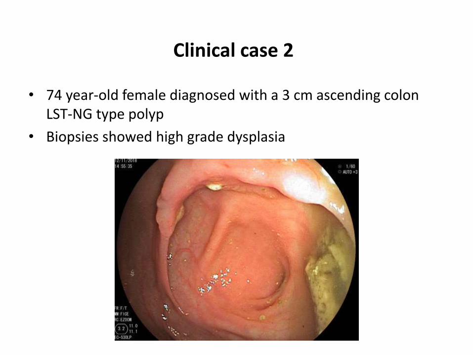
Clinical case 2
• 74 year-old female diagnosed with a 3 cm ascending colon LST-NG type polyp
• Biopsies showed high grade dysplasia

Treatment Decision
• It is not a cancer…
• Let’s try en-block removal for definite diagnosis

AdenoCa

Lesion morphologyParis Classification


Risk Stratification for Covert Invasive Cancer Among Patients Referred for Colonic Endoscopic Mucosal Resection:
A Large Multicenter Cohort Burgess N et al. Gastroenterology 2017;153:732–742

Magnification Chromoendoscopy
Kudo Pit-Pattern
Deep sm 28%
Deep sm
90%

Image Enhanced Endoscopy
overall accuracy for sm 70%


Surface/Vessel architectureMagnification virtual endoscopy


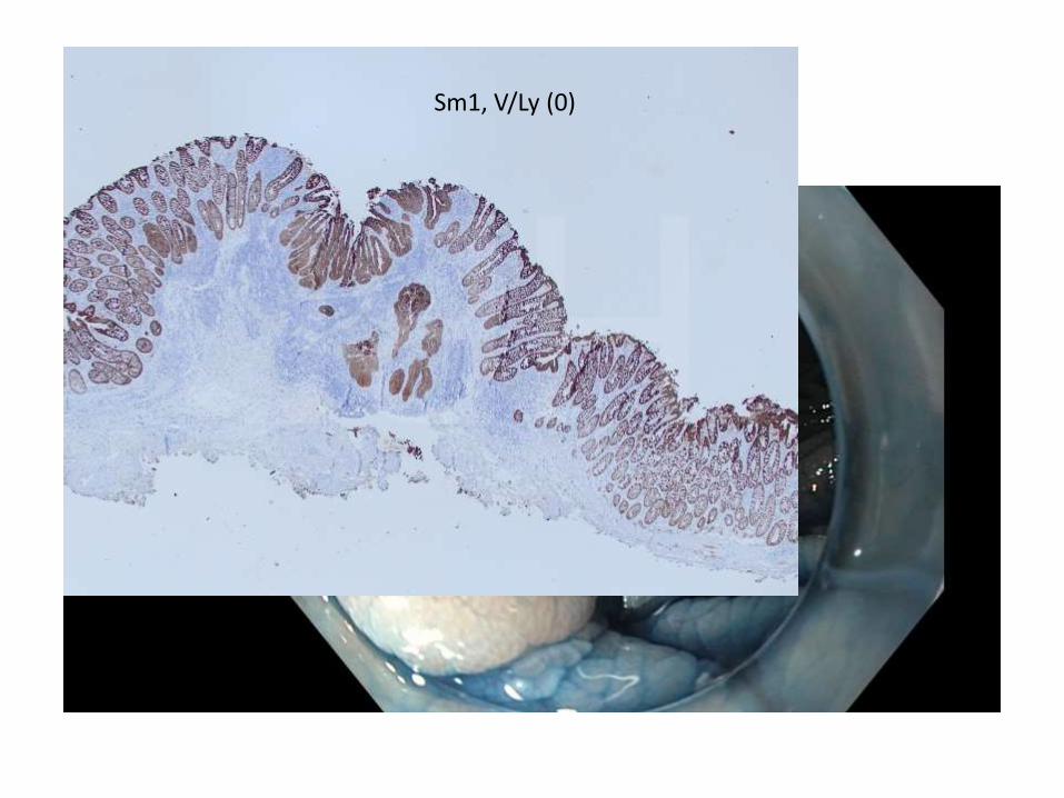
Sm1, V/Ly (0)


Conclusions
• Optical recognition is very important to determine diagnosis and choose the right therapy
• Use high definition endoscopes with image enhanced technology in cases of IIc-depressed lesions and LST-NG/G mixed types
• If this is not possible, just spray 0.3% Indigo Carmine for better interpretation
• Do not take multiple biopsies if you consider resectability. Fibrosis!! Do not tattoo under the lesion!
• One or two targeted samples are sufficient.
• Share your cases with other colleagues. We all get better endoscopists…

Optical recognition of polyps
Bassioukas P. Stefanos
Department of Advanced Therapeutic Endoscopy, Bioclinic Hospital Athens