Embed Size (px)
Citation preview

SCCS/1566/15 Revision of 16 March 2016
Version S
Scientific Committee on Consumer Safety
SCCS
OPINION ON
Hydroxyapatite (nano)
The SCCS adopted this opinion by written procedure
on 16 October 2015
Revision of 16 March 2016

SCCS/1566/15
Revision of opinion on hydroxyapatite (nano)
___________________________________________________________________________________________
2
About the Scientific Committees
Three independent non-food Scientific Committees provide the Commission with the
scientific advice it needs when preparing policy and proposals relating to consumer safety, public health and the environment. The Committees also draw the Commission's attention
to the new or emerging problems which may pose an actual or potential threat. They are: the Scientific Committee on Consumer Safety (SCCS), the Scientific Committee
on Health and Environmental Risks (SCHER) and the Scientific Committee on Emerging and Newly Identified Health Risks (SCENIHR) and are made up of independent experts.
In addition, the Commission relies upon the work of the European Food Safety Authority (EFSA), the European Medicines Agency (EMA), the European Centre for Disease prevention
and Control (ECDC) and the European Chemicals Agency (ECHA).
SCCS The Committee shall provide opinions on questions concerning all types of health and safety
risks (notably chemical, biological, mechanical and other physical risks) of non-food
consumer products (for example: cosmetic products and their ingredients, toys, textiles, clothing, personal care and household products such as detergents, etc.) and services (for
example: tattooing, artificial sun tanning, etc.).
Scientific Committee members
Ulrike Bernauer, Qasim Chaudhry, Pieter Coenraads, Gisela Degen, Maria Dusinska, Werner Lilienblum, Elsa Nielsen, Thomas Platzek, Christophe Rousselle, Jan van Benthem
Contact
European Commission Health and Food Safety
Directorate C: Public Health, country knowledge, crisis management Unit C2 – Country knowledge and scientific committees
Office: HTC 03/073 L-2920 Luxembourg
© European Union, 2016
ISSN ISBN
Doi: ND-
The opinions of the Scientific Committees present the views of the independent scientists
who are members of the committees. They do not necessarily reflect the views of the European Commission. The opinions are published by the European Commission in their
original language only.
http://ec.europa.eu/health/scientific_committees/consumer_safety/index_en.htm

SCCS/1566/15
Revision of opinion on hydroxyapatite (nano)
___________________________________________________________________________________________
3
ACKNOWLEDGMENTS
SCCS members
Dr. U. Bernauer (Rapporteur) Dr. Q. Chaudhry (Chairperson)
Dr. M. Dusinska Dr. W. Lilienblum
Prof. Th. Platzek Dr. J. van Benthem
SCENIHR members Prof. P. H. M. Hoet
Dr. K. Rydzynski
External experts Prof. A. Bernard
Dr. O. Bussolati Dr. N. von Götz
Dr. S. H. Doak
Dr. T. Jung
This opinion has been subject to a commenting period of eight weeks after its initial
publication. Comments received during this time have been considered by the SCCS and discussed in the subsequent plenary meeting. Where appropriate, the text of the relevant
sections of the opinion has been modified or explanations have been added. In the cases where the SCCS after consideration and discussion of the comments, has decided to
maintain its initial views, the opinion (or the section concerned) has remained unchanged.
Revised opinions carry the date of revision.
Keywords: SCCS, scientific opinion, hydroxyapatite, nano, Regulation 1223/2009, CAS
1306-06-5, EC 215-145-7
Opinion to be cited as: SCCS (Scientific Committee on Consumer Safety), Opinion on
hydroxyapatite (nano), 16 October 2015, SCCS/1566/15, revision of 16 March 2016

SCCS/1566/15
Revision of opinion on hydroxyapatite (nano)
___________________________________________________________________________________________
4
TABLE OF CONTENTS
1. BACKGROUND ............................................................................................. 5
2. TERMS OF REFERENCE .................................................................................. 5
3. OPINION ..................................................................................................... 6
3.1 Chemical and Physical Specifications ....................................................... 6
3.1.1 Chemical identity .................................................................... 6 3.1.2 Physical form ......................................................................... 6 3.1.3 Molecular weight .................................................................... 7 3.1.4 Purity, composition and substance codes .................................... 7 3.1.5 Impurities / accompanying contaminants ................................... 7 3.1.6 Solubility ............................................................................... 7 3.1.7 Partition coefficient (Log Pow) .................................................... 7 3.1.8 Additional physical and chemical specifications ............................ 7 3.1.9 Homogeneity and Stability ....................................................... 8
3.2 Function and uses ................................................................................ 9
3.3 Toxicological Evaluation ........................................................................ 9
3.3.1 Acute toxicity ......................................................................... 9 3.3.2 Irritation and corrosivity ........................................................ 12 3.3.3 Skin sensitisation .................................................................. 14 3.3.4 Dermal / percutaneous absorption........................................... 16 3.3.5 Repeated dose toxicity .......................................................... 16 3.3.6 Mutagenicity / Genotoxicity .................................................... 20 3.3.7 Carcinogenicity ..................................................................... 21 3.3.8 Reproductive toxicity ............................................................. 21 3.3.9 Toxicokinetics ...................................................................... 22 3.3.10 Photo-induced toxicity ........................................................... 23 3.3.11 Human data ......................................................................... 23 3.3.12 Special investigations ............................................................ 24 3.3.13 Safety evaluation (including calculation of the MoS) ................... 30 3.3.14 Discussion ........................................................................... 31
4. CONCLUSION ............................................................................................ 34
5. MINORITY OPINION .................................................................................... 36
6. REFERENCES ............................................................................................. 36
Annex ............................................................................................................... 45

SCCS/1566/15
Revision of opinion on hydroxyapatite (nano)
___________________________________________________________________________________________
5
1. BACKGROUND
Article 2(1)(k) of Regulation (EC) No 1223/2009 establishes that “nanomaterial" means an
insoluble or biopersistent and intentionally manufactured material with one or more external
dimensions, or an internal structure, on the scale from 1 to 100 nm.
That definition covers only materials in the nano-scale that are intentionally made, and are insoluble/partially-soluble or biopersistent (e.g. metals, metal oxides, carbon materials,
etc), and it does not cover those that are soluble or degradable/non-persistent in biological systems (e.g. liposomes, emulsions, etc). Article 16 of the Cosmetics Regulation requires
any cosmetic product containing nanomaterials to be notified to the Commission six months prior to being placed on the market, and Article 19 requires nano ingredients to be labelled
(name of the ingredient, followed by ‘nano’ in brackets). If there are concerns over the
safety of a nanomaterial, the Commission shall refer it to the Scientific Committee on Consumer Safety (SCCS) for a full risk assessment.
The Commission received 35 notifications of cosmetic products containing Hydroxyapatite
(CAS No 1306-06-5) in nano form. This ingredient is reported in the CosIng database without any reference to the nano form with the function of abrasive, bulking and emulsion
stabilising, but it is not regulated in Cosmetic Regulation (EC) No 1223/2009. According to the applicants, the ingredient is used in nano uncoated form both in leave-on and rinse-off
oral cosmetics products including toothpastes, tooth whiteners and mouth washes with
maximum reported concentration limit of 10% and specifications.
The Commission has concerns on the use of Hydroxyapatite in nano form because of the potential for nanoparticles of Hydroxyapatite to be absorbed and enter into the cells.
Therefore, we would like to request the SCCS a safety assessment of the nano form of Hydroxyapatite covered in the notifications listed in the annex to this mandate, in the
above-mentioned categories of products, taking into account the reasonably foreseeable exposure conditions.
2. TERMS OF REFERENCE
1. In view of above, and taken into account the scientific data provided, the SCCS is
requested to give its opinion on the safety of the nanomaterial Hydroxyapatite when used in oral cosmetics products including toothpastes, tooth whiteners and mouth washes with a
maximum concentration limit of 10%, taking into account the reasonably foreseeable exposure conditions.
2. SCCS is requested to address any further scientific concerns with regard to the use of
Hydroxyapatite in nano form in cosmetic products.

SCCS/1566/15
Revision of opinion on hydroxyapatite (nano)
___________________________________________________________________________________________
6
3. OPINION
At the Applicant’s request, the names of companies and materials have been coded in this opinion alphabetically (company names) and numerically (material names).
3.1 Chemical and Physical Specifications
3.1.1 Chemical identity
3.1.1.1 Primary name and/or INCI name
Hydroxyapatite
3.1.1.2 Chemical names
Hydroxyapatite
Calcium Phosphatetribasic Calcium Hydroxyphosphate
Hydroxylapatite
3.1.1.3 Trade names and abbreviations
Confidential
3.1.1.4 CAS / EC number
CAS: 1306-06-5
EC: 215-145-7
3.1.1.5 Structural formula
N.A.
3.1.1.6 Empirical formula
Ca5 (PO4)3 (OH)
3.1.2 Physical form
Materials added to products are listed either as powder (to be used in toothpastes) or suspension (mouthwash).
Suspension is described as an odourless white paste (Material codes 1000440; 1000320;
1001387; 1001388; 1001240; 1001241; 1001727); and all other materials as powder. The material is described to be composed of rod/thumb shaped particles that are in the
form of dispersed free particles and agglomerates (Company C).
Submission of company B refers to hydroxyapatite of any shape including nanofibres of 2-4 nm width and 20 – 50 nm in length.
All materials are stated to be in uncoated form.
SCCS comment: Hydroxyapatite is a naturally occurring mineral. Hydroxyapatite has a hexagonal crystal
structure comprising different crystalline phases. The OH- ions in hydroxyapatite can be replaced by different counter anions to form other members of the apatite group. Therefore

SCCS/1566/15
Revision of opinion on hydroxyapatite (nano)
___________________________________________________________________________________________
7
hydroxyapatite can bear a variable degree of other anions, most commonly carbonate, phosphate and fluoride, which leads to modified atomic composition and also properties
(e.g. see www.astm.org/Standards/F1185.htm).
There are several established pathways to produce hydroxyapatite, among them wet chemical synthesis and precipitation, biomimetic preparation and electrodeposition. A
number of different pathways also exist for preparing nanoforms of hydroxyapatite. The processing of natural hydroxyapatite sources like bovine bone to extract hydroxyapatite is
also used. There is a huge body of literature on hydroxyapatite, its manufacturing and biocompatibility due to use as an implant material and also on the generation of different
nanoforms.
According to the information provided by the applicants, the nano-hydroxyapatite in the raw
material is fully synthetic, made from inorganic reactants using food‐grade quality calcium
and phosphorous salts, i.e. it is not produced from bone material.'
3.1.3 Molecular weight
Molecular weight: 502.31 g/mol
3.1.4 Purity, composition and substance codes
Company A and C (material 1): Hydroxyapatite as the main component; Potassium chloride (KCl) as preservative and water as excipient.
Company B (material 2): “Artificially synthesized, amorphous, nano-disperse calcium hydroxyapatite in powder form or aqueous suspension does not contain reaction by-
products." Calcium content (by weight): ≥ 39.0%; phosphorus content (by weight): ≥ 18.0%.
3.1.5 Impurities / accompanying contaminants
Company A and C (material 1): Total heavy metals < 20 ppm.
Company B (material 2): Moisture content of the powder (by weight): ≤ 3.0%; content of heavy metals (by weight): ≤ 0.002%.
3.1.6 Solubility
Insoluble in water (aqueous solubility <0.01 mg/l).
3.1.7 Partition coefficient (Log Pow)
Log Pow: /
3.1.8 Additional physical and chemical specifications
Specific surface area:
Company A and C, material 1: 80 – 100 m2/g (according to BET nitrogen adsorption) Company B, material 2: BET specific surface area SSA: 147 m²/g
Volume specific surface area VSSA: 440 m²/cm³.
Zeta potential:
SCCS/1566/15
Revision of opinion on hydroxyapatite (nano)
___________________________________________________________________________________________
8
Company A and C, material 1: + 30 ± 1 mV Company B, material 2: not measurable.
Density: 1.1 – 1.2 g/cm3 (Company A and C, material 1)
Viscosity: 1.0 ± 0.3 Pa.s (assessed with a Physica UDS 200, oscillatory shear testing at 30ºC, deformation 100% at
10Hz) (Company A and C, material 1) pKa: /
Refractive index: / pH limits: 10.0 ± 0.5 (measured directly on the product) (Company
A and C, material 1)
Porosity: hydroxyapatite nanoparticles with mesopores (Company A and C, material 1)
UV_Vis spectrum: / Melting point: /
Boiling point: / Flash point: /
Vapour pressure: /
3.1.9 Homogeneity and Stability
Material 1: Hydroxyapatite is a chemically stable compound; therefore its degradation is not expected
under normal conditions of storage, avoiding freezing, and keeping the product in the original container at room temperature, in a clean, dry place.
To insure homogeneity of the material that contains the hydroxyapatite (nano) – (material 1, company A and C) - it should be stirred before every use. It is a thixotropic material,
which means that it is very viscous under normal conditions, but it becomes less viscous
over time when shaken, agitated, or otherwise stressed. However, this is a reversible microstructural change of the material. The shelf-life of this material is 18 months.
Material 2 (company B):
Calcium hydroxyapatite is chemically and thermally stable. Shelf-life of suspension: 2 months: shelf-life of powder: 2 years.
3.1.10 Particle Size Distribution and Crystalline Shape
Shape:
The reported particle shapes of the materials are either hexagonal plate, nanofibre, or amorphous, with some materials reported to be composed of nano-rod or nano-thumb
shaped particles. The hydroxyapatite nanoparticles are reported to be aggregated/agglomerated to larger
clusters. Needle-like hydroxyapatite particles have also been reported in scientific literature.
Company B (material 2) mentions: ’isometric aggregates consisting of particles of any
shape, sized 5-10 nm, and fibres formed from the same particles.’
Particle size/particle size distribution Company B (material 2)
According to TEM: nanofibre and plate form;
Nanofibres: 2 – 4 nm thickness; 20 – 50 nm length Plates: mean size 15 nm
Formation of aggregates in size range from 50 nm to 1.4 µm
According to DLS: size distribution 50 nm to 1.4 µm; mean size: 1 µm

SCCS/1566/15
Revision of opinion on hydroxyapatite (nano)
___________________________________________________________________________________________
9
Company A (material 1):
Form of nanoparticles: nanorod or nanothumb Malvern Mastersizer: no agglomerates with particle size < 100 nm; mean particle size of
agglomerates: 1.213 µm TEM: shows nanorod shaped entities with size below 100 nm; structures larger than 100
nm can be observed due to agglomeration of particles.
Company C Mean particle size of individualised particles measured by TEM in terms of particle number is
reported as 64 nm.
Primary particle size measured using Malvern Mastersizer 2000 is reported as 20 nm (lowest cut-off level); 60 to 400 nm (volume weighted median); and 30 nm to 80 nm (number
weighted median). Mean particle size of the agglomerates (in terms of particle number) is reported as 1.213 µm.
General comments on physicochemical characterisation The characterised materials do not necessarily correspond to those materials which have
been used for toxicity testing.
One submission refers to hydroxyapatite of any shape including nanofibres of 2-4 nm width and 20 – 50 nm in length.
3.2 Function and uses
Nano-hydroxyapatite is used as a drug delivery material and as a bone defect filling material. Oral care products containing nanoparticulate hydroxyapatite are intended to be
used for teeth remineralisation and apparently are already on the market. Products listed in the dossiers are either in the form of paste or suspension. All products listed in the dossiers
are intended for oral application. All products are listed as rinse off - except 3 mouthwash products that are listed by Company A as leave on. The material is intended for use in
toothpaste, tooth whitener and mouthwash products. Based on the explanation provided by
one Applicant (Company C) the concentration of hydroxyapatite of 3-15% refers to the concentration in the raw material 1, which contains 15.5% nano-hydroxyapatite. Therefore,
the 3-15% of the raw material 1 corresponds to 0.465 – 2.325% nano-hydroxyapatite in the final product. The submission also describes the use of 90% of raw material 1 which
corresponds to 13.95% of nano-hydroxyapatite (w/w) in the final product.
All materials are described as having no surface reactivity or photoreactivity.
SCCS comment:
Two applicants (company A and C) have used the same ingredient. One of the applicants (company A) later clarified that the minimum concentration used in final products can be as
low as 0.08% of the raw material (corresponding to 0.0125% nano-hydroxyapatite). The second Applicant (company C) clarified that although 90% raw material corresponds to
13.95% nano-hydroxyapatite (w/w), for this type of application, the expected maximum raw material concentration is 20%, which corresponds to 3.1% of nano-hydroxyapatite in
the final product. The applicants should provide further information on the level of any significant impurities
other than heavy metals, and provide an upper limit of total impurities in the raw material.
3.3 Toxicological Evaluation
3.3.1 Acute toxicity
SCCS/1566/15
Revision of opinion on hydroxyapatite (nano)
___________________________________________________________________________________________
10
3.3.1.1 Acute oral toxicity
A material named “Hydroxyapatite 5%, aqueous solution, 20 nm” was intra-gastrically administered in a dose range between 1,600 and 36,000 mg/kg bw to white rats of both
genders (strain unclear) and the animals were observed for 14 days. As described in the study report, death of the animals occurred from 10,000 mg/kg bw in both sexes as a result
of cardiac and respiratory arrest. Important findings on animals surviving 14 d: lung vessels were moderately congested, “alveoli filled with air”; signs of “fatty degeneration of the
liver”. The LD50 was 20,800 mg/kg bw in both sexes.
Ref.: Nechiporenko, S.P. and Stepanov, S.V. (2011)
SCCS comment:
The study did not adhere to any OECD or EU test guideline and it is not clear whether it was performed according to GLP principles. The test material was insufficiently characterised and
it is not clear whether the doses used correspond to test material as 5% aqueous solution or to the active ingredient. It is also not clear whether the tested material belongs to the
materials covered by the submission. The study is therefore of little value to this assessment.
3.3.1.2 Acute dermal toxicity
/
3.3.1.3 Acute inhalation toxicity
A material named “Hydroxyapatite 5%, aqueous solution, 20 nm” was investigated for acute inhalation toxicity in white mice. Signs of irritation of the upper respiratory tract were
observed in this study.
Ref.: Nechiporenko, S.P. and Stepanov, S.V. (2011)
SCCS comment:
Due to poor reporting and insufficient characterisation of test material, no conclusions can be drawn from that study.
3.3.1.4 Acute intravenous toxicity
Guideline: ISO 10993-11 Species/strain/sex: mouse, Hla®: (ICR) CVF®, male
Group size: 5 Test substance: Nano-Hydroxyapatite paste (a white viscous paste composed of
nanoparticles of hydroxyapatite and water; 38% hydroxyapatite)
Batch: 1309-12 Purity: > 95%
Vehicle: 0.9% Sodium Chloride in water
Dose levels: 10 g paste/kg bw Administration: intravenous
GLP: Yes Study period: September – October 2012
A single dose of test material extracted with 0.9% sodium chloride was intravenously
administered (no further information) at a dose of 50 ml/kg bw to five male animals, five further animals received vehicle control. Animals were observed for clinical reactions
SCCS/1566/15
Revision of opinion on hydroxyapatite (nano)
___________________________________________________________________________________________
11
immediately after and 4, 24, 48 and 72 hr after injection. Animals were weighed daily for three days after dosing. There was no mortality during the study and all animals (control
and treated) appeared normal during the course of the study. Treatment did not adversely affect the animals' body weight.
Ref.: NAMSA (2012a)
SCCS comment:
No material characterisation is given in the study report. However the applicant confirmed that this study had been performed with material 1 of the submission. The study did not
correspond to study types usually used to investigate acute toxicity of cosmetic ingredients
and the observation period after treatment was much shorter than usually applied in acute toxicity studies according to the OECD test guidelines. However, the study demonstrated
that a single intravenously administered dose of 10 g/kg bw nano-hydroxyapatite paste (corresponding to 3.8 g active ingredient) apparently did not adversely affect mice within an
observation period of 3 days. Apparently, this is the “acute systemic toxicity study” as described in Ding et al., 2012, where further study details are missing.
Information from open literature
In a publication from the open literature (article in Chinese, only some parts of the abstract in English), it was concluded that the “medium” (wording from the abstract) lethal dose of
nano-hydroxyapatite is 200 mg/kg bw. In this study, some clinical-chemical parameters were also investigated. It could be demonstrated that some enzymes dramatically increased
30 min after injection, reaching peaks at 2 hr after injection and returning to normal levels 24 hr after injection. BUN and ALP peaked 24 hr after injection and returned to normal
levels afterwards. As the study is in Chinese, no further detail on study conditions, material characterisation or explanation of abbreviations could be obtained.
Ref.: Liu, L.P. et al. (2005)
In a further publication from the open literature, 6 male Sprague-Dawley rats received
single injections of 50 mg/kg bw nano-hydroxyapatite (needle-like; long diameter: 80 nm, short diameter: 20 nm; hydrodynamic diameter in physiological saline: 245.1 nm (i.e.
agglomeration in physiological saline), Zeta potential in physiological saline: - 16.3 mV; purity: ≥ 99.0%) into the tail vein; 6 control rats received vehicle alone (physiological
saline). 48 hr later, blood samples were collected for haematological and clinical-
biochemical investigations and the left lateral lobe of the liver was removed for histopathological analysis and determination of oxidative biomarkers. Compared to control
livers which had normal histology, the liver tissues from the rats exposed to nano-hydroxyapatite showed inflammatory cell infiltration at the portal areas of the liver.
Haematological analysis demonstrated increased white blood cells, elevated levels of the inflammatory cytokine TNF-α, and increased alanine aminotransferase (ALT), aspartate
aminotransferase (AST), total bile acid (TBA), cholesterol, uric acid, lactate dehydrogenase (LDH) and low density lipoprotein (LDL) compared to controls. In the livers, increased levels
of H2O2 and MDA (malondialdehyde) and decreased levels of glutathione (p<0.05) were
observed. Ref.: Chen, Q. et al. (2014).
SCCS comment:
The study demonstrates that needle-shaped nano-hydroxyapatite can lead to biochemical changes and indications of oxidative damage after i.v. administration in rats within 48 hr.
3.3.1.5. Acute intraperitoneal toxicity

SCCS/1566/15
Revision of opinion on hydroxyapatite (nano)
___________________________________________________________________________________________
12
Guideline: ISO 10993-11 Species/strain/sex: mouse, Hla®: (ICR) CVF®, male
Group size: 5 Test substance: Nano-Hydroxyapatite paste (a white viscous paste composed of
nanoparticles of hydroxyapatite and water; strength: 38% hydroxyapatite)
Batch: 1309-12 Purity: > 95%
Vehicle: Sesame Oil Dose levels: 10 g/kg bw
Administration: intraperitoneal
GLP: Yes Study period: September – October 2012
Test article in sesame oil and sesame oil alone were “extracted” by continuous agitating for
24 hr at 70°C. The extracts were not centrifuged, filtered or otherwise altered prior to dosing (meaning that the test materials were in a suspension form).
A single dose of test material extracted with sesame oil was intraperitoneally administered (no further information) at 50 ml/kg to five male animals, 5 further animals received vehicle
control. Animals were observed for clinical reaction immediately after and 4, 24, 48 and 72
hr after injection. Animals were weighed daily for three days after dosing. There was no mortality during the study and all animals (control and treated) appeared normal during the
course of the study. Treatment did not adversely affect body weight in the animals.
Ref.: NAMSA (2012a)
SCCS comment: No material characterisation is given in the study report. However the applicant confirmed
that this study had been performed with material 1 of the submission. The study did not
correspond to the study types usually used to investigate acute toxicity of cosmetic ingredients and the observation period after treatment was much shorter than usually
applied in acute toxicity studies according to the OECD test guidelines. However, the study demonstrated that a single intraperitoneally administered dose of 10 g/kg bw nano-
hydroxyapatite paste (corresponding to 3.8 g active ingredient) apparently did not adversely affect mice within an observation period of 3 days.
SCCS conclusion on acute toxicity
The acute toxicity of nano-hydroxyapatite was investigated through the oral, inhalation, intraperitoneal and intravenous routes. No study was performed in accordance with any
OECD or EU Test guidelines and apart from one intravenous study published in the open literature (Chen et al., 2014), proper material characterisation was not given. Therefore, no
conclusions on acute toxicity can be drawn from these studies. The study from the open literature used needle-shaped nano-hydroxyapatite and indicated that needle-shaped
crystals of hydroxyapatite can be a concern in relation to acute toxicity.
3.3.2 Irritation and corrosivity
3.3.2.1 Skin irritation
Guideline: ISO 10993-10 Species/strain/sex: Rabbit, White New Zealand, male
Group size: 3 Test substance: Nano-Hydroxyapatite paste (a white viscous paste composed of
nanoparticles of hydroxyapatite and water; 38%
hydroxyapatite)

SCCS/1566/15
Revision of opinion on hydroxyapatite (nano)
___________________________________________________________________________________________
13
Batch: 1309-12 Purity: > 95%
Vehicle: 0.9% sodium chloride or sesame oil Dose level: approximately 40 mg/kg bw
Dose volume: 0.2 ml Observation: up to 72 hr after administration
GLP: Yes Study period: October 2012
Test article in the respective vehicles, and vehicles alone, were “extracted” by continuous agitating for 24 hr at 70°C. The extracts were not centrifuged, filtered or otherwise altered
prior to dosing (meaning that test materials were in a suspension form).
Groups of 3 animals received intracutaneous injections of substance dissolved in the respective vehicle at five sites of the clipped right back side of the animals; respective
vehicle controls were intracutaneously injected at five sites of the clipped left back side of the animals. The treatment sites were observed immediately after injections; animals were
then returned to cages and observations for edema and erythema (on a score from 0 to 4) were made 24, 48 and 72 hr after injection. Erythema and edema for test article and
vehicles at each scoring interval were calculated by adding erythema and edema scores. The differences between mean overall scores of treated versus vehicle scores were used to
assess local dermal irritation.
Results:
Two animals treated with the sesame oil vehicle had reduced faeces at the 24 hr examination, all other animals appeared normal. The score difference (treated versus
control sites) in animals treated with sodium chloride vehicle was zero, whereas score difference (treated versus control sites) in animals treated with sesame oil vehicle was 0.2.
A substance is considered to meet the requirements of this test when the difference between mean score of treated versus control is < 1). As differences were 0 and 0.2., the
material tested met the requirements of the test.
Ref.: NAMSA (2012b)
SCCS comment:
No material characterisation is given in the study report. However the applicant confirmed that this study had been performed with material 1 of the submission. The study did not
correspond to study types usually used to investigate skin irritation. However, it demonstrates that under the conditions used, intracutaneous injection of 40 mg/kg nano-
hydroxyapatite paste (corresponding to 15.2 mg a.i.) does not cause erythema and
oedema.
In another study, a material termed as “Hydroxyapatite 5%, aqueous solution, 20 nm” was investigated for skin effects in rats. Tails of white rats (no information on strain and sex)
were placed 2/3 in vials (no information on duration or the concentration of test material). Tails were examined for erythema and edema 1 hr and 16 hr after treatment. No changes of
tail morphology, epidermis and skin appendages were observed.
Ref.: Nechiporenko, S.P. and Stepanov, S.V. (2011)
SCCS comment: The study did not adhere to an OECD or EU test guideline and it is not clear whether it was
performed according to GLP principles. No conclusion can be drawn from this study.
For skin irritation in humans see section 3.3.11.

SCCS/1566/15
Revision of opinion on hydroxyapatite (nano)
___________________________________________________________________________________________
14
3.3.2.2 Mucous membrane irritation / Eye irritation
The eye irritation potential of material 1 was investigated in the Hen`s Egg Chorioallantoic Test. Three fertilised hen’s eggs were incubated with each substance respectively (material
1 at a concentration of 25%, diluted in water, and 2 positive controls (1% SDS and 0.1M NaOH) and 1 negative control (0.9% NaCl)) for nine days, and on the 10th day, the eggs
were opened and the CAM (chorioallantoic membrane) exposed. 0.3 g of the test substance was applied to the surface of the CAM and after a 20-second exposure period, the CAM was
rinsed with 5 ml of sterile Milli-Q water. Morphological analysis observing hyperaemia (increased blood flow), haemorrhage (blood from a ruptured vessel) and coagulation
(presence of blood clots) was performed 0.5, 2, and 5 minutes after rinsing-off the test
substance. Observations were subjectively assessed and scored according to published literature. Photographs were integrated in the report. According to the HET-CAM, 25% of
the material was considered weakly or slightly irritant with a score of 2.8 on the CAM, based on hyperaemia at the 2 min score.
Ref.: Inovapotek (2013a)
SCCS comment:
The HET-CAM test does not belong to the internationally accepted tests for eye irritation
testing. The study report contains no information on GLP-compliance. Reporting on findings on test material, positive and negative controls is considered insufficient by the SCCS as
e.g. no information on scores obtained from positive or negative controls is given. Also, there is no information on positive or negative scores obtained from historical controls.
Based on the findings of the study, mucous membrane irritation of material 1 of the submission cannot be excluded.
A material termed as “Hydroxyapatite 5%, aqueous solution, 20 nm” was investigated for
effects on mucous membrane of the eye. 2 drops of substance were instilled into the conjunctival sac of a rabbit (apparently one animal, no further information) which was
observed for 1 month. From the study it was concluded that the test material causes slight and reversible irritation.
Ref.: Nechiporenko, S.P. and Stepanov, S.V. (2011)
SCCS comment: The study did not adhere to an OECD or EU test guideline and it is not clear whether it was
performed according to GLP principles. The test material is insufficiently characterised and it
is not clear whether the tested material belongs to the materials covered by the submission. No conclusion can be drawn from this study.
SCCS conclusion on irritation and corrosivity No guideline compliant skin or mucous membrane irritation test was provided by the
applicants. The studies/study descriptions partly lacked proper material characterisation. A skin- and eye irritating potential cannot be excluded based on the studies provided.
3.3.3 Skin sensitisation
In a study from the open literature, needle-shaped nano-hydroxyapatite particles synthesised by a wet chemical method yielding particles of a size below 50 nm and a Zeta
potential of -13.4 mV in buffer (the authors state that the material was unstable in water due to a Zeta potential below 3.19 mV) were investigated for sensitising effects in Albino
Guinea pigs (Hartley) (sex not given). 10 test animals and 5 control animals were used. A paste of 80 mg nano-hydroxyapatite in physiological saline was applied topically to the
clipped back, covered and kept for 6 hr. The procedure was repeated three times a week for
three weeks. Two weeks after the last induction application, a challenge application was

SCCS/1566/15
Revision of opinion on hydroxyapatite (nano)
___________________________________________________________________________________________
15
made comparable to the induction applications. Following removal of the patch, the sensitisation potential was evaluated at 24, 48 and 72 hr after patch removal. No
indications for skin sensitisation were observed in the study. In the same animals, blood, livers and brains were taken at termination and haematological
and biochemical parameters were determined in blood. Parameters indicative of oxidative stress (total protein (TP), lipid peroxidation (malondialdehyde (MDA) formation), glutathione
reductase (GR), reduced glutathione (GSH), glutathione peroxidase (GPx), superoxide dismutase (SOD) and 8-hydroxyl-2-deoxyguanosine (8-OHdG)) were investigated in livers
and brains.
Results Haematology: whereas white blood cells were comparable between treated and untreated
animals, there was a statistically significant increase in red blood cells, haemoglobin and haematocrit and a statistically significant decrease in platelet counts in treated animals.
With respect to biochemical parameters, there were slight decreases in serum glutamic oxaloacetic transaminase, glucose and urea and statistically significant increases in serum
glutamic pyruvate transaminase and alkaline phosphatase in treated animals compared to
controls, however, the study authors considered these latter two variations as being in the normal range. Among the markers of oxidative stress investigated, there were no significant
changes between treated and control animals. The findings might be indicative of systemic effects after dermal administration of nano-hydroxyapatite.
Ref.: Geetha, C.S. et al. (2013)
SCCS comment:
It is not clear whether the study was performed according to an OECD/EU guideline and
according to GLP principles. According to OECD test guideline 406, the concentration to be used for induction should be the highest to cause mild irritation, whereas for challenge the
highest non-irritation dose should be used. It is not clear whether such considerations were made. The study is of limited value for assessment of the sensitising potential of nano-
hydroxyapatite. However, the findings might be indicative of systemic effects after dermal administration of needle-shaped nano-hydroxyapatite.
Another study used a material termed as “Hydroxyapatite 5%, aqueous solution, 20 nm” for
skin sensitisation in guinea pigs (no information on sex, strain and number of animals used). The test material was injected once at a dose of 20 mg/kg bw (unclear whether this
refers to test material or active ingredient) into the outer skin surface of the ear. 10 days later, a solution of the test material was applied to the skin (2 x 2 cm) of the shaved back
(100 mg/cm2). It is unclear whether this refers to test material or active ingredient. Control animals received distilled water. Blood was taken 3 hrs after skin application and analysed
for WBC, eosinophils, lymphocytes, monocytes and RSLL (no explanation is given for the abbreviations “RSLL” and “lysis” used in the report). No signs of irritation were observed in
treated skin, and there was no difference in the blood parameters investigated between
control and treated animals. Ref.: Nechiporenko, S.P. and Stepanov, S.V. (2011)
SCCS comment: The study did not adhere to an OECD or EU test guideline and it is not clear whether it was
performed according to GLP principles. The test material is insufficiently characterised and it is not clear whether the tested material belongs to the materials covered by the submission.
No conclusion can be drawn from this study.

SCCS/1566/15
Revision of opinion on hydroxyapatite (nano)
___________________________________________________________________________________________
16
SCCS conclusion on skin sensitization
No guideline-compliant study for skin sensitisation was provided. In a study from the open literature using needle-shaped nano-hydroxyapatite, there were no indications of skin
sensitisation. An in vivo study performed in guinea pigs cannot be used to assess skin sensitisation due to poor study description and material characterisation.
No conclusion on skin sensitisation can be drawn from the available information.
3.3.4 Dermal / percutaneous absorption
According to information from the applicant(s), a dermal absorption study is underway.
However, as submissions are intended for oral use, a dermal absorption study is not necessary for this type of use.
3.3.5 Repeated dose toxicity
3.3.5.1 Repeated Dose (short-term) toxicity
7-day study, intravenous administration
Two types of hydroxyapatite nanomaterials were used (number 1#: rod-like; 10 – 20 nm in
diameter; 30 – 50 nm in length; number 4#: rod-like, 20 – 40 nm in diameter; 70 – 90 nm in length). SD rats (no further explanation of abbreviation, most probably Sprague-Dawley
rats) from Shanghai XIPUER-BK Lab Animal Co. Ltd. were used. Nanomaterials were
sonicated for 30 min at 300W/50 kHz and suspended at a concentration of 1 mg/ml in normal saline solution containing 10% foetal calf serum. Groups of 5 male and 5 female
animals received one of the two types of nanomaterial at 10 mg/kg bw or vehicle control once daily for 7 days by the i.v. route. On day 8, blood was taken for determination of blood
cell count with differential and clinical-chemical analysis. Animals were killed; a variety of organs was taken and prepared for histopathological analysis. Haematology revealed no
statistically significant differences between control and treated animals. Clinical-chemical analysis revealed statistically significant differences from control of glucose and total
bilirubin for #1 material and statistically significant differences from control of glucose, ALT,
UA and Na (p< 0.05) (no explanations given for abbreviations. UA most probably means uric acid). Histopathological evaluation revealed “pseudotubercles in lung, performing
intracavity embolism, inflammatory cell infiltrate and hyperplasy of stroma cell.” 1# nanomaterials (i.e. the smaller ones) resulted in a “vacuolar degeneration of nephric tubule
epithelium in the kidney.” The authors of the publication conclude that a long-term study of systemic toxicity is needed and that the kidney may be the main organ of discharge of
nanoparticles. The authors also state that the smaller particles were more toxic than the larger particles and explained this by the larger surface area of the smaller particles.
Ref.: Ding, T. et al. (2012)
SCCS comment: Only the information as presented in the publication was available and there is no
information on GLP- or guideline adherence. From the design of the study (only one dose tested, only 7 d administration period, type of administration) the study cannot be taken to
determine a point of departure for toxicological risk assessment. However, it can be deduced from the study that smaller particles of nano-hydroxyapatite might be of higher

SCCS/1566/15
Revision of opinion on hydroxyapatite (nano)
___________________________________________________________________________________________
17
toxicological potency when systemically available. Furthermore, kidneys and lungs might be considered as target tissues for the systemically available nano-hydroxyapatite.
Oral 3-week study
Synthesised, needle-shaped hydroxyapatite nanoparticles (diameter: 3 – 7 nm; length: 27 – 46 nm; no further specification) were used for the study. No information on GLP is
available. The study design did not correspond to an OECD or EU guideline for repeat-dose testing. Groups (n=7) of female Sprague-Dawley rats were either kept untreated (control)
or received daily oral treatments (no further specification) with nano-hydroxyapatite at 150
or 300 mg/kg bw/d for 3 weeks. A further group was once injected subcutaneously with 600 mg/kg bw nano-hydroxyapatite and left for 5 weeks. Body weight was recorded weekly. At
the end of the experimental period, blood samples were taken for determination of alanine aminotransferase (ALT), aspartate aminotransferase (AST), alkaline phosphatase (ALP), uric
acid and urea, animals were killed and liver and kidney samples were taken for histopathology.
No statistically significant effects on body weight were observed. With respect to liver function tests (ALP, ALT and AST), statistically significant increases compared to controls
were observed in all treatment groups for ALT and AST. For ALP, statistically significant
increases compared to control were only observed in the oral high dose group. There was no change in kidney function parameters investigated apart from a statistically significant
decrease of urea in the s.c. group. Kidneys and livers of all animals showed normal structure.
Ref.: Hafez, A.I. et al. (2012)
SCCS comment:
Based on changes in liver function enzymes after oral administration of needle-shaped
nano-hydroxyapatite particles of the above-mentioned size, it can be deduced that the material investigated will lead to systemic effects related to hepatotoxicity. No
histopathological changes were detected in the treated animals' livers or kidneys. However, no other tissue was investigated in this study. Due to the poor description, the poor
material characterisation given in the publication and the design of the study, it is considered inappropriate for setting a point of departure for toxicological risk assessment.
Oral 20 day study
A material termed as “Hydroxyapatite 5%, aqueous solution, 20 nm” was intra-gastrically
administered for 20 days at a dose of 2,000 mg/kg bw to 3 white rats (strain and sex unclear). Animals were sacrificed one day after the last administration and a variety of
parameters were investigated (e.g. weights of certain organs, urinary protein, clinical-chemical parameters). Internal organs were examined macroscopically and lungs,
myocardium, livers, kidneys and gastric mucosa were examined histopathologically in treated animals. Amongst the parameters investigated, there was no apparent difference
between treated and control animals. In tissues examined histopathologically, there were no
abnormalities. Ref.: Nechiporenko, S.P. and Stepanov, S.V. (2011)
SCCS comment: The study did not adhere to an OECD or EU test guideline and it is not clear whether it was
performed according to GLP principles. The test material is insufficiently characterised and it is not clear whether the doses used correspond to test material as 5% aqueous solution or
to the active ingredient. It is also not clear whether the tested material belongs to the
materials covered by the submission. No conclusion can be drawn from this study.

SCCS/1566/15
Revision of opinion on hydroxyapatite (nano)
___________________________________________________________________________________________
18
Repeated (4 week) intraperitoneal (i.p.) study
In a study from the open literature, a group of male Sprague-Dawley rats (8 per dose group) received a freshly prepared and sonicated saline solution of nano-hydroxyapatite
(rod-shaped material with a mean size of 28.47 ± 0.4 nm (20 – 30 nm in length and 5 nm in width) at a concentration of 8.3 mg/ml, dose applied not mentioned), three times per
week for three weeks. Further groups of 8 animals received either saline alone, chitosan or a composite of nano-hydroxyapatite and chitosan. At the end of the 4th week, animals were
killed and blood was taken for analysis of biochemical markers. Livers and kidneys were
taken for histopathological investigation. After treatment with nano-hydroxyapatite, a statistically significant increase in sodium and
a statistically significant decrease in potassium (p<0.01) were observed. All other parameters investigated exhibited no significant change compared to control.
Histopathology revealed apoptotic cells in the kidneys and livers of animals treated with nano-hydroxyapatite.
Ref.: Wang, L. et al. (2012)
SCCS comment: The study was not performed according to a guideline and GLP adherence is unclear. The
exact dose administered is not clear. The study, however, demonstrates that systemically available nano-hydroxyapatite can induce apoptosis in the liver and kidney of rats. No other
organs were investigated. The finding of apoptosis confirms the findings of a panel of in vitro studies, where nano-hydroxyapatite induced apoptosis in a variety of cells. However,
the study cannot be used as a point of departure for risk assessment. The nanomaterials used in the publication were only poorly described. With respect to size and shape, they
might be of relevance for the materials considered for this opinion; however, Zeta potential
of the materials tested in the study is not comparable with that of the materials provided in the submissions by applicants (Company A and C, material 1).
Dermal 28-day study
In a study from the open literature, needle-shaped nano-hydroxyapatite particles
synthesised by a wet chemical method yielding particles of a size below 50 nm and a Zeta
potential of -13.4 mV in buffer (the material was stated to be unstable in water due to a Zeta potential below 3.19 mV) were investigated in a dermal 28-day repeat-dose study.
Groups of Wistar rats (5/sex/dose) received a paste of nano-hydroxyapatite in water at 0, 25, 50 and 100 mg/kg bw/d up to 6 hr/day for 7d/week for 28 days. At the end of the
exposure period, blood was taken for determination of haematological and biochemical parameters. Gross necropsy was performed in all animals, and major organs (heart, liver,
lungs, spleen and skin) were processed for histopathology. Liver homogenate was prepared and analysed for total protein, lipid peroxidation, reduced glutathione and antioxidant
enzymes (glutathione reductase, glutathione peroxidase and superoxide dismutase).
There was no effect on food intake, body weight or behaviour. Apart from a statistically
significant reduction in haemoglobin, platelets and mean corpuscular volume (MCV) at the highest dose, haematological investigations revealed no other statistically significant
differences between control and treated animals. With respect to biochemical parameters there were single incidences of differences between treated and control animals (for
chlorides at 50 mg/kg bw/d and alkaline phosphatase at 200 mg/kg bw/d). Gross pathology of major organs revealed no abnormalities. Upon histopathology, a mild infiltration of
mononuclear inflammatory cells was observed in the dermis of treated animals. In lungs,
seven cases of bronchial associated lymphoid tissue proliferation were observed.

SCCS/1566/15
Revision of opinion on hydroxyapatite (nano)
___________________________________________________________________________________________
19
Investigation of liver homogenates revealed changes in lipid peroxidation and reduced glutathione when compared to controls; however, there was no dose-dependency. All
antioxidant enzymes investigated were lower compared to control with a dose-dependency for glutathione reductase. Further, superoxide dismutase was decreased in homogenates
from treated animals. However, all changes in enzymes investigated did not reach statistical significance. The study authors conclude that the nano-material synthesised did not cause
any treatment-related adverse effects after 28 d repeated dermal administration to the skin of Wistar rats.
Ref.: Valappil, M.P. et al. (2014)
SCCS comment:
No information on guideline or GLP adherence is available. It remains to be clarified whether some of the effects observed (e.g. increased alkaline phosphatase, reduced glutathione
reductase) might be an indication of systemic effects. Also the lung effects observed after histopathological examination deserve clarification. As it is unclear whether the effects
observed could be substance-related or not and as the submissions are intended for oral administration, the study is not adequate for setting a point of departure for risk
assessment. From the shape and the Zeta potential, the material used is different from the
materials under consideration for the SCCS opinion.
3.3.5.2 Sub-chronic (90 days) toxicity (oral)
No data available
3.3.5.3 Chronic (> 12 months) toxicity
No data available
SCCS conclusion on repeat-dose toxicity
No guideline-compliant repeat-dose toxicity study was provided by the applicants or retrieved by literature search. However, some studies with repeated administration of nano-
hydroxyapatite have been performed for up to 28 days by the intravenous, intraperitoneal,
oral and dermal route. The studies mostly lack proper material characterisation and only a limited amount of parameters and tissues usually investigated in guideline-compliant
repeat-dose studies have been addressed in the available studies/study descriptions and only a few doses (in part only single doses) were tested.
The intravenous and intraperitoneal routes are considered as being of minor relevance for the risk assessment of cosmetics. A 7-day repeated dose intravenous study using two types
of rod-shaped nano-hydroxyapatite indicated that kidneys and lungs might be the target tissues for systemically available nano-hydroxyapatite. Apoptosis was observed in kidney
and liver cells from rats treated i.p. for 4 weeks with rod-shaped nano-hydroxyapatite,
although the informative value of this study is limited due to the administration route, insufficient material characterisation and the use of one dose only.
An oral 3-week study pointed to systemic effects of needle-like nano-hydroxyapatite due to increases in liver function enzymes; however, the questionable relevance of the material
used and the limited amount of parameters investigated render the study inappropriate for setting a point of departure for toxicological risk assessment. A further oral study is also
inappropriate due to major deficits (only one dose investigated, poor reporting, no material characterisation provided). In a dermal 28-day repeat dose toxicity study using needle-
shaped nano-hydroxyapatite, there were some indications of systemic effects. However,
again due to limited reporting, the questionable relevance of the material used for the

SCCS/1566/15
Revision of opinion on hydroxyapatite (nano)
___________________________________________________________________________________________
20
materials under consideration and the route of application different from that of the intended use, the study is not adequate for setting a point of departure for risk assessment.
In summary, there is no study available which would allow establishing a point of departure for risk assessment. However, the available studies indicate that needle-shaped nano-
hydroxyapatite might be of concern in relation to systemic availability and possible toxic effects.
3.3.6 Mutagenicity / Genotoxicity
3.3.6.1 Mutagenicity / Genotoxicity in vitro
Ames test, described in open literature
Two types of Hydroxyapatite nanomaterials were used (number 1#: rod-like; 10 – 20 nm in
diameter; 30 – 50 nm in length; number 4#: rod-like, 20 – 40 nm in diameter; 70 – 90 nm in length). Extracts of nanoparticles were prepared at 0.1 g/ml and 0.1 ml of test article
extract was used in the Ames test with S. typhimurium strains TA97, TA98, TA 100 and TA
102. S9 mix (no further details) was used as metabolic system. Based on the outcome of the test and comparison with concomitantly tested positive and negative controls (no
further information provided) it was concluded that the two nanomaterials investigated were negative in the Ames test.
Ref.: Ding, T. et al. (2012)
SCCS comment: Based on the poor reporting, no conclusions can be drawn from this study; in addition, the
SCCS notes that, although the bacterial reverse mutation test is a reliable genotoxicity
screen test for soluble chemicals, it does not appear to be suitable for nano-materials. This may in part be related to the size of bacteria, presence of the bacterial cell wall, and the
limited or total lack of uptake of nanoparticles by bacteria (Doak et al., 2012; Magdolenova et al., 2014). Consequently, the results obtained with the gene mutation test in bacteria
have no value for the assessment of the genotoxic potential of the material(s) tested.
Mouse lymphoma assay
Two types of hydroxyapatite nanomaterials were used (number 1#: rod-like; 10 – 20 nm in
diameter; 30 – 50 nm in length; number 4#: rod-like, 20 – 40 nm in diameter; 70 – 90 nm in length). Extracts of nanoparticles were prepared at 0.1 g/ml and 4.5 ml of test article
extract was used in the Mouse Lymphoma assay performed. As stated by the authors, the tests were carried out according to the OECD TG 476. MMS (abbreviation not explained in
the publication) at 10 µg/ml in treatment medium was used as positive control without S9 and CP (abbreviation not explained in the publication) at 3 µg/ml in treatment medium was
used as positive control with S9 mix. Incubation time was 3 hr with S9 and 24 hr without
S9. No further information on the source of S9 was given. Based on the outcome of the study, the authors concluded that the extracts of the two
nanomaterials investigated did not cause a positive response with or without S9.
Ref.: Ding, T. et al. (2012)
SCCS comment: Based on the poor description, no conclusions can be drawn from this study.
SCCS/1566/15
Revision of opinion on hydroxyapatite (nano)
___________________________________________________________________________________________
21
Turkez et al. (2014) investigated sister chromatid exchanges, micronucleus formation, chromosome aberration and 8-oxo-2-deoxyguanosine formation by needle-shaped nano-
hydroxyapatite with average particle diameters of 10 – 50 nm in cultured peripheral blood lymphocytes from 6 human donors. In comparison to untreated cultures, dose-dependent
increases - in part of statistical significance - of sister chromatid exchanges, micronuclei, chromosome aberration rates and 8-oxo-2-deoxyguanosine levels were observed in treated
cultures. Ref.: Turkez, H. et al. (2014)
SCCS comment:
The study used poorly characterised needle-like nano-hydroxyapatite, with limitations in both study design and description, so it is not of value for the SCCS opinion. However,
despite these limitations it documents potential genotoxic effects of needle-shaped nano-hydroxyapatite.
3.3.6.2 Mutagenicity / Genotoxicity in vivo
No data available
SCCS conclusions on Mutagenicity / Genotoxicity
Information on three genotoxicity studies (described above) performed with nano-hydroxyapatite was available from the open literature. It is not clear whether the studies
were performed in accordance with the respective OECD- or EU test guidelines. A gene mutation test performed in bacteria cannot be used for the assessment of genotoxicity of
nano-hydroxyapatite as this type of study is inappropriate for nanomaterials. No positive
response was observed in a mouse lymphoma assay performed with two types of rod-shaped nano-hydroxyapatite. In a study using needle-shaped nano-hydroxyapatite, dose-
dependent increases in sister chromatid exchanges, micronuclei, chromosome aberration rates and 8-oxo-2-deoxyguanosine levels were observed, pointing to genotoxic potential of
needle-shaped nano-hydroxyapatite. Due to poor material description and limitations in study design used in the mouse lymphoma assay and in the latter study, the relevance of
the findings for the materials of the submission remains unclear. No conclusion could thus be drawn on the genotoxicity/mutagenicity of nano-hydroxyapatite, although the available
information from Turkez et al. (2014) indicated that needle-shaped nano-hydroxyapatite
might be of concern in relation to genotoxicity.
3.3.7 Carcinogenicity
No data available
SCCS conclusion on carcinogenicity
Due to the absence of data, no conclusions can be drawn on the carcinogenicity of nano-hydroxyapatite.
3.3.8 Reproductive toxicity
3.3.8.1 Two generation reproduction toxicity

SCCS/1566/15
Revision of opinion on hydroxyapatite (nano)
___________________________________________________________________________________________
22
/
3.3.8.2 Other data on fertility and reproduction toxicity
No data available
3.3.8.3 Developmental Toxicity
No data available
SCCS conclusion on reproductive toxicity
Due to the absence of data, no conclusions can be drawn on the reproductive toxicity of
nano-hydroxyapatite.
3.3.9 Toxicokinetics
3.3.9.1 Toxicokinetics in laboratory animals
Absorption
A low oral bioavailability can be anticipated due to high molecular weight and low water solubility at least for the non-micronised and non-nano-sized forms of hydroxyapatite.
However, as described in Scheel and Hermann (2011), as the pH goes down in the acidic range (starting from pH values of appr. 5), hydroxyapatite becomes increasingly soluble,
thus explaining biodegradation by macrophages in the lysosomes. From the oral 3-week repeat-dose study described in section 3.3.5.1, it can be deduced that
needle-shaped hydroxyapatite nanoparticles (diameter: 3 – 7 nm; length: 27 – 46 nm) may
exert systemic effects, as statistically significant changes were observed in the liver markers ALT, AST and ALP after oral administration of this material. No conclusion on the oral
absorption is possible based on the available information.
In order to assess possible translocation into cells or cell layers, penetration through a mucosa-like human corneal epithelial (HCE) tissue model (SkinEthic®) was investigated.
Following pre-incubation of HCE tissues models in SkinEthic HCE culture medium overnight, models were treated with 50 µl of test samples (0.5% and 1% of each a hydroxyapatite-
protein composite (non nano-material), plate-like nano-hydroxyapatite (dimension in nm: 3
x 20 x 45) and needle-like nano-hydroxyapatite (dimensions in nm: 3 x 20 x 100) for 3 min by simulating slight mechanic stress (tooth brushing) by placing the cultures on a shaker
and rinsing two times afterwards with 0.5 ml phosphate-buffered saline. Afterwards, HCE models were investigated by Cryo-TEM analysis in the different compartments of the tissue.
No indication of penetration of the composite into tissue and cells could be detected in the TEM images, and only rarely there was an indication for penetration for the needle-shaped
material. Ref.: Scheel, J. and Hermann, M. (2011)
SCCS comment: Despite the insufficient description and the screening nature, the experiment demonstrates
that needle-shaped nano-hydroxyapatite may penetrate into mucosa-like human corneal epithelial cells.
Distribution

SCCS/1566/15
Revision of opinion on hydroxyapatite (nano)
___________________________________________________________________________________________
23
From the 7-day intravenous study described in section 3.3.5.1, it can be deduced that systemically available nano-hydroxyapatite particles might be distributed into lungs and
kidneys as histopathological changes were observed in these two organs.
Ref.: Ding, T. et al. (2012)
From a further intravenous study it can be inferred that nano-hydroxyapatite particles are transported to the liver and cause inflammatory cell infiltration in the portal areas and
oxidative damage. Ref.: Chen, Q. et al. (2014)
Metabolism and excretion
Nano-hydroxyapatite particles are assumed to be converted into Ca2+ and PO4
3- and
excreted afterwards. Ref.: Hafez, A.I. et al. (2012)
3.3.9.2 Toxicokinetics in humans
No information provided
SCCS conclusion on Toxicokinetics
The available information points to the fact that some forms of nano-hydroxyapatite might
exert systemic effects after administration by the dermal and oral route. Further, nano-hydroxyapatite might be taken up by mucosal–like corneal epithelial cells. These indications
mainly stem from studies performed with needle-shaped nano-hydroxyapatite. Apart from one study demonstrating uptake in human buccal cells of spherically shaped nano-
hydroxyapatite (see section 3.3.12.2), no information is available on the systemic effects or systemic uptake (absorption) of nano-hydroxyapatite in non-needle-like shape.
Based on the available information, systemic effects or systemic uptake of orally
administered nano-hydroxyapatite cannot be excluded. This is supported by the observed cellular uptake in in vitro studies performed with nano-hydroxyapatite. From the limited
information available it can be supposed, that if systemically available, nano-hydroxyapatite will be distributed to liver, kidneys and lungs.
Nano-hydroxyapatite particles have been assumed to be converted into Ca2+ and PO43- and
excreted afterwards.
3.3.10 Photo-induced toxicity
3.3.10.1 Phototoxicity / photo-irritation and photosensitisation
/
3.3.10.2 Photomutagenicity / photoclastogenicity
/
3.3.11 Human data
An acute cutaneous irritation test (patch test) was performed in order to evaluate the
cutaneous compatibility of material 1 (Company C) after a single application to the skin. The test was performed with 10 Caucasian healthy volunteers of both genders, aged between 18
and 65 years old selected according to inclusion/exclusion criteria as laid down in the report after informed consent. The test consisted of a single application of material 1 (Company C)

SCCS/1566/15
Revision of opinion on hydroxyapatite (nano)
___________________________________________________________________________________________
24
at 25% (aqueous solution with adjusted pH of 5.2) to the volunteers' backs with an occlusive patch (Finn chambers on Scanpor) for 48 hours. Purified water served as negative
control, SDS (1% in aqueous solution) served as positive control. The skin compatibility was assessed visually for erythema and oedema 30 minutes after patch removal and scored on a
four-point scale. The score obtained from negative controls was deduced from the scoring of the test material. Erythema score 1 was reported in 2 volunteers after deduction of negative
control score (no further information). Dryness of skin was reported for 5/10 volunteers. The study authors concluded that material 1 (Company B) 25% adjusted to pH 5.2 is non-
irritating.
Ref.: Inovapotek (2013b)
SCCS comment: A skin irritating potential of material 1 (Company B) 25% adjusted to pH 5.2 cannot be
excluded.
3.3.12 Special investigations
3.3.12.1 Cytotoxicity studies
Guideline: ISO 10993-5 Test system: L-929 mouse fibroblast cells
Replicates: 3 Test substance: Nano-Hydroxyapatite paste (a white viscous paste composed of
nanoparticles of hydroxyapatite and water; strength: 38%
hydroxyapatite) Batch: 1309-12
Purity: > 95% Concentrations:
Vehicle: 1X DMEM Treatment: 10 g paste/kg bw
Treatment period: GLP: Yes
Study period: September - October 2012
Test substance, vehicle control, positive control (powder free latex gloves (30.8 cm2 in 10
ml 1X DMEM) and negative control (high density polyethylene (60 cm2 in 10 ml 1X DMEM)) were “extracted” in vehicle by agitating for 24 hr at 37°C. Extracts were not centrifuged,
filtered or otherwise altered prior to dosing (i.e. the test material was in a suspension form). Triplicate, subconfluent monolayers of L-929 mouse fibroblast cells cultivated in 10 cm2
dishes were treated with 2.0 ml of either test substance extract (0.2 g substance/ml, corresponding to 0.076 g a.i./ml) or vehicle control or extracts of positive and negative
controls. Cell wells were then incubated at 37°C in 5% CO2 for 48 hr. Following incubation,
cells were examined microscopically to evaluate cellular characteristics and percent lysis. The test article showed no evidence of causing cell lysis or toxicity, no pH shift was
observed. In extracts from positive control on the other hand, rounding of cells, 100% of cells without intracytoplasmatic granules and 100% lysis was observed.
Ref.: NAMSA (2012c)
SCCS comment:

SCCS/1566/15
Revision of opinion on hydroxyapatite (nano)
___________________________________________________________________________________________
25
No material characterisation is given in the study report. However the applicant confirmed that this study had been performed with material 1 of the submission. There were no
indications of cytotoxicity of material 1 under the test conditions applied.
A description of a cytotoxicity assay with material 1 (Company C) in osteoblasts conducted in a university laboratory was provided by one of the applicants.
The following hydroxyapatite formulations were used: - hydroxyapatite spray dried powder with 5.0 µm (no further information)
- nano-hydroxyapatite paste at 30% The formulations were added to cells diluted in culture medium at concentrations of 50,
100, 500, 1000 and 5000 µg/mL and cells were then incubated for 1, 3 and 7 days. Control
cell cultures were also used, in the absence of the test formulations. The cell viability was assessed by the MTT assay, the cell’s cytoskeleton and nucleus were stained and observed
by confocal microscopy. At each time point cell viability was not affected when incubated with hydroxyapatite spray dried powder at concentrations up to 100 µg/mL and the
morphology of cells incubated at this concentration was similar to controls used. For higher concentrations, cell viability decreased in a dose-dependent manner. At a concentration of
5000 µg/mL the cell viability was impaired and the “cytoplasmatic volume” of the cells decreased. When the nano-hydroxyapatite paste at 30% was used, no significant alterations
on cell viability were observed up to a concentration of 1000 µg/mL and no morphologic
differences were observed up to this concentration. For both products at concentrations of 5000 µg/mL, the cytoplasmatic volume of cells decreased.
Ref.: submission dossier Company C
SCCS comment:
The study report was not provided for evaluation, the summary given by the applicant is considered insufficient. From the summary it can be deduced that concentrations above 100
and 1000 µg/ml of hydroxyapatite spray dried powder and nano-hydroxyapatite paste,
respectively, are toxic to osteoblasts.
Fan, et al. (2011) investigated the cytotoxicity of nano-hydroxyapatite (particle size about 67.8 ±22.5 nm; colloidal size about 93 nm, SSA about 32.2 m2/g, Zeta potential -10 ±1.8
mV (the medium in which Zeta potential was determined was not stated) in human bronchial epithelial BEAS-2B cells. Cytotoxicity was assessed by quantifying double-stranded
DNA by the Pico Green assay and by the WST-8 assay. Cells were grown in the presence of
30, 100 and 300 µg/ml nano-hydroxyapatite and readouts were made by fluorescence (Pico Green) or photometry (WST-8 assay). Although cell viability decreased dose-dependently by
both assays, there was no statistical significance of the findings.
Ref.: Fan, Q. et al. (2011)
SCCS comment: The study points to only mild cytotoxic effects of the material used. The Zeta potential of
the test materials is different from the materials of the submission.
The same study investigated the influence of a 1% solution of nano-hydroxyapatite on a commercially available clinical pulmonary surfactant. A time-dependent deterioration of the
surfactant was observed attributed to protein adsorption on the nanomaterials. When combining these results with the cytotoxicity experiments performed in this same study, the
authors conclude that determination of cytotoxicity alone might not be sufficient for concluding on toxicological effects in lung tissue.
Two independent measurements of cytotoxic effects of hydroxyapatite nanoparticles (no
further information given in the report) by the Neutral red uptake assay were performed in
SCCS/1566/15
Revision of opinion on hydroxyapatite (nano)
___________________________________________________________________________________________
26
BALB/c 3T3 cells. Sodium Lauryl Sulfate (SLS) was used as positive control. As a result, only raw data from microplate readings were given and no conclusions were drawn from
these readings by the study authors. Ref.: Feofanov, A.V. (2015)
SCCS comment:
The study report is of no value as no characterisation was provided for the material investigated.
3.3.12.2. Further in vitro investigations
The in vitro studies available in the published scientific literature are summarized in a Table (Table 2) annexed to this opinion. However, only the relevant in vitro studies from the large
number of available publications are described below.
In a study from the open literature, the subcellular distribution and apoptotic profile of nano-hydroxyapatite with a view to the use of the material in oral care products was
investigated in commercially available human buccal epithelial cells TR146. Commercially
available nano-hydroxyapatite of spherical shape, with a size of 51.1 ± 12.1 nm and a Zeta-potential of -5.41±0.59 mV in DMEM/F12 biological medium, was used. For determination of
cellular uptake, fluorescein isothiocyanate (FITC)-labelled nano-hydroxyapatite was prepared from the commercially obtained material (no further characterisation given). For
determination of cellular localisation, cells were cultivated for 12 hr with 125 and 1250 µM of FITC-labelled nano-hydroxyapatite, cytosolic and membrane fractions were prepared and
fluorescence measurements performed. Furthermore, cells treated for 12 hr with 1250 µM FITC-labelled nano-hydroxyapatite were analysed microscopically. Intracellular reactive
oxygen species was investigated by the 2′,7′-dichlorofluorescein diacetate assay (DFCH-DA
assay) after cultivating cells for 24 hr in the presence of 0, 62.5, 125, 250, 500, and 1250 μM nano-hydroxyapatite. To detect mitochondrial specific superoxide, cells were
subsequently counterstained with MitoSox Red and analysed by cytometer. Inflammatory response was investigated by IL-6 determination via PCR, whereas NF-κB was investigated
by reporter gene assay after 6 hr treatment of cells with 0, 62.5, 125, 250, 500 and 1250 µM nano-hydroxyapatite. Apoptosis and signalling pathways were investigated by a
commercially available Annexin V and dead cell assay kit and by immunoblotting.
Results:
At 125 μM of FITC-labelled nano-hydroxyapatite the distribution between cytoplasmic and membrane fractions was almost equal, whereas a significant retention in the membrane
fraction was observed at 1250 μM. Cross-sectional confocal acquisition of live cells incubated with FITC-labelled nano-hydroxyapatite revealed a higher amount of
nanomaterial located nearer to the apical (top) end of the cells. A significant, concentration-dependent increase in ROS formation was observed reaching a 40% increase at 1250 µM.
Counterstaining with MitoSox suggested that nano-hydroxyapatite stimulates mitochondrial superoxide production in TR146 cells. In the concentration range investigated, nano-
hydroxyapatite significantly increased IL-6 (up to 4-fold) expression and NF-κB
transcriptional activity compared to untreated controls. With respect to apoptosis, cell retained high viability (>90%) and percentage of necrotic cells did not change in the
concentration range investigated. Cells treated with nano-hydroxyapatite concentrations at 250 µM and higher displayed only a modest increase in early apoptotic cells by
approximately 4%, but a significant increase in percentage of late apoptotic cells by almost 2 fold compared to the untreated control.
Ref.: Tay, C. et al. (2014b)
SCCS comment:

SCCS/1566/15
Revision of opinion on hydroxyapatite (nano)
___________________________________________________________________________________________
27
The study demonstrates that FITC-labelled nano-hydroxyapatite can be taken up by human oral epithelial cells and preferentially accumulates near the apical cell membrane. In the
cells treated with non-FITC-labelled nano-hydroxyapatite, oxidative stress along with increased expression of inflammatory genes and apoptosis can be induced. The Zeta
potential of the material investigated differs from that reported for material 1 of the submissions.
Albrecht et al. (2009) investigated the biocompatibility of five hydroxyapatite materials of
different size and morphology as described in Table 1, i.e., nano/needle-shaped (HA-NN),
nano/rod-like (HA-NR), nano/plate-like (HA-NP), fine/dull needle-shaped (HA-FN), and a hydroxyapatite–protein-composite (HPC). Materials were investigated in rat NR8383 cells
and primary alveolar macrophages from female Wistar rats at concentrations up to 3000 µg/ml.
Table 1: Material characterisation for hydroxyapatite forms investigated in Albrecht et al., 2009.
Material Morphology Average particle size [nm]
Specific surface area [m2/g]
HPC (non-nano) irregular shaped 1200 x 2100 67
HA-NP mainly nano-sized plates
3 x 20 x 45 154
HA-NR mainly nano-sized
rods
5 x 90 166
HA-NN needles 3 x 20 x 100 106
HA-FN intermediate morphology between
rods and needles
95 x 740 27
Lipopolysaccharide (LPS) and DQ12 quartz served as positive controls. In the water-soluble
tetrazolium salt 1 (WST-1) and lactate dehydrogenase (LDH) assays with NR8383 cells, no cytotoxicity was observed for HPC and the pure hydroxyapatite samples up to 3000 µg/ml,
while HA-FN showed a significant effect at the highest dose in the LDH assay. In primary cells, no cytotoxicity based on LDH was observed with all samples up to 300 µg/ml. ROS
generation measured by electron paramagnetic resonance (EPR) technique was significantly
enhanced with HA-NN and HPC in NR8383 cells. No effect was detected in primary cells, which are considered more relevant to physiological conditions. All hydroxyapatites elicited
TNF-α release from the NR8383 cells, but with significantly lower potency than DQ12 quartz and LPS.
Ref.: Albrecht, C. et al. (2009)
SCCS comment: The original study report for this publication was not available for evaluation. The study
demonstrates that nano-hydroxyapatite needles enhanced ROS formation in macrophage
cell lines but not in primary cells. Furthermore, based on LDH, the highest dose of

SCCS/1566/15
Revision of opinion on hydroxyapatite (nano)
___________________________________________________________________________________________
28
hydroxyapatite nanoneedles was cytotoxic in cell lines. The Zeta potentials are not given in the publication. Therefore it remains unclear whether the results of this study are relevant
to the materials under evaluation and can be considered for the SCCS opinion.
The same hydroxyapatite materials as used in Albrecht et al. (2009) were investigated in
RAW264.7 macrophages, a murine macrophage cell line, for cytotoxicity (XTT release), inflammatory mediators (TNF-α-release) and nitrogen oxide production. RAW 264.7
macrophages were incubated with: - Nano-hydroxyapatite, rod-like (HA-NR),
- Nano-hydroxyapatite, plate-like (HA-NP) (glycerol/water dispersion with 5.3% HA-NP),
- Nano-hydroxyapatite, needle-shaped (HA-NN) - Fine hydroxyapatite, blunt-ended needles (HA-FN) and
- Hydroxyapatite–protein-composite (irregularly shaped - HPC) (glycerol/water dispersion with 9.5% HPC).
The used concentrations ranged from 50 to 5000 µg/ml. Cells were analysed for viability, cytokine production and induction of nitrogen oxide after 18 and 42 hr with DQ12 quartz
and lipopolysaccharide (LPS) as positive controls.
Results Cytotoxicity: Viability of RAW 264.7 macrophages was impaired above concentrations of 500 µg/ml by
the test samples at both time points.
Results TNF-α-release: HA-NR: significant induction after 18 hr at a concentration of 100 µg/ml (4.8 times
higher than the induction by DQ12 at 75 µg/ml, and about 28% of the TNF-α release induced by 10 µg/ml LPS). At 500 µg/ml, the signal induced by HA-NR was
significantly reduced compared to 100 µg/ml, and further decreased towards the
cytotoxic range. After 42 hr, a slight TNF-α secretion was found at the lowest concentration.
HA-NP: highest TNF-α release after 18 hr at a concentration of 500 µg/ml (slightly but significantly lower than for DQ12 quartz at 75 µg/ml). After 42 hr, detectable
induction through HA-NP could only be found at 1000 µg/ml. HA-NN: dose-dependent increase at both time points, peak-levels reached at 1000
µg/ml.
Results nitrogen oxide formation:
Nitrogen oxide release in RAW 264.7 macrophages after 18 hr was within the range between 0.4 and 8.4 nmol nitrogen oxide/ml for HA-NR, HA-NP and HA-NN, and was thus
an order of magnitude below the LPS-induced effects. Incubation of macrophages with HA-NR and HA-NN for 42 hr resulted in a slight increase in nitrogen oxide release relative to the
other test substances, in particular at 1000 and 5000 µg/ml.
Ref.: Scheel, J. et al. (2009)
SCCS comment:
The original study report from this publication was not available for evaluation. The study
demonstrates that the nanomaterials investigated have an effect on cell viability, TNF-α release and nitrogen oxide production (indicating macrophage activation). As Zeta
potentials are not given in the publication, it remains unclear whether the results of this study are relevant to the materials under evaluation and can be considered for the SCCS
opinion.
SCCS/1566/15
Revision of opinion on hydroxyapatite (nano)
___________________________________________________________________________________________
29
Four different nano-hydroxyapatite materials with different nanocrystal morphologies (short rod-like (SSA 68.45 m2/g), long rod-like (specific surface area (SSA) 45.00 m2/g), spherical
(SSA 122.48 m2/g) or needle-shaped (SSA 148.14 m2/g) crystals), and different sizes (10–20, 10–30 or 20–40 nm) were investigated in primary cultured rat osteoblasts with respect
to growth, inhibition and apoptosis. The osteoblasts were treated with the four types of nano-hydroxyapatite at various concentrations (20, 40, 60, 80 or 100mg/L). The cell growth
rate was detected using the MTT assay; apoptotic alterations and the level of reactive oxygen species in osteoblasts were measured using flow cytometry; the amounts of
apoptotic p53 and cytochrome c proteins were measured using Western Blotting. All four types of nano-hydroxyapatite inhibited the growth of osteoblasts in a dose-dependent
manner. These nano-hydroxyapatite materials significantly induced apoptosis in osteoblasts.
Nano-hydroxyapatite materials with smaller specific surface areas induced lower apoptosis rates. Reactive oxygen species production was significantly (p<0.01) increased compared to
negative control after treatment with all the nano-hydroxyapatite materials investigated. The needle-shaped and the short rod-like particles induced greater cellular injury than the
spherical and long rod-like particles, respectively. The increased apoptosis rates were accompanied by increased p53 and cytochrome c expression. These findings indicate that
nano-hydroxyapatite materials inhibit the proliferation of osteoblasts and induce their apoptosis in vitro.
Ref.: Xu, Z. (2012)
SCCS comment:
This study from the open literature points to cytotoxic effects of different forms of nano-hydroxyapatite to osteoblasts. However, Zeta potentials of the tested materials are not
given in the publication and it remains unclear whether the results of this study are relevant to the materials under consideration.
Liu et al. (2010) explored the effect of nano-hydroxyapatite (rod-like crystals, smallest
parts 20 – 30 nm; less than 10% > 100 nm) on steroid hormone production and apoptosis in human ovarian granulosa cells. Uptake of nanoparticles in human ovarian granulosa cells
was evaluated by transmission electron microscopy (TEM). The cell cycle was assessed with propidium iodide-stained cells (distribution of cells in G0/G1, S, and G2/M phases) by flow
cytometry. The pattern of cell death (necrosis and apoptosis) was analysed by flow cytometry with annexin V-FITC/PI staining. The expression of mRNAs encoding cholesterol
side chain cleavage enzyme (P450scc), P450 aromatase (P450arom) and steroidogenic acute regulatory protein (StAR) were determined by RT-PCR. Progesterone and estradiol
levels were measured by radioimmunoassay.
TEM results confirmed uptake of nano-hydroxyapatite into granulosa cells and distribution into membrane-bound compartments, including lysosomes, mitochondria and intracellular
vesicles. The increased percentage of cells in S phase when cultured with nanoparticles indicated that there was an arrest at the checkpoint from phase S-to-G2/M (from 6.28 ±
1.55% to 11.18 ± 1.73%, p < 0.05). The increased ratio of S/(G2/M) implied the inhibition of DNA synthesis and/or impairment in the transition from the S phase. The apoptosis rate
of normal granulosa cells was 7.83 ± 2.67% and increased to 16.53 ± 5.56% (p < 0.05) after the cells had been treated with 100 µM nano-hydroxyapatite for 48 hours. Treatment
of cells with nano-hydroxyapatite at concentrations between 10-100 µM did not significantly
change the progesterone or estradiol levels in culture medium, or the expression levels of mRNAs encoding P450scc, P450arom and StAR after 48 hr and 72 hr treatments.
Ref.: Liu, X. et al. (2010)
SCCS comment: This study from the open literature indicates uptake, apoptotic effects and effects on cell
cycle of nano-hydroxyapatite in human ovarian granulosa cells, but steroid hormone
production was unaffected. As Zeta potentials are not given in the publication, it remains

SCCS/1566/15
Revision of opinion on hydroxyapatite (nano)
___________________________________________________________________________________________
30
unclear whether the results of this study are relevant to the materials under evaluation and can be considered for the SCCS opinion.
Summary on in vitro studies (described in the above text and annexed in table 2 at the end of the opinion):
From the literature search performed on behalf of the Commission, a considerable number
of in vitro studies on nano-hydroxyapatite was identified and reviewed by the SCCS. A variety of different cell types were investigated in these studies. As material
characterisation in many of these studies was insufficient, it is not clear whether and to
what extent the results from these studies are of relevance to the materials under consideration and, therefore, for this SCCS opinion. However, the results of the in vitro
studies indicate a few important aspects relating to the safety assessment of hydroxyapatite materials which are summarised as follows:
Cellular uptake
Nano-hydroxyapatite materials are readily taken up by exposed cells. Some in vitro studies have shown preferential localisation close to the membrane, which might be accompanied
by formation of a special cellular structure called SCC (surface-connected compartment,
considered as a structure, where nano-hydroxyapatite might be sequestered). Other studies demonstrate the occurrence on nano-hydroxyapatite in the cytoplasm and also in the
nucleus. Studies also point to the fact that cellular uptake is dependent on physicochemical
parameters (e.g. shape, size, crystallinity) but also that (small) agglomerates of hydroxyapatite nanoparticles might be taken up by cells.
Studies using red blood cells have demonstrated that external adherence of nano-hydroxyapatite to erythrocytes (which might be of relevance to the nano-hydroxyapatite
material available in blood) might also have an impact on these cells (e.g. on
sedimentation, aggregation).
Cellular changes Studies have demonstrated morphological changes of cells after treatment with nano-
hydroxyapatite; contrasting results were reported for cytotoxicity. Whereas the majority of studies have reported cytotoxic effects when treating cells with nano-hydroxyapatite, some
studies report proliferative effects or absence of effects. Cytotoxicity seems to be dependent on the cell type used and physicochemical parameters (e.g. shape, size, crystallinity) of the
nano-hydroxyapatite material. It is also hypothesised that increased amounts of
intracellular calcium are responsible for cytotoxic effects. Studies in osteoblasts point to an influence of nano-hydroxyapatite on increasing cell
proliferation. Some studies indicate an influence of nano-hydroxyapatite on the cell cycle.
Downstream effects: Studies point to the induction of apoptosis as well as necrosis after treatment with nano-
hydroxyapatite. Studies further demonstrate the induction of oxidative stress and inflammatory changes in cells after treatment with nano-hydroxyapatite.
Thus, if systemically available, nano-hydroxyapatite might affect cells when it adheres to
the plasma membrane from outside (e.g. of blood cells) or when it reaches the intracellular compartment.
3.3.13 Safety evaluation (including calculation of the MoS)
There is no data available which would allow setting a point of departure for risk assessment. However, there are indications that nano-hydroxyapatite might exert systemic
effects after oral administration either via oral mucosa or by swallowing.

SCCS/1566/15
Revision of opinion on hydroxyapatite (nano)
___________________________________________________________________________________________
31
CALCULATION OF THE MARGIN OF SAFETY
Not possible
3.3.14 Discussion
Only a limited amount of data in line with section 1.3. of SCCS 1484/12 was provided. Almost all studies provided were not compliant with relevant test guidelines in terms of
study design and some of them did not use the materials of the submission (material 1 and
2). In some of the available studies/ study descriptions, material characterisation was poor or
absent, and it is not clear whether and to what extent the investigated materials corresponded to the materials of the submission.
No study has been identified that would allow the identification of a point of departure for risk assessment. On the other hand, there are indications from studies published in the
open literature for hydroxyapatite materials that are different from the materials 1 and 2 showing that they might be taken up locally (e.g. into buccal cells) and that nano-
hydroxyapatite might exert systemic effects after oral administration. As no information on
long-term exposure is available, it is not possible to draw any conclusion whether repeated, long-term systemic exposure to nano-hydroxyapatite (which can be anticipated from the
product type) would manifest in adverse effects as indicated in the scientific literature (e.g. expressed in Fox et al., 2012).
Also if locally taken up into cells, there are clear indications that this might be a concern as many nano-hydroxyapatite materials have been shown to be easily taken up into cells
where they have been shown to exert a variety of effects (cytotoxicity, induction of oxidative stress, apoptosis, inflammatory responses).
In summary, based on the information available, SCCS considers needle-like nano-hydroxyapatite as a concern in relation to potential toxic effects. It is of note that material 2
of the submission is stated to cover nano-hydroxyapatite of any shape and that it definitely contains nanofibres of 2-4 nm width and 20 – 50 nm in length which can be interpreted as
nanoneedles and which are very similar in shape to nano-needle-shaped material as described in the literature. However, whether other shapes of nano-hydroxyapatite including
material 1 of the submission might be of concern or not for the consumer when used up to a concentration of 10% in oral cosmetic products cannot be decided on the basis of the data
available.
Physicochemical properties
Hydroxyapatite is a naturally occurring, water-insoluble mineral of a molecular weight of 502.31 g/mol. Hydroxyapatite is of hexagonal crystal structure comprising different crystal
phases. The OH- ions in hydroxyapatite can be replaced by different counter anions to form other members of the apatite group. Nano-hydroxyapatite materials added to oral cosmetic
products are listed either as powder or suspension. Two different materials (material 1 and
2) are covered by the submission. Material 1 is characterised by a specific surface area of 80 – 100 m2/g and a Zeta potential
of + 30 ± 1 mV. The material is reported to be in the form of nanorod or nanothumb shape forming agglomerates with particle size > 100 nm, however according to TEM nanorod
shaped entities with size below 100 nm are observable. Primary particle size is reported as 20 nm (lowest cut-off level); 60 to 400 nm (volume
weighted median); and 30 nm to 80 nm (number weighted median). Material 2 is characterised by a specific surface area of 147 m²/g and Zeta potential is
stated not to be measurable. The material is reported to consist of ’isometric aggregates
consisting of particles of any shape and fibres formed from the same particles.’ In theory,

SCCS/1566/15
Revision of opinion on hydroxyapatite (nano)
___________________________________________________________________________________________
32
this could also include needle-shaped crystalline form of nano-hydroxyapatite. Size of nanofibres is reported to be 2 – 4 nm thickness and 20 – 50 nm length, whereas a mean
size of 15 nm is reported for plates. Material 2 forms aggregates in a size range from 50 nm to 1.4 µm.
Reported shelf-lives of both materials are > 18 months. These characterised materials however do not necessarily correspond to those materials
which have been used for toxicity testing.
Function and uses
Nano-hydroxyapatite is used as a drug delivery material and as a bone defect filling material. Oral care products containing nanoparticulate hydroxyapatite are intended to be
used for teeth remineralisation. The material is intended for use in toothpaste, tooth whitener and mouthwash products at a final concentration of up to around 3.1% of nano-
hydroxyapatite. Products listed in the dossiers are either in the form of paste or suspension. All products listed in the dossiers are intended for oral application.
Toxicological Evaluation
Acute toxicity
The acute toxicity of nano-hydroxyapatite was investigated by the oral, inhalation and
intravenous routes. Neither study was performed in accordance with an OECD or EU Test guideline and apart from one intravenous study published in the open literature (Chen et
al., 2014), proper material characterisation was not given. Therefore, no conclusions on acute toxicity could be drawn from these studies. The study from the open literature that
used needle-shaped nano-hydroxyapatite indicated that needle-shaped crystals of
hydroxyapatite can be a concern in relation to acute toxicity.
Irritation and Corrosivity
No guideline compliant skin or mucous membrane irritation test was provided by the applicants. The studies/study descriptions mainly lacked proper material characterisation.
No conclusions could be drawn with respect to skin irritation. There are however indications that nano-hydroxyapatite might be irritating to mucous membranes.
Dermal absorption
Not relevant for this opinion. However, one study using needle-shaped nano-hydroxyapatite
indicated systemic effects after dermal administration of nano-hydroxyapatite indicating potential dermal absorption.
Skin Sensitization
No guideline-compliant study for skin sensitisation was provided. In a study from the open
literature using needle-shaped nano-hydroxyapatite there were no indications of skin sensitisation. An in vivo study performed in guinea pigs could not be used to assess skin
sensitisation due to poor study description and material characterisation. No conclusion on skin sensitisation could be drawn from the available information.
Repeated-dose toxicity

SCCS/1566/15
Revision of opinion on hydroxyapatite (nano)
___________________________________________________________________________________________
33
No guideline-compliant repeat-dose toxicity study was provided by the Applicants or retrieved from literature search. However, some studies with repeated administration of
nano-hydroxyapatite have been performed for up to 28 days by the intravenous, intraperitoneal, oral and dermal route. These studies mostly lacked proper material
characterisation and only addressed a limited amount of parameters and tissues compared to those usually investigated in guideline-compliant repeat-dose studies and only few doses
(or only single doses) were tested. The intravenous and intraperitoneal routes are considered as being of minor relevance for
the risk assessment of cosmetics. However, a 7-day repeated dose intravenous study using two types of rod-shaped nano-hydroxyapatite indicated that kidneys and lungs might be
target tissues for systemically available nano-hydroxyapatite. Apoptosis was observed in
kidney and liver cells from rats treated i.p. for 4 weeks with rod-shaped nano-hydroxyapatite, although the informative value of this study is limited due to the
administration route, insufficient material characterisation and the use of one dose only. A 3-week oral study pointed to systemic effects of needle-like nano-hydroxyapatite due to
increases in liver function enzymes; however, the relevance of the material used and the limited amount of parameters investigated render the study inappropriate for setting a point
of departure for toxicological risk assessment. A further oral study is also inappropriate due to major deficiencies (only one dose investigated, poor reporting, no material
characterisation provided). In a dermal 28-day repeat dose toxicity study using needle-
shaped nano-hydroxyapatite there were some indications of systemic effects. However, again due to limited reporting, the questionable relevance of the material used for the
materials under consideration, and the route of application being different from that of the intended use, the study is not considered adequate for setting a point of departure for risk
assessment. In summary, there is no study available which would allow establishing a point of departure
for risk assessment. However, available studies indicate that needle-shaped nano-hydroxyapatite might be of concern in relation to systemic availability and possible toxic
effects.
Mutagenicity
Information on three genotoxicity studies performed with nano-hydroxyapatite was available from the open literature. It is not clear whether the studies were performed in
accordance with the respective OECD- or EU test guidelines. A gene mutation test performed in bacteria cannot be used for the assessment of genotoxicity of nano-
hydroxyapatite as this type of study is inappropriate for nanomaterials. No positive
response was observed in a mouse lymphoma assay performed with two types of rod-shaped nano-hydroxyapatite. In a study using needle-shaped nano-hydroxyapatite, dose-
dependent increases in sister chromatid exchanges, micronuclei, chromosome aberration rates and 8-oxo-2-deoxyguanosine levels were observed pointing to genotoxic potential of
needle-shaped nano-hydroxyapatite. Due to poor material description and limitations in study design used in the mouse lymphoma assay and in the latter study, the relevance of
the findings for the materials of the submission remains unclear. No conclusion could thus be drawn on the genotoxicity/mutagenicity of nano-hydroxyapatite. The available
information indicated that needle-shaped nano-hydroxyapatite might be of concern in
relation to genotoxicity.
Carcinogenicity
Due to the absence of data, no conclusions could be drawn on the carcinogenicity of nano-
hydroxyapatite.
Reproductive toxicity

SCCS/1566/15
Revision of opinion on hydroxyapatite (nano)
___________________________________________________________________________________________
34
Due to the absence of data, no conclusions could be drawn on the reproductive toxicity of
nano-hydroxyapatite.
Toxicokinetics
The available information points to the fact that nano-hydroxyapatite might exert systemic
effects after administration by the dermal and oral route. Further, nano-hydroxyapatite might be taken up by mucosal–like corneal epithelial cells. These indications mainly stem
from studies performed with needle-shaped nano-hydroxyapatite. Apart from one study
demonstrating uptake in human buccal cells of spherically shaped nano-hydroxyapatite, no information is available on possible systemic uptake (absorption) or systemic effects of
nano-hydroxyapatite with non-needle shape. Based on the available information, systemic effects or systemic uptake of orally
administered nano-hydroxyapatite could not be excluded. This is supported by the observed cellular uptake in in vitro studies performed with nano-hydroxyapatite. From the limited
information available it can be supposed, that if systemically available, nano-hydroxyapatite will be distributed to liver, kidneys and lungs.
Nano-hydroxyapatite particles have been assumed to be converted into Ca2+ and PO43- and
excreted afterwards.
Special investigations (in vitro studies)
Nano-hydroxyapatite materials are readily taken up into exposed cells. Some in vitro
studies showed preferential localisation close to the membrane, others demonstrate the occurrence on nano-hydroxyapatite in the cytoplasm and also in the nucleus.
Studies also point to the fact that cellular uptake is dependent on physicochemical
parameters (e.g. shape, size, crystallinity) but also that (small) agglomerates of hydroxyapatite nanoparticles might be taken up by cells. Studies using red blood cells have
demonstrated that external adherence of nano-hydroxyapatite to erythrocytes might also have an impact on these cells.
Studies have demonstrated morphological changes of cells after treatment with nano-
hydroxyapatite; contrasting results were reported for cytotoxicity, which seems to be dependent on the cell type used and physicochemical parameters (e.g. shape, size,
crystallinity) of the nano-hydroxyapatite material. It has been hypothesised that increased
amounts of intracellular calcium are responsible for cytotoxic effects. Some studies have indicated an influence of nano-hydroxyapatite on the cell cycle.
Studies have also pointed to the induction of apoptosis as well as necrosis after treatment
with nano-hydroxyapatite. Studies further demonstrated the induction of oxidative stress and inflammatory changes after treatment with nano-hydroxyapatite. Thus, if systemically
available, nano-hydroxyapatite might affect cells when it adheres to the plasma membrane (e.g. of blood cells) or when it reaches the intracellular compartment.
4. CONCLUSION
1. In view of above, and taken into account the scientific data provided, the SCCS is requested to give its opinion on the safety of the nanomaterial Hydroxyapatite when used in
oral cosmetics products including toothpastes, tooth whiteners and mouth washes with a maximum concentration limit of 10%, taking into account the reasonably foreseeable
exposure conditions.

SCCS/1566/15
Revision of opinion on hydroxyapatite (nano)
___________________________________________________________________________________________
35
Only a limited amount of data was provided by the Applicants that corresponded to the SCCS Guidance on Safety Assessment of Nanomaterials in Cosmetics (SCCS 1484/12). The
provided data were also not in line with the SCCS Memorandum on Relevance, Adequacy and Quality of Data in Safety Dossiers on Nanomaterials (SCCS/1524/13). To facilitate the
assessment, the SCCS therefore also considered additional information gathered through a search of the published scientific literature. However, after detailed evaluation, the SCCS
has concluded that the evidence, both that provided in the submission and that available in the scientific literature, is insufficient to allow drawing a conclusion on the safety of nano-
hydroxyapatite when used in oral cosmetic products. This is because:
The test materials used in toxicological studies mostly lacked information on
characterisation, were poorly described, or were different from those under evaluation (Materials 1 and 2). It is not clear in most cases whether and to what extent the
investigated materials correspond to the materials under evaluation.
Almost all of the toxicological studies provided were not compliant with relevant test
guidelines in terms of study design. In most cases, study reports included in the submission provided only a poor description of the studies. The quality of the
information from scientific publications could not be assessed because detailed study reports were not available.
No study, either from those provided by the Applicants or obtained from the scientific
literature, could be identified that would allow the identification of a point of departure for use in risk assessment.
Some studies published in the open literature for hydroxyapatite materials, which are different from the materials under evaluation, point to the possibility that nano-
hydroxyapatite might be taken up locally (e.g. into buccal cells), and that it might exert systemic effects after oral exposure. Since no information on long-term
exposure is available, it is not possible to draw any conclusion on whether repeated, long-term oral exposure to nano-hydroxyapatite would manifest in adverse effects as
indicated in the scientific literature (e.g. expressed in Fox et al., 2012).
Based on the information available, SCCS considers that the safety of nano-hydroxyapatite materials included in the submission to the consumer, when used up to a concentration of
10% in oral cosmetic products, cannot be decided on the basis of the data submitted by the applicants and that retrieved from literature search. Since the available data/ information
could not be related to the hydroxyapatite materials under evaluation, the SCCS will need toxicological data specific for the materials included in the submission for safety
assessment, unless a close similarity with the materials used in the available studies can be demonstrated to allow data read-across.
Guidance on the types of data important for safety evaluation of nanomaterials in cosmetic
products is detailed in the SCCS Nano-Guidance (SCCS/1484/12). Further clarification on certain aspects relating to relevance, adequacy and quality of the data required for safety
assessment of nanomaterials is provided in the SCCS Memorandum (SCCS/1524/13).
2. SCCS is requested to address any further scientific concerns with regard to the use of Hydroxyapatite in nano form in cosmetic products.
The available information indicates that nano-hydroxyapatite in needle-shaped form is of concern in relation to potential toxicity. Therefore, needle-shaped nano-hydroxyapatite
should not be used in cosmetic products. It is of note that Material 2 of the submission also includes nanofibres of needle-like structure.

SCCS/1566/15
Revision of opinion on hydroxyapatite (nano)
___________________________________________________________________________________________
36
5. MINORITY OPINION
6. REFERENCES
Submission files from applicants as well as references listed below were used.
Albrecht, C., Scherbart, A., van Berlo, D., Braunbarth, C., Schins, R. and Scheel, J. (2009): Evaluation of cytotoxic effects and oxidative stress with hydroxyapatite dispersions of
different physicochemical properties in rat NR8383 cells and primary macrophages.
Toxicology in vitro. 23, 520-530.
Banik, M. and Basu, T. (2014): Calcium phosphate nanoparticles: a study of their synthesis, characterization and mode of interaction with salmon testis DNA. Dalton Trans 34, 3244-
3259
Bauer, I., Li, S., Han, Y., Yuan, L. and Yin, M. (2008): Internalization of hydroxyapatite nanoparticles in liver cancer cells. J. Mater. Sci: Mater. Med. 19, 1091 - 1095.
Chen, L., Mccrete, J.M., Lee, J. C.-M., and Li, H. (2011): The role of surface charge on the uptake and biocompatibility of hydroxyapatite nanoparticles with osteoblast cells.
Nanotechnology 22, 105708. doi:10.1088/0957-4484/22/10/105708.
Chen, Q., Xue, Y. and Sun, J. (2014): Hepatotoxicity and liver injury induced by hydroxyapatite nanoparticles. J. Appl. Toxicol. 34, 1256 - 1264.
Chen, X., Deng, C., Tang, S. and Zhang, M. (2007): Mitochondria-dependent Apoptosis
Induced by Nanoscale Hydroxyapatite in Human Gastric Cancer SGC-7901 Cells. Biol.
Pharm. Bull 30, 128 - 132.
Chu, S.H., Feng, D.F., Ma, Y.B. and Li, Z.Q. (2012): Hydroxyapatite nanoparticles inhibit the growth of human glioma cells in vitro and in vivo. Int. J. Nanomedicine 7, 3659 - 3666.
Chu, S.H., Karri, S., Ma, Y.B., Feng, D.F. and Li, Z.Q. (2013): In vitro and in vivo
radiosensitization induced by hydroxyapatite nanoparticles. Neuro-Oncology 15, 880 - 890.
Costa-Rodrigues, J., Silva, A., Santos, C., Almeida, M., Costa, M. and Fernandes, M. (2014):
Complex effect of hydroxyapatite nanoparticles on the differentiation and functional activity of human pre-osteoclastic cells. J. Biomed. Nanotechnol. 10, 3590-3600.
Dey, S., Das, M. and Balla, V.K. (2014): Effect of hydroxyapatite particle size, morphology
and crystallinity on proliferation of colon cancer HCT116 cells. Materials Science and Engineering C39, 336 - 339.
Ding, T., Xue, Y., Lu, H., Huang, Z., and Sun, J. (2012): Effect of particle size of
Hydroxyapatite Nanoparticles on its biocompatibility. IEEE Transactions on Nanoscience 11
(4), 336 – 340).
Doak, S.H., Manshian, B., Jenkins, G.J. and Singh, N. (2012): In vitro genotoxicity testing strategy for nanomaterials and the adaptation of current OECD guidelines. Mutation
Research 745, 104-111. doi: 10.1016/j.mrgentox.2011.09.013
Feofanov, A.V. (2015): Report on measurements of cytotoxicity of hydroxyapatite calcium nanoparticles (lot 03315ETNA) on murine fibroblast 3T3 cells by neutral red uptake assay.
Shemyakin-Ovchinnikov Institute of Bioorganic Chemistry Russian Academy of Sciences.

SCCS/1566/15
Revision of opinion on hydroxyapatite (nano)
___________________________________________________________________________________________
37
Fox, K., Tran, P. and Tran, N. (2012): Recent Advances in Research Applications of
Nanophase Hydroxyapatite. Chem Phys Chem 13, 2495-2506.
Fu, Qiang, Rahaman, M., Zhou, N., Huang, W., Zhang, L. and Li., H. (2008): In vitro study on different cell response to spherical hydroxyapatite nanoparticles. Journal of Biomaterials
Applications 23, 37 - 50.
Geetha, C.S., Remya, N.S., Leji, K.B., Syama, S., Reshma, S.C., Sreekanth, P.J., Varma, H.K. and Mohanan, P.V. (2013): Cells-nano interactions and molecular toxicity after delayed
hypersensitivity, in Guinea pigs on exposure to hydroxyapatite nanoparticles. Colloids and
Surfaces B: Biointerfaces 112, 204 - 212.
Hafez, A.I., Hafez, F., Khedr, M., Ibrahim, O., Sabry, R. and Abdel-Wahhab, M.A. (2012): Safety evaluation of needle-like hydroxyapatite nanoparticles in female rats, Egyptian
Pharmaceutical Journal 11, 67–72.
Han, Y., Li, S., Cao, X., Yuan, L., Wang, Y., Yin, Y., Qiu, T., Dai, H. and Wang, X. (2014): Different Inhibitory Effect and Mechanism of Hydroxyapatite Nanoparticles on Normal Cells
and Cancer Cells in vitro and in vivo. Sci Rep. 2014 Nov 20; 4:7134. doi:
10.1038/srep07134
Han, Y., Wang, X., Dai, H. and Li., S. (2012): Nanosize and Surface Charge Effects of Hydroxyapatite Nanoparticles on Red Blood Cell Suspensions. ACS Applied Materials and
Interfaces 4, 4616 - 4622.
Hu, G., Jiao, B., Shi, X., Valle, R.P., Fan, Q. and Zuo, Y.Y. (2013): Physicochemical properties of nanoparticles regulate translocation across pulmonary surfactant monolayer
and formation of lipoprotein corona. American Chemical Society Nano. 10525-10533. doi:
10.1021/nn4054683
Huang, J., Best, S., Bonfield, W., Brooks, R.A., Rushton, N., Jayasinghe, S. and Edirisinghe, M. (2004): In vitro assessment of the biological response to nano-sized hydroxyapatite. J.
Mater. Sci. Mater. Med.15, 441-445.
Inovapotek (2013a): Final Report FR02B/P118B13.
Inovapotek (2013b): Final Report FR01A/P118B13.
Li., G., Huang, J., Li, Y., Zhang, R., Deng, B., Zhang, J. and Aoki, H. (2007): In vitro study
on influence of a discrete nano-hydroxyapatite on leukemia P388 cell behaviour. Bio-Medical Materials and Engineering 17, 321-327.
Liu, L.P., Xiao, Y., Xiao, Z., Wang, Z. et al. (2005): Toxicity of hydroxyapatite nanoparticles
on rabbits. J. of Hygiene Research 34, 474 – 476.
Liu, X. and Sun, J. (2014): Potential proinflammatory effects of hydroxyapatite
nanoparticles on endothelial cells in a monocyte-endothelial cell coculture model. International Journal of nanomedicine 9, 1261-1273. http://dx.doi.org/10.2147/IJN.S56298
Liu, X., Qin, D., Cui, Y., Chen, L., Li, H., Chen, Z., Gao, L., Li, Y. and Liu, J. (2010): The
effect of calcium phosphate nanoparticles on hormone production and apoptosis in human granulosa cells. Reproductive Biology and Endocrinology 8, 32-39.
Liu, X., Zhao, M., Lu, J., Ma, J., Wie, J. and Wie, S. (2012a): Cell responses to two kinds of
nanohydroxyapatite with different sizes and crystallinities. Int. J. Nanomedicine 7, 1239-
1250.

SCCS/1566/15
Revision of opinion on hydroxyapatite (nano)
___________________________________________________________________________________________
38
Liu, Z., Tang, S. and Ai, Z. (2003): Effects of hydroxyapatite nanoparticles on proliferation
and apoptosis of human hepatoma BEL-7402 cells. World J. Gastroenterol 9, 1968-1971. Liu, Z.S., Tang, S.L., and Ai, Z.L. (2003): Effects of hydroxyapatite nanoparticles on
proliferation and apoptosis of human hepatoma BEL-7402 cells. World J. Gastroenterol 9, 1968 - 1971.
Magdolenova, Z., Collins, A., Kumar, A., Dhawan, A., Stone, V. and Dusinska, M (2014):
Mechanisms of genotoxicity. A review of in vitro and in vivo studies with engineered nanoparticles. Nanotoxicology 8, 233-278. doi: 10.3109/17435390.2013.773464
Mestres, G., Espanol, M., Xia, W., Persson, C., Ginebra, M., Ott, M.K. (2014): Inflammatory Response to Nano- and Microstructured Hydroxyapatite. PLoS One. 2015 Apr
2;10(3):e0120381. doi: 10.1371/journal.pone.0120381
Motskin, M., Müller, K.H., Genoud, C. Monteith, A.G., and Skepper, J.N. (2011): The sequestration of hydroxyapatite nanoparticles by human monocyte-macrophages in a
compartment that allows free diffusion with the extracellular environment. Biomaterials 32, 9470 - 9482.
Motskin, M., Wright, D.M., Muller, K., Kyle, N., Gard, T.G., Porter, A.E. and Skepper, J.N. (2009): Hydroxyapatite nano and micro-particles: Correlation of particle properties with
cytotoxicity and biostability. Biomaterials 30, 3307 - 3317.
Müller, K.H., Motskin, M., Philpott, A., Routh, A., Shanahan, C., Duer, M. and Skepper, J. (2014): The effect of particle agglomeration on the formation of a specific surface-
connected compartment induced by hydroxyapatite nanoparticles in human monocyte-
derived macrophages. Biomaterials 35, 1074-1088.
NAMSA (2012a): GLP report ISO Systemic Toxicity Study in Mice. Nano-Hydroxyapatite Paste (Sponsor confidential). NAMSA, 6750 Wales Road, Northwood, OH 43619. Report
integrated in: answers cpnp _Hydroxyapatite-SCCS may 2015.pdf
NAMSA (2012b): GLP report ISO Intracutaneous Study in Rabbits. Nano-Hydroxyapatite Paste (Sponsor confidential). NAMSA, 6750 Wales Road, Northwood, OH 43619. Report
integrated in: answers cpnp _Hydroxyapatite-SCCS may 2015.pdf
NAMSA (2012c): Cytotoxicity Study Using the ISO Elution Method. Nano-Hydroxyapatite
Paste (Sponsor confidential). NAMSA, 6750 Wales Road, Northwood, OH 43619. Report integrated in: answers cpnp _Hydroxyapatite-SCCS may 2015.pdf
Nechiporenko, S.P. and Stepanov, S.V. (2011): Report on the study of toxicological and
hygienic characteristics of the drug “Hydroxyapatite 5% aqueous solution” (technical specifications 9325-005-71623124-04). Federal Biomedical Agency, Federal State
Institution of Science, Institute of Toxicology (Primary National Registration number
1937825009090), St. Petersburg.
Opačić-Galić, V., Petrović, V., Zivković, S., Jokanović, V., Nikolić, B., Knežević-Vukčević, J. and Mitić-Ćulafić, D. (2013): New nanostructural biomaterials based on active silicate
systems and hydroxyapatite: characterization and genotoxicity in human peripheral blood lymphocytes. Int. Endod. J. 46, 506-516. doi: 10.1111/iej.12017
Palanivelu, R. and Kumar, A. (2014): Synthesis, characterization, in vitro anti-proliferative
and hemolytic activity of hydroxyapatite. Spectrochimica Acta Part A: Molecular and
Biomolecular Spectroscopy 127, 434-438.
SCCS/1566/15
Revision of opinion on hydroxyapatite (nano)
___________________________________________________________________________________________
39
Qing, F., Wang, Z., Hong, Y., Liu, M., Guo, B., Luo, H. and Zhang, X. (2012): Selective effects of hydroxyapatite nanoparticles on osteosarcoma cells and osteoblasts. J. Mater. Sci:
Mater. Med. 23, 2245-2251.
Remya, N.S., Syama, S., Gayathri, V., Varma, H.K. and Mohanan, P.V. (2014): An in vitro study on the interaction of hydroxyapatite nanoparticles and bone marrow mesenchymal
stem cells for assessing the toxicological behaviour. Colloids and Surfaces B: Biointerfaces 117, 389 - 397.
Sánchez Lafarga, A., Pacheco Moisés, F., Gurinov, A., Ortiz, G., and Arízaga, G. (2015):
Dual responsive dysprosium-doped hydroxyapatite particles and toxicity reduction after
functionalization with folic and glucuronic acids. Materials Science and Engineering C 48, 541-547.
Scheel, J., Weimans, S., Thiemann, A., Heisler, E. and Hermann, M. (2009): Exposure of
the murine RAW 264.7 macrophage cell line to hydroxyapatite dispersions of various composition and morphology: Assessment of cytotoxicity, activation and stress response.
Toxicology in vitro 23, 531-538.
Scheel, J. and Hermann, M. (2011): Integrated risk assessment of a hydroxyapatite-
protein-composite for use in oral care products: A weight-of-evidence cas study. Reg. Tox. Pharmacol. 59, 310-323.
Shi, Z., Huang, X., Cai, Y., Tang, R., and Yang, D. (2009): Size effect of hydroxyapatite
nanoparticles on proliferation and apoptosis of osteoblast-like cells. Acta Biomaterialia 5, 338-345.
Shi, Z., Huang, X., Liu, B. and Tao, H. (2010): Biological response of osteosarcoma cells to
size-controlled nanostructured hydroxyapatite. Journal of Biomaterials Applications 25, 19-
37.
Sun, W., Chu, C., Wang, J. and Zhao, H. (2007): Comparison of periodontal ligament cells responses to dense and nanophase hydroxyapatite. J. Mater. Sci: Mater. Med. 18, 677-683.
Sun, J. and Ding, T. (2009a): p53 Reaction to apoptosis induced by hydroxyapatite
nanoparticles in rat macrophages. J. Biomed. Mater. Res. A. 88, 673-679. doi: 10.1002/jbm.a.31892
Sun, J. and Ding, T. (2009b): Differences in DNA Damage Pathways Induced by Two Ceramic Nanoparticles. IEEE Transactions on Nanobioscience 8, 78-82.
Tang, W., Yuan, Y., Liu, C., Wu, Y., Lu, X. and Qian, J. (2014): Differential cytotoxicity and
particle action of hydroxyapatite nanoparticles in human cancer cells. Nanomedicine 9, 397-412.
Tay, C., Cai, P., Setyawati, M., Fang, W., Tan, L., Hong, C., Chen, X., and Leong, D.
(2014a): Nanoparticles Strengthen Intracellular Tension and Retard Cellular Migration. Nano
Lett. 14, 83-88.
Tay, C., Fang, W., Setyawati, M., Chia, S.L., Tan, K.S., Hong, C.H. and Leong, D.T. (2014b): Nano-hydroxyapatite and nano-titanium dioxide exhibit different subcellular
distribution and apoptotic profile in human oral epithelium. ACS Appl Mater Interfaces 6, 6248-6256. doi: 10.1021/am501266a.
Turkez, H., Yousef, M.I., Sönmez, E., Togar, B., Bakan, F., Sozio, P. and Di Stefano, A.
(2014): Evaluation of cytotoxic, oxidative stress and genotoxic responses of hydroxyapatite
nanoparticles on human blood cells. Journal of Applied Toxicology 34, 373 - 379.
SCCS/1566/15
Revision of opinion on hydroxyapatite (nano)
___________________________________________________________________________________________
40
Valappil, M.P., Santhakumar, S. and Arumugam, S. (2014): Determination of Oxidative
Stress Related Toxicity on Repeated Dermal Exposure of Hydroxyapatite Nanoparticles in Rats. International Journal of Biomaterials, Volume 2014, Article ID 476942;
http://dx.doi.org/10.1155/2014/476942
Wang, L., Zhou, G., Liu, H., Han, J., Zheng, L. and Fan, Y. (2012): Nano-hydroxyapatite particles induce apoptosis on MC3T3-E1 cells and tissue cells in SD rats. Nanoscale 4, 2894-
2899.
Xu, J., Xu, P., Li, Z., Huang, J. and Yang, Z. (2011): Oxidative stress and apoptosis induced
by hydroxyapatite nanoparticles in C6 cells. J. Biomed. Mater. Res. A. 100, 738-745. doi: 10.1002/jbm.a.33270
Xu, Z., Liu, C., Weib J., and Sun, J. (2012): Effects of four types of hydroxyapatite
nanoparticles with different nanocrystal morphologies and sizes on apoptosis in rat osteoblasts. Journal of Applied Toxicology 32, 429–435.
Xu, Z., Zhang, Y., Song, C., Wu, L. and Gao, H. (2012): Interactions of Hydroxyapatite with
Proteins and Ist Toxicological Effect to Zebrafish Embryos Development. PLoS ONE 7, (4)
e32818.
Xue, Y., Wu, J. and Sun, J. (2012): Four types of inorganic nanoparticles stimulate the inflammatory reaction in brain microglia and damage neurons in vitro. Tox. Lett. 214, 91-
98.
Yin, M., Han, Y., Bauer, I.W., Chen, P. and Li, S. (2006): Effect of hydroxyapatite nanoparticles on the ultrastructure and function of hepatocellular carcinoma cells in vitro.
Biomed. Mater. 1, 38-41
Yuan, Y., Liu, C., Qian, J., Wang, J. and Zhang, Y. (2010): Size-mediated cytotoxicity and
apoptosis of hydroxyapatite nanoparticles in human hepatoma HepG2 cells. Biomaterials 31, 730 - 740.
Zhang, M., Liu, S., Xu, G., Fu, J. and Zhang, D. (2014): Cytotoxicity and apoptosis induced
by nanobacteria in human breast cancer cells. International Journal of Nanomedicine 9, 265-271.
Zhao, X., Heng, B.C., Xiong, S., Guo, J., Tan, T.T., Boey, F.Y.C., NG, K.W. and Loo, J.S.C. (2011): In vitro assessment of cellular responses to rod-shaped hydroxyapatite
nanoparticles of varying lengths and surface areas. Nanotoxicology 5, 182 - 194.
Zhao, X., Ng, S., Heng, B., Guo, J., Ma, L., Tan, T., Ng, K. and Loo, S. (2013): Cytotoxicity of hydroxyapatite nanoparticles is shape and cell dependent. Arch.Toxicol. 87, 1037-1052.
Zhao, Y., Huang, Y., Zhu, J., Zhu, S., Li, Z. and Zhou, K.

SCCS/1566/15 Revision of 16 March 2016
Annex
Table 2: Overview on in vitro studies performed with nano-hydroxyapatite:

SCCS/1566/15 Revision of 16 March 2016
Reference Endpoint(s) investigated Cell type / Model description Hydroxyapatite characterisation Experimental procedure/Testing
conditions Results and conclusion
Banik, M. and Basu, T. (2014):
Calcium phosphate
nanoparticles: a study of their
synthesis, characterization and
mode of interaction with salmon
testis DNA. Dalton Trans 34,
3244-3259.
Interaction of nano-HYAP with
DNA.
Salmon testis DNA (no further
information on source).
Synthesized, citrate-stabled nano-HYAP (co-
precipitation), characterized by DLS, XRD,
atomic force microscopy (AFM), SEM-EDX
(energy-dispersive X-ray); TEM, FT-IR
differential thermal and thermo-gravimetric
analysis. Shape: round; mean hydrodynamic
radius 160 ±5 nm; calculated size (Debye-
Scherrer) 36.4 nm, zeta potential: -23 mV at
pH 8, average size (AFM): 45 nm. Authors
state that material is stable for several days.
nano-HYAP-DNA interaction assessed by
UV-absorption, CD-spectrum, viscometry,
DNA melting, gel retardation, FTIR,
voltametry, AFM digestion.
The results revealed that calcium phosphate
nanoparticles interacted with DNA with ∼1 : 3.3
stoichiometry with a binding constant of the order of
104 M−1 through groove-interacting mode and a single
nanoparticle covered about 6.2 base pairs of the DNA
chain. Binding interaction was spontaneous,
cooperative, exothermic and enthalpy-driven and some
electrostatic nature of the binding was also evident;
however, the non-polyelectrolyte contribution was
dominant. The binding interaction finally caused an
increase in the melting temperature of DNA from 70.8
°C to 75 °C and alteration of its secondary structure
from the naturally occurring B-form to C-form.
Bauer, I.W., Li, S., Han, Y.,
Yuan, L. and Yin, M. (2008):
Internalization of hydroxyapatite
nanoparticles in liver cancer
cells. J. Mater. Sci: Mater. Med.
19, 1091 - 1095.
Internalisation of nano-HYAP into
cells.
Hepatocellular carcinoma cell lines
(not further specified).
Synthesized nano-HYAP characterised by X-
ray diffraction, TEM and particle-size
distribution. Particle size < 100 nm,
agglomeration in cell culture medium; no
further characteristics given, however as
mentioned in the text, apparently needle-
shaped.
Determination of intracellular calcium
depletion after 1 hr incubation with nano-
HYAP sol at 1.4 mg/ml.
nano-HYAP particle agglomerates are taken up by
cells via clathrin-mediated endocytosis (nano-HYAP
internalized in intracellular vacuoles (i.e. vesicles of
100 - 800 nm in diameter)); potassium-depleted cells
did not internalize nanoparticles.
Chen, L., Mccrete, J.M., Lee, J.
C.-M., and Li, H. (2011): The
role of surface charge on the
uptake and biocompatibility of
hydroxyapatite nanoparticles
with osteoblast cells.
Nanotechnology 22, 105708.
doi:10.1088/0957-
4484/22/10/105708.
Cell viability and uptake of
untreated und surface-treated nano-
HYAP. For surface treatment,
three different carboxylic acid
compounds with very similar
molecular structure except for one
functional group (amine group,
carboxyl group, or methyl group)
were selected and grafted on
HYAP nanoparticles to provide
different nanoparticles surface
charge, while keeping other
properties unchanged, in order to
systematically investigate the
influence of surface charge with
minimum interference from other
factors.
Commercially available MC3T3-E1
cells (osteoblast cell line).
Untreated and 3 different surface-modified
nano-HYAP materials used characterised by
FT-IR, Zeta-potential, XRD and TEM; Zeta
potentia (in PBS at pH 7.4): untreated nano-
HYAP: −11.7±0.7mV; negatively charged nano-
HYAP: −28.3±3.9mV; neutral nano-HYAP:
−11.4±2.5mV; positively charged nano-HYAP:
48.6±5.2mV; Compared to untreated nano-
HYAP, no significant difference on size and
morphology was observed on the nano-HYAP
modified with 12-aminododecanoic acid
(positive) and dodecanedioic acid (negative).
nano-HYAP size increased from 100nm in
length, 20nm in diameter to about 150 nm in
length and 50 nm in diameter after the surface
modification with dodecanoic acid (neutral).
MTT and LDH assays performed after 4 and
7 days of exposure; concentrations: 0.05,
0.1, 0.5 and 1.0 mg/ml in MTT assay; LDH
assay: 1.0 mg/ml.
Positively charged surface modified nano-HYAP is
more easily taken up into cells than negatively charged
or unmodified nano-HYAP; all nano-HYAP forms
investigated could be internalized by the cells.
Cytotoxicity assays revealed proliferative effects of
nano-HYAP materials investigated.
Chen, Q., Xue, Y. and Sun, J.
(2014): Hepatotoxicity and liver
injury induced by hydroxyapatite
nanoparticles. J. Appl. Toxicol.
34, 1256 - 1264.
Cell viability, apoptosis, MAPK
signalling pathway, cellular
uptake.
Commercially available Buffalo rat liver
(BRL) cells.
Commercially available nano-HYAP; needle-
like; long diameter: 80 nm, short diameter: 20
nm; hydrodynamic diameter in cell culture
medium: 278.2 nm, Zeta potential in cell
culture medium: - 12.4 mV; Zeta potential in
physiological saline: -16.3 mV; purity: ≥ 99.0
%; agglomeration in cell culture medium.
Cellular uptake: by TEM after 1 hr
incubation; cell viability: CCK-8 assay and
LDH leakage in a concentration range of
nano-HYAP of 25 - 800 µg/ml, incubation
period 24 hr; apoptosis: Annexin V-
FITC/propidium iodide detection kit and flow
cytometry; analysis of p-ERK, ERK, p-p38,
p38, p-JNK and JNK by Western Blotting.
Cellular uptake: aggregates in cytoplasmatic vesicles
and in cytoplasm. Cell viability: concentration-
dependent loss of cell survival from 200 µg/ml and
concomitantly increased LDH leakage pointing to
damage of cell membrane. Apoptosis: dose-dependent
increase of apoptotic and necrotic cells at 400 and 800
µg/ml. Dose-dependent increase of p38
phosphorylation at 200 and 800 µg/ml, increased
phosphorylation of ERK and JNK at 400 and 800
µg/ml; no change in total p38, ERK and JNK.

SCCS/1566/15
Revision of opinion on hydroxyapatite (nano)
___________________________________________________________________________________________
Chen, X., Deng, C., Tang, S.
and Zhang, M. (2007):
Mitochondria-dependent
Apoptosis Induced by
Nanoscale Hydroxyapatite in
Human Gastric Cancer SGC-
7901 Cells. Biol. Pharm. Bull 30,
128 - 132.
Cell viability, apoptosis. Commercially available human gastric
cancer SGC-7901 cells.
nano-HYAP (purity ≥ 99.9 %; ɸ ≤ 50 nm), no
further information given.
Determination of cell viability by MTT assay
at 25, 50, 100, 150, 200 µg/ml for 0, 24, 48,
and 72 hr. Apoptosis: Caspase inhibition by
flow cytometry, TEM, DNA-fragmentation,
Mitochondrial membrane potential; Western
Blot analysis for Bcl-2, Bax and
Cytochrome c.
Dose- and time-dependent decrease of cell viability.
Induction of apoptosis. Activation of Caspase-3 and
caspase-9 but not of caspase-8; down-regulation of
Bcl2, upregulation of Bax, decrease of mitochondrial
membrane potential, release of cyctochrome c from
mitochondria into cytosol.
Chu, S.H., Feng, D.F., Ma, Y.B.
and Li, Z.Q. (2012):
Hydroxyapatite nanoparticles
inhibit the growth of human
glioma cells in vitro and in vivo.
Int. J. Nanomedicine 7, 3659 -
3666.
Cell viability, apoptosis. Human glioma 251 and human glioma
SHG44 cells.
Unclear, only information on size is given: 50
nm ("very uniform size").
Cell viability: MTT assay, concentrations: 0,
15, 30, 60, 120, and 240 mg/L, duration:
24, 48 and 72 hr. Apoptosis: TEM, flow
cytometry and Western Blotting,
concentrations: 0, 120 and 240 mg/l,
duration: 48 hr.
Nano-HYAP induced decrease in cell viability in a dose-
dependent manner. At 120 and 240 mg/l, apoptotic
morphological changes in cells were seen. The
expression of c-Met, SATB1, Ki-67, and bcl-2 protein
decreased, and the expression of SLC22A18 and
caspase-3 protein decreased noticeably. The findings
indicate that nano-HYAP has an evident inhibitory
action and induces apoptosis of human glioma cells in
vitro.
Costa-Rodrigues, J., Silva, A.,
Santos, C., Almeida, M., Costa,
M. and Fernandes, M. (2014):
Complex effect of hydroxyapatite
nanoparticles on the
differentiation and functional
activity of human pre-
osteoclastic cells. J. Biomed.
Nanotechnol. 10, 3590-3600.
Osteoclastic differentiation. Peripheral blood mononuclear cells
(PBMC cells) (containing CD14+
osteoclastic precursors) isolated from
blood of male healthy donors aged 25 -
35 years und cultured either
unstimulated or under
osteoclastogenic-induced conditions.
Synthesized (hydrothermal precipitation
method, citric acid assisted), elongated rod-
like particles characterized by XRD, TEM,
EDX, BET, FT-IR, Zeta sizing. Crystalline
phase: hexagonal; specific surface area: 55
m2/g; Zeta potential: -60 mV at pH 7.5;
average size (length x width): 100 x 25 nm.
PBMC cells were cultivated either
unstimulated or under osteoclastogenic
conditions. In both conditions, nano-HYAP
(1 - 100 µg/ml) was added at different time-
points (day 1, 7 or 14) of a 21 day culture
time. Cultures in the absence of nano-
HYAP served as negative controls.
Osteoclastic cell response was determined
on days 7, 14 and 21 by using different
markers of osteoclastic cell response (e.g.
tartrate resistant acid phosphatase (TRAP),
osteoclast-related gene expression).
Results showed that nano-HYAP modulated the
differentiation and function of osteoclastic cells in a
dose- and time-dependent manner. Effects were
dependent on the stage of osteoclastic differentiation.
In unstimulated PBMC, nano-HYAP significantly
increased osteoclastogenesis, leading to the formation
of mature osteoclasts, as evident by the significant
increase of TRAP activity, number of TRAP-positive
multinucleated cells, osteoclastic gene expression and
resorbing ability. In a population of mature osteoclasts
(formed in osteoclastogenic-induced PBMC cultures),
nano-HYAP caused a dose-dependent decrease on the
osteoclastic-related parameters. These results
highlight the complex effects of nano-HYAP in
osteoclastic differentiation and activity and suggest the
possibility of nano-HYAP to modulate/disrupt
osteoclastic behavior, with eventual imbalances in the
bone metabolism. This should be carefully considered
in bone-related and other established and prospective
biomedical applications of nano-HYAP.
Dey, S., Das, M. and Balla,
V.K. (2014): Effect of
hydroxyapatite particle size,
morphology and crystallinity on
proliferation of colon cancer
HCT116 cells. Materials Science
and Engineering C39, 336 - 339.
Cell growth and cell viability. Commercially available human colon
carcinoma cells HCT116.
3 different synthesized HYAP materials:
micropartiples (MP), nanoparticle (NP) and
nanorods (NRD), characterized by X-ray
diffraction, FTIR and SEM. Crystallite size
(nm): MP: 177.2, NP: 11.2, NRD: 22.1 nm;
crystallinity: MP: 0.92, NP: 0.12; NRD: 0.43.
Cell viability: MTT assay at HYAP
concentration of 0.1 mg/ml, incubation
times: 24, 48 and 72 hr.
Cell viability significantly reduced with all HYAP
materials used. The lower the particle size, the higher
the decrease in viability. Rod-shaped material
decreased viability to the greatest extent. In addition to
size, also crystallinity has an influence on cell viability.
Fu, Qiang, Rahaman, M., Zhou,
N., Huang, W., Zhang, L. and
Li., H. (2008): In vitro study on
different cell response to
spherical hydroxyapatite
nanoparticles. Journal of
Biomaterials Applications 23, 37
- 50.
Cytotoxicity and cell proliferation. Commercially available mouse
fibroblasts (L929) and commercially
available osteosarcoma U2-OS cells.
Synthesized material, characterised by
scanning electron microscopy (SEM), X-ray
diffraction (XRD), FT-IR analysis and BET
analysis: particle size range: 20 - 40 nm;
specific surface area: 75 m2/g. Nearly
spherical shape. Commercially available
HYAP (no further information, apparently non-
nano) was used for comparison.
Concentrations of nano-HYAP tested in
L929 cells: 62.5, 125, 250 and 500 µg/ml
for 1 - 10 days.
Inhibition of proliferation and induction of necrosis in
U2OS cells by nano-HYAP (necrosis discussed as
consequence of increased Calcium uptake into cells).
In L929 cells, nano-HYAP was less toxic than
commercially available HYAP.

SCCS/1566/15
Revision of opinion on hydroxyapatite (nano)
___________________________________________________________________________________________
Geetha, C.S., Remya, N.S.,
Leji, K.B., Syama, S., Reshma,
S.C., Sreekanth, P.J., Varma,
H.K. and Mohanan, P.V. (2013):
Cells-nano interactions and
molecular toxicity after delayed
hypersensitivity, in Guinea pigs
on exposure to hydroxyapatite
nanoparticles. Colloids and
Surfaces B: Biointerfaces 112,
204 - 212.
Cytotoxicity and oxidative
damage.
L929 fibroblast cells (no information
on source given) for cytotoxicity
determination; rat liver homogenate
from Wistar rats (no information on
sex) for determination of oxidative
stress.
Synthesized, needle-shaped nano-HYAP
particles characterised by TEM, FT-IR, XRD,
Zeta sizing. Particles size: < 50 nm, Zeta
potential -13.4 mV in buffer , Zeta potential <
3.19 mV in water.
Cytotoxicity: MTT assay and cell
morphology at 10, 25, 50, 100, 200, 400,
600 and 700 µg/mL, incubation time: 24±1
hr. Oxidative stress: photometric
determination of total protein (TP), lipid
peroxidation (malondialdehyde (MDA)
formation), glutathione reductase (GR),
GSH, glutathione peroxidase (GPx),
superoxide dismutase (SOD) and 8-
hydroxyl-2-deoxyguanosine (8-OHdG) after
3 hr incubation with 12.5, 25, 50 and 100
mg/ml nano-HYAP.
Cytotoxicity: some decrease of viability at the highest
concentration in MTT assay, cell morphology remained
normal. Oxidative damage: significantly increased
MDA at 100 µg/ml, non-significantly increased GPx at
100 µg/ml; no significant change in GR, SOD, 8-
OHdG.
Han., Y., Wang, X., Dai, H. and
Li., S. (2012): Nanosize and
Surface Charge Effects of
Hydroxyapatite Nanoparticles on
Red Blood Cell Suspensions.
ACS Applied Materials and
Interfaces 4, 4616 - 4622.
Effects of micro- and nano-HYAP
(surface-modified and non-
modified) on red blood cells.
Red blood cell (RBC) suspension from
one male rabbit (strain not specified) .
Synthesized non-modified and surface-
modified nano-HYAP; non-modified nano-
HYAP: needle-shaped, 18-30 nm x 45-120
nm; SSA: 139.7 m2/g, Zeta potential: -7.3
mV; heparin-modified nano-HYAP: needle-
shaped, 10-20 nm x 40-100 nm; SSA: 30.0
m2/g, Zeta-potential: -50 mV; micro-HYAP:
spherical shape, diameter: 0.15 - 0.3 µm,
SSA: 10.9 m2/g, Zeta-potential: -17.7 mV.
Non-modified nano-HYAP and micro-HYAP
form aggregates in aqueous solution. Zeta
potential determined in aqueous solution.
Investigation of RBCs by haemolysis assay
(4 hr incubation in concentration range
0.056 - 0.28 mg/ml), optical microscopy
(after 1 and 1 hr 15 min incubation), TEM
observations, spectrophotometric
determination of adsorption on Sialic acid
(SA, a membrane component of RBCs).
No haemolysis, however increase erythrocyte
sedimentation induced by nano-HYAP. TEM reveals
RBC aggregation after 1 hr incubation with non-
modified nano-HYAP, no aggregation with micro-
HYAP, lower aggregation with surface-modified nano-
HYAP. Non-modified nano-HYAP interacts with RBC
and finally leads to membrane-bound localization. This
was not observed with micro-HYAP and to a lower
extent with surface-modified nano-HYAP. However,
surface-modified nano-HYAP does not belong to the
materials of the submission.
Huang, J., Best, S., Bonfield,
W., Brooks, R.A., Rushton, N.,
Jayasinghe, S. and Edirisinghe,
M. (2004): In vitro assessment of
the biological response to nano-
sized hydroxyapatite. J. Mater.
Sci. Mater. Med.15, 441-445.
Cytotoxicity, cell growth, cytokine
release.
Human monocyte-derived
macrophages (HMM, no information
on source) and bone-forming HOB
cells (commercially obtained).
Synthesized nano-HYAP (precipitation
method), characterized by TEM and XRD: rod-
like nano-HYAP, size: about 50 - 80 nm in
length.
Cytotoxicity determined in HMM by LDH
release (commercial kit; concentration
range: 1 - 100 mio particles per 0.5 mio
cells; incubation time 24 hr). TNF-α-release
determined in the same cultures by ELISA.
Growth of bone-forming HOB cells
determined on glass substrates with
electrosprayed nano-HYAP on it. Growth
assessed by Alamar Blue assay,
cytoskeletal assessment and SEM.
Some evidence for LDH release in HMM cells, no
significant release of TNF-α. Good attachment and
growth of BOB cells on glass substrates with
electrospayed nano-HYAP on it. Authors conclude that
material is suited for biomedical applications.
Li., G., Huang, J., Li, Y., Zhang,
R., Deng, B., Zhang, J. and
Aoki, H. (2007): In vitro study on
influence of a discrete nano-
hydroxyapatite on leukemia
P388 cell behaviour. Bio-Medical
Materials and Engineering 17,
321-327.
Cytotoxicity, cell proliferation,
apoptosis.
Leukemia P388 cells from BALB mice
(unclear, whether prepared or from
commercial source).
Synthesized nano-HYAP, characterised by
XRD, IR, TEM. The crystal size was
estimated less than 20 nm by Scherrer's
equation. Material did not aggregate in saline.
Cell viability assessed by MTT
(concentration range: 8 - 40 µg/ml;
incubation time: 48 hr) and TEM
(concentration: 35 µg/ml, 24 hr incubation).
Apoptosis: concentration: 35 µg/ml,
incubation: 24, 48 and 72 hr; Annexin -
propidium iodide method.
Cell viability: IC50 = 37.79 µg/ml; dose-dependent
decrease in cell viability for the concentration range
investigated.TEM: expansion of endoplasmatic
reticulum in cells, swelling of mitochondria, further
changes in cell structure. Apoptosis: compared to
untreated cells, treated cell exhibited highest
apoptosis rates at 24 and 48 hr, G1 phase arrest.
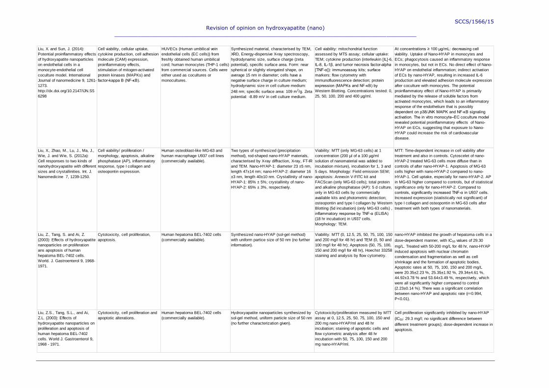
SCCS/1566/15
Revision of opinion on hydroxyapatite (nano)
___________________________________________________________________________________________
Liu, X. and Sun, J. (2014):
Potential proinflammatory effects
of hydroxyapatite nanoparticles
on endothelial cells in a
monocyte-endothelial cell
coculture model. International
Journal of nanomedicine 9, 1261-
1273.
http://dx.doi.org/10.2147/IJN.S5
6298
Cell viability, cellular uptake,
cytokine production, cell adhesion
molecule (CAM) expression,
proinflammatory effects,
stimulation of mitogen-activated
protein kinases (MAPKs) and
factor-kappa B (NF-κB).
HUVECs (Human umbilical vein
endothelial cells (EC cells)) from
freshly obtained human umbilical
cord; human monocytes (THP-1 cells)
from commercial sources. Cells were
either used as cocultures or
monocultures.
Synthesized material, characterised by TEM,
XRD, Energy-dispersive X-ray spectroscopy,
hydrodynamic size, surface charge (zeta
potential), specific surface area. Form: near
spherical or slightly elongated shape, on
average 15 nm in diameter; cells have a
negative surface charge in culture medium;
hydrodynamic size in cell culture medium:
248 nm; specific surface area: 109 m2/g. Zeta
potential: -8.89 mV in cell culture medium.
Cell viability: mitochondrial function
assessed by MTS assay; cellular uptake:
TEM; cytokine production (interleukin [IL]-6,
IL-8, IL-1β, and tumor necrosis factor-alpha
[TNF-α]): immunoassay kits; surface
markers: flow cytometry with
immunofluorescence detection; protein
expression (MAPKs and NF-κB) by
Western Blotting. Concentrations tested: 0,
25, 50, 100, 200 and 400 µg/ml.
At concentrations ≥ 100 μg/mL: decreasing cell
viability. Uptake of Nano-HYAP in monocytes and
ECs; phagocytosis caused an inflammatory response
in monocytes, but not in ECs. No direct effect of Nano-
HYAP on endothelial inflammation; indirect activation
of ECs by nano-HYAP, resulting in increased IL-6
production and elevated adhesion molecule expression
after coculture with monocytes. The potential
proinflammatory effect of Nano-HYAP is primarily
mediated by the release of soluble factors from
activated monocytes, which leads to an inflammatory
response of the endothelium that is possibly
dependent on p38/JNK MAPK and NF-κB signaling
activation. The in vitro monocyte–EC coculture model
revealed potential proinflammatory effects of Nano-
HYAP on ECs, suggesting that exposure to Nano-
HYAP could increase the risk of cardiovascular
disease.
Liu, X., Zhao, M., Lu, J., Ma, J.,
Wie, J. and Wie, S. (2012a):
Cell responses to two kinds of
nanohydroxyapatite with different
sizes and crystallinities. Int. J.
Nanomedicine 7, 1239-1250.
Cell viability/ proliferation /
morphology, apoptosis, alkaline
phosphatase (AP), inflammatory
response, type I collagen and
osteopontin expression.
Human osteoblast-like MG-63 and
human macrophage U937 cell lines
(commercially available).
Two types of synthesized (precipitation
method), rod-shaped nano-HYAP materials,
characterised by X-ray diffraction, X-ray, FT-IR
and TEM. Nano-HYAP-1: diameter 23 ±5 nm,
length 47±14 nm; nano-HYAP-2: diameter 16
±3 nm, length 40±10 nm. Crystallinity of nano-
HYAP-1: 85% ± 5%; crystallinity of nano-
HYAP-2: 65% ± 3%, respectively.
Viability: MTT (only MG-63 cells) at 1
concentration (200 µl of a 100 µg/ml
solution of nanomaterial was added to
incubation mixture), incubation for 1, 3 and
5 days. Morphology: Field emission SEM;
apoptosis: Annexin V-FITC kit and
FACScan (only MG-63 cells); total protein
and alkaline phosphatase (AP): 5 d culture,
only in MG-63 cells by commercially
available kits and photometric detection;
osteopontin and type I collagen by Western
Blotting (5d incubation) (only MG-63 cells) ,
inflammatory response by TNF-α (ELISA)
(18 hr incubation) in U937 cells.
Morphology: TEM.
MTT: Time-dependent increase in cell viability after
treatment and also in controls. Cytoscelet of nano-
HYAP-2 treated MG-63 cells more diffuse than in
controls of after nano-HYAP-1. Apoptosis of MG-63
cells higher with nano-HYAP-2 compared to nano-
HYAP-1. Cell uptake, especially for nano-HYAP-2. AP
in MG-63 higher compared to controls, but of statistical
significance only for nano-HYAP-2. Compared to
controls, significantly increased TNF-α in U937 cells.
Increased expression (statistically not significant) of
type I collagen and osteopontin in MG-63 cells after
treatment with both types of nanomaterials.
Liu, Z., Tang, S. and Ai, Z.
(2003): Effects of hydroxyapatite
nanoparticles on proliferation
ans apoptosis of human
hepatoma BEL-7402 cells.
World. J. Gastroenterol 9, 1968-
1971.
Cytotoxicity, cell proliferation,
apoptosis.
Human hepatoma BEL-7402 cells
(commercially available).
Synthesized nano-HYAP (sol-gel method)
with uniform partice size of 50 nm (no further
information).
Viability: MTT (0, 12.5, 25, 50, 75, 100, 150
and 200 mg/l for 48 hr) and TEM (0, 50 and
100 mg/l for 48 hr); Apoptosis (50, 75, 100,
150 and 200 mg/l for 48 hr), Hoechst 33258
staining and analysis by flow cytometry.
nano-HYAP inhibited the growth of hepatoma cells in a
dose-dependent manner, with IC50 values of 29.30
mg/L. Treated with 50-200 mg/L for 48 hr, nano-HYAP
induced apoptosis with nuclear chromatin
condensation and fragmentation as well as cell
shrinkage and the formation of apoptotic bodies.
Apoptotic rates at 50, 75, 100, 150 and 200 mg/L
were 20.35±2.23 %, 25.35±1.92 %, 29.34±4.61 %,
44.92±3.78 % and 53.64±3.49 %, respectively, which
were all significantly higher compared to control
(2.23±0.14 %). There was a significant correlation
between nano-HYAP and apoptotic rate (r=0.994,
P<0.01).
Liu, Z.S., Tang, S.L., and Ai,
Z.L. (2003): Effects of
hydroxyapatite nanoparticles on
proliferation and apoptosis of
human hepatoma BEL-7402
cells. World J. Gastroenterol 9,
1968 - 1971.
Cytotoxicity, cell proliferation and
apoptotic alterations.
Human hepatoma BEL-7402 cells
(commercially available).
Hydroxyapatite nanoparticles synthesized by
sol-gel method, uniform particle size of 50 nm
(no further characterization given).
Cytotoxicity/proliferation measured by MTT
assay at 0, 12.5, 25, 50, 75, 100, 150 and
200 mg nano-HYAP/ml and 48 hr
incubation; staining of apoptotic cells and
flow cytometric analysis after 48 hr
incubation with 50, 75, 100, 150 and 200
mg nano-HYAP/ml.
Cell proliferation significantly inhibited by nano-HYAP
(IC50: 29.3 mg/l; no significant difference between
different treatment groups); dose-dependent increase in
apoptosis.
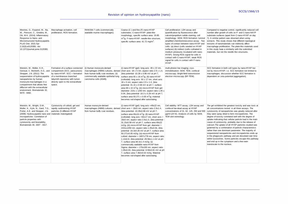
SCCS/1566/15
Revision of opinion on hydroxyapatite (nano)
___________________________________________________________________________________________
Mestres, G., Espanol, M., Xia,
W., Persson, C., Ginebra, M.,
Ott, M.K. (2014): Inflammatory
Response to Nano- and
Microstructured Hydroxyapatite.
PLoS One. 2015 Apr
2;10(3):e0120381. doi:
10.1371/journal.pone.0120381
Macrophage activation, cell
proliferation, ROS formation.
RAW264.7 cells (commercially
available murine macrophages).
Coarse (C ) and fine (F) nano-HYAP
substrates; C-nano-HYAP: plate-like
morphology, specific surface area: 19.85
m2/g. F-nano-HYAP: needle-like structure,
specific surface area: 41.72 mg/m2.
Cell proliferation: LDH assay and
quantification by fluorescence after
calcein/propidium iodide staining; cell
morphology: SEM; ROS formation: luminol
amplified chemiluminescence assay . Two
types of contact between nano-HYAP and
cells: (a) direct (cells seeded on HYAP
surfaces) (b) indirect (cells cultivated in
medium previously incubated with nano-
HYAP). Strong ROS signal for cells in
contact with C-nano-HYAP, weak ROS
signal for cells in contact with F-nano-
HYAP.
Compared to negative control, significantly reduced cell
number after growth of cells on F- and C-nano-HAYP
substrate surfaces (apart from C-nano-HYAP on day
7). A similar pattern was observed when using
extracts. This study shows that different topological
textures of nanomaterials can indirectly affect
macrophage proliferation. The plate-like materials used
in this study have a similarity with the submitted
materials, but not the needle like structures.
Motskin, M., Müller, K.H.,
Genoud, C. Monteith, A.G., and
Skepper, J.N. (2011): The
sequestration of hydroxyapatite
nanoparticles by human
monocyte-macrophages in a
compartment that allows free
diffucion with the extracellular
environment. Biomaterials 32,
9470 - 9482.
Formation of a surface connected
compartment (SCC, patocytosis)
by nano-HYAP. SCC = formation
of a membranous branched
labyrinth repository with lumen
directly open to the extracellular
space.
(i) Human monocyte-derived
macrophages (HMM) cultures, derived
from human buffy coat residues; (ii)
commercially available epithelial lung
carcinoma cells (A549).
(i) nano-HYAP (gel): long axis: 49 ± 22 nm,
short axis: 18 ± 5 nm; aspect ratio 2.9 ± 1.4,
Zeta potential -10.28 ± 0.86 mV at pH 7,
surface area 65 ± 16 m2/g; (II) nano-HYAP
(colloidal): long axis: 50 ± 17 nm, short axis:
18 ± 5 nm; aspect ratio 2.9 ± 1.0, Zeta
potential -31.15 ± 0.58 mV at pH 7, surface
area 69 ± 22 m2/g; (iii) micro-HYAP from gel:
diameter: 2151 ± 1250 nm; aspect ratio 1.05 ±
0.04, Zeta potential -16.3 ± 0.26 mV at pH 7,
surface area 93.271 ± 0.05 m2/g; material
becomes rod-shaped after autoclaving.
multi-photon live imaging; cryo-
immobilisation; SEM, TEM, confocal
microscopy; Bright-field transmission
electron microscopy (BF-TEM).
SCC formation in both cell types by nano-HYAP but
not by micro-HYAP; i.e. SCC formation not limited to
macrophages; discussion whether SCC formation is
dependent on zeta potential (aggregation).
Motskin, M., Wright, D.M.,
Muller, K., Kyle, N., Gard, T.G.,
Porter, A.E. and Skepper, J.N.
(2009): Hydroxyapatite nano and
microparticles: Correlation of
particle properties with
cytotoxicity and biostability.
Biomaterials 30, 3307 - 3317.
Cytotoxicity of colloid, gel and
rapidly sedimenting HYAP
particles (5 different HYAP
materials investigated)
Human monocyte-derived
macrophages (HMM) cultures, derived
from human buffy coat residues.
(i) nano-HYAP (gel): long axis =49±22 nm,
short axis = 18±5 nm; aspect ratio 2.9±1.4,
Zeta potential -10.28±0.86 mV at pH 7,
surface area 65±16 m2/g; (II) nano-HYAP
(colloidal): long axis =50±17 nm, short axis =
18±5 nm; aspect ratio 2.9±1.0, Zeta potential -
31.15±0.58 mV at pH 7, surface area 69±22
m2/g; (iii) micro-HYAP from gel: diameter =
2151±1250 nm; aspect ratio 1.05±0.04, Zeta
potential -16.3±0.26 mV at pH 7, surface area
93.271±0.05 m2/g; (iv) micro-HYAP from
colloid: diameter = 1657±778 nm; aspect ratio
1.12±0.8, Zeta potential -29.56±1.1 mV at pH
7, surface area 60.3±1.4 m2/g; (v)
commercially available nano-HYAP from
Sigma: diameter = 170±100 nm; aspect ratio
1.05±0.03, Zeta potential -9.59±0.81 mV at pH
7, surface area 7.362±0.02 m2/g. Material
becomes rod-shaped after autoclaving.
Cell viability: MTT assay, LDH assay and
confocal live-dead assay at HYAP
concentrations of 31, 62, 125, 250 and 500
µg/ml (24 hr). Analysis of cells by SEM,
TEM and stereology.
The gel exhibited the greatest toxicity and was toxic at
all concentrations tested in all three assays. The
cytotoxicity of nanoparticles was greatly reduced after
they were spray dried to form microparticles. The
degree of toxicity correlated well with the degree of
uptake indicating that cellular particle load is the main
cause of cytotoxicity, probably due to the release of
calcium.The uptake of all HYAP particles studied was
governed by a combination of particle characteristics
rather than one dominant parameter. The majority of
sequestered nanoparticles and microparticles ends up
in the phagocytic pathway and are dissolved over time
within lysosomes. Some particles escape this pathway
and end up in the cytoplasm and a few even
translocate to the nucleus.

SCCS/1566/15
Revision of opinion on hydroxyapatite (nano)
___________________________________________________________________________________________
Müller, K.H., Motskin, M.,
Philpott, A., Routh, A.,
Shanahan, C., Duer, M. and
Skepper, J. (2014): The effect of
particle agglomeration on the
formation of a specific surface-
connected compartment induced
by hydroxyapatite nanoparticles
in human monocyte-derived
macrophages. Biomaterials 35,
1074-1088.
Influence of particle agglomeration
on cellular uptake, cytotoxicity
and formation of a surface-
connected compartment (SCC).
In vitro cultures of mature human
macrophages (HMM) isolated from
human buffy coat residues.
5 different nano-HYAP materials (4
synthesized, 1 commercially available). All
synthesizds materials were fine needle-like to
plate-like and became longer, thicker and
more lozenge-shaped after autoclavation.
Characterized by BF-TEM (bright-field TEM),
BET, Zeta-potential (in the presence or
absence of the dispersant D7) , DLS, SEM.
Synthesized material: large, round particles.
Material 1: non-autoclaved, non citrated
(NANC): long axis: 30 ± 5 nm, short axis: 8 ±
2 nm; aspect ratio: 3.9 ± 1.1; zeta potential (-
D7): -17 ± 1.42 mV; (+D7): -36.11 ± 2.98 mV;
surface area: 121.4 ± 0.6 m2/g.
Material 2: non-autoclaved, citrated (NAC):
long axis: 27 ± 7 nm, short axis: 8 ± 2 nm;
aspect ratio: 3.6±1.2; zeta potential (-D7): -
14.48 ± 0.92 mV; (+D7): -24.08 ± 1.29 mV;
surface area: 156.4 ± 1.5 m2/g.
Material 3: autoclaved, non-citrated (ANC):
long axis: 54 ± 19 nm, short axis: 18 ± 5 nm;
aspect ratio: 3.2 ± 1.2; zeta potential (-D7): -
10.56 ± 0.84 mV; (+D7): -55.84 ± 1.2 mV;
surface area: 64.9 ± 0.35 m2/g.
Material 4: autoclaved, citrated (AC): long
axis: 46± 18 nm, short axis: 17± 5 nm;
aspect ratio: 32.8±0.95; zeta potential (-D7):
-24.29±0.75 mV; (+D7): -43.6±2.48 mV;
surface area: 52.8±0.3 m2/g. I.e. NAC and
AC as uncoated materials relevant
for the opinion.
Experiments were performed either in the
presence or in the absence of dispersant
D7. Cellular uptake: determination of
calcium in lysed cells by ICP-MS after 1 hr
incubation with 125 µg/ml nano-HYAP.
Cytotoxicity: MTT assay, concentrations: 0,
31.25, 62.5, 125, 250 and 500 µg/ml,
incubation time 24 hr. Agglomeration: DLS;
SCC formation after 2 hr incubation with 30,
60 or 125 µg/ml nanomaterial. Imaging: BF-
TEM.
In the absence of disperser, formation of large
agglomerates in NANC and ANC with NANC forming
larger agglomerates.Statistically significant reduction
in viability after NANC and ANC, citration and
dispersion reduced cytotoxicity. SCC formation:
Formation of large, interconnected vacuoles lining the
membrane and occupying larger parts of the cells with
increasing concentration; SCC open to extracellular
space. SCC formation/particle internalization reduced
by D7 or citration. Change in particle morphology in the
SCC (from needle to plate, considered as
sequestration). High cellular uptake of NANC and ANC
in contrast to citrated or dispersed materials.
Agglomerate dispersion prevented the SCC from
forming, but did not completely inhibit nanoparticle
uptake by other mechanisms.
Opačić-Galić, V., Petrović, V.,
Zivković, S., Jokanović, V.,
Nikolić, B., Knežević-Vukčević,
J. and Mitić-Ćulafić, D. (2013):
New nanostructural biomaterials
based on active silicate systems
and hydroxyapatite:
characterization and
genotoxicity in human peripheral
blood lymphocytes. Int. Endod.
J. 46, 506-516. doi:
10.1111/iej.12017
Cell viability and genotoxicity
(Comet Assay).
Human lymphocytes from 3 donors. Not given. The genotoxic potential of HA was tested in
the concentration range 0.05–50
mg/ml,using lymphocytes from three
donors.
Equivocal results were observed: after the treatment of
lymphocytes originating from donor 1, nano-HYAP
exhibited statistically significant genotoxicity (P <
0.05) at all tested concentrations in comparison with
negative control. However, nano-HYAP did not induce
DNA-damage in the two other donors; moreover, the
percentage of DNA damage in nano-HYAP-treated
cells was decreased compared to controls.
Palanivelu, R. and Kumar, A.
(2014): Synthesis,
characterization, in vitro anti-
proliferative and hemolytic
activity of hydroxyapatite.
Spectrochimica Acta Part A:
Molecular and Biomolecular
Spectroscopy 127, 434-438.
Viability, morphology, haemolytic
activity.
A549 cells for MTT assay; fresh
human red blood cells for haemolytic
activity (no information on donor(s)).
Synthesized nano-HYAP (precipitation
method), characterised by XRD, FT-IR, FT-
Raman spectroscopy, SEM and TEM.
According to SEM: Particles have uniform rod-
like structure with pores in between; size: 35 -
65 nm in length. According to TEM: size: 65 x
21 nm; agglomeration.
Viability: MTT assay (concentration range
100 - 1000 µg/ml, incubation time 24 hr).
Haemolytic assay: photometric assay
(concentrations: 50, 100, 150 and 200
µg/ml; incubation time 1 hr).
Dose-dependent increase in cell death and haemolytic
activity (no statistical analysis).

SCCS/1566/15
Revision of opinion on hydroxyapatite (nano)
___________________________________________________________________________________________
Qing, F., Wang, Z., Hong, Y.,
Liu, M., Guo, B., Luo, H. and
Zhang, X. (2012): Selective
effects of hydroxyapatite
nanoparticles on osteosarcoma
cells and osteoblasts. J. Mater.
Sci: Mater. Med. 23, 2245-2251.
Cell viability and apoptosis. Human osteosarcoma MG63 cells
(commercially available) and normal
human osteoblasts (from a 12 year
old female traumatic patient).
Synthesized nano-HYAP (hydrothermal
process); characterisation by XRD, TEM and
SEM. Nano-HYAP particles of spherical
shape and average particle size of about 40
nm.
Viability: MTT assay and TEM analysis
after fluorescent staining (concentrations:
100, 250 and 500 µg/ml; incubation time: 1,
3 and 5 days). Apoptosis: TEM.
MTT assay: inhibition of cell growth in MG63 cells;
stimulation of cell growth in osteoblasts. TEM: uptake
of nano-HYAP into MG63 cells, mostly in lysosomes,
indication of apoptosis, mitochondrial swelling,
extreme vacuolation at the highest dose tested (500
µg/ml); formation of myelinosomes; after 5 d: nano-
HYAP enters nucleus. No apoptotic changes observed
in osteoblasts, less changes in osteoblasts; nano-
HYAP entered cytoplasm and lysosomes. In
summary, nano-HYAP had different effects to the two
different kinds of cells investigated.
Remya, N.S., Syama, S.,
Gayathri, V., Varma, H.K. and
Mohanan, P.V. (2014): An in
vitro study on the interaction of
hydroxyapatite nanoparticles
and bone marrow mesenchymal
stem cells for assessing the
toxicological behaviour. Colloids
and Surfaces B: Biointerfaces
117, 389 - 397.
Cytotoxicity, Cellular uptake,
oxidative stress, apoptosis,
cytoskeletal arrangement.
Mesenchymal stem cells isolated
from pooled bone marrow of 6 mice
(no further information on animals
(e.g. sex or strain). Abbreviated:
BMSC cells.
Synthesized nano-HYAP (precipitation
method), characterised by XRD, FT-IR, TEM,
SEM, zeta potential and energy dispersive
spectroscopy (EDS). Nano-HYAP of rod-
shaped structure, size below 50 nm,
agglomeration abserved. Zeta-potential in
buffer: -14.4 mV (i.e. stable in buffer); Zeta
potential in water 3.19 mV (i.e. unstable in
water).
Cell viability: MTT assay and microscopic
evaluation, concentrations: 10, 50, 100, 500
and 1000 µg/ml; incubation time 24 hr.
Cellular uptake: light microscopy after
Giemsa staining and fluorescence staining
and confocal microscopy after incubation of
cells with fluorescence-labelled nano-HYAP
(concentration: 100 µg/ml; incubation time:
0, 2, 4, 12 and 24 hr). Apoptosis: Caspase
assay after overnight incubation with 10,
50, 100, 500 and 1000 µg/ml nano-HYAP
and annexin/propidium iodide staining and
flow cytometry after 24 hr incubation with
50, 100, 500, 800 and 1000 µg/ml nano-
HYAP; oxidative stress: formation of
reactive oxygen species by DCF-DA assay.
Cytotoxicity: impairment of cell viability at the highest
concentration; uptake of nano-HYAP into cells starting
at 2 hr; occurrence of nano-HYAP in cytoplasm, as
agglomerates/aggregates. Statistically significant
increase in ROS formation at the highest dose. No
indications of apoptosis by Caspase assay;
annexin/propidium iodide staining: presence of early
apoptotic cells after incubation with 50, 100 and 500
µg/ml, but not at higher concentrations. No late
apoptotic or necrotic cells at all concentrations tested,
i.e. no apoptosis.
Sánchez Lafarga, A., Pacheco
Moisés, F., Gurinov, A., Ortiz,
G., and Arízaga, G. (2015): Dual
responsive dysprosium-doped
hydroxyapatite particles and
toxicity reduction after
functionalization with folic and
glucuronic acids. Materials
Science and Engineering C 48,
541-547.
Ex vivo determination of different
biochemical parameters
(membrane fluidity, endproducts of
lipid peroxidation, nitrite and
nitrate values, glutathione
peroxidase activity, ATPase
activity).
Homogenates from kidneys, livers and
lungs of Sprague-Dawley rats
prepared 7 days after i.p. treatment
with 10 or 20 mg nano-HYAP
(untreated nano-HAYP, nano-HYAP
doped with dysprosium cations, nano-
HYAP functionalized with folic acid or
glucuronic acid); 4 animals per dose
group.
Only untreated nano-HYAP described here as
modified nano-HYAP does not belong to the
materials of the submission. Synthesized
material characterised by XRD,
thermogravimetric analysis, fluorescence. No
information on size given.
Membrane fluidity: fluorimetric method; end-
products of lipid peroxidation
(malondialdehyde (MDA) and 4-
hydroxyalkenals (4-OHA)): photometric
assay kit. Nitrite and nitrate levels: Griess
reaction; glutathione peroxidase activity:
photometric method. ATPase activity:
method not described.
Only untreated nano-HYAP results reported here.
Compared to untreated controls, dose-dependent
increase of products of lipid peroxidation in liver, kidney
and lung; significantly increased nitrate and nitrite
levels in kidneys, livers and lungs; significantly reduced
membrane fluidity in liver, kidney and lung; increase in
ATPase activity in liver, kidney and lung (statistically
significant after 20 mg in liver and after 10 and 20 mg in
kidneys); statistically significantly increased
glutathione peroxidase activity in erythrocytes from
rats treated with 20 mg nano-HYAP.
Shi, Z., Huang, X., Cai, Y.,
Tang, R., and Yang, D. (2009):
Size effect of hydroxyapatite
nanoparticles on proliferation
and apoptosis of osteoblast-like
cells. Acta Biomaterialia 5, 338-
345.
Cell proliferation and apoptosis. Human osteoblast-like MG-63 cells;
commercially available.
Synthesized nano-HYAP of 2 different sizes
characterised by TEM, XRD, DLS, field
electron scanning microscopy (FESEM), FT-
IR. Sphere-like nano-HYAP of 20 ± 5 nm
(np20) and 80 ± 12 nm (np80) diameter and
weak crystallinity; micro-sized HYAP used for
comparison.
Cell proliferation: acridine orange staining
and fluorescence microscopy; FESEM,
TEM; Apoptosis: propidium iodide staining
and FACScan; analysis of apoptosis-
related genes by RT-PCR. Analyses were
performed on glass films coated with 1%
suspension of HYAP materials; incubation
was up to 5 days.
Cell proliferation: compared to untreated controls,
proliferation was decreased with all HYAP materials
investigated; the most profound effect was observed
with microsized material. Morphological changes of
cells observed after treament with np80 and microsized
HYAP, but not with np20. Cellular uptake - on the other
hand - only observed with np20. Apoptosis: increased
rate of apoptosis with increasing particle size;
decreased Bcl-2 levels with increasing particle size.

SCCS/1566/15
Revision of opinion on hydroxyapatite (nano)
___________________________________________________________________________________________
Shi, Z., Huang, X., Liu, B. and
Tao, H. (2010): Biological
response of osteosarcoma cells
to size-controlled nanostructured
hydroxyapatite. Journal of
Biomaterials Applications 25, 19-
37.
Cell proliferation and apoptosis. Human osteoblast-like MG-63 cells;
commercially available.
Synthesized nano-HYAP of 2 different sizes
characterised by TEM, XRD, DLS, field
electron scanning microscopy (FESEM), FT-
IR. Sphere-like nano-HYAP of 20 ± 6 nm
(NanoHA-S) and 80 ± 14 nm (NanoHA-L)
diameter and weak crystallinity; micro-sized
HYAP used for comparison.
Cell proliferation: modified MTT assay and
acridine orange staining and fluorescence
microscopy. Apoptosis: Annexin V and
propidium iodide staining and FACScan;
Western Blot analysis of caspase-8 and
caspase-9. Analyses were performed on
glass films coated with 1% suspension of
HYAP materials; incubation was up to 5
days.
Cell proliferation: compared to untreated controls, cell
growth was reduced after nano-HYAP treatment.
Compared to controls, increased apoptosis rates
observed in particle treated cells (larger particles
induced higher apoptosis rates). Activation of
procaspase-9 into caspase-9 by nano-HYAP, but not
in untreated controls. Caspase-8 detected in none of
the samples.
Sun, J. and Ding, T. (2009a):
p53 Reaction to apoptosis
induced by hydroxyapatite
nanoparticles in rat
macrophages. J. Biomed. Mater.
Res. A. 88, 673-679. doi:
10.1002/jbm.a.31892
Cytotoxicity, Apoptosis,
Morphological changes.
Macrophages from male Sprague-
Dawley rats.
Commercially available nano-HAYP,
sonicated. Diameter between 30 and 80 nm
(no further information given).
Cytotoxicity: determined by response index
zone/lysis and microscopy; concentrations:
10, 20, 100 and 200 µg/ml, incubation time:
24 hr. Morphological changes determined
by TEM; Apoptosis analysed by p53
measurements using semiquantitative RT-
PCR and Western Blotting.
The results showed a dose-dependent proliferative
inhibition of macrophages by nano-HYAP at
concentrations between 20 and 200 µg/ml. The
characteristic morphological changes of apoptosis
were observed in macrophages after treatment with
nano-HYAP for 24 hr, p53 expression was induced
after treatment with nano-HYAP.
Sun, J. and Ding, T. (2009b):
Differences in DNA Damage
Pathways Induced by Two
Ceramic Nanoparticles. IEEE
Transactions on Nanobioscience
8, 78-82.
Expression of p53, p21 and heat
shock protein 70. Growth arrest,
DNA damage 45 (Gadd45).
Macrophages prepared from male
Sprague-Dawley rats
Commercially available nano-HAYP,
sonicated. Diameter between 30 and 80 nm
(no further information given).
RT-PCR after incubation of macrophages for
24 hr with 10, 20, 100 and 200 µg/ml nano-
HYAP.
Dose-dependent increase in p53 expression; dose-
dependent increase in p21 expression up to 100 µg/ml,
then decrease to negative control levels at 200 mg/ml;
no effect on Gadd45 expression; dose-dependent
increase in HSP70 expression.
Sun, W., Chu, C., Wang, J. and
Zhao, H. (2007): Comparison of
periodontal ligament cells
responses to dense and
nanophase hydroxyapatite. J.
Mater. Sci: Mater. Med. 18, 677-
683.
Cell proliferation and differentiation. Human periodontal ligament fibroblast-
like cells (PDLC) prepared from
patients undergoing surgical
extraction of teeth.
Synthesized nano-HYAP (sol-gel method),
characterised by TEM and energy-dispersive X-
ray (EDAX); particle size in average less than
50 nm, spontaneous congregation (no further
information on shape or size dimensions).
Commercially available micro-sized HYAP
was used for comparison.
Cell proliferation: incubation with 1 mg/ml
HYAP materials, cultivation for 5 days and
analysis by SEM, TEM and EDAX. MTT:
after cultivation for 4 days. Cell
differentiation: alkaline phosphatase activity
after cultivation for 5 and 8 days; flow
cytometric analysis after cultivation for 5
days.
Uptake of nano-HYAP into cells, but not of micro-sized
material. Alkaline phosphatase not statistically
significantly increased after nano-HYAP when
compared to micro-sized material and controls. Cell
proliferation increased by nano-HYAP.
Tang, W., Yuan, Y., Liu, C.,
Wu, Y., Lu, X. and Qian, J.
(2014): Differential cytotoxicity
and particle action of
hydroxyapatite nanoparticles in
human cancer cells.
Nanomedicine 9, 397-412.
Cytotoxicity, Apoptosis, cellular
uptake, oxidative stress.
Human gastric cancer cells (MGC80-
3); human cervical adenocarcinoma
cells (HeLa); human hepatocellular
carcinoma cells (HepG2); normal
human liver cells (L-02).
Synthesized (aqueous precipitation method)
nano-HYAP; characterized by TEM and XRD.
Rod-shaped; length x width 50 x 20 nm (no
further information).
FITC-labelled nano-HYAP prepared and used
for cellular uptake.
Cytotoxicity: MTT Assay ; concentrations:
62, 125, 250, 500, 750 and 1000 µg/ml;
incubation time: 24, 48 and 72 hr; nuclear
morphology: fluorescence microscopy after
treatment with 500 µg/ml nano-HYAP for 72
hr. Apoptosis: AnnexinV/propidium iodide
assay after treatment with 250 and 500
µg/ml for 48 and 72 hr; caspase 3, 8 and 9
determination by fluorimetric assay kits
after treatment with 250 or 500 µg/ml for 72
hr; oxidative stress: fluorescence assay
(DCFH-DA) for intracellular ROS and GSH
assay kit for intracellular GSH after
treatment with 250 or 500 µg/ml for 72 hr.
Cellular uptake: flow cytometry and
fluorescence microscopy of FITC-labelles
nano-HYAP; fluo-3 labelling for intracellular
calcium.
nano-HYAP significantly inhibited cell proliferation and
induced apoptosis in cancer cells with an order of
MGC80‑3 > HepG2 > HeLa, but had no impact on
normal hepatic cells (L‑02). The increase in apoptosis
was accompanied by activation of caspase 3 and 9,
but not caspase 8. nano-HYAP induced formation of
reactive oxygen species and decreased intracellular
glutathione in cancer cells (highest reactive oxygen
species burst in HeLa cells). No correlation observed
between degree of cytotoxicity and cellular uptake.
After 48 hr incubation, nano-HYAP was detected in all
three cancer cell lines both in cytoplasm and nucleus.
Highest uptake in nucleus found in NGC80-3 cells. In
normal cells, nano-HYAP was not located in nucleus.
An increase in the intracellular calcium level was
observed in all cells (highest level in MGC80‑3). The
study authors conclude that various types of cancer
cells react diffferently to nano-HYAP and they
hypothesize that elevated calcium concentration and
nuclear localization of the particles might be the main
mechanism of growth inhibition by nano-HYAP.

SCCS/1566/15
Revision of opinion on hydroxyapatite (nano)
___________________________________________________________________________________________
Tay, C., Cai, P., Setyawati, M.,
Fang, W., Tan, L., Hong, C.,
Chen, X., and Leong, D. (2014a):
Nanoparticles Strengthen
Intracellular Tension and Retard
Cellular Migration. Nano Lett.
14, 83-88.
Cell migration. TR146 oral mucosa cells. Commercial grade nano-HYAP (no information
on source), spherical shape, characterised by
TEM; primary particle diameter: 49 ± 14 nm;
hydrodynamic diameter in cell culture
medium: 236 ± 9 nm (i.e. agglomeration); Zeta
potential (determined in serum containing cell
culture medium): -6.5 ± 2 mV.
Combination of different culture experiments
and visualization techniques.
nano-HYAP inhibits cell migration; internalisation as
prerequisite for inhibition of migration; interference of
nano-HYAP with microtubule assembly.
Turkez, H., Yousef, M.I.,
Sönmez, E., Togar, B., Bakan,
F., Sozio, P. and Di Stefano, A.
(2014): Evaluation of cytotoxic,
oxidative stress and genotoxic
responses of hydroxyapatite
nanoparticles on human blood
cells. Journal of Applied
Toxicology 34, 373 - 379.
Cytotoxicity, Oxidative stress,
Genotoxicity.
Human peripheral blood lymphocyte
cultures prepared from blood samples
from 6 healthy, non-smoking men.
Sythesized needle-like nano-crystals; average
diameter 10 - 50 nm (TEM).
Cytotoxicity: MTT and LDH assay. Total
antioxidant capacity (TAC) and Total
oxidative stress (TOS) determined by
commercially available kits. Chromosome
aberration (CA), Micronucleus (MN)
formation (Cytochalasin B addition after 44
hr of incubation with nanomaterial), Sister
Chromatide-Exchange (SCE) for
assessment of genotoxicity. 8-OH-dG
formation determined by commercially
available kit. Concentrations tested: 5, 10,
20, 50, 75, 100, 150, 300, 500 and 1000
ppm; incubation time: 72 hr.
Cytotoxicity: statistically significantly decreased cell
viability at concentrations ≥ 150 ppm (MTT) and ≥ 300
ppm (LDH). Antioxidant capacity: change in TAC levels
at concentrations ≥ 150 ppm and in TOS levels at
concentrations ≥ 300 ppm. Increase in 8-OH-dG at
concentrations ≥ 300 ppm. Statistically significant
differences of SCE (at 150, 300 and 500 ppm), MN (at
500 ppm) and CA (at 300 and 500 ppm) compared to
control .
Wang, L., Zhou, G., Liu, H.,
Han, J., Zheng, L. and Fan, Y.
(2012): Nano-hydroxyapatite
particles induce apoptosis on
MC3T3-E1 cells and tissue cells
in SD rats. Nanoscale 4, 2894-
2899.
Cell proliferation and apoptosis. MC3T3-E1 cells (commercially
available osteoblast cell line).
Synthesized nano-HYAP (precipitation
method) characterised by TEM, XRD and FT-
IR. Shape: rod, mean size: 28.47 ±0.4 nm.
Cell proliferation: MTT assay,
concentrations: 0, 10, 100, 1000 and 10000
µg/ml, incubation time: 24 hr. Apoptosis:
TEM (concentrations unclear; incubation
time: 24 hr).
Dose-dependent and statistically significant decrease
in cell viability in the concentration range observed.
Apoptosis: after incubation with 10000 µg/ml (highest
dose), chromatin condensation, chromosomal DNA-
fragmentation, membrane shrinking and formation of
apoptotic bodies were observed.
Xu, J., Xu, P., Li, Z., Huang, J.
and Yang, Z. (2011): Oxidative
stress and apoptosis induced by
hydroxyapatite nanoparticles in
C6 cells. J. Biomed. Mater. Res.
A. 100, 738-745. doi:
10.1002/jbm.a.33270
Cytotoxicity, oxidative stress,
apoptosis.
C6 cells (commercially available rat
glioma cell line).
Synthesized nano-HYAP (precipitation
method); size: 50 - 80 nm (no further
information).
Cytotoxicity: MTT assay (concentrations:
10, 100, 250 and 500 µg/ml, incubation
time: 24, 36 and 48 hr). Apoptosis: nuclear
staining by Hoechst 33342 and by flow
cytometry after Annexin-V-FITC and
propidium iodide staining. Oxidative stress:
DCFH-DA-assay and T-SOD (superoxide
dismutase) assay.
Compared to untreated controls, concentration- and
time-dependent significant decreases in cell viabilities
which could be prevented by using a ROS scavenger.
Compared to controls, dose-dependent increase in
apoptosis which could be reduced by using a ROS
scavenger. Apoptosis was confirmed by morphology.
Concentration-dependent significant increase in ROS
formation by nano-HYAP. Dose-dependent decrease
(statistically significant from 100 µg/ml) in SOD.
Authors conclude that nano-HYAP reduces growth of
C6 cells and levels of SOD in these cells. It increases
ROS levels and induces apoptosis.

SCCS/1566/15
Revision of opinion on hydroxyapatite (nano)
___________________________________________________________________________________________
Xue, Y., Wu, J. and Sun, J.
(2012): Four types of inorganic
nanoparticles stimulate the
inflammatory reaction in brain
microglia and damage neurons
in vitro. Tox. Lett. 214, 91-98.
Release of proinflammatory
cytokines and chemokines.
Primary microglia cells prepared from
brains of Sprague-Dawley rats and
commercially available PC12 cell line.
Nano-HYAP obtained from a university,
characterized by TEM, zeta potential, XRD.
Needle-shaped material, length 60 nm,
diameter 12 nm, Zeta-potential (in medium)
17.8 mV
Microglia cells were incubated with 0.25
and 50 mg/ml nano-HYAP for 24 hr.
Supernatants were used to stimulate PC12
cells. Lipopolysaccharide was used as
positive control. In microglia, production of
nitrite was determined by photometry. Nitric
acid synthase, MCP-1 and MIP-1α were
determined by RT-PCR, nitric acid
synthase also by Western blotting; NF-kB
activation in microglia by electrophoretic
mobility shift assay; cytokines (TNF-α, IL-
1β and IL-6) in microglia supernatants were
determined by ELISA, cytotoxicity in PC12
cells after incubation with microglia
supernatant was determined by MTT assay.
Compared to negative controls, significantly increased
nitrite and nitric acid synthase. Significantly
upregulated MCP-1 and MIP-1α production. Increased
expression of NF-kB; significantly increased levels of
cytokines (TNF-α, IL-1β and IL-6). Effects in PC12
cells incubated with supernatants from nano-HYAP
treated microglia cells: significantly reduced viability.
Yin, M., Han, Y., Bauer, I.W.,
Chen, P. and Li, S. (2006):
Effect of hydroxyapatite
nanoparticles on the
ultrastructure and function of
hepatocellular carcinoma cells in
vitro. Biomed. Mater. 1, 38-41.
Cytotoxicity, cellular uptake and
morphology, cell cycle analysis
Bel-7402 human hepatocellular
carcinoma cells.
Synthesized nano-HYAP (precipitation
method), analyzed by TEM. Rod- and ball-
shaped, average size about 15 x 60 nm
(apparently superficial positive charge , see
section 3.2).
Cytotoxicity: MTT assay (concentrations: 0,
0.14, 0.28 and 0.56 mmol/l; incubation
time: 1, 2, 3, 4, 5, 6, 7 days). Cell
morphology: after incubation with 0.56
mmol/l. Apoptosis: propidium iodide
staining of 3, 4 and 5 day incubations.Cell
counts: all 1-5 day incubations. Influence of
a not further speciofied stabilizer on cellular
uptake was also investigated.
Cellular adherence affected by nano-HYAP due to
attachment of nano-HYAP on cell membrane followed
by internalisation (uptake in mambrane, endocytosis).
After 4 days, structural changes of cells (e.g.
mitochondrial vacuolization, swell of endoplasmatic
reticulum, nuclear blebs. Dose- and time-dependent
decrease in cell number and cell growth. Time-
dependent increase of cells in G1 phase versus
decrease of cels in S and G2/M phase. the authors
conclude that nano-HYAP easily enters human
hepatocellular carcinoma cells, change their
ultrastructure and suppress their proliferation.
Yuan, Y., Liu, C. , Qian, J.,
Wang, J. and Zhang, Y. (2010):
Size-mediated cytotoxicity and
apoptosis of hydroxyapatite
nanoparticles in human
hepatoma HepG2 cells.
Biomaterials 31, 730 - 740.
Cell viability, apoptosis, cellular
uptake
Human hepatocellular carcinoma
HepG2 cells and normal human liver L-
02 cells.
Synthezised nano-HYAP particles of different
size (26 nm; 45 nm, 78 nm, 175 nm) and FITC-
labelled nano-HYAP particles.
Characterization: X-ray diffraction, Dynamic
light Scattering, morphology analysis by
Nanoscope ØAFM. Particle shape only for 45
nm material explicitly mentioned: spherical.
Cellular uptake and nuclear morphology:
microscopy. Cell viability: MTT assay.
Apoptosis: FITC Annexin staining (flow
cytometry). Caspase activities
(fluorescence); PARP, pro-caspase 3, Bax,
Bcl-2, Bid and cytochrome c by Western
Blotting. Concentration: 50, 100 and 200
µg/ml; incubation time: 24, 48 and 72 hr.
Cell viability measured in both cell types; all
other tests performed in HepG2 cells only.
Cell viability decreases with decreasing particle size;
HepG2 cells more affected than L-02 cells. In HepG2
cells, apoptosis strongly depended on the size (45-
nm> 26-nm > 78-nm > 175-nm). Particles ranging from
20 - 80 nm in size activate caspase 3 and 9, decrease
Bcl-2 and increase levels of Bax, Bid and the release
of cytochrome c from mitochondria into cytoplasm,
with the best efficiency from 45-nm nano-HYAP.
Correlating the cellular response with the cellular
internalization, it can be inferred that the size of HYAP
and thereby the cellular localization had a predominant
effect on the HAYP-induced cytotoxicity, apoptotis,
and the levels of the apoptotic proteins in HepG2 cells.
Zhang, M., Liu, S., Xu, G., Fu, J.
and Zhang, D. (2014):
Cytotoxicity and apoptosis
induced by nanobacteria in
human breast cancer cells.
International Journal of
Nanomedicine 9, 265-271.
Cell viability, apoptosis. MDA-MB-231 human breast cancer
cell line (commercially available).
Commercially obtained nano-HYAP, particle
size determined by SEM revealed a size
range from 50 to 200 nm (no further
information on characterization).
Viability determined by CCK-8 kit, exposure
concentraions unclear, duration of
incubation: 72 hr. Apoptosis determined by
(FITC)-annexin V and propidium iodide
staining and flow cytometry. Cell
morphology investigated by phase contrast
and electron microsopy after 72 hr
incubation with nano-HYAP.
Uptake of nano-HYAP into cells and morphological
changes in cells. Compared to controls, significantly
decreased cell viability; induction of early apoptosis but
not of late apoptosis or necrosis. Mitochondrial
swelling.

SCCS/1566/15
Revision of opinion on hydroxyapatite (nano)
___________________________________________________________________________________________
Zhao, X., Heng, B.C., Xiong, S.,
Guo, J., Tan, T.T., Boey, F.Y.C.,
NG, K.W. and Loo, J.S.C.
(2011): In vitro assessment of
cellular responses to rod-shaped
hydroxyapatite nanoparticles of
varying lengths and surface
areas. Nanotoxicology 5, 182 -
194.
Cell viability, oxidative stress,
cellular uptake.
BEAS-2B (an immortalized human
bronchial epithelial cell line),
RAW264.7 (an immortalized mouse
monoyte/macrophage cell line) and
HepG2 cells.
Synthesized rod-shaped nano-HYAP of
different sizes; characterized by FT-IR, X-ray
diffraction, BET analysis, TEM, DLS.
Agglomeration observed in PBS, therefore
medium containing SHMP (sodium
hexametaphosphate) was used.
Characterisation: nHA60: average length 60 ±
10 nm; average width 25 ± 5 nm (TEM);
average size in 0.1% SHMP: 79 nm; zeta
potential -55.4 mV in SHMP and -0.4 mV in
water; specific surface area 47.02 cm2/g.
nHA120: average length 120 ± 15 nm; average
width 30 ± 5 nm (TEM); average size in 0.1%
SHMP: 96 nm; zeta potential -36.3 mV in
SHMP and -5.7 mV in water; specific surface
area 23.33cm2/g. nHA240: average length
240 ± 30 nm; average width 20 ± 5 nm (TEM);
average size in 0.1 % SHMP: 112 nm; zeta
potential -45.8 mV in SHMP and -5.3 mV in
water; specific surface area 46.12 cm2/g.
Cell viability: WST-8 assay, concentration
range: 10 - 300 µg/ml, incubation time: 24
hr. Intracellular ROS formation: DFDA
assay (a fluorescence method),
concentration range: 10 -300 µg/ml,
incubation time: 4 hr. Alizarin S-assay (for
calcium mineral histochemistry),
concentration: 100 µg/ml, incubation time:
4 hr.
Cell viability: compared to positive controls, only minor
impairment of cell viability in BEAS-2B and HepG2
cells, in RAW264.7 cells, viability decreased with
increasing concentrations of nano-HYAP reaching
statistical significance at 100 and 300 µg/ml (for all
sizes of nano-HYAP); in general, lower cell viabilities
with nHA20 compared to other sizes. ROS generation:
increased ROS in all three cell lines with all sizes of
nano-HYAP. Most significant increase in BEAS-2B
cells with nHA 240 at 30 and 100 µg/ml. In RAW264.7
and HepG2: most significant increase with nHA60 at
300 µl. Alizarin Red-S: compared to untreated controls:
increased staining for calcium in all three cell lines,
comparable staining for nHA 60 and nHA240, lower
staining for nHA120.
Zhao, X., Ng, S., Heng, B., Guo,
J., Ma, L., Tan, T., Ng, K. and
Loo, S. (2013): Cytotoxicity of
hydroxyapatite nanoparticles is
shape and cell dependent.
Arch.Toxicol. 87, 1037-1052.
Apoptosis, DNA-content,
Intracellular ROS generation,
Relase of IL-6 in BEAS-2B cells
and of TNF-α in RAW264 cells,
intracellular particle distribution,
particle-cell association.
RAW264.7 cells (commercially
available murine macrophages) and
BEAS-2B cells (commercially
available human bronchial epithelial
cells).
Synthesized nano-HYAP of four different
shapes (rod-like (RD), needle-shaped (DN),
spherical (SP) and plate-shaped (PL)),
characterised by BET, XRD, TEM, DLS, zeta
potential, FT-IR. Nano-HYAP-ND: average
size 74.9 nm (in 0.1 % SHMP), Zeta potential -
4.8 ± 0.3 mV (in deionised water), specific
surface area 74.88 cm2/g. Nano-HYAP-RD:
average size 96 nm (in 0.1 % SHMP), Zeta
potential -5.7 ± 0.6 mV (in deionised water),
specific surface area 23.33 cm2/g. Nano-
HYAP-SP: average size 92.8 nm (in 0.1 %
SHMP), Zeta potential -14.1 ± 0.3 mV (in
deionised water), specific surface area 32.823
cm2/g. nano-HYAP-PL: average size 108.7
nm (in 0.1 % SHMP), Zeta potential -9.7 ± 0.3
mV (in DI water), specific surface area 61.75
cm2/g.
DNA-content: pico-green assay after 24 hr
incubation in concentration range 10 - 300
µg/ml. Apoptosis/necrosis: Annexin-
V/propidium iodide staining and flow
cytometry/ FACScan after 24 hr incubation.
Intracellular ROS generation:
dihydrofluorescein diacetate (DFDA) assay
after 4 hr incubation and photometric
reading. Relase of IL-6 in BEAS-2B cells
and TNF-α in RAW264 cells: ELISA after
24 hr incubation in concentration range 10 -
300 µg/ml. Particle-cell association:
Alizarin red S staining after 4- and 24 hr
incubation with nano-HYAP.
Viability and DNA-content: compared to the positive
control nano-ZnO, only minor concentration-dependent
decreases in viability observed for all materials in
BEAS-2B cells, viability not changed in RAW264.7
cells. No significant ROS formation in both cell types;
highest cytotoxicity observed for needle- and plate-
shaped nano-HYAP. Cytokine production: induction of
IL-6 in BEAS-2B cells comparable to positive control
(Lipopolysaccharide); highest induction for needle- and
platelet shaped material. TNF-α in RAW264 cells:
dose-dependent increase, but no difference between
shapes. Cellular uptake: higher uptake in RAW264.7
cells, incubation-time dependent increase in uptake;
rod-shaped particles generally exhibited higher levels of
cell-particle association than rod-shaped material.
Conclusion: cytotoxicity of nano-HYAP first dependent
on cell type, then on shape. ROS generation in this
study most probably not the cause for cell death. Role
of inflammatory cytokines in cell death to be further
elucidated; particle agglomeration influences cellular
uptake.
Higher uptake does not necessarily lead to higher
cytotoxicity, as uptake was highest for rod-shaped
material.

SCCS/1566/15
Revision of opinion on hydroxyapatite (nano)
___________________________________________________________________________________________
Information on solvent used for determination of Zeta potential was given when available (it was not always given in the publications)
Abbreviations used in Table 2 (if not explained in Table 2):
BET: Brunauer, Emmett and Teller
EDX: Energy Dispersive X-Ray FT-IR: Fourier Transform Infrared Spectroscopy
HYAP: Hydroxyapatite IR: Infrared Spectroscopy
RT-PCR: Real-Time Polymerase Chain Reaction ROS: Reactive Oxygen Species
SEM: Scanning Electron Microscopy
SSA: Specific Surface Area TEM: Transmission Electron Microscopy


![Synthesis and Characterizations of Hydroxyapatite Derived ...ripublication.com/ijac16/ijacv12n4_04.pdf528 Bulkis Musa, Indah Raya and Hasnah Natsir granosa) [9,10]. Nano-sized hydroxyapatite](https://img.dokumen.tips/doc/110x75/5cad6ca688c99376788dc707/synthesis-and-characterizations-of-hydroxyapatite-derived-bulkis-musa-indah.jpg)