Embed Size (px)
Citation preview
CONCEPTS OF BIOLOGY
Chapter 16 THE BODY’S SYSTEMSPowerPoint Image Slideshow

FIGURE 16.1
An arctic fox is a complex animal, well adapted to its environment. (credit: Keith
Morehouse, USFWS)

FIGURE 16.2
The body is able to regulate temperature in response to signals from the nervous
system.

FIGURE 16.3
The human excretory system is made up of the kidneys, ureter, urinary bladder, and
urethra. The kidneys filter blood and form urine, which is stored in the bladder until it is
eliminated through the urethra. On the right, the internal structure of the kidney is
shown. (credit: modification of work by NCI, NIH)

FIGURE 16.4
The components of the human digestive
system are shown.

FIGURE 16.5
(a) Digestion of food begins in the mouth.
(b) Food is masticated by teeth and moistened by saliva secreted from the salivary glands. Enzymes in the saliva begin to digest starches and fats. With the help of the tongue, the resulting bolus is moved into the esophagus by swallowing. (credit: modification of work by Mariana Ruiz Villareal)

FIGURE 16.6
The large intestine reabsorbs water from undigested food and stores waste until it is
eliminated. (credit: modification of work by Mariana Ruiz Villareal)
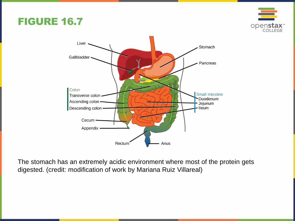
FIGURE 16.7
The stomach has an extremely acidic environment where most of the protein gets
digested. (credit: modification of work by Mariana Ruiz Villareal)

FIGURE 16.8
For humans, a balanced diet includes fruits, vegetables, grains, protein, and
dairy.(credit: USDA)

FIGURE 16.9
Air enters the respiratory system through
the nasal cavity, and then passes
through the pharynx and the trachea into
the lungs. (credit: modification of work by
NCI)

FIGURE 16.10
The heart is divided into four chambers, two atria, and two ventricles. Each chamber is
separated by one-way valves. The right side of the heart receives deoxygenated blood
from the body and pumps it to the lungs. The left side of the heart pumps blood to the
rest of the body.

FIGURE 16.11
In each cardiac cycle, a series of contractions (systoles) and relaxations (diastoles) pumps blood through the heart and through the body.
(a) During cardiac diastole, blood flows into the heart while all chambers are relaxed.
(b) Then the ventricles remain relaxed while atrial systole pushes blood into the ventricles.
(c) Once the atria relax again, ventricle systole pushes blood out of the heart.
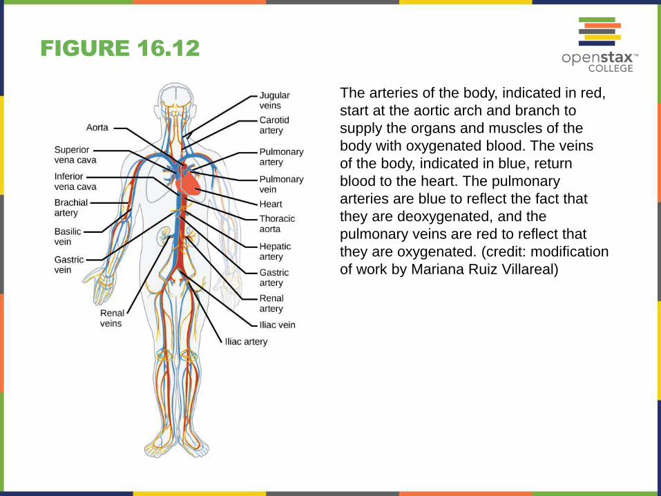
FIGURE 16.12
The arteries of the body, indicated in red,
start at the aortic arch and branch to
supply the organs and muscles of the
body with oxygenated blood. The veins
of the body, indicated in blue, return
blood to the heart. The pulmonary
arteries are blue to reflect the fact that
they are deoxygenated, and the
pulmonary veins are red to reflect that
they are oxygenated. (credit: modification
of work by Mariana Ruiz Villareal)
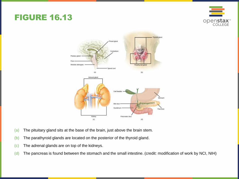
FIGURE 16.13
(a) The pituitary gland sits at the base of the brain, just above the brain stem.
(b) The parathyroid glands are located on the posterior of the thyroid gland.
(c) The adrenal glands are on top of the kidneys.
(d) The pancreas is found between the stomach and the small intestine. (credit: modification of work by NCI, NIH)

FIGURE 16.14
The anterior pituitary stimulates the
thyroid gland to release thyroid
hormones T3 and T4. Increasing levels of
these hormones in the blood result in
feedback to the hypothalamus and
anterior pituitary to inhibit further
signaling to the thyroid gland. (credit:
modification of work by Mikael
Häggström)
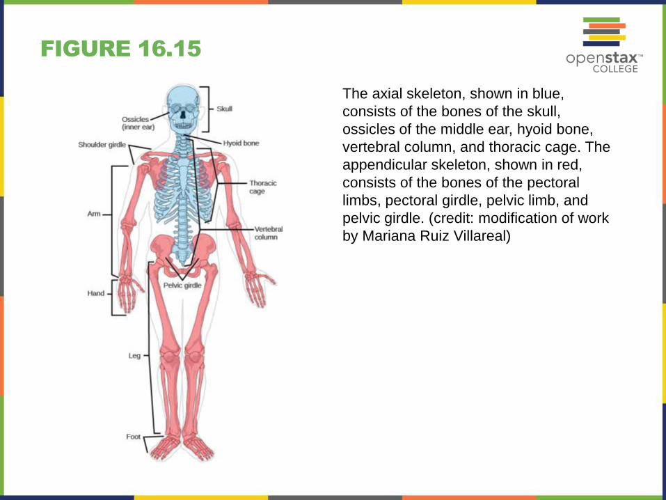
FIGURE 16.15
The axial skeleton, shown in blue,
consists of the bones of the skull,
ossicles of the middle ear, hyoid bone,
vertebral column, and thoracic cage. The
appendicular skeleton, shown in red,
consists of the bones of the pectoral
limbs, pectoral girdle, pelvic limb, and
pelvic girdle. (credit: modification of work
by Mariana Ruiz Villareal)

FIGURE 16.16
(a) Sutures are fibrous joints found only in the skull.
(b) Cartilaginous joints are bones connected by cartilage, such as between vertebrae.
(c) Synovial joints are the only joints that have a space or “synovial cavity” in the joint.

FIGURE 16.17
The body contains three types of muscle tissue: skeletal muscle, smooth muscle, and cardiac muscle. Notice that skeletal muscle cells are long and cylindrical, they have multiple nuclei, and the small, dark nuclei are pushed to the periphery of the cell. Smooth muscle cells are short, tapered at each end, and have only one nucleus each. Cardiac muscle cells are also cylindrical, but short. The cytoplasm may branch, and they have one or two nuclei in the center of the cell. (credit: modification of work by NCI, NIH; scale-bar data from Matt Russell)

FIGURE 16.18
A skeletal muscle fiber is surrounded by a plasma membrane called the sarcolemma,
with a cytoplasm called the sarcoplasm. A muscle fiber is composed of many fibrils
packaged into orderly units. The orderly arrangement of the proteins in each unit,
shown as red and blue lines, gives the cell its striated appearance.

FIGURE 16.19
Neurons contain organelles common to other cells, such as a nucleus and
mitochondria. They also have more specialized structures, including dendrites and
axons.
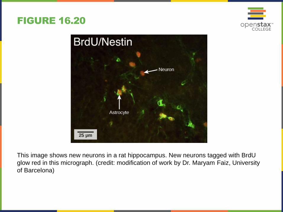
FIGURE 16.20
This image shows new neurons in a rat hippocampus. New neurons tagged with BrdU
glow red in this micrograph. (credit: modification of work by Dr. Maryam Faiz, University
of Barcelona)

FIGURE 16.21
The cerebral cortex is covered by three layers of meninges: the dura, arachnoid, and
pia maters. (credit: modification of work by Gray’s Anatomy)

FIGURE 16.22
The human cerebral cortex includes the frontal, parietal, temporal, and occipital lobes.

FIGURE 16.23
A cross-section of the spinal cord shows gray matter (containing cell bodies and
interneurons) and white matter (containing myelinated axons).

FIGURE 16.24
In the autonomic nervous system, a
preganglionic neuron (originating in the
CNS) synapses to a neuron in a ganglion
that, in turn, synapses on a target organ.
Activation of the sympathetic nervous
system causes release of norepinephrine
on the target organ. Activation of the
parasympathetic nervous system causes
release of acetylcholine on the target
organ.

FIGURE 16.25
The sympathetic and parasympathetic
nervous systems often have opposing
effects on target organs.

This PowerPoint file is copyright 2011-2013, Rice University. All
Rights Reserved.