Embed Size (px)
Citation preview

OPEN ACCESS ATLAS OF OTOLARYNGOLOGY HEAD AND
NECK OPERATIVE SURGERY
SURGICAL MANAGEMENT OF SKIN CANCERS OF THE HEAD AND NECK
André Bandiera de Oliveira Santos, Claudio Roberto Cernea
Skin cancer is the most common cancer
among Caucasians. The most common
histological types are basal cell carcinoma
(BCC), squamous cell carcinoma (SCC)
and melanoma. Head and Neck Surgery
(HNS) has its role in the treatment of
advanced cases of the most frequent types
and in the treatment of rare tumors.1
BCC
The incidence of BCC is the highest of all
malignant tumors. However, it is difficult
to quantify, as notification is not manda-
tory in most countries. It is estimated to
have an incidence between 500 and 1000
cases / 100,000 inhabitants, i.e. at least 100
times higher than the incidence of oral
cancer in the United States. 2
There are several treatment modalities with
cure rates of the order of 95%. Eighty
percent of BCCs are of the head and neck.
Curettage, cryotherapy, electrocoagulation,
photodynamic therapy and topical chemo-
therapy can be performed with high suc-
cess rates, especially in early cases. The
most commonly used treatment is conven-
tional surgical excision.3
The small number of cases referred for
surgery are advanced, sometimes due to
the patient's neglect of the tumour,
difficulty in accessing specialised care, and
failure of previous treatments. The most
common site is the nose, and there are
reports of younger mean age for patients
treated by head and neck surgery specia-
lists, when compared to those treated by
dermatologists.1,4
There are at least 8 histological subtypes of
BCC: nodular, superficial, morpheaform
(sclerodermiform), metatypical, micronod-
ular, sclerosing, pigmented, and adenoid.
The occurrence of more than one subtype
in a single tumour is common. The nodular
and superficial subtypes are the most
prevalent and the least aggressive, with a
lower probability of relapse. The subtypes
micronodular, metatypical and morphea-
form present more aggressive charac-
teristics and are more likely to relapse.5
Clinically, nodular BCC presents as an
elevated, nodular lesion, with well-defined
borders, a reddish-brown, pearly, shiny
colour and with teleangiectases on the
borders, sometimes ulcerated. The superfi-
cial BCC has the same characteristics
without the appearance of a nodule. Much
less characteristic, the morpheaform sub-
type presents as a reddish, brown or even
whitish plaque, resembling sclerodermia,
with a poorly defined border. This charac-
teristic is due to the presence of collage-
nase at the extremities of the tumours
which may present with finger-like exten-
sions, some-times called pseudopods. 4 In
these cases, the margins of surgical
resection are uncertain and should be
confirmed by frozen section examination
until complete resection of the lesion. Most
of the time the diagnosis is made only after
biopsy and the professional can only then
plan the surgery.6
In cases of locally advanced tumors, there
is a need for histopathological confirma-
tion with incisional biopsy by an experien-
ced pathologist, to determine a diagnosis
and permit the correct therapeutic plan-
ning. The clinical characteristics described
above do not always apply to large lesions,
since there is often significant tissue
destruction and loss of tissue differentia-
tion.7

2
For surgical planning, computed tomogra-
phy with contrast should be performed in
cases where there is suspicion of invasion
of deep planes, especially bony, cartilagi-
nous and parotid. In cases of suspected
invasion of the skull base, MRI should be
requested.8
The location of a BCC is important
because of the likelihood of recurrence.
High risk areas are nose, eyelids, periorbi-
ta, periorally, chin, mandibular, preauricu-
lar, postauricular, ear and temporal. Fac-
tors such as immunosuppression and pre-
vious radiotherapy also increase the chance
of relapse, as well as the occurrence of
scleroderma, metatypic or micronodular
subtypes at any point of the tumour.9
Almost all of the tumours treated by head
and surgeons can be classified into the
high-risk group. For these lesions, excision
with circumferential and deep free margins
confirmed by frozen section is recommen-
ded. A 1cm margin is desirable, but is not
always possible, especially on the face.
Elective neck dissection is not indicated.
We observed an incidence of less than 1%
of cervical metastases, which is therefore a
rare occurrence.10
Radiotherapy may be indicated in surgical
cases with compromised margins where
reoperation is not an option, major bone
resections and invasion of the skull base.
There is a good response of BCC to
radiotherapy in the rare cases where
surgery is not the treatment of choice.11
Inhibitors of the Hedgehog pathway (HH),
such as Vismodegib, are oral chemothe-
rapy options for advanced and irresectable
cases. Well-tolerated, the medication
presents objective results in selected cases,
at a high cost. 12
SCC
SCC is derived from the spinous layer of
the epidermis and accounts for 20% of
malignant skin tumours. It has a metastatic
potential of around 10%. The precusor
lesion is actinic keratosis. One of the
lesions that presents a difficult differential
diagnosis is the keratoacanthoma, a benign
lesion with fast growth.2
Clinically, cutaneous squamous cell carci-
noma may appear as a plaque or a papule.
Bowen's disease and Queyrat's erythro-
plasia are different forms of SCC in situ.
The invasive form is firm and often ulcer-
ated. Although the majority of cases
referred to the head and neck surgeon
already has diagnosed by biopsy, it is
preferable that the biopsy be reviewed or
repeated in doubtful cases.
The risk factors for recurrence in ECC are:
• Location in zone "H"
• Immunosuppression
• Previous radiotherapy
• Recurrence in an operated site
• Poorly defined borders
• Rapid growth
• Neurological symptoms
• Little or moderately differentiated
• Acantholytic, adenosquamous, meta-
plastic or desmoplastic subtypes
• Invasion level corresponding to Clark
IV or V or Breslow> 4mm
• Vascular or perineural invasion
Classically, elective neck dissection is not
indicated for cutaneous SCC. The search
for sentinel lymph nodes in high-risk SCC
is not routine and its benefit is
questionable. With cervical metastases,
neck dissection should be comprehensive;
and with parotid metastasis, at least a
supraomohyoid neck dissection should be
done, in addition to parotidectomy. 13

3
Radiotherapy can be used as primary treat-
ment in nonsurgical cases. As adjuvant
treatment, it is beneficial in cases of
cervical metastasis revealed by neck
dissection or parotidectomy, especially if
there is extracapsular extension. Other
indications are involved margins and
perineural invasion.11
TNM Staging for Nonmelanoma Skin
Cancer (2017)
T1 < 2cm
T2 > 2 cm e <4 cm
T3 ≥4 cm, deep invasion (> 6mm or beyond
subcutaneous), bone erosion or perineural
invasion
T4 T4a: Gross invasion of cortical bone or
marrow
T4b: Invasion of the base of the skull or
foramina of the base of the skull
N1 ipsilateral lymph node <3cm, without
extracapsular extension
N2a ipsilateral lymph node between 3 and 6 cm,
without extracapsular extension
N2b more than 1 lymph node, ipsilateral, <6cm,
without extracapsular extension
N2c Contralateral or bilateral lymph node(s)
<6cm, without extracapsular extension
N3 N3a: lymph node ≥ 6 cm, without
extracapsular extension
N3b: lymph node (s) with extracapsular
extension
Surgical treatment of BCC and SCC
Resection of high-risk head and neck
tumours should be planned with 10mm
circumferential and deep free margins.
Often the planning of this type of resection
involves exenteration of the orbit, resection
of bone, by a multimultidisciplinary team
to permit large surgical approaches (Figure
1).
Resulting deformity requires a common
sense approach by the surgeon and a frank
discussion with the patient. 14 Curative
treatment is not always the best option and
palliative alternatives should be consider-
ed.
With surgical planning, involvement of the
surgical teams from other specialties such
as plastic surgery, neurosurgery and oto-
rhinolaryngology is required to optimise
surgical outcome and functional rehabilita-
tion (Figure 2).
b
a a

4
Figure 1 a-e: Recurrent BCC of glabella
with invasion of glabella, orbit and skull
base
c
d
e
a
5
Figure 2 a-c: Patient with SCC of eyelid
with orbital and skull base invasion,
reconstructed with anterolateral thigh flap
With the patient anaesthetised, the borders
of the tumour including the peripheral rim
of erythema should be carefully marked.
The margin should be drawn with a
surgical pen or other marker before
infiltration of local anaesthetic. Drawing
the specimen and the defect for the
pathologist is desirable and assists in
describing the margins (Figure 3). Infiltra-
tion with vasoconstrictor-containing solu-
tion can be employed as it reduces the need
for electrocoagulation of the margins,
contributing to better histopathological
analysis. To remove the tumour, a double-
bladed scalpel (1mm distance between the
blades) 15 may be used, which can be made
by attaching a second blade with sterile
adhesive tape.
b
c
a
b

6
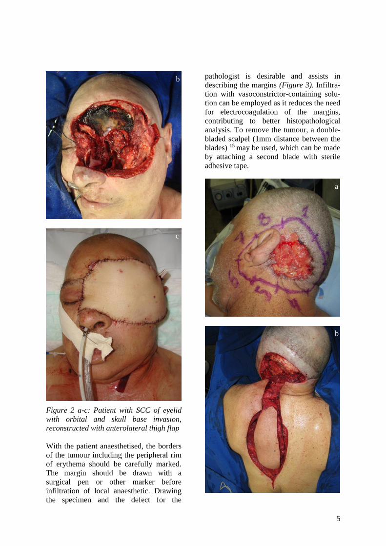
Figure 3 a-c: Retro-auricular SCC recon-
structed with a trapezus flap
This technique allows the removal of
margins of skin that can be immediately
taken to the pathologist, even prior to
removal of the tumour. After removal of
the lesion, the deep margin should be
assessed, if possible also with frozen
section.
Melanoma
Melanocytes are cells originating from the
neural crest that occupy the epidermis in
its basal layer. In part because of their
distinct embryological origin, melanomas
have very pronounced metastatic potential.
Although it accounts for 4% of skin
tumours, melanoma is the most lethal skin
tumour. Its prognosis is related to tumour
thickness from the granulosa layer to the
deepest part of the primary lesion (Breslow
index). The higher the Breslow index in
millimeters, the greater the chance of lym-
phatic and distant metastases, especially
above 1mm.16
Clinically, any melanocytic lesion that
changes its growth characteristics, changes
colour or has pruritus should be suspected.
The ABCDE rule exemplifies such
characteristics - Asymmetry, irregular
Borders, multiple Colours, Diameter
>6mm and Evolution. The diagnosis of
melanoma is made with a biopsy,
preferably excisional, or incisional in cases
where the resulting defect is unacceptable.
Most of the time the head and neck sur-
geon receives the diagnosis of melanoma
following a prior biopsy. The treatment to
be followed depends fundamentally on the
Breslow index.17 It is necessary to resect a
margin of macroscopic normal skin around
the biopsy scar, according to Breslow
index below:
Breslow (mm) Margin (cm)
In situ 0,5
< 1 1
1-2 1 a 2
2-4 2
>4 2 or greater
Wide excision is the main treatment in
patients without metastases. Treatment of
the lymphatics depends on sentinel lymph
node screening.
Sentinel lymph node biopsy is indicated in
all patients with melanoma with a Breslow
index above 1 mm without detectable
metastases. Cases with Breslow between
0.75mm and 1mm should indicate if there
is ulceration or a mitotic index other than
zero.
On the day prior to surgery, the patient
should have lymphoscintigraphy with tech-
netium. The technetium is injected around
the scar and the image obtained locates the
first lymph node that drains the region. The
nuclear physician then marks the patient's
skin overlying the lymph node confirmed
by the probe (probe attached to the
c

7
measuring device). After anesthesia, the
patent blue dye is injected (patent blue dye
is permeable to lymphatic vessels, unlike
methylene blue and other dyes). The
recommended volume is 1mL, preferably
in insulin syringe, but it is not always
possible to inject all the contents, and there
is the risk of permanently "tattooing" the
skin in regions such as the face. For best
results, the injection should be performed
intradermally - below this level the
drainage is impaired. Leaks from the
syringe should be carefully avoided to
avoid compromising the procedure.
Close proximity of the lymph node and the
primary lesion (rarely with lesions outside
the head and neck) may impair the investi-
gation when using the probe. In some
cases, margin resection may be necessary
prior to sentinel lymph node biopsy. Intra-
operative frozen section examination of
sentinel lymph nodes is not currently
recommended, since it may impair the
study of the whole lymph node with hema-
toxylin-eosin and immunohistochemical
study (HMB 45, melan A and protein
S100), which is best performed in a block
of paraffin.
Classically, the finding of a positive cervi-
cal sentinel lymph node is an indication for
neck dissection of all 5 levels of the neck.
Recently, in a prospective, randomised
study, survival proved to be equal between
serial ultrasound observation and elective
neck dissection in cases with positive
sentinel nodes, 18 and continues to be a
subject of debate today. 17
With parotid lymph nodes, parotidectomy
and supraomohyoid neck dissection should
be performed. Much controversy exists
about what to do in relation to the facial
nerve with intraparotid sentinel lymph
nodes - due to the need for reoperation if
the node is positive. We perform super-
ficial parotidectomy if nerve dissection is
required, which has been shown to be rare
- in most cases the lymph nodes are close
to the parotid capsule and can be safely
biopsied without nerve dissection.
In cases of cervical macrometastases, neck
dissection is performed according to the
standard procedure for squamous cell
carcinoma of the mucosa of the head and
neck. Preservation of structures not affec-
ted by metastases (modified radical cervi-
cal dissection) may be performed. Loco-
regional control is important in melanoma
and the presence of distant metastases does
not contraindicate neck dissection by itself.
Selected cases also benefit from metasta-
sectomy in extra-cervical sites. The beha-
viour of distant metastases may be
aggressive, but is not infrequently slow
and indolent. 17
After neck dissection for macro-metas-
tases, complementary cervical radiotherapy
is indicated. This approach is well accep-
ted for extracapsular extension, but some
studies have shown better control also for
macro-metastases. 17
Staging
T Breslow
(mm)
T1 ≤ 1.0
A: 0- 0.8mm without ulceration
B: 0.8-1mm or <0.8 with
ulceration
T2 1.01-2.0 A – With ulceration
B – Without ulceration T3 2.01-4.0
T4 > 4.0
Number of metastatic lymph nodes
Number of
metastatic lymph
nodes
Metastatic lymph node
mass
N1
1 lymph node
0 lymph node
A: micrometastasis *
B: macrometastasis†
C: metastasis or in transit
without metastatic lymph
nodes

8
N2
2-3 lymph nodes
1 lymph node
A: micrometastasis*
B: macrometastasis†
C: metastasis or in transit
without metastatic lymph
nodes
N3
4 or more
2 or more
A: positive sentinel lymph
nodes
B: 4 or more, at least 1
clinically detectable or
any number of coalescing
lymph nodes
C: two or more, with
satellite metastasis or
associated in transit
Local DHL sérica
M1a Skin distant,
subcutaneous, or lymph
node metastasis
M1b Pulmonary metastasis (0) DHL normal
M1c All other visceral
metastases
(1) DHL elevated
M1d Metastasis to Central
Nervous System
* diagnosed after sentinel lymph node or elective
lymphadenectomy
† defined as metastatic lymph node clinically
confirmed by therapeutic lymphadenectomy or
when lymph node metastasis exhibits gross
extracapsular extravasation
Levels of histological invasion (Clark)
I in situ. Entire lesion is intraepidermal II Beyond basement membrane. Occupies
papillary dermis, does not reach the border
between papillary and reticular III Occupies papillary dermis; reaches border
between papillary dermis and reticular,
without invading the reticular IV Includes reticular dermis V invades hypodermis
Clinical stages of melanoma: UICC 2017
Melanomas with Breslow > 4mm present a
>40% chance of distant metastasis. They
are sometimes mistakenly thought to have
such a bad outcome that it would not be
worthy resecting an additional margin and
treating the cervical nodes. Such a practice
is unfortunately common and worsens the
prognosis. Even in advanced cases, surgi-
cal treatment should always be considered,
since local control is very important for
treatment.
Surgery according to tumor location
The nose is the site of highest incidence of
skin tumours on the face. Simple suturing
is usually preferred when possible. Nasal
tip defects smaller than 1 cm without carti-
lage invasion can be reconstructed with a
bilobed flap from the nose itself (Figure
4).
a
b

9
Figures 4 a-d: Patient of 4 years with
congenital nevus reconstructed with
bilobed flap
One option is the nasal sliding flap, using
part of the glabella (Figure 5). Small nasal
wing defects can be reconstructed with a
superior or inferior pedicled nasogenial
flap.
Small nasal dorsal defects can be recon-
structed with a glabellar flap. With large
resections of nasal skin without removal of
cartilage, the frontal (or Indian) flap is a
good option, requiring two surgical visits,
the second time being scheduled three to
four weeks later with transection of the
pedicle. Removing nasal cartilage implies
a greater complexity of reconstruction,
which should include the nasal lining and
possibly using auricular cartilage to
replace a nasal wing.
Figure 5 a-b: Patient with melanoma of
nasal tip, Breslow 1,3mm, reconstructed
with nasal flap
With total or near total rhinectomy, nasal
reconstruction should be an exception, as
keeping the defect open may be the best
option, being careful not to keep the bone
exposed (often it is necessary to remove
part of the nasal bone) and posterior
reconstruction with a prosthesis.
c
d
a
b

10
With the scalp, the thickness of the prima-
ry lesion and its extension into the pericra-
nium is important. If the pericranium can
be preserved, skin grafting is a good option
for most cases. However, when there is
invasion and hence resection of pericra-
nium, a scalp flap is needed to cover the
exposed bone and a skin graft for the donor
area.
In the malar region a number of recon-
struction options may be considered. The
Mustardé flap is a good option, especially
for defects near the lower eyelid (Figure
6). Large bilobed or rhomboid flaps can
also be used, always taking care to avoid
ectropion due to tension on the lower
eyelid.
Figures 6 a-c: Patient with eyelid mela-
noma, Breslow 1mm, reconstruction with
Mustardé flap
Figure 7 is an example of lip reconstruc-
tion with V-Y flaps bilaterally. The frontal
and temporal regions are regions where
skin grafting lends itself well, with
aesthetic and functional results superior to
grafting on other parts of the face and
neck. Still, flap coverage should be
preferred whenever possible.
a
b
a
c

11
Figures 7 a,b: Resection of SCC of the
upper lip, reconstruction with bilateral V-Y
flap
A defect arising from orbital exenteration
may be skin grafted directly or onto a
temporalis muscle flap in order to better
adapt the prosthesis
References
1. Andrade N, Santos A, Lourenco S, esta
Neto C, Cernea C, Brandao L.
Epidemiological and histopathological
profile of 642 cases of basal cell
carcinoma of head and neck surgery in
a tertiary institution Sao Paulo: Rev.
Bras. Cir. Cab. Pesc.; 2011:148-53
2. Madan V, Lear JT, Szeimies RM. Non-
melanoma skin cancer. Lancet
2010;375:673-85
3. Zou Y, Zhao Y, Yu J, et al. Photodyna-
mic therapy versus surgical excision to
basal cell carcinoma: meta-analysis. J
Cosmet Dermatol 2016;15:374-82
4. Rubin AI, Chen EH, Ratner D. Current
concepts - Basal-cell carcinoma. N
Engl J Med 2005;353:2262-9
5. Sexton M, Jones DB, Maloney ME.
Histologic pattern analysis of basal cell
carcinoma. Study of a series of 1039
consecutive neoplasms. J Am Acad
Dermatol 1990;23:1118-26
6. Cernea CR, Dias FL, Lima RA, et al.
Atypical facial access: an unusually
high prevalence of use among patients
with skull base tumors treated at 2
centers. Arch Otolaryngol Head Neck
Surg 2007;133:816-9
7. Costantino D, Lowe L, Brown DL.
Basosquamous carcinoma-an under-
recognized, high-risk cutaneous neo-
plasm: case study and review of the
literature. J Plast Reconstr Aesthet
Surg 2006;59:424-8
8. Williams LS, Mancuso AA, Menden-
hall WM. Perineural spread of cuta-
neous squamous and basal cell
carcinoma: CT and MR detection and
its impact on patient management and
prognosis. Int J Radiat Oncol Biol
Phys 2001;49:1061-9
9. Bastiaens MT, Hoefnagel JJ, Bruijn
JA, Westendorp RG, Vermeer BJ,
Bouwes Bavinck JN. Differences in
age, site distribution, and sex between
nodular and superficial basal cell carci-
noma indicate different types of
tumors. J Invest Dermatol 1998;110:
880-4
10. Santos AB, Andrade NM, Brandão LG,
Cernea CR. Which features of advan-
ced head and neck basal cell carcinoma
are associated with perineural inva-
sion? Braz J Otorhinolaryngol 2017;
83:94-7
11. Mendenhall WM, Ferlito A, Takes RP,
et al. Cutaneous head and neck basal
and squamous cell carcinomas with
perineural invasion. Oral Oncol
2012;48:918-22
12. Sekulic A, Migden MR, Basset-Seguin
N, et al. Long-term safety and efficacy
of vismodegib in patients with
advanced basal cell carcinoma: final
update of the pivotal ERIVANCE BCC
study. BMC Cancer 2017;17:332
13. O'Hara J, Ferlito A, Takes RP, et al.
Cutaneous squamous cell carcinoma of
the head and neck metastasizing to the
parotid gland - a review of current
b

12
recommendations. Head Neck 2011;33:
1789-95
14. Cernea CR, Ferraz AR, de Castro IV,
et al. Perineural Invasion in Aggressive
Skin Carcinomas of the Head and
Neck. Orl-J Oto-Rhino-Laryngology
and Its Related Specialties 2009;71:21-
6
15. Cernea CR, Velasco O, Gomes MQ, et
al. Double-bladed scalpel: a new option
for harvesting margins in head and
neck cancers. ORL J Otorhinolaryngol
Relat Spec 2006;68:83-7
16. Balch CM, Gershenwald JE, Soong SJ,
et al. Final version of 2009 AJCC
melanoma staging and classification. J
Clin Oncol 2009;27:6199-206
17. Coit DG, Thompson JA, Algazi A, et
al. Melanoma, Version 2.2016, NCCN
Clinical Practice Guidelines in Oncolo-
gy. J Natl Compr Canc Netw 2016;14:
450-73
18. Faries MB, Thompson JF, Cochran AJ,
et al. Completion Dissection or Obser-
vation for Sentinel-Node Metastasis in
Melanoma. N Engl J Med 2017;376:
2211-22
Authors
André Bandiera de Oliveira Santos
Head and Neck Surgeon,
Faculty of Medicine
University of São Paulo
Brazil
Claudio Roberto Cernea
Professor
Faculty of Medicine
University of São Paulo
Brazil
Editor
Johan Fagan MBChB, FCORL, MMed
Professor and Chairman
Division of Otolaryngology
University of Cape Town
Cape Town, South Africa
THE OPEN ACCESS ATLAS OF
OTOLARYNGOLOGY, HEAD &
NECK OPERATIVE SURGERY www.entdev.uct.ac.za
The Open Access Atlas of Otolaryngology, Head & Neck Operative Surgery by Johan Fagan (Editor) [email protected] is licensed under a Creative Commons Attribution - Non-Commercial 3.0 Unported License