Embed Size (px)
Citation preview

One Man’s View of One HealthTranslational Lessons Learned from a Canine Model of
Duchenne Muscular Dystrophy
Joe N. Kornegay, DVM, PhD, DipACVIM (Neurology)Texas A&M University
AAVMC 2014 Recognition LectureAlexandria, VA
March 15 , 2014

http://onehealthinitiative.com/about.php
⁄Genetic diseases
One Genome
One Medicin
e

Veterinary Medicine and Human HealthCalvin Schwabe
• Ist Edition (1964) had sections on population medicine, epidemiology, and food and hygiene.
• 2nd Edition (1969) included, under Epidemiology, a subsection on “Comparative Approaches to Diseases of Unknown Etiology” in which cardiovascular
disease and cancer were discussed.
• 3rd Edition (1984) included an initial
section on the “Challenges of One Medicine”
and a multifaceted discussion of
animal models.

“My second fixed idea is the uselessness of men above sixty years of age, and the incalculable benefit it would be in commercial, political, and in professional life, if as a matter of course, men stopped work at this age.”
William Osler – The Father of Modern
Medicine
(Vol. I, Ch. 24 : The Fixed Period –
The Life of Sir
William Osler; 1925)

Biomedical ModelsNational Research Council (1985, 1998)
• “A surrogate for a human being, or a human biologic system, that can be used to understand normal and abnormal function from gene to phenotype and to provide a basis for preventive or therapeutic intervention in human diseases.”
• “Can be many types – from animal models of human diseases to animal, in vitro, or modeling systems for studying any aspect of human biology or disease.”

Muscular Dystrophy• Group of inherited, progressive
myopathies in which major signs relate primarily to skeletal muscle.
• Originally classified based on clinical features (pattern of inheritance, age at onset, and muscles involved).
• Classification system has been revised based on molecular testing.
• Duchenne muscular dystrophy (DMD) – proximal distribution.
ED LG
FSHD Distal OP
Emery AEH: Lancet 359: 687–95, 2002.
DMD
Dystrophin-Glycoprotein Complex
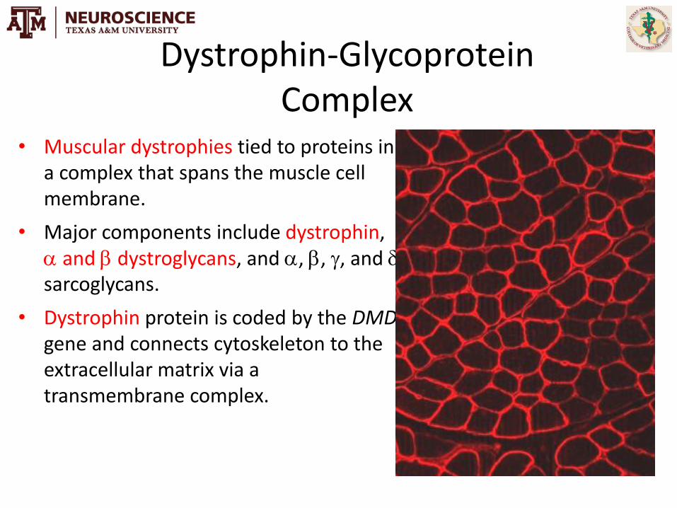
• Muscular dystrophies tied to proteins in a complex that spans the muscle cell membrane.
• Major components include dystrophin, and dystroglycans, and , , , and sarcoglycans.
• Dystrophin protein is coded by the DMDgene and connects cytoskeleton to the extracellular matrix via a transmembrane complex.
From Michele DE & Campbell KP: J Biol Chem 278:15457-60, 2003.

• X-linked; males affected,
females are carriers.
• ~ 1 in 5,000 live male births.
• Boys in wheelchairs by teens;
most die by early 20s.
• Cardiomyopathy; respiratory.
Duchenne Muscular Dystrophy
SartoriusGracilis
• Spinal lordosis.
• Pseudohypertrophy of the
calf muscles.
• Selective muscle sparing.
• Agonist-antagonist imbalance
contributes to contractures.

Mammalian Animal Models of DMD
• Mdx mouse – a nonsense point mutation causes premature termination of translation within exon 23. Mild phenotype.
• Feline hypertrophic muscular dystrophy – deletion of the dystrophin muscle and cerebellar Purkinje cell promoters in one case.
• GRMD – splice site mutation in intron 6 causes exon 7 to be skipped in transcription and a stop codon; multiple other dog breeds.

GRMD Colony Development
• 1981 – GRMD littermates (Rusty and Dusty) seen at UGA (Kornegay JN et al: Muscle Nerve 11:1056-1064, 1988).
• 1982 – Rusty and Dusty transferred to NC State.
• 1985 – GRMD dog (Rusty) first seen at UGA provided to Barry Cooper at Cornell to develop colony.
• 1987 – GRMD dog and carriers provided to NCSU for second colony.
• 1994 – NCSU colony to Missouri.
• 2007 – Missouri colony to UNC-CH.
• 2012 – UNC-CH colony to Texas A&M.
• Colonies in Australia (closed), France, Japan, Brazil, the Netherlands, Missouri, and at the Fred Hutchinson Cancer Center (Seattle).

The Role of Animal Models in Treatment Development for DMD
• In vitro studies (cell culture, etc).
• In vivo systems not involving specific X-linked animal models.
• Mdx mouse; mdx/utrophin DKO mouse.
• GRMD dog and/or primate.
• DMD patients.
Predictive value of animal models. Will the mdx or GRMD model better predict outcome in DMD?
A balancing act – should the dog be driving the bus (tricycle)?

GRMD – General Features• Progressive disease
• Postural instability/contractures
• Muscle atrophy and hypertrophy
• Kyphosis/lordosis
• Respiratory
• Cardiomyopathy
Phenotypic Variation: Species, Individual, and Muscle (confounds preclinical trials but
offers insight on disease pathogenesis).
Primary vs. Secondary Effects of Dystrophin Deficiency and Identification of
Modifier Genes as Druggable Targets

Therapeutic Approaches• Gene Therapy
- Viral Vectors
- Plasmid DNA
- Antisense oligos
• Cell Therapy
- Myoblasts
- Stem cells
• Pharmacologic
- Prednisone (Std of Care for DMD)
- ↑ utrophin (surrogates)
- NF-кB inhibitors
- Calpain inhibitors
- Membrane sealants
- Myostatin (GDF-8) inhibition
AAV-minidystrophin, Neonatal Intravenous
Kornegay JN, et al: Molecular Therapy 18:1501-1508, 2010.
Mesoangioblast Therapy in GRMD
Sampaolesi M, et al: Nature 444:574-579, 2006

Myostatin Inhibition
• Myostatin (Growth/differentiation factor 8; GDF-8). Negative regulator of muscle growth; mutations lead to muscle hypertrophy (double muscled cattle; human; sheep; whippets).
• Knocking out myostatin improves function in mdx mice BUT…
• Adult dystrophy myostatin-inhibition (MYO-29) trial ambiguous
• Murine models of LGMD and CMD either did not improve or had differential effects in young vs. old mice and/or muscles.
Schulke M, et al: NEJM 350:2682-8, 2004

Hypertrophy of the GRMD Cranial Sartorius (and Other Flexor Muscles) May Have Deleterious Effects.
Kornegay JN, et al: Lab Anim Sci 44:331-333, 1994.
GRMD – 3 Months
GRMD – 6 Months
Kornegay JN, et al: Neuromuscul Disord 13:493-500, 2003.

GRMD – Postural Changes
GRMD
Normal
Brumitt JW, et al: Vet Radiol Ultrasound 47:574-580, 2006.Kornegay JN, et al: Lab Anim Sci 44:331-333, 1994.
GRMD – 3 Months
GRMD – 6 Months

Myostatin Inhibition
• GRMD data suggest muscle hypertrophy can be harmful, BUT….
• Whippet dogs that are heterozygous for myostatin are better athletes; homozygous-null dogs have gross muscle hypertrophy, i.e. so-called bully whippets.
• Transgenic/knockout technology in dogs is not widely utilized.
• Potential to cross breed GRMD and heterozygous myostatin whippet dogs (GRippets).
Mosher DS, et al: PLoS Genet 3:779-786, 2007.

Myostatin-Heterozygous (Mstn+/-) GRMD Dogs; GRippets
• Collaboration with Kathryn Wagner and Se-Jin Lee of Johns Hopkins.• First litter – GRMD carrier bred to sire (Mstn+/-) of “Bully Whippet” (Mstn-/-).• Second litter – GRMD male bred to Speedy (double mutation).
Table 1. GRMD-Myostatin Status
Dog Name Gender GRMD Status Myostatin Status
First Litter (“Racing”)
Racer Male Normal Normal
Flash Male Affected Normal
Dash Male Affected Heterozygote
Speedy Female Carrier Heterozygote
Lightning Female Carrier Normal
Zippy Female Normal Heterozygote
Second Litter (“Bewitched”)
Endora Female Carrier Normal
Esmerelda Female Carrier Heterozygote
Samantha Female Affected Normal
Hagatha Female Affected Normal
Tabitha Female Affected Heterozygote
Derrwood Male Affected Heterozygote
Abner Male Affected Heterozygote
Non-dystrophic Controls (n = 3)
GRMD Myostatin normal (n = 3)
GRMD Myostatin heterozygous (n = 4)
Kornegay JN, et al, submitted.

Myostatin-Heterozygote (Mstn+/-) GRMD (Litter 1 – “Racing”)
• Similar phenotype for Flash and Dash until ~ 4.5 mos.
• Dash developed contractures.
• Hypertrophy of the cranial sartorius, semitendinosus, and semimembranosus muscles.
• Dash had features of a severe GRMD phenotype.
• Contractures – unbalanced agonist/antagonist muscles.
• Bigger is not necessarily better.
Racer Flash Dash
Dash Flash
Racer Flash Dash

Segmentation
• Overall body-weight-corrected (mm3/kg) muscle mass comparable among the three groups of dogs.
• Marked variation among muscles; trend whereby relative lack of myostatin exaggerates pre-existing trends for muscle atrophy/hypertrophy.
Kornegay JN, et al: Phys Med Rehabil Clin N Am 23:149-72, 2012.

The Realities of Drug Development• The success rate for Phase II human clinical trials fell
from 28% in 2006-2007 to 18% for 2008-2009 (Arrowsmith 2011).
• Over half (51%) of 108 reported Phase II failures occurred due to insufficient efficacy, even though most drugs were assessed in animal models (Plenge et al. 2013).
• Only 28 of 76 (37%) of highly-cited studies that investigated a preventive or therapeutic intervention in an in vivo animal model over the 1980-2000 period were replicated in human randomized trials (Hackam and Redelmeier 2006).

Lost in Translation(Ergorul and Levin 2013)
• The Butterfly Effect (chaotic behavior whereby small differences in the animal model lead to substantial differences in clinical results);
• The Princess and the Pea problem based in variability of effect size when progressing from biochemical findings through tissue culture and animal and human studies (the pea does not indent the mattress to the same degree as the princess);
• The Two Cultures problem evident in preclinical and clinical research (need for more rigorous experimental design in preclinical studies).

• Biological (animal) models can be based in analogy or homology.
- Analogy implies a point-by-point relationship.
- Homology implies a shared evolutionary history and DNA makeup.
To be functionally useful, homologous models must be good models by analogy.
• Models may be one-to-one (disease in humans and a species that share the same clinical features) or many-to-many (findings from more than one species or organ system model disease features).
Biomedical ModelsNational Research Council (1985, 1998)

GRMD – General Features• Progressive disease
• Postural instability/contractures
• Muscle atrophy and hypertrophy
• Kyphosis/lordosis
• Respiratory
• Cardiomyopathy
Phenotypic Variation: Species, Individual, and Muscle (confounds preclinical trials but
offers insight on disease pathogenesis).
Primary vs. Secondary Effects of Dystrophin Deficiency and Identification of
Modifier Genes as Druggable Targets

Traditional Drug Targeting – Histopathologic Lesions and Presumed Pathogenetic Mechanisms
A New Model – Targeting Genetic Mechanisms such as Modifier Genes through mRNA Array and GWAS Studies
Kornegay et al: ILAR J, in press.

Phenotypic Data from GRMD Dogs Assessed with mRNA Microarrays
• Collaboration with Scott Schatzberg and Peter Nghiem at UGA and Eric Hoffman at Children’s National Medical Center in Washington, DC.
• 8 GRMD and 4 normal dogs with muscle biopsies at 8 wks and 6 months.
• Biopsies from the vastus lateralis, cranial sartorius, and long digital extensor muscles.
GRMD Dogs
(6 mos)
Left TTJ Angle
(degrees)
Tetanic
Extensor
Force
(N/kg)
Tetanic
Flexor Force
(N/kg)
Body
Weight
(kg)
CS Circumference
(mm/kg)
Overall Rank
(1= least severe, 8=
most severe
phenotype)
Tico 159 3.138 0.296 15.2 2.303 1
Joker 155 2.135 0.331 16.3 2.653 2
Peggy 155 2.109 0.489 13.1 2.774 3
Jeannette 151 1.669 0.489 13.2 3.102 4
Huckleberry 141 1.144 0.508 13.2 3.298 5
Porsche 130 1.041 0.575 14.6 3.493 6
Janet 142 1.583 0.667 12.6 3.598 7
Betty 102 2.29 0.72 9.3 6.156 8

1
3
2
4
1 = Normal CS 4-9 Wk (n = 4)2 = GRMD CS 4-9 Wk (n = 8)3 = Normal CS 6 Mo (n = 4)4 = GRMD CS 6 Mo (n = 8)
Supervised hierarchical clustering of 250 genes from cranial sartorius (CS) array correlated to CS size (p = 0.001; r = > 0.94) in CS profiles of normal and GRMD at
4-9 Wk and 6 Mo Red = up-regulated; Green = down-regulated
Nghiem PP, et al: Am J Pathol 183:1411-24, 2013.

Ingenuity PathwayAnalysis (IPA) of 250genes associatedwith CS hypertrophygenerated the top-ranked network,DAG1 & LARGE.
Nghiem PP, et al: Am J Pathol 183:1411-24, 2013.
Nghiem PP, et al: Am J Pathol 183:1411-24, 2013.

Table 2. Total Spectral Counts for Spectrin, Myotrophin, andLaminin-α2 from Proteomic Profiling Results
Genotype Age SPTAN1 SPTBN1 MTPN LAMA2
Normal4-9 wks
43 7 7 54
6 mos 36 1 5 3
GRMD4-9 wks
65 19 12 12
6 mos 65 3 15 0
Additional Studies
Sum of spectral counts for each group is shown. There are n = 3 profiles for normal and GRMD at 4 to 9 weeks and at 6 months (12 total profiles). LAMA2, laminin-α2; MTPN, myotrophin; SPTAN1, α-spectrin; SPTBN1, β-spectrin.
• Proteomic profiling (mass spectrometry) was done to identify additional dystrophin surrogates and/or hypertrophic factors.
• Membrane proteins α- and β-spectrin, as well as the muscle growth factor, myotrophin, were selectively upregulated in the CS (Confirmed by IHC and/or Western blots).
Nghiem PP, et al: Am J Pathol 183:1411-24, 2013.

What about Myostatin?
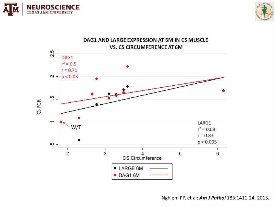
• Myostatin was downregulated on qPCR in all GRMD muscles but especially in the CS.
• Myostain mRNA levels were inversely correlated with CS muscle circumference at 6 mos.
C: MSTN qPCR expression revealed decreased expression in GRMD muscle, with the greatest decrease in the hypertrophied CS at 6 months (6M; n = 8 arrayed dogs). D: MSTN mRNA was inversely correlatedwith CS size at 6 months in the CS of GRMD dogs of the discovery data set (r = 0.73; r2 = 0.53; P < 0.05; n = 8 arrayed dogs). *P < 0.05, **P < 0.01, and ***P < 0.001.
C D
Nghiem PP, et al: Am J Pathol 183:1411-24, 2013.

Overall Conclusion: Cranial sartorius hypertrophy in the GRMD model is
driven and supported by a complex set of genes whose manipulation could
have (favorable or deleterious) clinical significance.

Summary
• Animal models are a powerful example of the one medicineconcept.
• Preclinical studies often do not translate to humans.
• Homologous genetic models are not necessarily analogous.
• Genetic studies, including mRNA analysis, offer an additional tool to identify potential drug targets, especially if coupled with phenotypic data.

Acknowledgements
Kornegay Lab: Janet and Dan Bogan, Jennifer Dow, David Detwiler
CNMC-DC and GWU:
Eric Hoffman, Priya Mittal, Heather Gordish-Dressman,
Svetlana Ghimbovschi, Kanneyboyina Nagaraju, Zuyi
Wang, and Peter Nghiem
UGA-Athens: Scott Schatzberg and Peter Nghiem
Johns Hopkins:
Kathryn Wagner, Leigh Warsing, & Emily Cousins
MRI: Martin Styner, Jane Fan, and Jiahui Wang
GRMD AAV-Minidys: Xiao Xiao and Juan Li
UNC-Chapel Hill
Texas A&MCandice Brinkmeyer-
Langford, Mandy Bettis, Cindy Balog

Funding• Muscular Dystrophy Association
• Association Francaise contre les Myopathies
• Parent Project Muscular Dystrophy
• National Institutes of Health (NIAMS, NINDS, NHLBI, NCMRR)

Thank You
Thank You!QUESTIONS?