Embed Size (px)
Citation preview

M i c r o s c o p y f r o m C a r l Z e i s s
On the Trail of Life Itself
Individual Microscope Systems for All Micromanipulation
Applications in In-vitro-Fertilisation, Transgenic Techniques,
Stem Cell Research and Developmental Biology


3
Content
Micromanipulation Systems from Carl Zeiss 4-5
IVF, ICSI and IMSI 6-7
Recommended Microscopes 8-9
Transgenic Techniques, Stem Cell Research 10-11
Recommended Microscopes 12-13
Developmental Biology 14-15
Recommended Microscopes 16-17

4
Micromanipulation Systems Must Satisfy Many Requirements.The Most Important One? Being Right for Your Application.
Flexible microscope systems are based on the principles of well-considered modu-
larity and complete integration. Carl Zeiss is able to provide at least one appropriate
solution for each application – with a confi guration that suits your requirements
and also leaves plenty of upgrade options open for future tasks.
applications and highly complex stem cell research ex-periments through to high-throughput applications in developmental biology – with the latest generation of instruments all the possibilities of micromanipulation are open to you.
IVF, ICSI and IMSIBesides conventional (and in most laboratories already routine) IVF applications, using the systems from Carl Zeiss it is also possible to perform additional applications such as ICSI (Intracytoplasmic Sperm Injection) and IMSI (Intracytoplasmic Morphologically Selected Sperm Injec-tion). Equipment developed specifi cally for this purpose streamlines and optimizes the workfl ow considerably, while short operating procedures reduce the time it takes to carry out ICSI and IMSI to a minimum. Each system can also be expanded to include incubation and cameras for documentation. All commonly used manipulators can be adapted. Recommended microscopes:
• Axiovert 40, the inverse routine microscope for cell biology offering excellent optical performance and numerous functions.
• The Axio Observer.A1 (manual) research platform for demanding routine tasks or, for example, the motorized Axio Observer.Z1 with maximum ease of operation and fl exibility for speed and ergonomics.
• The compact, cost-effective Stemi 2000 stereomicro-scope with brilliant 3D optics for the cleaning of egg cells and subsequent assessment of embryos.
Transgenic techniques and stem cell researchIn the case of applications such as PN injections, nuclear transfer and reporter gene analysis, which become vis-ible using fl uorescence and the appropriate HE fi lter sets, numerous confi gurations are available to the microscope user for tasks ranging from routine high-throughput
More possibilities: the ideal solution for every budgetNumerous microscope stands with countless interfaces allowing individual adjustment to any experiment – with Carl Zeiss micromanipulation systems you can choose from a wide variety of manual and motorized stereo-microscope and inverse fl uorescence microscope systems. The advantage: in consultation with you, your Carl Zeiss advisor can put together a system confi guration tailored to your specifi c application. Irrespective of size or price, all microscopes include impressive performance features which make even the very fi nest structures visible and allow living cells or gene material to be manipulated while offering the sample maximum protection:
• Ergonomic operating concept• Brilliant optics• Guaranteed reproducibility• Vibration-free• Various contrast techniques: HMC, PlasDIC, DIC,
Phase contrast• Maximum precision
A common feature of all systems is the modular system architecture which can be adapted fl exibly to meet your changing requirements. This means that you can expand your system at any time with precisely the component that your application demands – and only this compo-nent, giving you a system that is extremely simple to use and uncomplicated, and which can be confi gured in no time at all. This is a micromanipulation system “to go”.
A wider range of uses: all applications from IVF to embryologyThe range of uses for Carl Zeiss micromanipulation microscopes is as diverse as the range of tasks faced in their various areas of application. From routine IVF

5
• Stemi 2000 for simple manipulation, e.g. of frogs’ eggs, meal worms, etc.
• SteREO Discovery.V12 for detailed manipulation at high magnifi cations, e.g. for zebra fi sh, C. elegans and Drosophila applications.
• SteREO Discovery.V20 for applications involving extremely fi ne structures at very high magnifi cations, e.g. for the manipulation of fi ne tissue structures in zebra fi sh.
More integration: system software and a variety of componentsAn economic advantage is full integration into AxioVision. With the possibility of adding a wide range of modules, right through to an analysis platform, even the functions of the basic version are perfectly suffi cient for all relevant applications. AxioVision LE can be downloaded from the Internet free of charge for simple image acquisition. Applications under physiological conditions can sub-stantially increase success rates and, for this reason, Carl Zeiss has developed a complete portfolio of incuba-tion components which simulate the in-vivo conditions of the cells almost perfectly in the system. For simple documentation using AxioVision LE you can connect the digital camera of your choice. Besides the tried-and-tested cameras of the AxioCam family, consumer and video cameras made by other manufacturers are also compatible – for simple image acquisition, image pro-cessing and measuring.
workfl ows to those requiring high-end technology and involving incubation, documentation and analysis soft-ware. As in IVF/ICSI/IMSI it is also possible to work with various manipulators here. Recommended microscopes:
• Axiovert 40 (see above)• The Axio Observer.D1 high-end research microscope
offering supreme ease of use and increased fl exibility. With this microscope motorized selection of the re-fl ector turret, condenser and refl ected-light beam path is possible.
• The SteREO Discovery.V20 high-end stereomicro-scope with brilliant optics and the highest resolution of its class for a splendid 3D display and strong 3D impression even at high magnifi cations.
Developmental biologyCarl Zeiss has the perfect confi guration for all applications in developmental biology. From samples such as small organelles, embryos and egg cells, or whole organisms such as Drosophila, C. elegans and zebra fi sh, through to genetic manipulation and knockouts, stereomicroscopes of all classes deliver precisely the brilliant 3D display that such applications demand. This product spectrum stands for maximum precision, brilliant optics, a high degree of reproducibility and automated processes in the high-end range. For excellent optical performance, speed and ergonomics in routine applications. And for meaningful scientifi c results in all cases. Recommended microscopes:
Suitable for all micromanipulation requirements: microscopes from Carl Zeiss are tailored to each micromanipulation application

6
A cost-effective decision: Axiovert 40 for IVFThe compact inverse Axiovert 40 is particularly impressive in terms of its price-performance ratio. With manipula-tors that can be selected in accordance with your own requirements, this compact solution is just as stable as the Axio Observer research platform. This “budget so-lution” also impresses with the contrast techniques it offers: with Axiovert 40 you have access to PlasDIC and phase contrast – perfectly suffi cient for routine IVF/ICSI. Heating and documentation are also possible with Axiovert 40.
One research stand – all contrasts for IVF, ICSI and IMSIFor successful ICSI (Intracytoplasmic Sperm Injection), clearly visible structures such as the zona pellucida and polar body of the egg cell are essential. With IMSI (In-tracytoplasmic Morphologically Selected Sperm Injec-tion), you can also assess the shape and vacuole count of the sperm cells. In borderline cases, several contrast techniques have to be possible that can deliver scientifi -cally unambiguous, high-contrast images. In this area, the Axio Observer research microscope is unique: all relevant contrast techniques are available on one single stand. Hoffmann Modulation Contrast, PlasDIC and DIC are all possible and, in addition, phase contrast can be combined with all methods.
The majority of ICSI experiments take place in plastic dishes, for which DIC is not suitable. That’s why Carl Zeiss developed PlasDIC – using this patented con-trast technique, egg and sperm cells can be observed in the same plane. PlasDIC produces a relief that is similar to DIC and delivers an impressive three-dimensional display. The advantage: a total solution that is geared precisely to your needs. Plus a completely vibration-free stand and high-resolution, needle-sharp DIC-like images.
Where Every Success is so Signifi cant, Leading-edge Technology is Essential.Because It Allows Use of the Most Advanced Techniques.
In scarcely any other area are the pressure to succeed and expectations quite so high as
in the fi eld of reproductive medicine. The best possible support for the responsible work
carried out by attending physicians and their MTAs is a microscope system that enables the
latest methods to be employed, as well as offering good ergonomics and ease of operation.
Human sperm cells at 40x PlasDICAstri Wold, Trondheim University Hospital, Norway
Dividing human embryo at 40x PlasDICAstri Wold, Trondheim University Hospital, Norway
Dividing human embryo at 40x HMC. Less three-dimensional and less relief in comparison to PlasDIC.Astri Wold, Trondheim University Hospital, Norway

7
from high to medium magnifi cation is particularly bene-fi cial for the subsequent assessment of the embryo. What is more, having IMSI and ICSI on a single microscope makes your workfl ow more effi cient than ever before, and offers the sensitive egg cells optimal protection.
To complete the ICSI laboratory: Stemi 2000The Stemi 2000 dissection stereomicroscope – which can be mounted in a Laminar Flow Box for optimized condi-tions – makes a useful and cost-effective addition to your ICSI/IMSI system. This compact stereomicroscope gen-erates brilliant three-dimensional images. Egg cells can therefore be isolated quickly and without complications (egg recovery) and the embryos can be assessed quickly following injection. The operation of the microscope is as easy as it is ergonomic. With this instrument your ICSI/IMSI workstation is complete – including success moni-toring.
Egg cell is positioned so that the polar body is at a 90° angle to the insertion
Insertion of the capillary tube, containing a sperm cell, through the zona pellucida
Injection of the sperm cell into the oolemma of the egg cell
Optimal workfl ow: the research platform for ICSI + IMSIToday, some laboratories are already using IMSI (Intracy-toplasmic Morphologically Selected Sperm Injection) as a fi rst step in order to signifi cantly raise the success rate of ICSI. The numbers speak for themselves: in practice, prior selection of suitable sperm cells increases the chances of successful fertilization considerably. This presents a great challenge for the microscope systems involved, as extremely high magnifi cations are required to allow a morphological assessment of the sperm cells. DIC with oil immersion and special, high-performance objectives – ideally in combination with incubation – is perfectly suit-ed to IMSI. The ultimate in convenience and economy: based on the partly motorized Axio Observer.D1 research platform, Carl Zeiss has developed a combined system that allows the sperm cell to be injected into the egg cell immediately after selection. The large fi eld of view pro-vides a clear picture of the egg and sperm cell with the injectors. Incubation can then be continued in an incuba-tor with no problem at all. The ability to switch quickly
IVF, ICSI, IMSI
Ergonomics on the Axiovert 40, shown here with Eppendorf manipulators

8
Axiovert 40for ICSI with Narishige Micromanipulation, with Brightfi eld, Phase and Relief Contrast (PlasDIC) and Various Options (Heating, Camera, High-quality Optics)
Stemi 2000Dissection Stereomicroscope for Egg Cell Isolation and Embryo Monitoring
Axiovert 40
491202-0007-000 Axiovert 40 for ICSI (32x)491202-0008-000 Axiovert 40 for ICSI (40x)491202-0009-000 Axiovert 40 for ICSI (32x, heating)491202-0010-000 Axiovert 40 for ICSI (40x, heating)491202-0011-000 Axiovert 40 for ICSI (32x, heating frame)491202-0012-000 Axiovert 40 for ICSI (40x, heating frame)
491202-0013-000Axiovert 40 for ICSI (Plan-NEOFLUAR, heating)
491202-0014-000Axiovert 40 for ICSI (Plan-NEOFLUAR, heating frame)
if manipulation is required:
490002-0031-000 Narishige manipulat. MWO-202D (Axiovert 40)
Other manipulators, e.g. Eppendorf manipulators, are of course compatible.
if documentation is required:490002-0035-000 Video documentation (Axiovert 40)
490002-0036-000 Digital documentation(for FL, Axiovert 40)
490002-0037-000 Digital documentation(Canon, Axiovert 40)
Stemi 2000
495005-0006-000 Stemi 2000 dissection stereo-microscope
9
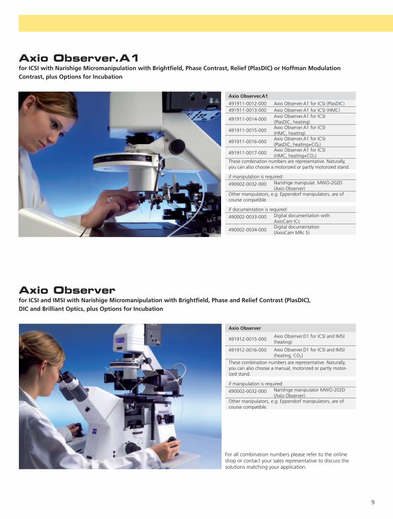
Axio Observer.A1for ICSI with Narishige Micromanipulation with Brightfi eld, Phase Contrast, Relief (PlasDIC) or Hoffman Modulation Contrast, plus Options for Incubation
Axio Observer.A1
491911-0012-000 Axio Observer.A1 for ICSI (PlasDIC)491911-0013-000 Axio Observer.A1 for ICSI (HMC)
491911-0014-000Axio Observer.A1 for ICSI (PlasDIC, heating)
491911-0015-000Axio Observer.A1 for ICSI(HMC, heating)
491911-0016-000Axio Observer.A1 for ICSI (PlasDIC, heating+CO2)
491911-0017-000Axio Observer.A1 for ICSI (HMC, heating+CO2)
These combination numbers are representative. Naturally, you can also choose a motorized or partly motorized stand.
if manipulation is required:
490002-0032-000 Narishige manipulat. MWO-202D (Axio Observer)
Other manipulators, e.g. Eppendorf manipulators, are of course compatible.
if documentation is required:
490002-0033-000 Digital documentation withAxioCam ICc
490002-0034-000Digital documentation(AxioCam MRc 5)
Axio Observer
491912-0015-000 Axio Observer.D1 for ICSI and IMSI (heating)
491912-0016-000 Axio Observer.D1 for ICSI and IMSI (heating, CO2)
These combination numbers are representative. Naturally, you can also choose a manual, motorized or partly motor-ized stand.
if manipulation is required:
490002-0032-000 Narishige manipulator MWO-202D (Axio Observer)
Other manipulators, e.g. Eppendorf manipulators, are of course compatible.
Axio Observerfor ICSI and IMSI with Narishige Micromanipulation with Brightfi eld, Phase and Relief Contrast (PlasDIC), DIC and Brilliant Optics, plus Options for Incubation
For all combination numbers please refer to the online shop or contact your sales representative to discuss the solutions matching your application.

10
1 2 3
Research at the Limits of Technology Demands Tools That Go Beyond Boundaries.With Performance That Impresses in Every Detail.
Transgenic techniques and stem cell research are the research fi elds of the future.
Characterized by the extremely high demands placed on people and technology,
this area of research is continually highlighting new ways ahead. This can only be
achieved using technology at the very limits of what is possible – a task to which
Carl Zeiss is fully committed.
touchscreen allows you to operate the system con-veniently and directly without the need for any time-consuming advance confi guration.
Best optics and maximum magnifi cationsTransgenic techniques and stem cell research demand the most powerful microscopes that technology has to offer. Transferring genetic material and isolating and manipulating stem cells require brilliant optics, the high-est possible magnifi cations and contrast methods that can be variably combined in order to make the fi nest structures, clearly visible. Objectives representing the very best in quality right across all classes are perfectly tailored to the applications and contrast methods in-volved. PlasDIC is particularly well-suited to plastic Petri dishes and DIC to PN injection in glass dishes. In addi-tion, phase contrast can also be performed. For research with transgenic animals, many scientists employ the SteREO Discovery stereomicroscope, as it delivers de-tailed three-dimensional information, and the egg cell and material to be injected are visible in a single plane. This is an extremely economical option for specifi c visual requirements. All in all, numerous high-performance
Technology of the future for the tasks of the futureUnderstanding and curing diseases – a considerable chal-lenge in a fi eld that is unsurpassed in terms of its com-plexity and demands. Virtually all areas of research come together here. At present we only have a vague notion of the potential of transgenic techniques and stem cell research. No other research area offers so many positive opportunities and possibilities for medicine and for us as humans. It also presents a signifi cant challenge as far as technology is concerned: manipulating genetic material or stem cells calls for microscopes that can be adapted quickly and fully to ever-increasing requirements.
The solution: individually confi gurable system solutionsfrom Carl Zeiss. The corresponding research platforms, based on motorized, vibration-free stands, offer out-standing optics with extremely high numerical aperturesand special HE (High Effi ciency) fl uorescence fi lters.Manipulation and various incubation systems with stablegas concentrations can be easily combined with theseplatforms. Physiological conditions can therefore alsobe maintained during manipulation, guaranteeing the survival of sensitive stem cells. A TFT display with

11
4 5 6 7
systems are available that are immediately ready to use and offer the widest possible range of applications.
A system world for top-level researchEach individual application requires dedicated compo-nents. No matter which Carl Zeiss microscope stand you opt for, the full integration of all components into the AxioVision software platform will provide you with a system that saves you the need for complex, time-con-suming confi guration and is simple to operate. Specially developed components, such as the Colibri LED light source, guarantee optimal protection of the sample. The problem of bleaching is minimized, which is a con-siderable advantage in the case of weak fl uorescence. Monochrome cameras for fl uorescence images with an optimal signal-to-noise ratio, incubation components and contrast techniques are perfectly tailored to one an-other. And AxioVision not only allows you to perform automated online experiments, it also offers you direct analysis and evaluation.
Simple sample changing on Axio Observer, shown here with Narishige manipulators
1)-6) Transgenic mouse embryos in various stages of development in DIC or PlasDIC, 40x magnifi cation, 7) Mouse sperm cell in PlasDIC, 40x magnifi cation1)-5) and 7) Dr. Ropeter, Dragon-IVF, Dr. Michelmann, Frauenklinik Göttingen and Ms. Buhtz, University of Göttingen, Germany6) Dr. Luís-Miguel Criado Rodríguez, Transgenesis Department, Fundación Centro Nacional de Investigaciones Cardiovasculares Carlos III, Madrid, Spain
More than just one option: a whole world ofincubationTransgenic techniques and stem cell research frequently require incubation. In this area Carl Zeiss has a unique range of different components available, which are of-fered with all recommended microscope stands. From simple heating using stage incubators for manipulation with CO2 incubation right through to incubators that set the necessary CO2 atmosphere and reduction in the O2 concentration for long-term experiments, everything is covered. This ensures the best possible conditions for cul-tivating sensitive stem cells in particular. In this complex area too, it is possible to tailor the confi guration precisely to each individual requirement. Only by perfectly imitat-ing physiological conditions and rapidly changing con-trasts and components is it possible to guarantee that your experiment will be a success. No other system offers you greater reliability or more effi cient workfl ows.
Transgenic Techniques

12
Axiovert 40 + manipulation
491202-0017-000 Axiovert 40 Ph, PlasDIC, FL491202-0018-000 Axiovert 40 Ph, PlasDIC, heating491202-0019-000 Axiovert 40 Ph, PlasDIC, heating frame
if manipulation is required:
490002-00031-000 Narishige manipulat. MWO-202D (Axiovert 40)
Other manipulators, e.g. Eppendorf manipulators, are of course compatible.
if documentation is required:490002-0035-000 Video documentation (Axiovert 40)
490002-0036-000 Digital documentation (FL, Axiovert 40)
490002-0037-000 Digital documentation (Canon, Axiovert 40)
490002-0046-000 Digital documentation for Axiovert 40 (AxioCam ICc1)
Axiovert 40for Rapid Control of Cells and Their Growth
Axiovert 40for Transgenic Applications and Stem Cell Manipulation
Axiovert 40
491202-0015-000 Axiovert 40 Ph491202-0016-000 Axiovert 40 Ph, FL

13
Axio Observerfor Transgenic Applications and Stem Cell Manipulation
Axio Observer.D1
491911-0018-000Axio Observer.D1 (FL and DIC)+heating
491911-0019-000 Axio Observer.D1 (FL and DIC)+heating+CO2
491911-0020-000 Axio Observer.D1 (FL and DIC)+heating+CO2+O2
if manipulation is required:
490002-0032-000 Narishige manipulat. MWO-202D (Axio Observer)
Other manipulators, e.g. Eppendorf manipulators, are of course compatible.
if documentation is required:
490002-0033-000 Digitale documentation with AxioCam ICc
490002-0034-000 Digitale documentation(AxioCam MRc 5)
SteREO Discovery.V20
495016-0013-000 SteREO Discovery.V20 transgenic
495016-0014-000 SteREO Discovery.V20 transgenic +heating
if manipulation is required:
490002-0038-000 Narishige manipulation for SteREO Discovery
490002-0039-000 Narishige manipulation for SteREO Discovery temp
Other manipulators, e.g. Eppendorf manipulators, are of course compatible.
if documentation is required:
490002-0040-000 Digitale documentation SteREO Discovery
SteREO Discovery.V20for Transgenic Applications
For all combination numbers please refer to the online shop or contact your sales representative to discuss the solutions matching your application.

14
1 2 3
5
4
The Range of Applications Determines the Range of Confi gurations.Or Redefi nes Them.
Embryonic development, cancer research, neurobiology – it is in the fi eld of develop-
mental biology that the most diverse range of micromanipulation applications is en-
countered. Versatility is therefore a key factor for your stereomicroscopes. From brilliant
3D images through to extremely fast systems, all-round performance is required.
phenotypes, interventions in embryogenesis through the injection of DNA into egg cells or of genetic material into larval stages, and the injection of chemical or biological agents for manipulating germlines of adult animals are taken to a whole new level. With unmatched resolution,SteREO Discovery.V12 and SteREO Discovery.V20 allow you to observe objective structures down to 0.5 µm in their three-dimensional context. For the manipulation of your objects at magnifi cations up to 345x (10x eye-piece), this is possible using the SteREO Discovery.V20 research microscope. New: a stereomicroscope with an additional microobjective that allows magnifi cations up to 525x. This is a huge benefi t for research involving C. elegans, as all workfl ows can be performed on just one stand.
Depth of focus in 3D for fascinating imagesWhat happens during the embryonic development of a living organism that has a deformed eye or a stunted leg as an adult animal? Why does one animal group show resistance to environmental infl uences and not the other? If you want to answer these questions, reliable re-producibility, clear and highly resolved images and maxi-mum precision are essential. With the rigorous demands of high-end research in mind, Carl Zeiss has developed a new generation of stereomicroscopes that signifi cantly increase the limit of resolution: the SteREO Discovery product family. The 3D display offered by these stereo-microscopes makes it possible to view thick samples, such as entire organisms or eggs. During micromanipula-tion it is possible to see more quickly where you need to inject. Applications such as knockouts and their resulting
1) Embryonic stage of a horseshoe crab2) Two horseshoe crabs3) Embryonic stage of a squid4) Different embryonic stage of a squid5) mHIP Xenopus where mRNA has been injected (Hedgehog Interacting Protein); half of animal is deformed6) Reproductive organs of a polychaete worm7) Adult brine shrimp8) Sea urchin embryo9) Frog embryos stage 21, stained with Rhodamine Dextran
1-4 and 6-8: Dr. Cassandra Extravour, Department of Organismic and Evolutionary Biology, Cambridge, Massachusetts, USA
5 and 9: Dr. Andres Collazo, Inner Ear Development Group, House Ear Institute, Los Angeles, USA

15
7 8 96
New insights into developmental biology with SteREO DiscoveryWith color-accurate, high-contrast 3D images that have excellent depth of focus, the SteREO Discovery stereo-microscopes offer you outstanding image information, as well as supreme ease of operation thanks to, amongst other things, the large working area with space for nu-merous sample holders. The option of combining the microscopes with a heating plate is also available, en-abling you to work effi ciently in high-throughput ap-plications under optimal physiological conditions. This heating plate and a non-heatable plate are available in a magnetic variant. You can therefore place the manipula-tors close to the sample for ergonomic working. Sepa-rate magnetic plates are also available for positioning the manipulator next to the stand.
Impressively powerful as part of a systemAs an option, ZEISS offers two Narishige manipulators as part of a system solution. The fi rst is a fi ne manipula-tor (MN-151) for manipulating fi ne structures, e.g. in C. elegans, Drosophila or zebra fi sh. The second is a simple, economical manipulator (YOU-1) for performing injec-tions into frogs’ eggs or various larvae. When it comes to documentation, even the basic version of AxioVision of-fers comprehensive functions that meet the requirements
A system solution from Carl Zeiss that ensures an optimum workfl ow
of most applications. Thanks to the modular structure, functions can be easily added as you come up against new tasks. And it goes without saying that a suitable camera is also available. In short, Carl Zeiss can offer you an individually confi gurable system solution for any task in developmental biology.
Compact yet powerful solution for routineapplicationsThe compact Stemi 2000 stereomicroscope with 7.7x zoom has all the features needed to carry out coarse ma-nipulation during routine applications in developmental biology. It can be operated quickly, ergonomically and precisely and is therefore highly effi cient to use. If you require transmitted-light, it is possible to choose from a range of options, from a simple attachment for high-con-trast brightfi eld through to the transmitted-light unit S with brightfi eld, darkfi eld and oblique-light contrast. A further impressive feature of the Stemi 2000 is its out-standing optical quality, which is close to matching that of a high-end microscope. Just like any other Carl Zeiss microscope, Stemi 2000 can also be adapted precisely to suit your requirements, allowing you to perform a whole host of applications – and all at a surprisingly impressive price-performance ratio.
Developmental Biology

16
SteREO Discovery.V12 + 3.5x Micro objective + 1.0x Macro objective
495008-0010-000 SteREO Discovery.V12 stereomicroscope
495008-0011-000 SteREO Discovery.V12 stereomicroscope+heating
if manipulation is required:490002-0041-000 Narishige manipulator manual coarse490002-0042-000 Narishige manipulator fi ne |490002-0043-000 Narishige manipulator fi ne ||Other manipulators, e.g. Eppendorf manipulators, are of course compatible.
if documentation is required:490002-0044-000 Digital documentation for color images490002-0045-000 Digital documentation for FL images
SteREO Discovery.V12for Finer Structures in C. Elegans, Zebra Fish and Drosophila

17
Stemi 2000Dissection Stereomicroscope for Larger Structures Such as Larvae and Frogs’ Eggs
Stemi 2000
495005-0007-000 Stemi 2000 dissection stereo-microscope
if manipulation is required:490002-0041-000 Narishige manipulator manual coarse490002-0042-000 Narishige manipulator fi ne I
SteREO Discovery.V8 + TL base 450
495015-0014-000 SteREO Discovery.V8 with transmitted-light and bifocal refl ected-light
SteREO Discovery.V8for Routine Dissections
For all combination numbers please refer to the online shop or contact your sales representative to discuss the solutions matching your application.

Carl Zeiss MicroImaging GmbH07740 Jena, Germany
BioSciences | Göttingen LocationPhone: +49 551 5060 660Telefax: +49 551 5060 464E-Mail: [email protected]
www.zeiss.de/micro-applications Info
rmat
ion
subj
ect t
o ch
ange
.Pr
inte
d on
env
ironm
enta
lly fr
iend
ly p
aper
blea
ched
with
out c
holo
rine.
60-2
-000
2/e
– pr
inte
d 06
.08


