Embed Size (px)
Citation preview
On the origin of autismby Pierluigi Fortini
Department of Physics - Via Saragat 1University of Ferrara - Ferrara 44100-Italy
1 Summary of the research on autism con-
ducted so far
I shall start from the published papers by three groups lead by Courchesne,Rodier and Gillberg: the reason for this choice is due to the fact that thefundamental assumpions of my paper are based on their works.
E. Courchesne 1 by means of an fMRI examined the brain of autisticchildren from the age of three years up to sexual maturity. He got thefollowing results2:
A) 90% of autistic children aged 3-4 have a volume of the brain greaterthan that of normal children of the same age (see fig.1 pag. S22)3
B) The almost totality of cases examined postmortem showed a reductionof Purkinje cells. This result has been confirm by fMRI measurements4.
This means that the cerebral capacity is reduced and this fact was con-firmed in the paper of G.Allen et al.5
C) The autistic people in the range of 2-3 years have a surplus of 12% ofgrey matter and of 18% of white matter in the cerebellum and 39% of whitematter in the brain compared with normal children of the same age. On theother hand the older and adolescent autistics do not have such surplus ofgrey and white matter (see fig 1 pag. S22)
D)This means that the autistic brain follows different patterns in its de-
1Center for Research on Autism, 8110 La Jolla Shores Drive, La Jolla, CA 92037 (USA)-E-mail: [email protected]
2See for instanceE. Courchesne: Abnormal early brain development in autism - Molecular Psychiatry 7,
S21-S23 (2002)3Lemons J. et al. Human Biology 53, 351-354 (1981)
Courchesne E. et al. Neurology 57, 245-254 (2001)4Greg Allen, Eric Courchesne: Differential effects of developmental cerebellar abnor-
mality on cognitive and motor functions in the cerebellum: an fMRI study of autism.Am.J.Psychiatry 160, 262-273 (2003)
5G.Allen, R. Mueller, E. Courchesne: Cerebellar function in autism: fMRI activationduring a simple motor task. Biol.Psychiatry 56, 269-278 (2004)
1
velopment: with age the surplus of grey and white matters tend to level onnormal values.
C. Gillberg 6 realized that autism is also present in individuals affectedby malformations concerning the brain stem which points to a presence ofan embryonic damage. Gillberg’ s group was particularly interested in thethalidomide epidemic happened of the 60s which had affected 10.000 peopleamong which 6000 survived7. The Swedish scholars noted that alongsidethalidomide there were cases of autism, of Mobius syndrome and of Golden-har syndrome.
Statistic involvement of the cranial nerve, in the cases more studied, wereapproximately in the proportion:
thalidomide:involvement of the abducens nerve (VI)(44%)involvement of the facial nerve (VII) (20%)involvement of the hypoglossal nerve (XII) (19%)Mobius:involvement of the abducens nerve (VI)(totality)involvement of the facial nerve (VII)(totality)involvement of the hypoglossal nerve (XII) (64%)In all syndromes studied the presence of ocular disorders is particularly
evident.All the investigates cases bear witness to the link between autism and
XII cranial nerve disorders. The problem is then to find an association (ifthere is one!) between them: the purpose of this paper therefore is to find aconnection among these facts.
PM. Rodier 8 starts from a similar approach to that investigated by Gill-berg with the difference that Rodier takes care of teratology i.e. the studyof the congenital anomalies and their genetic and environmental causes9
Rodier’s position, as is in general the American trend, considers autismdepending on environmental causes and in her papers she brings forwardthe experiments in favour of this theory . These tests consist in exposingexperimental animals to the action of valproic acid (which is a powerful
6Department of child and adolescent psychiatry, Goteborg University, Sweden andSt. George’s Hospital Medical School, University of London, UK; E-mail: [email protected]
7C. Gillberg et al.: Autism associated with conditions characterized by developmentalerrors in early embryogenesis: a mini review - Int. J.Devl. Neurol. 23, 201-219 (2005)
8Department of Obstetrics and Gynecology, University of Rochester School of Medicine,New York 14642, USA; E-mail: Patricia [email protected]
9SeeTL. Arndt, CJ. Stodgell and PM. Rodier:The teratology of autism - Int. J. Devl.Neuroscience 23, 189-199 (2005)
2

teratologic factor on the mammal embryos) and then compare these resultswith autistic post mortem exhibits. The most interesting thing in Rodier’spapers is that there are lesions in the 12 cranial nerves along the lines ofGillberg’s papers. So, for instance, there is a post mortem finding in whichthe facial nucleus and the superior olive are lacking.
Continuing along this line Rodier noted that the exposition to valproicacid caused a reduction in the number of motor neurons in the nuclei ofnerves V, XII, VI, III and VII10.
Besides Rodier’s group has noticed that when a female rat is exposedto valproic acid at the time of the closure of the neural tube the nuclei aredestroyed.
2 Assumptions of this paper
In the preceding section we have seen that the experimental research wasconcerned with two areas in the cerebral axis: on one side the cerebellumwith the involvements in the front brain (Courchesne) and on the other sidethe more caudal part with its 12 nerves which are created near the cerebellum(Gillberg, Rodier). These parts (front brain and 12 nerves) are anatomicallylinked between them and cerebellum plays a fundamental role. In fact theanomalies of the frontal part of the brain are linked directly with cerebellumby means of various pathways. Just to give an example of what we mean,recalling the cerebello - rubro - thalamo - cortical pathway which originates inthe cerebellum, makes synapses in the red nucleus, goes to the thalamus andfinally finishes in the cerebral cortex11. Starting from similar considerationsGillberg says in his paper: “ The anomalies of the cerebral axis don’t excludedysfunctions of the front part of the brain”.
Starting from this position it’ s possible to unite the results by Courch-esne, Gillberg e Rodier and it is possible to get a global view of autism. Tothis end it is sufficient to consider the XII cranial nerves as “spies” of thepathological status of the matter of the brain plus cerebellum system. In factthe XII cranial nerves originate from cerebral axis and not from the spine,according to the classical division of any book of anatomy.
According to this view the 12 cranial nerves must be considered as anintegral part of the autistic disease together with the frontal part of the brain.
10Rodier PM etal.: Embryological origin for autism: developmental anomalies of cranial nerve motor
nuclei - J.Comp. Neurol. 370 (2), 247-61 (1996)11See:http://en.wikipedia.org/wiki/Neural pathway
3

The latter is affected, through the motor part of the cortex, indirectly by thecerebellum. Today we are accustomed to consider autism a mental disease.In this way we put the lid on other very serious disorders (for example ocular,intestinal disorders, the lack of speech etc.) which have the same importanceas mental retardation. In our view we must put all disorders of autism onthe same footing because they are all neurological and linked by pathwaysamong them12.
According to this opinion if one wants to understand the autism syndromeone must consider the system (brain, cerebellum, XII cranial nerves) as awhole entity. In fact all three parts are linked by the motor part of the nervoussystem. We can therefore reasonably think of this “overgrowth” of brain massas having produced a kind of chronic inflammation; in the following we shallindifferently speak of autism or “chronic inflammation”. This situation isresponsible for all the troubles both of nervous (lack of speech, intestinaltrouble etc.) and psychological origin (lack of social interaction, recognitionof faces linked to the social interaction and in general linked with humansocial life etc.). In support of the fact that autism has a completely nervousorigin and not a “psychological” one, see the paper of Porges13. He, accordingto a point of view which goes back to Charles Darwin, claims that socialinteraction is due to the vagus nerve and this in the whole class Mammalia14.This position is also supported by Paul MacLean15.
Let us summarize: we have reduced the result of many experiments(Courchesne, Gillberg and Rodier) to only one: autism ( or chronic inflamma-tion) is a disease which affects the nervous system through brain, cerebellum
12See the pathways which connect the cerebellum with the motor cortexhttp://en.wikipedia.org/wiki/Motor cortexhttp://en.wikipedia.org/wiki/Primary motor cortexhttp://en.wikipedia.org/wiki/Premotor cortexhttp://en.wikipedia.org/wiki/Supplementary motor areaand with the spino-cerebellar tract:http://en.wikipedia.org/wiki/Spinocerebellar tracthttp://en.wikipedia.org/wiki/Mossy fiber %28cerebellum%29http://en.wikipedia.org/wiki/Purkinje cellhttp://en.wikipedia.org/wiki/Vestibular nervehttp://en.wikipedia.org/wiki/Dentate nucleus13Porges S.W.: The polyvagal theory: phylogenetic contribution to social behaviour -
Physiology and Behavior 79, 503-513 (2003)14Darwin C.: The expression of emotions in man and animals. London: John Murray.
1.st edition (1872)- Look for instance p. 69Orhttp://darwin-online.org.uk/contents.html15seehttp://en.wikipedia.org/wiki/Paul D. MacLean
4

and XII cranial nerves.But now we have to discuss the nature of this disease: is it genetic or
what? Fortunately this problem has been solved in the last 30 years.Michael Rutter has solved this problem applying a well known procedure
of experimental technique16. He was able to show that, taken two identicaltwins, neither are autistic or both are. This kind of measurement is based onthe principle “all or nothing”. The idea was originally due to physicists inXIX century by which Eotvos measured the torsion of a wire in 1908 whichallowed Einstein to found the theory of General Relativity17. The difficultywith such measurements is that one has to do with human beings (the twins)who have almost an infinity of variables (not only one i.e. the torsion ona wire!). Rutter repeated many times this measurement overcoming manydifficulties and the results were invariably the same. His results will be takenas a point of departure for this paper and autism must be considered agenetic disease even if certain facets make it slightly different from what weunderstand as a genetic disease.
Coming back to the results by Courchesne ( see Courchesne’ s papers) wesaid that the inflammation slightly reduces with age in autistic people. Thisdoesn’t mean that an autistic person, becoming older, can recover completely(such cases are absent from scientific literature and this fact is in agreementwith the fact that autism is genetic) but what can happen is that an autisticperson improves slightly. This behaviour finds an explanation in the data offMRI and the growth curves of cerebral matter.
The main contributions given by Rodier and Gillberg are to have broughtto light the fact that many syndromes (thalidomide poisoning, syndrome ofMobius, valproic acid poisoning etc.) have in common with autism certainautistic symptoms. This means that the autistic syndrome has its originin the formation of the nervous system which appears at the beginning ofgestation.
Research in this sense has begun by a group of italian researchers incollaboration with other groups18
Particularly interesting is the first paper in which the authors find an
16See for Rutter’s work my sitehttp://xoomer.alice.it/bioresautism/padre.html17Seehttp://en.wikipedia.org/wiki/Equivalence principle18M. Elia et al.: A genetic variant that disrupts MET transcription isassociated with autism - PNAS ( Proceedings of the National Academy of Sciences of
the USA)- 1 September 2006M. Conciatori et al.: Association between the HOXA1 A218G plymorphism and in-
creased head circumference in patients with autism - Biol. Psychiatry 55, 413-419 (2004)
5

association with autism of MET, which is an oncogenetic factor called mes-enchymal epithelial transition. This receptor has an important role in variousfunctions among which there is a gastrointestinal repair and the growth ofthe cerebellum. Considering the importance of cerebellum in the researchon autism we shall call this factor MET factor to underline the importancethat this gene could have in the pursuit of the gene (or genes) of autism. Inmy paper this “MET factor” will be responsible for various troubles in theinteractions of brain, cerebellum and 12 cranial nerves.
Apropos of the diction “MET factor” we must make some very impor-tant clarifications. With these words we don’t mean in general that particularoncogenetic factor taken into account in the paper of E.Elia et al. but wemean the gene, still not known, responsible for autism. Therefore the words“MET factor” remain in general for this gene we are looking for. So for ex-ample in the paper by D.M.Sikore et al.19 the authors, taking into accountthe Smith-Lemli-Opitz syndrome20 , which turns up through a deficency incholesterol metabolism linked to the 7-dehydrocholesterol (7-DHC) reduc-tase, note that this syndrome is associated with the majority of symptoms ofautism. This situation is much similar to the syndromes observed by Gillbergfor thalidomide and Rodier for valproic acid. The deficiency of 7-DHC forcholesterol gives similar symptoms in autism but it does not coincide withautism itself. Therefore the words “MET factor”, in my draft, can includealso 7-DHC reductase as a first “approximation” toward the identification ofthe autism gene. In fact I am considering in a forthcoming paper the possiblelinks between autism and the chemistry and biology of cholesterol: in thiswork I am still using the words “MET factor”.
A consequence of this new outlook is that many theories on autism ,sometimes very bizarre indeed, are no longer sustainable. Here I give oneexample of this kind of theory which, though not bizarre, must be discardedapplying the so called “Occam’s razor”21
Recently Pierce et al.22 used fMRI to examine the behaviour of autistic
19Sikora D.M., Pettit-Kekel K., Penfield J., Merkens L.S., Steiner R.D. 2006. Thenear universal presence of autism spectrum disorders in children with Smith-Lemli-Opitzsyndrome. Am. J. Med. Genet. Part A 140A:1511-1518.
20See for examplehttp://www.geneclinics.org/profiles/slo/details.htmlhttp://en.wikipedia.org/wiki/Smith-Lemli-Opitz syndrome21This principle is due to William of Occam (c. 1288 - c. 1347) an English Franciscan
friar and scholastic philosopher. In explaining some phenomenon one must be very carefulto eliminate everything that make no difference in the observable prediction. In Latin is”entia non sunt multiplicanda praeter necessitatem”, or ”entities should not be multipliedbeyond necessity”.
22Pierce et al.: The brain response to personally familiar faces in autism: findings of
6

children compared with normal children. They were shown the faces in pic-tures of family members (mother, father etc.): the answers of autistic childrenwere lacking in activity of the frontal lobe (see figure 8 pag. 161 of the quotedpaper). The absence of a response from autistic children to optical stimulihas been explained by Courchesne and co-workers to the partial absence inthe cortex structure of minicolumns, which are particular structures in pri-mate primary visual cortex23. The introduction of further structures (like the“mirror neurons”) for explaining the lack of recognition of the faces of themembers of the family is typically an assumption which must be abolishedapplying the “Occam razor”: the assumption of “mirror neurons” becomescompletely useless.
Our next task in the following of this draft will be1) autism can be explained with the anatomy of cranial nerves in the
presence of a still unknown gene.2) there is a particular test which is based on Rutter’ s experiments which,
if improved, can replace fMRI for a rapid and safe diagnosis of autism.3) if all the assumptions are correct, we shall have a clue for looking in
the right place to discover the still unknown gene.
3 Autism and theory of cranial nerves
We will fix our attention on that part of the human brain which contains thecerebellum that is rhombencephalon.
The rhombencephalon contains, besides cerebellum, also the medullaoblongata and the pons Varolii.
Now we’ ll give a concise explanation of the figures which are useful tolocalize the anatomical site where autism takes place.
In figure 1 at page 8 we’ ll get a glance at the human brain.We’ ll look at that part of the brain called rhombencephalon (figure 2 at
page 9). Moreover, in this part, we shall consider that part which is beneaththe cerebellum and is between pons Varolii and medulla oblonga (figure 3at page 10). In this area the majority of 12 cranial nerves have their origin( some of these nerves are show in figure 4 at page 11).
Going from the cranial part to the caudal part we meet the XII cranialnerves in this succession:
I – olfactory nerveII– optical nerve
fusiform activity and beyond. Brain, 127,2703-2716 (2004)23Seehttp://en.wikipedia.org/wiki/Cortical minicolumn
7

Figure 1: In this figure there are the various parts of the brain. The rhomben-cephalon is in the lower part of this figure. There are highlighted the pons,medulla oblongata and the cerebellum. (Gray678.eps)
8

Figure 2: In this figure there is the rhombencephalon more enlarged (thelower part of the preceding figure).(Gray768.eps)
9
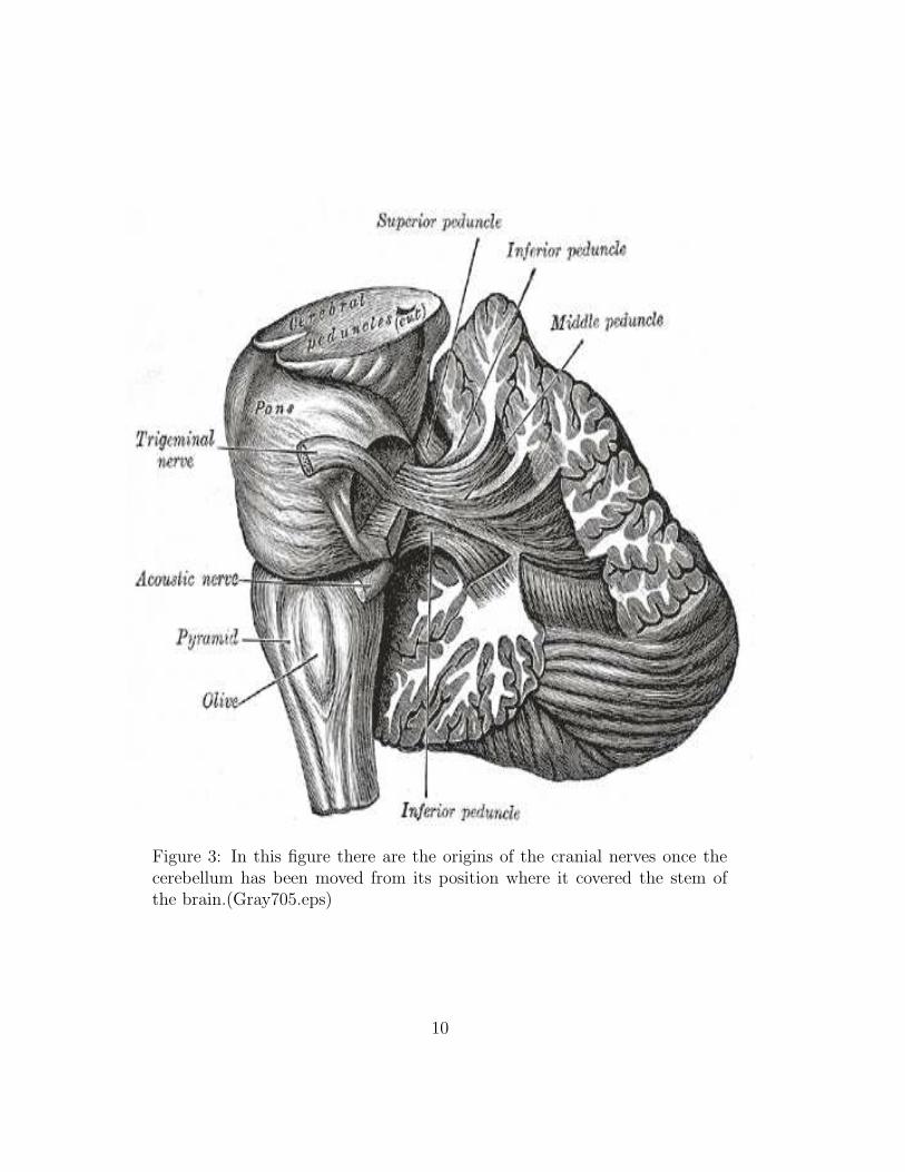
Figure 3: In this figure there are the origins of the cranial nerves once thecerebellum has been moved from its position where it covered the stem ofthe brain.(Gray705.eps)
10

Figure 4: In this figure the cerebellum was completely removed; in this waythe origin of some of 12 cranial nerves are highlighted.(Gray719.eps)
11

III– oculomotor nerveIV – trochlear nerveV – trigeminal nerveVI – abducens nerveVII – facial nerveVIII – acoustic nerve or vestibulocochlear nerveIX – glossopharyngeal nerveX – vagus nerve or pneumogastric nerveXI – spinal accessory nerveXII – hypoglossal nerveAs we shall see in the next section, the nerves from VI to X are the
nerves which are more important in our considerations: the other nervesare involved in autism but take part in the inflammation indirectly. We saythey are “dragged” because of topographic proximity of nerves belonging toneighbouring zones (from V up and from XI down).
Now we shall examine which kind of disorders can be explained with thetheory of cranial nerves.
3.1 Abducens nerve (VI) - Other nerves depending onit: II,III,IV
This nerve concerns the autistic disorder which affects the eye; more exactlywe refer to the muscular apparatus which moves the eye by means of theextra-ophthalmic six muscles. As we shall see in the following, the autisticdisorder is essentially more motorial than sensorial in nature i.e., in general,the MET factor concerns mainly the conjunction of nerve with the muscle.
In figure 5 at page 13 the six ocular muscles are shown; the lateralrectus muscle ( fifth in the figure) is the only one which is innervated by theabducens nerve; the other muscles are innervated by nerves II, III e IV.
According to our assumptions, the others nerves (II,III,IV) have not beenparticularly hit by the MET factor and only the VI nerve was hit. This meansthat the movement, by which the autistic person looks laterally, is, in a cer-tain measure, prevented. At this level of our diagnosis we are obliged touse sentences of the kind “in a certain way” waiting for the analysis madeby means of sophisticated devices i.e. using PET or MRI, now completelylacking. These apparati are necessary to measure the ability of the abducensnerve in behaving normally because the way of looking of small autistic chil-dren is definitely dysfunctional.
In fact small children (of the age of about 3-4 years) have a strange wayof looking which upsets parents and in general adults. This ”strange way of
12

Figure 5: Muscles: 2-higher;3-lower;4-median;5- lateral. Other muscles:9-lift of the eyelid. Other structures: 1-Zinn’s ring;7-trochlea;10-highertarsus;11-sclera;12-optical nerve. (Eyemuscles.eps)
13
behaving” consists in running to and fro very near a wall trying to look inthe opposite directions of their motion. The more obvious explanation of thisbehaviour is that the child, who is not able to speak, sees the objects havinga non natural placement in space and so he/she tries to find, subconsciouslyof course, a different solution to this problem. He/she is trying to rotate theeyes sideways accompanying them with the mouvement of the head; in thisway he/she gets over the difficulty of rotating the eyes sideways.
When the optical system tends to normalize because of development withage, as we have seen previously, there is always, in the autistic way of ob-serving, a strong incapacity of looking sideways. Therefore the autistics,if called by a person at their side, find it difficult to make this movement:keeping their head still rotates the eyes to look but he finds it better to doit this way: rotating their head keeping his eyes looking straight in front.
To this end we notice that the complete interruption of the VI nervecauses a diplopia (i.e. a double vision) due to the lack of the action of thelateral rectus muscle: the eye is compelled to stay in a median position inthe orbit. This situation is very similar to the autistic’s way of looking. Thedifference to normal people is however that the autistic person is not able totell other people the discomfort of his situation. So, I suppose, he is runningalong a wall trying to correct this “double vision” as mentioned above.
We notice that the abducens (VI), facial (VII), oculomotor (III) andtrochlear (VI) nerves are very close in a very small area. This area is there-fore particularly crowded with nuclei of nerves. So it would be enough forthe “MET factor” to cause the inflammation of a single nerve that such aninflammation would also affect other nerve nuclei nearby (see figure 6 atpage 15).
3.2 Facial nerve (VII). Other nerves depending on it:
V, VIII
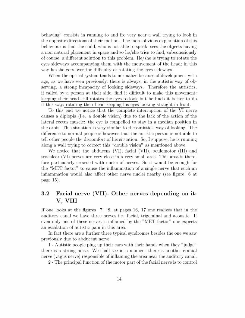
If one looks at the figures 7, 8, at pages 16, 17 one realizes that in theauditory canal we have three nerves i.e. facial, trigeminal and acoustic. Ifeven only one of these nerves is inflamed by the ”MET factor” one expectsan escalation of autistic pain in this area.
In fact there are a further three typical syndromes besides the one we sawpreviously due to abducent nerve.
1 - Autistic people plug up their ears with their hands when they ”judge”there is a strong noise. We shall see in a moment there is another cranialnerve (vagus nerve) responsible of inflaming the area near the auditory canal.
2 - The principal function of the motor part of the facial nerve is to control
14

Figure 6: The nuclei which give origin to the abducens and facialnerves.(Brainstem Abducens.eps)
15

Figure 7: Part of the auditory canal; next figure shows the internal of theauditory canal with the nerves.(Gray138.eps)
16
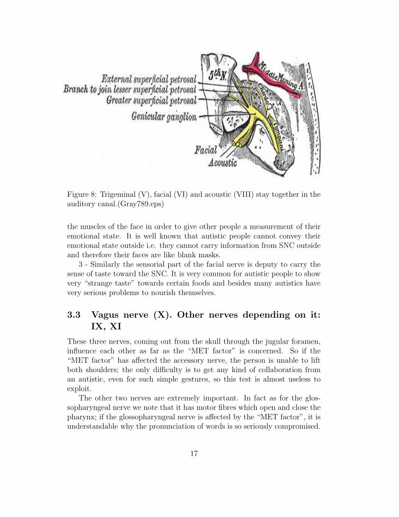
Figure 8: Trigeminal (V), facial (VI) and acoustic (VIII) stay together in theauditory canal.(Gray789.eps)
the muscles of the face in order to give other people a measurement of theiremotional state. It is well known that autistic people cannot convey theiremotional state outside i.e. they cannot carry information from SNC outsideand therefore their faces are like blank masks.
3 - Similarly the sensorial part of the facial nerve is deputy to carry thesense of taste toward the SNC. It is very common for autistic people to showvery “strange taste” towards certain foods and besides many autistics havevery serious problems to nourish themselves.
3.3 Vagus nerve (X). Other nerves depending on it:IX, XI
These three nerves, coming out from the skull through the jugular foramen,influence each other as far as the “MET factor” is concerned. So if the“MET factor” has affected the accessory nerve, the person is unable to liftboth shoulders; the only difficulty is to get any kind of collaboration froman autistic, even for such simple gestures, so this test is almost useless toexploit.
The other two nerves are extremely important. In fact as for the glos-sopharyngeal nerve we note that it has motor fibres which open and close thepharynx; if the glossopharyngeal nerve is affected by the “MET factor”, it isunderstandable why the pronunciation of words is so seriously compromised.
17

This situation is even worse because of the presence of the vagus nerve. Thisnerve in fact has a branch going to the larynx and the other going to thepharynx. If the “MET factor” prevents these branches to operate correctly,together with the glossopharyngeal nerve, it is obvious that it becomes almostimpossible to pronounce words so that autism is a typical disease affecting“dumb people”.
4 Vagus nerve and gastrointestinal tract
As it is well known the vagus nerve is responsible for a lot of innerva-tions. Are due to the vagus nerve the heartbeat, gastrointestinal peristal-sis, perspiration, movements of some muscles of the mouth etc. Besidesthis the vagus nerve receives sensations from the external ear by means ofAlderman’s nerve.
The most important branches of the vagus nerve are: 1 innervation of theheart and 2 innervation of the intestine.
4.1 Innervation of the heart
It is well known that the innervation of the heart, though being due to thevagus nerve, forms a subsystem of the nerve itself for the following reasons: 1the cardiac muscle is more similar to a skeletal muscle; 2 the action potentialwhich is responsible for the heartbeat is generated inside the heart itself; 3the transfer of these impulses happens for the whole cardiac muscle causingin this way a synchronous wave which goes from auricles to ventricles.
For these reasons the cardiac muscle cannot behave like the rest of othermuscles. So certain cardiac dysfunctions can be detected by means of thepulmonary system which is similarly innervated by the same branch of thevagus nerve.
In our italian association of parents (Angsa: Associazione Nazionale Gen-itori Soggetti Autistici), one year ago a case came to light which can beprobably explained by means of our theory. An 24 year old autistic boy diedin this way: one month before his feet swelled up and his outer ear swelledtoo assuming a blue colour, then he went to an emergency unit and there hedied. The post-mortem examination showed a pulmonary cardiac oedema.
These facts put together, can be explained, I suppose, assuming that,according to our theory, the vagus system was inflamed. I know quite wellthat a single case is not enough to have the absolute certainty that somethingis going wrong. However one must be very cautious and always alert in thecase of autistics who are not able to convey their state of health. As far
18

as I know both parents and doctors give no importance to the health of thehearts of autistic people they are so much concerned only by the psychologicalproblems. For the same reason we lack statistical data on heart disease ofautistic people so that anything can happen like what happened to thatItalian autistic boy.
There is another disorder, linked to the vagus system, that is a kind ofintestinal trouble which is considered by medical doctors as one unimportantcolitis. The man who stressed the importance of this kind of colitis for autisticpeople was Prof. Kalle Reichelt of the University of Oslo: it is due, as weshall see according to the present theory, to the particular conformation ofthe intestinal nervous system.
4.2 Enteric nervous system (ENS)
It is well known that the autonomic nervous system (ANS) is formed bythe sympathetic system and the parasympathetic system: the sympatheticganglions are near the backbone and the parasympathetic ganglions are nearthe organ of which they are targets (heart, salivary gland etc.)(see figure 9page 20).
Research in gastroenterology came recently to the conclusion that therelative part to the vagus nerve, which innervates the intestine, must beseparate from the ANS and was called enteric nervous system (ENS). Sucha system is endowed of so many peculiarities that it is also called “secondbrain”24.
In particular it is able to: 1 - regulate the endocrine function of theintestine in a completely independent way to other parts of nervous system.2 - regulate the nerve-muscle relationship within the intestine. 3 - secretevarious very important neurotransmitters, as for instance serotonin.
4.3 Vagus nerve and intestine: some observational re-sults
Let us remember briefly the path of the vagus nerve. From figure 10 page22 we can see that this nerve goes from the skull to the stomach runningparallel to the oesophagus (let me call this part “superior vagus”) and thengoes from the duodenum to the second half of the transverse colon (let mecall this part “inferior vagus”).
24Seehttp://en.wikipedia.org/wiki/Enteric nervous system
19

Figure 9: Autonomic nervous system. Blue: parasympathetic ; Red: sympa-thetic . The vagus is drawn in various branches. The vagus which innervatethe intestine is drawn in blue with the colon icon.(Gray839.eps)
20

From this point down to the anus the innervation is due to the pelvicnerve.
Dr. Wakefield was the first to examine colonscopies of autistic childrenand found the intestinal tube inflamed. As this inflammation was of unknownorigin, he called this “autistic enterocolitis”. During the same period he madethe hypothesis that autism was due to a great extent to measles vaccination.In a recent paper25 both Wakefield’s hypotheses (1 - autistic enterocolitis; 2- measles vaccination) turned out to be false. The first in particular was nota colitis at all but the origin of this inflammation remains a mystery. Herewe bring forward the following assumption: the inflammation is caused bythe “MET factor” which can be studied where the vagus nerve is visible i.e.in the intestinal tube.
In the paper by Balzola Federico et al.26 you can see the first pictures ofthis mysterious inflammation which has the following characteristics (see forinstance figure 11 page 23):
1 - From some gastroscopies it turns out that the oesophagus is inflameddown to the duodenum
2 - the same situation is present in the ileum-colon tract. This inflamma-tion stopped before the middle transverse colon.
3 - the descendant colon down to the anus was without inflammation.Of course we need further confirmation.
5 Conclusions and expectations
In the end we must draw some important observations.
Autism is a genetic disease
It has been clear since ’70 that autism is a genetic disease (as we said atthe beginning) but we have no clue as to the particular gene to investigate(see measurements by Rutter). If we however believe in the present theory,we have some ideas to look at. It is very important to repeat the measure-ments by Rutter in the light of our theory i.e. we must use gastro-scopies and colonscopies to carry out the measurements of the intestines inidentical twins. If the intestines are “identical” (i.e. the inflammations of the
25Gillian Baird et al. Measles vaccination and antibody response in autism spectrumdisorders - Arch.Dis.Child published online 5 Feb.2008
26Balzola R. et alii: Autistic enterocolitis: further confirmation in Italian autisticchilden. Meeting on “Digestive and liver disease” - (P02.87, S137, vol. 37 Suppl. 1)- March 2005
21

Figure 10: Vagus path between skull and stomach.(Gray793)
22
Figure 11: Tract of an intestine of a 30 years old autistic.
23

intestines are identical) one can draw the following conclusions: 1 - autism isgenetic and one can discard other places to look for the gene and concentrateon the intestine; 2 - if one accepts the present theory one can focus on thevagus nerve and therefore on the XII cranial nerves.
Autism is not a mental disease
Autism is a disease of the motor system linked to 1 - the XII cranial nervesand 2 - the motor system of the brain but not to the cerebral cortex. In factreferring to mental disorders we usually intend a sickness which affects thecerebral cortex, according the theory of my paper. But in our case autismaffects only the motor system of the brain, leaving undamaged the cerebralcortex in its functions.
In my opinion, if this is really the situation (i.e. autism is not a mentaldisease) it can be seen from these further following considerations.
A) In 1985 the child psychiatrist Michele Zappella, who is working at theHospital of Siena (Italy), tried to release the ability of speaking using thefollowing approach called “holding”. The child is laid down and immobilizedby other people while another person, blocking the mouth of the child, isspeaking to him/her; from time to time the mouth is freed in order to seehis/her reactions. Some parents who tried this approach gave a positivereport but the majority of them were unable to stand their son/daughterin a such piteous situation and the “holding” was quickly abandoned. Thepositive results were: 1 - the releasing of speech started a) pronouncing veryfew words and b) complicating the sentences and 2 - in a few months thechild learnt to read and write.
B) From what precedes we can fairly be sure that autism is not a mentaldisease: 1 - the releasing of speech means that “stimulating” the vagus nervewe were able to soften the inflammation and the system larynx-pharynx waspartly unblocked allowing the words to be formed. 2 - after the inflammationwas partly relieved, the very refined mechanism of reading and writing began.At the same time the neural apparatus proved to be undamaged when thecerebellum and/or brain gave the ok to the cerebral cortex.
C) Autism must not be mistaken with mental disease as there is no cere-bral lesion in the superior hemispheres of the brain. No one has been able todemonstrate that such a lesion exists; only Courchesne has proved that thebrain has a smaller volume but this does not prove that a “piece of brain”is lacking (as was claimed by people sponsoring of the theory of “mirrorneurons”).
Conclusion: autism is not a mental disease
24

Treatment of autism: what can be done?
If we are right then: 1 - autism must not be treated as a mental dis-ease; 2 - autism is a genetic disease but “sui generis”. Autism is a kindof dysfunction which acts directly on the motor part of the nervous systemand generates movements not well-coordinated. In fact all functions are un-damaged but it was as if they were plunged into a “ground noise” or, saidin other words, they are inflamed. As a consequence it is necessary to usedrugs against inflammation of the nervous system. One must take into ac-count also that this inflammation is spread over the whole organism (the XIInerves practically are everywhere because of the vagus nerve), so it is notenough to give a small dose of drug but the dose must be proportional tothe body weight. From this simple consideration one sees that autism is animportant area of research for the pharmaceutical industry.
Nowadays lacking drugs for autism, we must recur to the only drug ex-isting which soften the symptom of autism i.e. bromelain. It is a naturalcompound derived from fruits of pineapples formed by 10 or 20 enzymes (theexact composition of bromelain is not yet known; see27). Bromelain is lack-ing harmful effects on human beings at the dose of 2.5-3.0 grams/die whichshould be taken to get a positive effect on autism. We point out that, at amuch smaller dose, bromelain is used in dentistry for the treatment of thenerves of teeth irritated by infection.
As a last topic I wish to recall that a particular diet was prescribed forautistic people by Prof. Kalle Reichelt of the University of Oslo. Accordingto this diet foods containing gluten and casein should be prohibited. In Italyover the last 20 years many people have tried this diet (essentially becauseof my intervention as President of ANGSA) with good results. But this dietis more widespread in the USA and in 2008 (or 2009) the National Instituteof Health (NIH) is expected to publish the following experimental question:Is Reichelt’s diet effective in weakening the symptom of autism?
In any case it is not at all clear why a diet of any kind should work. In myopinion the only answer to this question comes from the present theory. Infact if one believes that autism is a mental disease there is no way to answerthe question, while if one believes that autism is produced by the XII nervesa hint to a solution of this puzzle is possible. What I mean is the role ofthe vagus nerve which is 1) present in the whole intestine as a unique nerve
27http://xoomer.alice.it/bioresautism/theory-app-A.html.See also: http://xoomer.alice.it/bioresautism/theory-int.html;http://xoomer.alice.it/bioresautism/theory-1.html andhttp://xoomer.alice.it/bioresautism/theory-2.htmlfor discussions with Rimland Association and Reichelt’s hypotesis
25

and 2) if this nerve has some dysfunction of a genetic nature then what canhappen is 3) the digestion of particular foods can be made difficult or evenact as a poison for the intestine.
Personally I “believe” in this diet simply because it has saved my son fromgoing into a mental hospital. Obviously a single case cannot be generalized(so we must wait for the results of the NIH ) so medical interventions mustbe applied even if they only bring benefit to the single person.
Conclusion: apart from the psychological interventions the biologicalinterventions are: 1 - use of bromelain; 2 - Reichelt’s diet (after favourableopinion of NIH); 3 - the “holding” (where possible). All the three points arein agreement with the present theory.
26