Embed Size (px)
Citation preview

on June 9, 2018http://rsob.royalsocietypublishing.org/Downloaded from
rsob.royalsocietypublishing.org
ReviewCite this article: Bayliss R, Fry A, Haq T, Yeoh
S. 2012 On the molecular mechanisms of
mitotic kinase activation. Open Biol 2: 120136.
http://dx.doi.org/10.1098/rsob.120136
Received: 18 September 2012
Accepted: 12 October 2012
Subject Area:structural biology/biochemistry/cellular
biology/molecular biology
Keywords:mitosis, protein kinases, regulation, protein
structures
Author for correspondence:Richard Bayliss
e-mail: [email protected]
& 2012 The Authors. Published by the Royal Society under the terms of the Creative Commons AttributionLicense http://creativecommons.org/licenses/by/3.0/, which permits unrestricted use, provided the originalauthor and source are credited.
On the molecular mechanisms ofmitotic kinase activationRichard Bayliss1, Andrew Fry1, Tamanna Haq1
and Sharon Yeoh2
1Department of Biochemistry, and 2Centre for Translational Therapeutics,Henry Wellcome Laboratories for Structural Biology, University of Leicester,Lancaster Road, Leicester LE1 9HN, UK
1. SummaryDuring mitosis, human cells exhibit a peak of protein phosphorylation that
alters the behaviour of a significant proportion of proteins, driving a dramatic
transformation in the cell’s shape, intracellular structures and biochemistry.
These mitotic phosphorylation events are catalysed by several families of
protein kinases, including Auroras, Cdks, Plks, Neks, Bubs, Haspin and
Mps1/TTK. The catalytic activities of these kinases are activated by phos-
phorylation and through protein–protein interactions. In this review, we
summarize the current state of knowledge of the structural basis of mitotic
kinase activation mechanisms. This review aims to provide a clear and compre-
hensive primer on these mechanisms to a broad community of researchers,
bringing together the common themes, and highlighting specific differences.
Along the way, we have uncovered some features of these proteins that have
previously gone unreported, and identified unexplored questions for future
work. The dysregulation of mitotic kinases is associated with proliferative dis-
orders such as cancer, and structural biology will continue to play a critical role
in the development of chemical probes used to interrogate disease biology and
applied to the treatment of patients.
2. Protein kinases in mitotic regulationThe eukaryotic cell cycle can be very broadly divided into two main stages:
mitosis and interphase. During mitosis, the condensed chromosomes segregate
into two genetically identical daughter cells so that each daughter cell contains
a full set of chromosomes and a single centrosome. Chromosome segregation is
carried out by the mitotic spindle, a dynamic molecular machine formed from
microtubules and hundreds of other proteins. The fidelity of chromosome seg-
regation is enhanced by cell-cycle checkpoints such as the spindle assembly
checkpoint (SAC), which monitors the assembly of the mitotic spindle and
delays chromosome segregation until every chromosome is correctly attached
to spindle microtubules via kinetochores [1]. Mitotic events require precise con-
trol and coordination, involving a large number of proteins, including protein
kinases, working together to choreograph the interplay between chromosomes
and microtubules (figure 1). The activities of a subset of protein kinases are cru-
cial for the proper regulation of mitosis, and disruption of any of these proteins
can generate an aberrant mitotic phenotype. Dysregulation of mitotic kinases
can result in severe mitotic defects that result in aneuploidy and genetic
instability. These defects can subsequently give rise to a variety of diseases of
chromosomal aberrations, such as cancer and Down’s syndrome [2–4]. Mitotic
kinases are attractive cancer drug targets as alternatives to current anti-mitotic
drugs used in the clinic, which function by binding microtubules. The

PLK1 AURA AURBMPS1
CDK1 AURB MPS1BUB1 BUB1B
PLK1 AURB HaspinNEK6 NEK7
CDK1 PLK1 AURAAURB MPS1 Haspin
BUB1 BUB1B
CDK1 PLK1 AURAAURB
NEK2 NEK6 NEK9
anaphase
spindleassemblycheckpoint
metaphase
prophasetelophase and cytokinesis
interphaseDNAdamagecheckpoint
Figure 1. An illustration of the protein kinases that are discussed in this review and the mitotic events they coordinate. Many of the kinases play roles in multiplestages of mitosis. Progress through the cell cycle is restricted by checkpoints that serve to maintain the fidelity of genetic transmission (red boxes).
rsob.royalsocietypublishing.orgOpen
Biol2:120136
2
on June 9, 2018http://rsob.royalsocietypublishing.org/Downloaded from
expectation is that compounds targeting mitotic kinases
should exhibit fewer dose-limiting side effects as they
would not target non-dividing cells [5]. The elaboration of
the functions of mitotic kinases is a highly active subject of
research and the state of play has been recently reviewed [6].
The activities of mitotic kinases are regulated in both
space and time by post-translational modifications and
protein–protein interactions. The most common forms of
post-translational modification are phosphorylation (usually
activating the kinase) and ubiquitination (generally targeting
the kinase to the proteasome for degradation). Protein-
binding partners can either stimulate or activate kinase
activity, and can specify the subcellular localization of a
population of the kinase. Unravelling these mechanisms of
regulation will be crucial for understanding the aetiology of
some human diseases and developing new treatments for
patients. The regulatory mechanisms of these kinases have
previously been compared individually with those of the
archetypal examples such as protein kinase A (PKA) or
cyclin-dependent kinases (Cdks). We feel that a cross-
comparison of mitotic kinases will be helpful for those in
the mitosis field, and to highlight the ways in which these
kinases diverge from archetypal kinases.
In this review, we appraise the available structural infor-
mation on the mitotic kinases and describe how this has
revealed mechanisms of regulation at the molecular level.
There are many protein kinases that influence mitotic events,
but for reasons of brevity and clarity, this review will focus
on the subset of kinase families that have members that are
generally agreed to carry out their main function during mito-
sis, and for which there are available crystal structures: Cdk,
Plk, Aurora, Nek, Bub, Haspin and Mps1/TTK. We will
describe the crystal structures that have been determined,
summarize the important conserved structural features that
are crucial for activity and activation, and explain the key
mechanisms of their regulation (table 1).
3. Families of mitotic kinasesThere are 518 known protein kinases in the human kinome
that are divided by primary sequence into seven groups [7].
With the exception of Cdks and Lats kinases, which belong
in the CMGC and AGC families, respectively, most mitotic
kinases are somewhat divergent from archetypal members
of the seven groups. As we will see, this divergence is also
reflected in their structures and specific regulatory mechan-
isms. We briefly outline the functions and regulation of
selected mitotic kinase families below, and refer to the
many excellent reviews that are available for further details.

Tabl
e1.
Sum
mar
yof
key
mot
ifsfo
und
inm
itotic
prot
einkin
ases
,with
non-
cano
nica
lres
idue
sem
phas
ized
inbo
ld.
kina
sesp
ineb
4,a
C,DF
GHR
DDF
GHR
Dac
tivat
ion
loop
phos
phor
ylat
edLy
s–Gl
upa
irco
ordi
nate
phos
?(a
C,b
9,HR
D,ot
her)a
GTm
otif
CDK2
(Hom
osa
piens
)
1HCK
(aut
oinhi
bite
d)
1FIN
(Cyc
linA)
1JST
(Cyc
linA,
pT16
0)
Leu6
6
Leu5
5
Phe1
46
His1
25
DFG:
145–
147
HRD:
125–
127
Thr1
60Ly
s33
Glu5
1
Arg5
0
Arg1
50
Arg1
26
Val1
64
Thr1
65
Auro
ra-A
(Hom
osa
piens
)
1MUO
,1M
Q4,1
OL6,
2WTV
1OL5
(TPX
2,pT
288)
1OL7
(pT2
88)
Leu1
96
Gln1
85
Phe2
75
His2
54
DFG:
274–
276
HRD:
254–
256
Thr2
87,T
hr28
8Ly
s162
Glu1
81
Arg1
80
Val2
79
Arg2
55
Arg2
86(A
L)
Gly2
91
Thr2
92
Auro
raB
(Xen
opus
laevis
)
2BFX
(INCE
NP,p
T232
)
Leu1
40(M
et15
6)
Gln1
29(G
ln14
5)
Phe2
19(P
he23
5)
His1
98(H
is214
)
DFG:
218–
220
(DFG
:234
–23
6)
HRD:
198–
200
(HRD
:214
–21
6)
Thr2
32
(Thr
248)
Lys1
06
Glu1
25
(Lys
122
Glu1
41)
Arg2
46
Val2
39
Arg2
15
Arg2
46(A
L)
Gly2
35
Thr2
36
Plk1
(zeb
rafish
)
3D5W
(pho
spho
)
3D5U
(non
-pho
spho
)
Phe1
16(P
he10
2)
His1
05(H
is91)
Phe1
95(P
he18
1)
His1
74(H
is160
)
DFG:
194–
196
(DFG
:180
–18
2)
HRD:
174–
176
(HRD
:160
–16
2)
Thr2
10
(Thr
196)
Lys8
2
Glu1
01
(Lys
68Gl
u87)
Met
100
(Thr
86)
Thr1
99(T
hr18
5)
Arg1
75(A
rg16
1)
Lys2
08(L
ys19
4)(A
L)
Gly2
13
Thr2
14
Nek2
(Hom
osa
piens
)
2W5B
,2JA
V(in
activ
e)
Tyr7
0
Leu5
9
Phe1
60
His1
39
DFG:
159–
161
HRD:
139–
141
Thr1
70,S
er17
1,Th
r175
Lys3
7
Glu5
5
Ala4
7
Arg1
64
Arg1
40
Gly1
78
Thr1
79
Nek7
(Hom
osa
piens
)
2WQM
Tyr9
7
Leu8
6
Leu1
80
His1
59
DLG:
179–
181
HRD:
159–
161
Ser1
95Ly
s63
Glu8
2
Lys8
1
Arg1
60
Arg1
84
Gly1
98
Thr1
99
Hasp
in(H
omo
sapie
ns)
2VUW
,2W
B8
Leu5
59
Ser5
39
Tyr6
88
His6
47
DYT:
687–
689
HRD:
647–
649
notp
hosp
hory
lated
Lys5
11
Glu5
35
Pro5
34
Arg6
92
Arg6
48
Tyr7
22b
(Con
tinue
d.)
rsob.royalsocietypublishing.orgOpen
Biol2:120136
3
on June 9, 2018http://rsob.royalsocietypublishing.org/Downloaded from

Tabl
e1.
.(Co
ntin
ued.
)
kina
sesp
ineb
4,a
C,DF
GHR
DDF
GHR
Dac
tivat
ion
loop
phos
phor
ylat
edLy
s–Gl
upa
irco
ordi
nate
phos
?(a
C,b
9,HR
D,ot
her)a
GTm
otif
Bub1
(Hom
osa
piens
)
3E7E
Phe8
52
Gly8
34
Leu9
47
His9
15
DLG:
946–
948
HGD:
915–
917
notp
hosp
hory
lated
Glu8
30
Lys8
21
Notp
hosp
hory
lated
Gly9
59
Thr9
60
Glu9
67
Thr9
68
Mps
1(H
omo
sapie
ns)
2ZM
C(a
po)
3H9F
(pho
spho
)
Leu5
88
Leu5
75
Phe6
65
His6
45
DFG:
664–
666
HSD:
644–
647
Thr6
75,T
hr67
6,Th
r686
Lys5
53
Glu5
71
Asn5
70
Asn6
69
Ser6
46
Lys6
81(A
L)
Lys7
08,L
ys71
0(se
e
text
)
Gly6
85
Thr6
86
aa
Cis
resi
du
ep
rece
din
gG
lu,b
9is
resi
du
eD
FGþ
3,A
Lis
acti
vat
ion
loo
ptw
ore
sid
ues
pre
ced
ing
ph
osp
ho
ryla
ted
Ser
/T
hr,
ital
ics
sho
wp
red
icte
dre
sid
ues
wh
ere
no
rele
van
tst
ruct
ure
exis
ts.
bF
un
ctio
nal
lyeq
uiv
alen
tto
GT
mo
tif
inth
atth
esi
de
chai
nfo
rms
H-b
on
dw
ith
cata
lyti
cA
sp64
9.
rsob.royalsocietypublishing.orgOpen
Biol2:120136
4
on June 9, 2018http://rsob.royalsocietypublishing.org/Downloaded from
3.1. Cyclin-dependent kinasesCdks are a large family of serine/threonine kinases that have
been well studied since their discovery in the 1970s [8]. Cdks
become active when bound to their cognate cyclin partner
protein, and subsequently by phosphorylation by a Cdk-
activating kinase (CAK) [9,10]. As their name suggests, the
level of cyclin protein expression, and hence Cdk activity,
varies through the cell cycle. Hence, different Cdk/cyclin
complexes function in distinct phases of the cell cycle, with,
for example, Cdk2/Cyclin E associated with G1/S pro-
gression, and Cdk1/Cyclin B associated with M-phase entry.
Cdk1 was first discovered in Saccharomyces cerevisiae(cdc28) and in Schizosaccharomyces pombe (cdc2), and is the
key regulator of the G2/M transition, when it is subject to
control by the DNA damage checkpoint [11]. Cdk1 is regu-
lated by inhibitory phosphorylation on Thr14 and Tyr15,
which is reversed by phosphatases of the Cdc25 family [9].
Cdk1 forms a complex with Cyclin B, and is then phosphory-
lated by CAK. Cdk1/Cyclin B activity drives cells into
mitosis through phosphorylation of many substrates that con-
tribute to chromosome condensation, nuclear envelope
breakdown and spindle assembly [11]. Cdk1/Cyclin B
also stimulates activation of the anaphase-promoting
complex/cyclosome (APC/C), which is required to trigger
chromosome segregation through proteolytic degradation of
securin, and mitotic exit through degradation of cyclin B,
thereby leading to the loss of cellular Cdk1 activity.
3.2. Polo-like kinasesThere are four Polo-like kinases (Plk1–4), all of which are pro-
posed to have functional roles within the cell cycle [12,13]. Plks
are serine/threonine kinases, which possess an N-terminal cat-
alytic domain, a destruction box (D-box) and a C-terminal
region containing either one or two polo-box domains [14].
The best-characterized member of the family is Polo-
like-kinase-1 (Plk1), which is known to play an essential role
in many processes associated with cell division, including
mitotic entry, centrosome separation, bipolar spindle assembly
and cytokinesis. Expression of Plk1 is highest in late G2, where it
regulates Cdk1/Cyclin B activity through phosphorylation
of the phosphatase Cdc25, and in M phase, where it has many
substrates. Plk1 is activated through phosphorylation on its acti-
vation loop by upstream kinases including Aurora-A, and
phosphorylation of Plk1 peaks between metaphase and
anaphase [15]. In late mitosis, Plk1 is ubiquitinated by the
APC/C, leading to its degradation by the proteasome [12,16].
3.3. Aurora kinasesThe first members of the Aurora family of serine/threonine
protein kinases were discovered in yeast and Drosophila[13,17,18]. Although yeast have only a single Aurora kinase,
and Drosophila have two, there are three members of this
family in humans: Aurora-A, -B and -C. Aurora-A is associ-
ated with centrosomes and the mitotic spindle, and
functions in mitotic entry, centrosome maturation and mitotic
spindle assembly, and phosphorylates microtubule-associ-
ated proteins such as TACC3 [19,20]. Aurora-A also has
non-mitotic roles in neurite extension and in stimulating the
disassembly of cilia [21,22]. During early mitosis, Aurora-B
associates with chromosomes, and phosphorylates histone

rsob.royalsocietypublishing.orgOpen
Biol2:120136
5
on June 9, 2018http://rsob.royalsocietypublishing.org/Downloaded from
H3 on Ser10 and Ser28 [23]. Aurora-B is part of the chromo-
some passenger complex (CPC), a key component of the SAC
and also functions in cytokinesis [24]. Aurora-C is expressed
only in a few tissues and is believed to fulfil a similar function
to Aurora-B [25].
All three Aurora kinases are activated by phosphorylation
and through the binding of partner proteins, and are targeted
for degradation by the APC/C. They have two domains: an
N-terminal domain that includes regulatory elements and a
C-terminal catalytic domain. The regulatory domain varies
greatly among the family, whereas the C-terminal catalytic
domains are about 70 per cent homologous [26].
3.4. Never in mitosis A-related kinasesNever in mitosis A (NIMA)-related serine/threonine kinases
(Neks) were first identified in Aspergillus nidulans, where
NIMA was shown to be essential for progression through
the cell cycle [27,28]. There are 11 members of the Nek
family in humans, of which Neks 2, 6, 7 and 9 are strongly
implicated in mitotic function [29]. Of the human Nek
kinases, all but Nek6 and Nek7 have a C-terminal region
that encodes coiled-coil motifs and/or b-propeller domains
of the regulator of chromosome condensation 1 family.
Nek2 and Nek9 are believed to form homodimers through
their coiled-coil motifs [30,31].
Nek2 has the highest homology at an amino acid level to
the archetypal NIMA protein from Aspergillus and functions
in centrosome separation by phosphorylating tethering pro-
teins such as C-Nap1 [32,33]. Nek2 activity peaks during
the transition from G2 to M phase after protein phosphatase
1 (PP1) inactivation and phosphorylation [34]. Once cells
enter mitosis, Nek2 is ubiquitinated by APC/C-Cdc20 and
then degraded [35,36]. Nek9 is upstream of Neks 6 and 7 in
a recently characterized mitotic pathway that functions in
bipolar spindle assembly and centrosome separation [29,37].
Nek9 is thought to activate Nek6 and Nek7 through phos-
phorylation and allosteric mechanisms [38,39]. Very few
downstream substrates of these kinases have been identified,
with the exception of the microtubule motor protein Eg5,
which is a Nek6 substrate in centrosome separation, and
Nedd1, which is a Nek9 substrate in the recruitment of
g-tubulin to centrosomes [40,41].
3.5. Budding uninhibited by benzimidazolesBudding uninhabited by benzimidazole (Bub) proteins were
first discovered in S. cerevisiae, and there are two known
members of this family of serine/threonine protein kinases
in humans: Bub1 and Bub1B/BubR1. There are several
recent reviews that describe their functions, which we briefly
describe below [42–44].
Bub1 and BubR1 both localize to kinetochores and are
thought to play dual roles, contributing to the SAC and to
the regulation of kinetochore–MT attachments. The precise
contribution of the catalytic activities of Bubs to the SAC is
under active investigation, and many substrates of these
kinases are yet to be discovered. These proteins also have
scaffolding roles. Bub1 localizes to kinetochores in early pro-
phase, and determines kinetochore recruitment of a number
of proteins, including BubR1 and an adaptor protein Bub3.
The kinetochore localization of Bub1 and BubR1 depend on
their N-terminal TPR domains. Both kinases interact with
the b-propeller domain of Bub3 through their Gle2-binding
sequence motifs. These interactions have been characterized
by X-ray crystallography [45–47].
3.6. Haploid germ-cell-specific nuclear protein kinaseHaploid germ-cell-specific nuclear protein kinase (Haspin) is a
serine/threonine protein kinase that has an atypical catalytic
domain that is highly divergent from other eukaryotic protein
kinases at the sequence level [48]. Haspin was first identified
in mice as the product of the germ-cell-specific gene-2 and is
conserved in other eukaryotes, including S. cerevisiae. Haspin
depletion results in defective chromosome alignment on the
metaphase plate and a delay in mitotic exit due to activation
of the SAC, thought to arise in part from loss of chromosome
cohesion. The most clearly defined substrate of Haspin is
residue Thr3 of histone H3 (HH3-Thr3), a mitosis-specific
modification that is required to localize the CPC to centromeres
and activate Aurora-B [49,50]. The domain structure of Haspin
comprises a C-terminal catalytic domain and an N-terminal
region of unknown structure that is highly divergent between
Haspin homologues. Haspin protein levels and catalytic
activity appear to be constant through the cell cycle. Indeed,
although Aurora-B phosphorylates Haspin, leading to an
increase in the phosphorylation of HH3-Thr3, this is not
through an increase in the intrinsic activity of Haspin [51].
Therefore, regulation of the phosphorylation of Haspin
substrates such as HH3-Thr3 is thought to occur through
alternative mechanisms such as phosphatases, or through the
binding of a protein that inhibits Haspin activity.
3.7. Monopolar spindle-1Monopolar spindle-1 (Mps1) is a dual-specificity kinase that
was first identified in S. cerevisiae as having multiple cell-
cycle functions, including duplication of the spindle pole
body, the functional equivalent of the centrosome and the
SAC (reviewed by Liu & Winey [52]). Human Mps1, also
known as TTK, is required for proper chromosome alignment
on the metaphase plate and for the fidelity of chromosome
segregation, and might also have a role at centrosomes. Evi-
dence is clearest for a role in the SAC, as depletion of Mps1
by RNAi or blocking Mps1 activity using small molecule
inhibitors abrogates the checkpoint and markedly reduces the
duration of mitosis (reviewed by Lan & Cleveland [53]). One
key contribution of Mps1 to the SAC is in recruitment of
other checkpoint components to the kinetochore, including
Mad1, Mad2, Bub1, BubR1 and Bub3.
The catalytic domain of Mps1 is in the C-terminal region,
and the N-terminal region includes TPR repeats that promote
homodimer formation, which is important for proper Mps1
function [54]. Mps1 becomes phosphorylated on multiple
sites during mitosis, which is at least partly due to auto-
phosphorylation, and phospho-Mps1 is localized to both
kinetochores and centrosomes [55]. In late mitosis, Mps1 is
ubiquitinated by the APC/C and subsequently degraded [56].
4. The hallmarks of active kinasearchitecture
The first crystal structure of a protein kinase was of PKA in
its active form, determined by the Taylor laboratory [57].

DFG
APE
P + 1
a ctiva
tion
loop
P
HRD
b9
ATP
P
aC
aC
aC
N-lobe
C-lobe
hinge
P-loop(a) (b)
(c) (d)
substrateprotein
ATP binding cleft
HRDmotif
DFGmotif
Lys–Glusaltbridge
hydrophobic spine
phosphorylated phosphorylated residueresidue
activationsegment
GT-HRD H-bond
Lys–Glusalt bridge
HRD–phosphateinteraction
substrateprotein
substrateSer/Thr
activationsegment
N-lobe
C–lobe
b4
aC
Figure 2. Conserved structural features of the active conformation of a protein kinase. The four panels use the following colour scheme: kinase main chain and Lys/Glu pair (beige), HRD motif (blue), DFG motif side chains (red), activation segment (cyan), phosphorylated residue on activation loop (yellow), hydrophobic spine(green), ATP (magenta), substrate protein (grey). (a) Architecture of the archetypal protein kinase A (PKA) catalytic domain in cartoon representation, with keyresidues shown as sticks and the ADP ligand shown as spheres (PDB code 1JBP). The side chains of the four residues that comprise the hydrophobic spine aresurrounded by a wire mesh. This and all subsequent structure figures were produced using PYMOL. (b) Magnified view of the active site and residues involved inregulation. Key interactions between residues are indicated with dashed black lines; these do not necessarily imply hydrogen bonds. (c) Schematic that summarizesthe major features of the kinase active conformation. Versions of this diagram will be used throughout the text to illustrate differences between inactive and activestates of protein kinases. Three key interactions are labelled in black. (d ) Schematic diagram to illustrate the canonical interactions between the phosphorylatedresidue of the activation loop and basic residues in the aC-helix, b9 and the HRD motif. These interactions are not all conserved in mitotic kinases. For example,Aurora-A and Plk1 lack the basic residue on b9, and instead have a basic residue on the activation loop (shown with fainter colouring). The different parts of theactivation segment are labelled, such as the Pþ1 loop that recognizes the Pþ1 residue of the substrate protein.
rsob.royalsocietypublishing.orgOpen
Biol2:120136
6
on June 9, 2018http://rsob.royalsocietypublishing.org/Downloaded from
This structure serves as a model for understanding the
features of an active kinase. Structures of well over a hun-
dred protein kinases have been determined, and these
have yielded valuable insights into their mechanisms of
catalysis and regulation [58–60]. The protein kinase fold
is composed of two sub-domains: the N-terminal lobe
(N-lobe) and the C-terminal lobe (C-lobe), connected by a
flexible hinge region (figure 2a). The N-lobe is made up of
a five-stranded b-sheet and (usually) two a-helices, of
which the aC-helix is the structurally most conserved,
whereas the larger C-lobe is mainly a-helical. A molecule
of ATP binds in the cleft between the two lobes. The adeno-
sine base forms H-bonds with the kinase hinge region, the
ribose moiety binds to the ribose binding pocket and
the phosphate groups interact with the Gly-rich loop,
which is also called the phosphate-binding loop (P-loop).
Sites phosphorylated by kinases usually lie within unstruc-
tured regions of proteins, and structural studies using
substrate peptides have shown that the C-lobe recognizes
the sequence on either side of the phosphorylated residues
[61,62]. In this review, we will not go into any further
depth regarding the catalytic mechanism of kinases or
how they recognize ATP and protein substrates, which
have been reviewed elsewhere [59]. Instead, we will focus
on the key features of protein kinase structure, and explain
how structural changes are related to different states of
activity among mitotic kinases. A set of highly conserved
motifs form the catalytic core and key regulatory features
of protein kinases, as detailed below (figure 2b,c). Intrigu-
ingly, most mitotic kinases are divergent in at least one of
these canonical motifs, perhaps reflecting their unusual
regulatory mechanisms (table 1).

rsob.royalsocietypublishing.orgOpen
Biol2:120136
7
on June 9, 2018http://rsob.royalsocietypublishing.org/Downloaded from
The HRD and DFG motifs are defining features of protein
kinases, and the aspartic acids in these motifs are essential cat-
alytic residues. The aspartate residue of the HRD motif acts as a
base catalyst that deprotonates the substrate side chain serine/
threonine, and this region of the kinase is sometimes referred to
as the catalytic loop [63]. The aspartate of the DFG motif
coordinates a magnesium ion that activates the gamma phos-
phate of ATP, and this region of kinases is often called the
magnesium (Mg2þ)-binding loop. As with any enzyme, these
residues must be positioned correctly and in the appropriate
chemical environment in order for the kinase to catalyse phos-
phate transfer. In active kinases, the DFG motif adopts the
DFG-in conformation, and contacts the HRD motif through a
hydrophobic interaction. Displacement of these motifs into
positions incompatible with catalysis is part of the regulatory
mechanism of many kinases and can also be induced by
small molecule inhibitors of kinases, as exemplified by the
DFG-out conformation of cAbl that is induced by Imatinib/
Glivec, used in the treatment of chronic myelogenous
leukaemia and gastrointestinal stromal tumours [64].
The lysine–glutamate salt bridge between the ammonium
group of the lysine and the carboxylate group of the gluta-
mate is a characteristic feature of active protein kinases. The
lysine is located on the b3-strand and the glutamate is located
on the aC-helix, and the bridge thus links these two impor-
tant structural elements of the N-lobe together. The lysine
also interacts with the terminal phosphate groups of ATP,
stabilizing them in the correct position for catalysis [59].
The hydrophobic spine is a recently defined feature of active
kinase structures and is made up of the side chains of
four hydrophobic residues (figure 2 and table 1). The first
side chain comes from the b4 strand in the N-terminal
lobe; the second comes from the adjacent aC helix; the
third is the phenylalanine residue from the DFG motif; and,
finally, the fourth member of the spine is the histidine residue
from the HRD motif [65]. The relative positions of these four
residues are strikingly similar in active kinase structures,
where they form a continuous hydrophobic chain, or
‘spine’, shown as a green wire mesh in figure 2b.
The activation segment starts at the DFG motif and ends
at a sequence that has the consensus alanine–proline–gluta-
mic acid, called the APE motif (figure 2d ). It is often called
the T-loop or activation loop, but we will reserve this latter
term for a portion of the activation segment that contains
the primary site of activating phosphorylation, following
the same naming convention as Nolen et al. [60]. The acti-
vation segment includes several important features that are
conserved in active kinase structures. Strand b9 pairs with
b6 within the catalytic loop, immediately N-terminal to
the HRD motif. The activation loop is variable in length
and sequence, and is the primary site of regulatory phos-
phorylation [59]. The P þ 1 loop forms a pocket that
interacts with the P þ 1 site of substrates, and must therefore
form the right conformation for efficient catalysis. In serine/
threonine kinases, this requires a conserved H-bond between
a threonine (or serine) within the P þ 1 loop, which is
usually preceded by a glycine (forming a conserved GT
motif ), and the aspartic acid of the HRD motif. In tyrosine
kinases, the residue equivalent to the threonine of the GT
motif is usually a proline.
The phosphorylated residue on the activation loop is typically
coordinated by the side chains of basic residues at specific
positions on three different structural features, namely the
residue preceding the glutamic acid on aC, the residue
three positions C-terminal to the DFG motif on b9 and the
arginine residue of the HRD motif (figure 2d ). This three-
point attachment helps to stabilize the activation segment in
the correct conformation for substrate binding.
Every mitotic kinase that has been crystallized diverges
from the canonical sequence in at least one of these motifs
(highlighted in bold in table 1), and the implications of this
will be discussed throughout the review. For example, the
mitotic kinases Nek6, Nek7, Nek9, Haspin and Bub1 all
have non-canonical DFG motifs, although all conserve the
aspartic acid (table 1). However, in this case, there is as yet
no evidence that these divergent DFG motifs confer a sub-
stantial biochemical difference. Indeed, when the divergent
leucine of Nek7 was mutated to the more common phenyl-
alanine, the mutant protein exhibited comparable activity to
the wild-type protein [39].
At present, there are crystal structures of active and inac-
tive forms of Aurora-A kinase and Plk1. The structure of
Haspin has been resolved in an active form, whereas those
of Aurora-B and Bub1 are only available for a partially
active form. Mitotic kinases with only the inactive structure
include Nek2, Nek7, Haspin and Mps1/TTK. The molecular
detail obtained from the structures of these kinases in active
and inactive conformations, in conjunction with information
from biochemical assay methods, have greatly facilitated
the elucidation of protein kinase regulatory mechanisms.
Mitotic kinases for which there are currently no structures
available include Cdk1, Aurora-C, Plk2, Plk3, Nek6, Nek9,
Lats1, Lats2, Bub1b and Greatwall.
5. Common themes among the regulatorystrategies of protein kinases
Looking at mitotic kinases from a structural perspective, it
becomes apparent that kinase activation is not a binary process
(from inactive to active), but that there are intermediate states
that may exhibit varying degrees of activity [66]. We will refer
to these as either ‘inactive’ or ‘partially active’ states, because it
is difficult to judge how much catalytic activity these states
possess, based on structures alone. However, it is straightfor-
ward to identify which conformational changes need to
occur in order to bring about a fully active conformation,
and hence the mechanisms of activation can be inferred.
Kinases can adopt a variety of inactive conformations, and
the intricate control mechanisms of protein kinases are extre-
mely diverse. Nevertheless, the strategies used to regulate
the catalytic activities of a protein can readily be categorized
into phosphorylation (and consequently dephosphorylation),
autoinhibition and the binding of a partner protein [58,59].
Some kinases may use more than one of these mechanisms
in order to change their conformation from an inactive struc-
ture to an active structure and thereby achieve full activation.
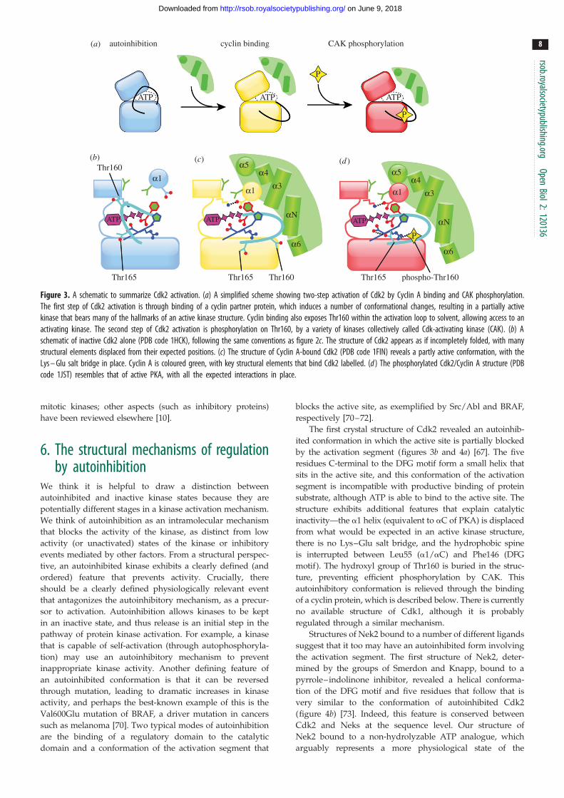
All three mechanisms are used in the regulation of Cdk2,
which on its own exists in an inactive, autoinhibited state,
achieves partial activity upon binding of Cyclin A and
achieves full activity only when this complex is phosphory-
lated on the activation loop (figure 3) [67–69]. The activation
of Cdk2 has been extensively characterized, and it thus
serves as a useful basis for the analysis of other kinase acti-
vation mechanisms. In this review, we will only discuss
aspects of Cdk2 regulation that have clear relevance to the
(a)
(b) (c) (d )a5
a3
aN
a6
a4
a5
a3
aN
a6
a4
ATP
ATP ATP ATP
P
cyclin binding CAK phosphorylation
P
autoinhibition
a1
ATP ATP
a1
P
a1Thr160
Thr165 Thr160Thr165 phospho-Thr160Thr165
Figure 3. A schematic to summarize Cdk2 activation. (a) A simplified scheme showing two-step activation of Cdk2 by Cyclin A binding and CAK phosphorylation.The first step of Cdk2 activation is through binding of a cyclin partner protein, which induces a number of conformational changes, resulting in a partially activekinase that bears many of the hallmarks of an active kinase structure. Cyclin binding also exposes Thr160 within the activation loop to solvent, allowing access to anactivating kinase. The second step of Cdk2 activation is phosphorylation on Thr160, by a variety of kinases collectively called Cdk-activating kinase (CAK). (b) Aschematic of inactive Cdk2 alone (PDB code 1HCK), following the same conventions as figure 2c. The structure of Cdk2 appears as if incompletely folded, with manystructural elements displaced from their expected positions. (c) The structure of Cyclin A-bound Cdk2 (PDB code 1FIN) reveals a partly active conformation, with theLys – Glu salt bridge in place. Cyclin A is coloured green, with key structural elements that bind Cdk2 labelled. (d ) The phosphorylated Cdk2/Cyclin A structure (PDBcode 1JST) resembles that of active PKA, with all the expected interactions in place.
rsob.royalsocietypublishing.orgOpen
Biol2:120136
8
on June 9, 2018http://rsob.royalsocietypublishing.org/Downloaded from
mitotic kinases; other aspects (such as inhibitory proteins)
have been reviewed elsewhere [10].
6. The structural mechanisms of regulationby autoinhibition
We think it is helpful to draw a distinction between
autoinhibited and inactive kinase states because they are
potentially different stages in a kinase activation mechanism.
We think of autoinhibition as an intramolecular mechanism
that blocks the activity of the kinase, as distinct from low
activity (or unactivated) states of the kinase or inhibitory
events mediated by other factors. From a structural perspec-
tive, an autoinhibited kinase exhibits a clearly defined (and
ordered) feature that prevents activity. Crucially, there
should be a clearly defined physiologically relevant event
that antagonizes the autoinhibitory mechanism, as a precur-
sor to activation. Autoinhibition allows kinases to be kept
in an inactive state, and thus release is an initial step in the
pathway of protein kinase activation. For example, a kinase
that is capable of self-activation (through autophosphoryla-
tion) may use an autoinhibitory mechanism to prevent
inappropriate kinase activity. Another defining feature of
an autoinhibited conformation is that it can be reversed
through mutation, leading to dramatic increases in kinase
activity, and perhaps the best-known example of this is the
Val600Glu mutation of BRAF, a driver mutation in cancers
such as melanoma [70]. Two typical modes of autoinhibition
are the binding of a regulatory domain to the catalytic
domain and a conformation of the activation segment that
blocks the active site, as exemplified by Src/Abl and BRAF,
respectively [70–72].
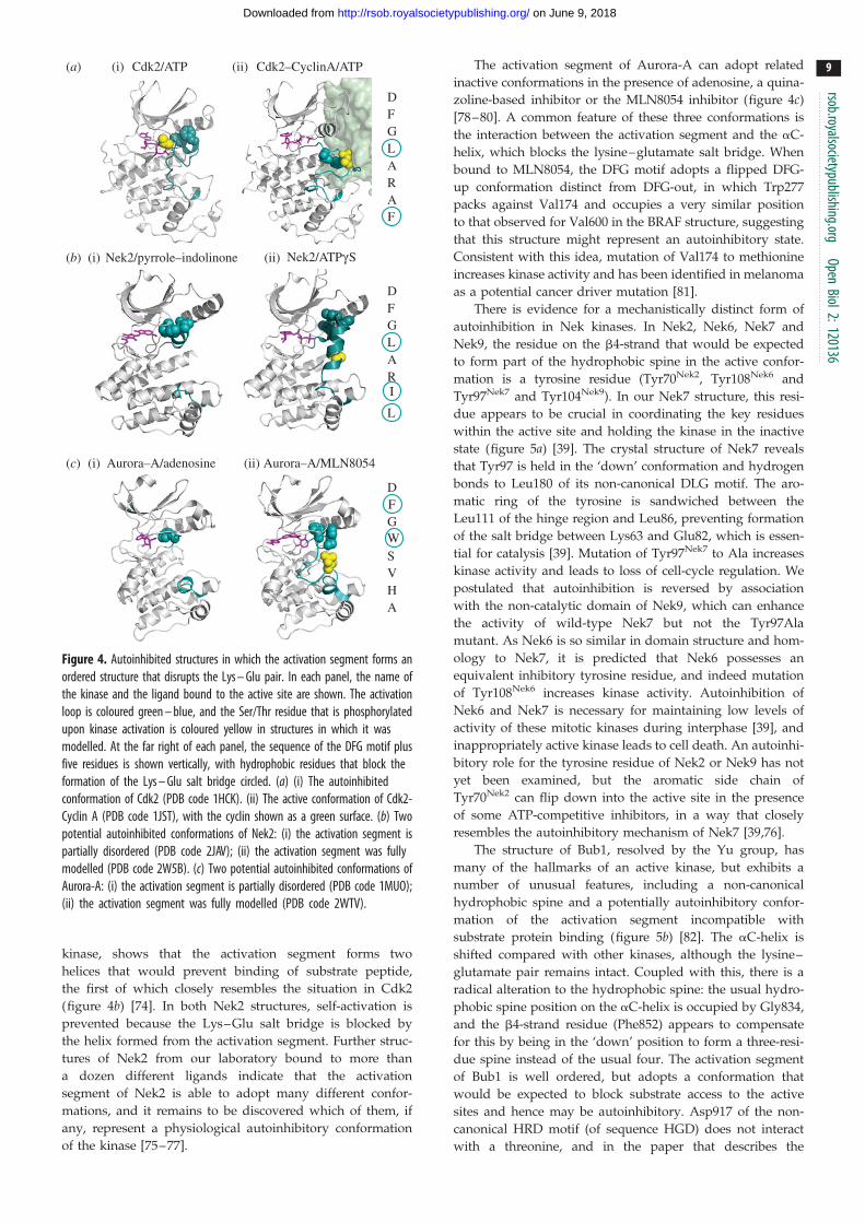
The first crystal structure of Cdk2 revealed an autoinhib-
ited conformation in which the active site is partially blocked
by the activation segment (figures 3b and 4a) [67]. The five
residues C-terminal to the DFG motif form a small helix that
sits in the active site, and this conformation of the activation
segment is incompatible with productive binding of protein
substrate, although ATP is able to bind to the active site. The
structure exhibits additional features that explain catalytic
inactivity—the a1 helix (equivalent to aC of PKA) is displaced
from what would be expected in an active kinase structure,
there is no Lys–Glu salt bridge, and the hydrophobic spine
is interrupted between Leu55 (a1/aC) and Phe146 (DFG
motif). The hydroxyl group of Thr160 is buried in the struc-
ture, preventing efficient phosphorylation by CAK. This
autoinhibitory conformation is relieved through the binding
of a cyclin protein, which is described below. There is currently
no available structure of Cdk1, although it is probably
regulated through a similar mechanism.
Structures of Nek2 bound to a number of different ligands
suggest that it too may have an autoinhibited form involving
the activation segment. The first structure of Nek2, deter-
mined by the groups of Smerdon and Knapp, bound to a
pyrrole–indolinone inhibitor, revealed a helical conforma-
tion of the DFG motif and five residues that follow that is
very similar to the conformation of autoinhibited Cdk2
(figure 4b) [73]. Indeed, this feature is conserved between
Cdk2 and Neks at the sequence level. Our structure of
Nek2 bound to a non-hydrolyzable ATP analogue, which
arguably represents a more physiological state of the
Cdk2/ATP(a) (i) (ii)
(b)
(c)
Nek2/pyrrole–indolinone Nek2/ATPgS
Aurora–A/adenosine
DFGLARAF
DFGLARI
L
DFGWSVHA
Cdk2–CyclinA/ATP
Aurora–A/MLN8054
(i) (ii)
(i) (ii)
Figure 4. Autoinhibited structures in which the activation segment forms anordered structure that disrupts the Lys – Glu pair. In each panel, the name ofthe kinase and the ligand bound to the active site are shown. The activationloop is coloured green – blue, and the Ser/Thr residue that is phosphorylatedupon kinase activation is coloured yellow in structures in which it wasmodelled. At the far right of each panel, the sequence of the DFG motif plusfive residues is shown vertically, with hydrophobic residues that block theformation of the Lys – Glu salt bridge circled. (a) (i) The autoinhibitedconformation of Cdk2 (PDB code 1HCK). (ii) The active conformation of Cdk2-Cyclin A (PDB code 1JST), with the cyclin shown as a green surface. (b) Twopotential autoinhibited conformations of Nek2: (i) the activation segment ispartially disordered (PDB code 2JAV); (ii) the activation segment was fullymodelled (PDB code 2W5B). (c) Two potential autoinhibited conformations ofAurora-A: (i) the activation segment is partially disordered (PDB code 1MUO);(ii) the activation segment was fully modelled (PDB code 2WTV).
rsob.royalsocietypublishing.orgOpen
Biol2:120136
9
on June 9, 2018http://rsob.royalsocietypublishing.org/Downloaded from
kinase, shows that the activation segment forms two
helices that would prevent binding of substrate peptide,
the first of which closely resembles the situation in Cdk2
(figure 4b) [74]. In both Nek2 structures, self-activation is
prevented because the Lys–Glu salt bridge is blocked by
the helix formed from the activation segment. Further struc-
tures of Nek2 from our laboratory bound to more than
a dozen different ligands indicate that the activation
segment of Nek2 is able to adopt many different confor-
mations, and it remains to be discovered which of them, if
any, represent a physiological autoinhibitory conformation
of the kinase [75–77].
The activation segment of Aurora-A can adopt related
inactive conformations in the presence of adenosine, a quina-
zoline-based inhibitor or the MLN8054 inhibitor (figure 4c)
[78–80]. A common feature of these three conformations is
the interaction between the activation segment and the aC-
helix, which blocks the lysine–glutamate salt bridge. When
bound to MLN8054, the DFG motif adopts a flipped DFG-
up conformation distinct from DFG-out, in which Trp277
packs against Val174 and occupies a very similar position
to that observed for Val600 in the BRAF structure, suggesting
that this structure might represent an autoinhibitory state.
Consistent with this idea, mutation of Val174 to methionine
increases kinase activity and has been identified in melanoma
as a potential cancer driver mutation [81].
There is evidence for a mechanistically distinct form of
autoinhibition in Nek kinases. In Nek2, Nek6, Nek7 and
Nek9, the residue on the b4-strand that would be expected
to form part of the hydrophobic spine in the active confor-
mation is a tyrosine residue (Tyr70Nek2, Tyr108Nek6 and
Tyr97Nek7 and Tyr104Nek9). In our Nek7 structure, this resi-
due appears to be crucial in coordinating the key residues
within the active site and holding the kinase in the inactive
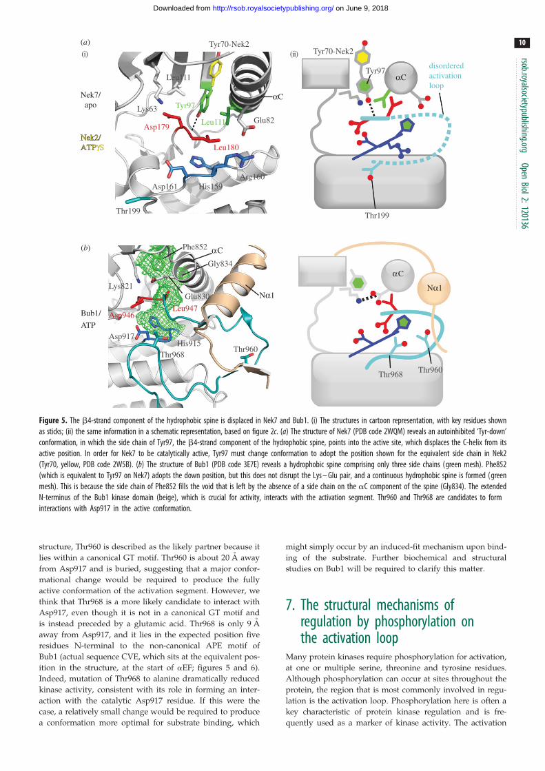
state (figure 5a) [39]. The crystal structure of Nek7 reveals
that Tyr97 is held in the ‘down’ conformation and hydrogen
bonds to Leu180 of its non-canonical DLG motif. The aro-
matic ring of the tyrosine is sandwiched between the
Leu111 of the hinge region and Leu86, preventing formation
of the salt bridge between Lys63 and Glu82, which is essen-
tial for catalysis [39]. Mutation of Tyr97Nek7 to Ala increases
kinase activity and leads to loss of cell-cycle regulation. We
postulated that autoinhibition is reversed by association
with the non-catalytic domain of Nek9, which can enhance
the activity of wild-type Nek7 but not the Tyr97Ala
mutant. As Nek6 is so similar in domain structure and hom-
ology to Nek7, it is predicted that Nek6 possesses an
equivalent inhibitory tyrosine residue, and indeed mutation
of Tyr108Nek6 increases kinase activity. Autoinhibition of
Nek6 and Nek7 is necessary for maintaining low levels of
activity of these mitotic kinases during interphase [39], and
inappropriately active kinase leads to cell death. An autoinhi-
bitory role for the tyrosine residue of Nek2 or Nek9 has not
yet been examined, but the aromatic side chain of
Tyr70Nek2 can flip down into the active site in the presence
of some ATP-competitive inhibitors, in a way that closely
resembles the autoinhibitory mechanism of Nek7 [39,76].
The structure of Bub1, resolved by the Yu group, has
many of the hallmarks of an active kinase, but exhibits a
number of unusual features, including a non-canonical
hydrophobic spine and a potentially autoinhibitory confor-
mation of the activation segment incompatible with
substrate protein binding (figure 5b) [82]. The aC-helix is
shifted compared with other kinases, although the lysine–
glutamate pair remains intact. Coupled with this, there is a
radical alteration to the hydrophobic spine: the usual hydro-
phobic spine position on the aC-helix is occupied by Gly834,
and the b4-strand residue (Phe852) appears to compensate
for this by being in the ‘down’ position to form a three-resi-
due spine instead of the usual four. The activation segment
of Bub1 is well ordered, but adopts a conformation that
would be expected to block substrate access to the active
sites and hence may be autoinhibitory. Asp917 of the non-
canonical HRD motif (of sequence HGD) does not interact
with a threonine, and in the paper that describes the
Thr960Thr968
Asp917His915
Phe852
Glu830Lys821
Gly834
Asp946Leu947
Bub1/
ATP
Thr199
Leu111
Lys63Glu82
Tyr97
Tyr70-Nek2(i) (ii)
Leu111Asp179
Leu180
Asp161 His159Arg160
Nek7/apo
(a)
(b)
αC
αC
αC
αC
Tyr70-Nek2
Tyr97
Thr199
disorderedactivation loop
Nα1Nα1
Thr960Thr968
Nek2/Nek2/ATPATPgS
Figure 5. The b4-strand component of the hydrophobic spine is displaced in Nek7 and Bub1. (i) The structures in cartoon representation, with key residues shownas sticks; (ii) the same information in a schematic representation, based on figure 2c. (a) The structure of Nek7 (PDB code 2WQM) reveals an autoinhibited ‘Tyr-down’conformation, in which the side chain of Tyr97, the b4-strand component of the hydrophobic spine, points into the active site, which displaces the C-helix from itsactive position. In order for Nek7 to be catalytically active, Tyr97 must change conformation to adopt the position shown for the equivalent side chain in Nek2(Tyr70, yellow, PDB code 2W5B). (b) The structure of Bub1 (PDB code 3E7E) reveals a hydrophobic spine comprising only three side chains (green mesh). Phe852(which is equivalent to Tyr97 on Nek7) adopts the down position, but this does not disrupt the Lys – Glu pair, and a continuous hydrophobic spine is formed (greenmesh). This is because the side chain of Phe852 fills the void that is left by the absence of a side chain on the aC component of the spine (Gly834). The extendedN-terminus of the Bub1 kinase domain (beige), which is crucial for activity, interacts with the activation segment. Thr960 and Thr968 are candidates to forminteractions with Asp917 in the active conformation.
rsob.royalsocietypublishing.orgOpen
Biol2:120136
10
on June 9, 2018http://rsob.royalsocietypublishing.org/Downloaded from
structure, Thr960 is described as the likely partner because it
lies within a canonical GT motif. Thr960 is about 20 A away
from Asp917 and is buried, suggesting that a major confor-
mational change would be required to produce the fully
active conformation of the activation segment. However, we
think that Thr968 is a more likely candidate to interact with
Asp917, even though it is not in a canonical GT motif and
is instead preceded by a glutamic acid. Thr968 is only 9 A
away from Asp917, and it lies in the expected position five
residues N-terminal to the non-canonical APE motif of
Bub1 (actual sequence CVE, which sits at the equivalent pos-
ition in the structure, at the start of aEF; figures 5 and 6).
Indeed, mutation of Thr968 to alanine dramatically reduced
kinase activity, consistent with its role in forming an inter-
action with the catalytic Asp917 residue. If this were the
case, a relatively small change would be required to produce
a conformation more optimal for substrate binding, which
might simply occur by an induced-fit mechanism upon bind-
ing of the substrate. Further biochemical and structural
studies on Bub1 will be required to clarify this matter.
7. The structural mechanisms ofregulation by phosphorylation onthe activation loop
Many protein kinases require phosphorylation for activation,
at one or multiple serine, threonine and tyrosine residues.
Although phosphorylation can occur at sites throughout the
protein, the region that is most commonly involved in regu-
lation is the activation loop. Phosphorylation here is often a
key characteristic of protein kinase regulation and is fre-
quently used as a marker of kinase activity. The activation

Figure 6. Sequence alignment of mitotic kinase activation segments, compared with protein kinase A (PKA). The alignment was produced using CLUSTALW2.Absolutely conserved residues are marked with an asterisk, conservatively substituted residues with a colon and semi-conservative substitutions with a dot. Potentialsites of activating phosphorylation are highlighted in yellow. Sites that have been confirmed to have an activating role in a crystal structure are marked in bold type.The ‘GT’ motif in the Pþ1 loop is underlined.
rsob.royalsocietypublishing.orgOpen
Biol2:120136
11
on June 9, 2018http://rsob.royalsocietypublishing.org/Downloaded from
loops of kinases vary in length and often contain multiple
serine, threonine or tyrosine residues, making it difficult to
predict the sites of activating phosphorylation based purely
on sequence (figure 6). Phosphorylation sites can be ident-
ified by mass spectrometry, which are then validated, most
commonly using site-specific mutagenesis. In principle,
mutating the site of activating phosphorylation to alanine
should render the kinase inactive (or at least decrease
activity), and mutating to aspartate/glutamate should acti-
vate the kinase (at least to higher than the activity of
unphosphorylated kinase). In practice, this can yield false
results because serine/threonine residues can play structural
or catalytic roles in the kinase mechanism (e.g. the conserved
threonine in the P þ 1 loop) and aspartate/glutamate can be
a poor phosphomimic [83]. Of course, these ambiguities are
resolved if you can obtain a crystal structure of the kinase
phosphorylated specifically on the appropriate residue that
shows how the modification enhances activity. This can be
technically challenging, and has thus far only been done
for Plk1, Aurora-A and Aurora-B of the mitotic kinases
[84–86]. These structures provide a mechanistic basis for
understanding the role of phosphorylation in the activation
of mitotic kinases.
Kinases that are activated through phosphorylation of
their activation loop usually have a canonical HRD motif,
and the arginine residue within the motif recognizes the
phosphorylated activation loop [60]. This interaction stabilizes
the activation segment in a conformation that efficiently recog-
nizes substrate. The HRD motif is thus crucial in maintenance
of the kinase active state as well as being important in cataly-
sis. In many kinases, the phosphorylated side chain forms
interact with other arginine/lysine residues that also stabilize
the kinase in its active conformation, such as in the b9
strand of the activation segment or in the aC-helix. In the
absence of phosphorylation, these basic side chains repel
each other, destabilizing the active kinase conformation. The
presence or absence of the residues that recognize the primary
phosphorylation site reveal whether or not a kinase is likely to
be regulated by phosphorylation on the activation loop.
Phosphorylation of Cdk2/Cyclin A complex is essential for
full activity, although because CAK is constitutively active, this
is not a regulatory step in the strictest sense. In the structure of
the phosphorylated Cdk2/Cyclin A complex (figure 4a),
pThr160 is mostly buried, where it interacts with three
arginine residues at the canonical positions (aC, b9, HRD).
There is ongoing debate regarding the kinases upstream of
Plk1, which include Aurora-A, Aurora-B, Plkk1 (Plk kinase-1,
also known as Snf1 in humans) and PKA [12,87–90]. The
structural mechanism of Plk1 activation is more straightfor-
ward—its conformation is largely unchanged during the
activation process and appears to be regulated only by phos-
phorylation (figure 7). During mitosis, and more specifically
at the G2/M phase boundary, Plk1 undergoes phosphoryl-
ation at Thr210 in the activation loop [91,92]. Structures
resolved by the Romanowski group show that this threonine
residue moves a distance of 2.9 A upon phosphorylation
(PDB entries 3D5U, 3D5W) [84]. The structure of unpho-
sphorylated Plk1 has many of the features of an active
kinase, including an intact hydrophobic spine and the Lys–
Glu salt bridge, and structural changes upon phosphoryl-
ation are largely confined to the activation segment. It is
tempting to speculate that an additional H-bond interaction
between His105, part of the hydrophobic spine, and the b4
strand main chain contributes to the stability of the unpho-
sphorylated Plk1 structure (figure 7). The phosphate group
on Thr210 interacts with the side chain of Arg175 in the
HRD motif, but Plk1 lacks basic residues on the canonical
positions of the aC helix and b9, and so this interaction
appears to be sufficient to lock the activation segment in
the active conformation, such that Thr214 can form an
H-bond with the catalytic aspartic acid of the HRD motif.
Plk1 does have basic residues at other positions that are in
proximity to the phosphate group (figure 7c and table 1).
Hence, the presence of the phosphate group neutralizes
the positive charge of this region, in addition to promoting
stabilizing interactions.
Aurora-A and Aurora-B kinase are phosphorylated at
threonine residues within their activation loops at Thr288
and Thr232, respectively, which increases catalytic activity
[85,86]. Although Aurora-A and Aurora-B can be phosphory-
lated by other kinases, the principal mechanism in cells
appears to be autophosphorylation [90,93,94]. Like Plk1, the

Lys97
Thr210Thr214
Arg175Asp176
PLK1-unphos PLK1-phos
ATP
ATP
ATPATP
PP
P
TPX2
TPX2
P
Arg180Asp256
Thr292Arg255
P
TPX2
ATPATP
P AURA-phos/
TPX2
AURA-unphos
AURA-phos
PLK1-unphos PLK1-phos
AURA-phos AURA-phos/TPX2
PLK1-unphos
(i) (ii)
(i) (ii)
PLK1-phos
AURA-phos AURA-phos/TPX2
Arg286
Lys194
(a) (b)
(c)
(e) ( f )
(d )AURA-unphos / AURA-unphos / TPX2
pThr210pThr210
pThr288pThr288
pThr288(+TPX2)pThr288(+TPX2)
aCaC
aC aC
Figure 7. The mechanism of Plk1 and Aurora-A activation is mainly through stabilization of the activation segment conformation. (a) Summary of the one-stepactivation mechanism of Plk1, based on the crystal structures in unphosphorylated state (PLK1-unphos, blue, PDB code 3D5U) and phosphorylated state (PLK1-phos,orange, PDB code 3D5W). Upon phosphorylation, the activation loop changes conformation from an inactive position inconsistent with substrate protein binding(dashed line) to an active position (solid line). (b) Summary of the two-step activation mechanism of Aurora-A, based on crystal structures of the unphosphorylatedstate (AURA-unphos, blue, PDB codes 1MUO, 1MQ4), phosphorylated state (AURA-phos, orange, PDB code 1OL7) and phosphorylated state in complex with TPX2(AURA-phos/TPX2, red, PDB code 1OL5). Note that there is currently no crystal structure of unphosphorylated Aurora-A bound to TPX2 (yellow). The activationsegment of unphosphorylated Aurora-A is partially disordered; the activation segment of phosphorylated Aurora-A is ordered, but in an inactive conformationincompatible with substrate protein binding; and the addition of TPX2 locks the activation segment into a conformation compatible with protein substrate binding.(c) Superposed crystal structures of PLK1-phos (orange) and PLK1-unphos (blue), shown in the vicinity of the activation segment. Phosphorylated Thr210 is shown inyellow, and key interactions are shown as dashed lines. Conformational differences are mostly restricted to the activation segment. In addition, the side chain ofLys97 also moves closer to the phosphorylated form of Thr210, although the functional significance of this is unclear. (d ) Schematic diagrams summarizing the keyfeatures of (i) PLK1-unphos and (ii) PLK1-phos. Note that the unphosphorylated Plk1 structure has many of the features of an active kinase, with the exception ofthe activation loop conformation. (e) Superposed crystal structures of phosphorylated Aurora-A alone (orange) and bound to TPX2 aa1 – 43 (red). PhosphorylatedThr288 is shown in yellow, and the position of pThr288 in the presence of TPX2 is labelled (þTPX2). Key interactions are shown as dashed lines. The side chain ofArg286 is only shown in the TPX2-bound form of Aurora-A because, if it were to be shown in the other structure, its position would obscure the interactions ofpThr288. ( f ) Schematic summarizing the key features of (i) AURA-phos and (ii) AURA-phos/TPX2. Note that the AURA-phos structure bears a close resemblance toPLK1-unphos, and AURA-phos/TPX2 is similar to PLK1-phos.
rsob.royalsocietypublishing.orgOpen
Biol2:120136
12
on June 9, 2018http://rsob.royalsocietypublishing.org/Downloaded from
structure of unphosphorylated Aurora-A has an intact hydro-
phobic spine and Lys–Glu salt bridge, suggesting that its
activation occurs primarily through stabilization of the acti-
vation segment. Aurora kinases have canonical basic
residues for recognition of phosphate in the HRD motif and
the aC helix, but, like Plk1, lack a canonical basic residue
on the b9 strand, which is perhaps compensated for by the
presence of an additional basic residue on the activation
loop. Remarkably, despite the presence of these three basic
residues, our structure of phosphorylated Aurora-A shows
that the activation segment does not adopt a conformation
similar to that observed for active kinase structures—the
phosphate group on Thr288 does not interact with Arg255
of the HRD motif, and Thr292 does not form the expected
H-bond with Asp256 in the HRD motif (figure 7e,f ). The
final stabilization of the activation segment is brought
about by binding of TPX2, which will be described in the fol-
lowing section. There is no structure of phosphorylated
Mps1/TTK
Haspin
Thr686
Asp647
Ser646
His645
Lys553
(a)
(b)
Leu588(i) (ii)
(i) (ii)
Leu575
Phe665
Asp664 Glu571
Asp687His647
Tyr722
Trp733
Arg648
Leu559
Ser539Glu535
Lys511
Asp649
Tyr688
disorderedactivationloop
Thr686
aC
aC
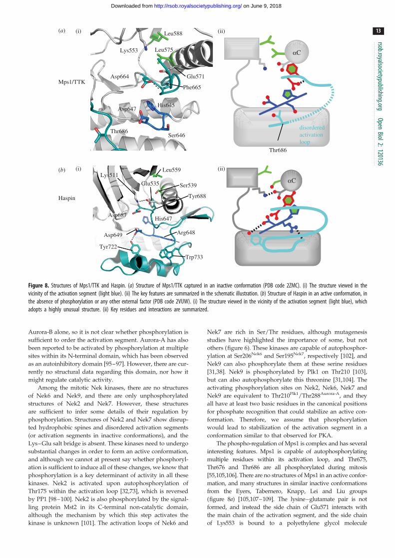
Figure 8. Structures of Mps1/TTK and Haspin. (a) Structure of Mps1/TTK captured in an inactive conformation (PDB code 2ZMC). (i) The structure viewed in thevicinity of the activation segment (light blue). (ii) The key features are summarized in the schematic illustration. (b) Structure of Haspin in an active conformation, inthe absence of phosphorylation or any other external factor (PDB code 2VUW). (i) The structure viewed in the vicinity of the activation segment (light blue), whichadopts a highly unusual structure. (ii) Key residues and interactions are summarized.
rsob.royalsocietypublishing.orgOpen
Biol2:120136
13
on June 9, 2018http://rsob.royalsocietypublishing.org/Downloaded from
Aurora-B alone, so it is not clear whether phosphorylation is
sufficient to order the activation segment. Aurora-A has also
been reported to be activated by phosphorylation at multiple
sites within its N-terminal domain, which has been observed
as an autoinhibitory domain [95–97]. However, there are cur-
rently no structural data regarding this domain, nor how it
might regulate catalytic activity.
Among the mitotic Nek kinases, there are no structures
of Nek6 and Nek9, and there are only unphosphorylated
structures of Nek2 and Nek7. However, these structures
are sufficient to infer some details of their regulation by
phosphorylation. Structures of Nek2 and Nek7 show disrup-
ted hydrophobic spines and disordered activation segments
(or activation segments in inactive conformations), and the
Lys–Glu salt bridge is absent. These kinases need to undergo
substantial changes in order to form an active conformation,
and although we cannot at present say whether phosphoryl-
ation is sufficient to induce all of these changes, we know that
phosphorylation is a key determinant of activity in all these
kinases. Nek2 is activated upon autophosphorylation of
Thr175 within the activation loop [32,73], which is reversed
by PP1 [98–100]. Nek2 is also phosphorylated by the signal-
ling protein Mst2 in its C-terminal non-catalytic domain,
although the mechanism by which this step activates the
kinase is unknown [101]. The activation loops of Nek6 and
Nek7 are rich in Ser/Thr residues, although mutagenesis
studies have highlighted the importance of some, but not
others (figure 6). These kinases are capable of autophosphor-
ylation at Ser206Nek6 and Ser195Nek7, respectively [102], and
Nek9 can also phosphorylate them at these serine residues
[31,38]. Nek9 is phosphorylated by Plk1 on Thr210 [103],
but can also autophosphorylate this threonine [31,104]. The
activating phosphorylation sites on Nek2, Nek6, Nek7 and
Nek9 are equivalent to Thr210Plk1/Thr288Aurora-A, and they
all have at least two basic residues in the canonical positions
for phosphate recognition that could stabilize an active con-
formation. Therefore, we assume that phosphorylation
would lead to stabilization of the activation segment in a
conformation similar to that observed for PKA.
The phospho-regulation of Mps1 is complex and has several
interesting features. Mps1 is capable of autophosphorylating
multiple residues within its activation loop, and Thr675,
Thr676 and Thr686 are all phosphorylated during mitosis
[55,105,106]. There are no structures of Mps1 in an active confor-
mation, and many structures in similar inactive conformations
from the Eyers, Tabernero, Knapp, Lei and Liu groups
(figure 8a) [105,107–109]. The lysine–glutamate pair is not
formed, and instead the side chain of Glu571 interacts with
the main chain of the activation segment, and the side chain
of Lys553 is bound to a polyethylene glycol molecule

rsob.royalsocietypublishing.orgOpen
Biol2:120136
14
on June 9, 2018http://rsob.royalsocietypublishing.org/Downloaded from
originating from the crystallization solution. The hydrophobic
spine is disrupted between Phe665 of the DFG motif and
His645 of the HRD motif. The mechanism by which activation
loop phosphorylation of Mps1 activates the kinase has not been
resolved, but it must be highly unusual because Mps1 lacks the
usual motifs that might interact with the phosphorylated acti-
vation loop. It is not an RD kinase (the Arg is replaced by
Ser), the two basic residues on the aC-helix are positioned on
the opposite side of the helix to the activation loop, and there
is no basic residue in the b9 strand. There is a basic residue
(Lys681) at the equivalent position on the activation loop to
that involved in phosphate recognition in Plk1/Aurora-A/
Aurora-B; however, this residue is disordered in all current
Mps1 structures, and its role in kinase activation is unknown
(figure 6). It has been proposed that a basic patch C-terminal
to the activation segment might interact with the phosphory-
lated activation loop, and indeed mutations at Lys708 and
Lys710 reduce kinase activity [105]. However, the only crystal
structure of Mps1 in which the phosphorylated side chains of
Thr675, Thr676 and Ser677 are ordered also shows an inactive
conformation, with all three phosphates positioned out into sol-
vent (PDB code 3H9F). In this case, an inactive conformation of
the activation loop is probably stabilized by interactions
with the ligand bound to the active site [109]. More surpris-
ingly, there is strong evidence that Thr686 within the GT
motif is phosphorylated during mitosis and that this form of
the kinase is active. This is an unprecedented modification in
a protein kinase, and suggests that this modification might
switch the specificity from serine/threonine to tyrosine sub-
strates [105]. Structural studies on specific phospho-states of
Mps1 will clarify the mechanism of activation, but will be
technically challenging.
8. The structural mechanisms of regulationby binding partners
Cdks are the best-known examples of protein kinases that are
activated by the binding of a partner protein, which in this
case are cyclins, and the details of the mechanism have been
extensively reviewed [9,10]. Cyclin A forms an extensive inter-
face with unphosphorylated Cdk2, interacting primarily with
the a1/aC helix and activation segment (figures 3 and 4a).
This releases the activation segment from its autoinhibitory
conformation, and remoulds the conformation of Cdk2 into a
partially active form, in which the Lys–Glu salt bridge is
formed, the hydrophobic spine is assembled and Thr160 is
accessible for phosphorylation. One way of thinking about
the function of the cyclin is that it completes the proper folding
of the kinase domain, and that the Cdk on its own has an
incomplete catalytic domain because it requires the cyclin for
it to fold properly and have activity [10].
The catalytic activity of Aurora kinases depends on the
binding of a number of protein-binding partners and crystal
structures of the Aurora-A/TPX2 and Aurora-B/inner centro-
mere protein (INCENP) complexes have shed light on their
respective activation mechanisms (figures 7 and 9) [85,86].
TPX2 is a microtubule-associated protein [110], and
INCENP is another component of the CPC [111]. Residues
7–21 of TPX2 bind to the N-lobe of Aurora-A in an extended
conformation, and residues 30–40 form a short helix
that binds between the N-lobe and the activation loop
(figure 9a) [85]. This interaction generates conformational
changes in the activation loop of Aurora-A, leading to a
fully active conformation, in which key interactions are estab-
lished, between pThr288 and Arg255, Arg180 (aC-helix) and
Arg286, as well as between Thr292 and Asp256 (figure 7e,f ).
When TPX2 is bound to Aurora-A, the phosphate group on
Thr288 is buried, whereas without TPX2, this phosphate is
accessible to phosphatases (figure 7e). This explains why
dephosphorylation of Aurora-A by PP1 is less efficient in
the presence of TPX2.
The structure of Aurora-B bound to INCENP, from the
Musacchio group, shows a similar conformation of the acti-
vation segment to that observed in the Aurora-A/TPX2
complex (figure 9). Residues 798–841 of INCENP bind to
the N-lobe of Aurora-B, forming a series of three helices,
the final two of which occupy a site similar to that of residues
7–21 of TPX2 [86]. The first helix of INCENP does not resemble
TPX2, but instead wraps around the other side of the N-lobe,
forming a contact with the C-terminal region of Aurora-B.
The crystal structure does not capture a fully active confor-
mation of Aurora-B, because the aC-helix and C-lobe are
both rotated 158 away from what is found in the structure of
Aurora-A/TPX2, and one consequence of this is that the
Lys–Glu salt bridge is not formed. It has been proposed that
phosphorylation of a TSS motif in INCENP changes the pos-
ition of Phe837, allowing the aC-helix to rotate into its active
position. Aurora-A and -B are more than 70 per cent identical
in their catalytic domains. TPX2 can discriminate between
Aurora-A and -B on the basis of a single residue, Gly198,
which is Asn in Aurora-B [112]. Indeed, a Gly198 to Asn
mutation in Aurora-A not only ablates TPX2 binding, but
confers regulation by INCENP, so that the mutant somewhat
resembles Aurora-B in its cellular behaviour [113,114].
More recently, the contribution of TPX2 binding and phos-
phorylation to Aurora-A activity have been thoroughly
explored. TPX2 can stimulate the activity of unphosphorylated
Aurora-A, and its energetic contribution to catalysis is 2.1–
2.3 kcal mol21, independent of the phosphorylation state of
Aurora-A [66]. Our working model is that TPX2 activates
unphosphorylated Aurora-A through partial stabilization of
the activation segment. Indeed, we postulate that the first step
of Aurora-A activation is partial stabilization of the activation
segment, whether by phosphorylation or by protein partner
binding. This model is in contrast to the activation pathway
of Cdks, in which cyclin binding and phosphorylation have
distinct and sequential roles.
9. Mitotic kinases with constitutively activecatalytic domains
Bub1 and Haspin do not appear to be obviously regulated
through any of the three common mechanisms discussed
above. This is perhaps not surprising because they diverge
hugely in their regulatory motifs (table 1). Although the
structure of Bub1 was previously mentioned as an example
of autoinhibition, this assignment is debatable and it could
well be regarded as an example of a constitutively active
kinase, with a highly unusual activation segment.
The primary sequence of Haspin is divergent from other
protein kinases, including the catalytic and regulatory motifs
(table 1). Crystal structures from the Knapp and Musacchio
laboratories revealed a recognizable kinase fold, albeit highly
modified, with three major differences from that adopted by

a1
P
a2a3
AURKB/INCENPAURKA/TPX2
Phe837
Phe837
TPX2 INCENP
aC
a1
a2a3
(Met156)
Gln129
Glu125
Asp218 Phe219
His198
Arg199Asp200
Thr236
pThr232pThr232
Thr236
Lys106
aC aC
(a) (b)
(c) (d)
Figure 9. Comparison of activation mechanisms of Aurora-A and Aurora-B. (a) Crystal structure of phosphorylated Aurora-A (red, activation segment in green – blue)bound to TPX2 (green), based on PDB code 1OL5. (b) Structure of phosphorylated Aurora-B (grey, activation segment in green – blue) bound to INCENP (green),based on PDB code 2BFX. The three a-helices of the INCENP fragment are labelled a1, a2 and a3. (c) Structure of INCENP/Aurora-B complex in the vicinity of theactivation segment. It is not known whether INCENP affects the conformation of the activation segment, which it does not directly contact. It is thought that Phe837of INCENP causes a rotation of the aC helix that prevents Lys – Glu salt bridge formation. Note that Xenopus laevis Aurora-B has residue Met156, equivalent tohuman Leu140; all the other residues are labelled with human protein numbering. (d ) Schematic illustration of the INCENP/Aurora-B complex.
rsob.royalsocietypublishing.orgOpen
Biol2:120136
15
on June 9, 2018http://rsob.royalsocietypublishing.org/Downloaded from
other kinases (figure 8b) [115,116]. First, the N-lobe is larger,
with elements such as an a-helix inserted between b4 and
b5 that stabilizes the Gly-rich loop. Second, the activation seg-
ment is highly divergent and lacks an APE motif. Third, the
C-lobe is modified by the addition of a two-strand b-sheet
that interacts with the loop between aC and b4, and by the
deletion of the aG helix, which usually abuts the activation
segment. The structures appear to show a constitutively
active kinase, with most of the key catalytic elements such as
the Lys–Glu salt bridge in place, albeit with a few interesting
twists. The aC component of the hydrophobic spine is a Ser,
and the void is partly filled by water molecules and partly
by the side chain of Tyr from the DYT motif that replaces
DFG at the start of the activation segment. The impact of
this on Mg/ATP binding is unclear, although it has been
suggested that, unlike in other kinases, ATP might be recog-
nized directly by the catalytic Asp of the HRD motif and a
histidine residue close by. The activation segment is fully
ordered in a conformation that is highly divergent from
other active kinases, but which appears to be consistent with
substrate binding. The activation loop has no GT motif, and
instead the Tyr722 side chain forms a hydrogen bond with
the catalytic Asp649. During mitosis, Haspin is highly phos-
phorylated, but the structures suggest that Haspin is
probably not regulated by phosphorylation on the activation
loop, even though it conserves two out of three canonical resi-
dues involved in phosphate recognition (Arg648 of the HRD
motif and Arg692 on b9). Indeed, both basic side chains form
salt bridges with glutamic acid residues, and hence their posi-
tive charges are already neutralized and they fulfil structural
roles in the absence of phosphorylation. In addition, the side
chain of Arg648 forms interactions with the main chain at
Trp733 and Glu735. Furthermore, there is only one suitable
phosphorylatable residue on the activation segment (Ser705),
which has not been observed to be phosphorylated [115].
10. Summary and outstanding questionsDuring mitosis, there is a peak in cellular phosphorylation,
which is thought to effect a wholesale transformation in the
activities of cellular proteins [117]. These phosphorylation
events are catalysed by a subset of protein kinases that are
activated during mitosis. Recent structural studies have pro-
vided molecular insights into the activation mechanisms of
these kinases. Structural studies provide essential validation
of key regulatory steps and contribute to the eventual aim
of understanding these pathways at a systems level; and

rsob.royalsocietypublishing.orgOpen
Biol2:120136
16
on June 9, 2018http://rsob.royalsocietypublishing.org/Downloaded from
yet there are many questions left unresolved. For a start, there
are no structures of key mitotic kinases such as Lats kinases
or Greatwall, and even in the case of kinases with numerous
entries in the Protein Data Bank, there remain many mechan-
istic questions that require a structural biology approach. In
this concluding section of the review, we will highlight
some of the outstanding questions that need to be addressed
by structural biologists who study mitotic kinases.
Protein crystallography has revealed that active protein
kinases adopt a specific conformation with respect to a key
set of features, including the lysine–glutamate pair, the
HRD and DFG motifs, the hydrophobic spine and a set of
three basic residues that coordinate the activating phosphate
group. These features are mostly conserved at the sequence
level in mitotic protein kinases, although all mitotic kinases
are divergent in at least one feature, which influences how
they achieve an active conformation. Of the mitotic kinases,
only Aurora-A, Plk1 and Haspin have been crystallized in
fully active conformations, although the structures of
Aurora-B/INCENP and Bub1 do exhibit most of the features
of an active kinase. Perhaps most strikingly of all, structures
of Haspin and Bub1 show how an active kinase conformation
(or almost active) can be achieved with highly divergent
kinases despite lacking one of the key hydrophobic spine resi-
dues (Bub1) or several of the usual motifs (Haspin). In crystal
structures of kinases in inactive conformations, these features
are displaced from their active positions in a variety of ways,
which have illuminated their mechanisms of regulation; for
example, in the identification of a novel mechanism of auto-
inhibition in Nek7 and explaining why phosphorylation is
insufficient for full activity in Aurora-A. Other structures
raise the prospect of the existence of autoinhibited states
(Nek2, Aurora-A) or highlight the requirement for further
conformational changes to achieve full activity (Aurora-B/
INCENP, Bub1).
10.1. Autoinhibition and other mechanisms thatprevent activity
Mechanisms that prevent the untimely activation of kinases
are crucial to ensure the fidelity of cell division. Autoinhibi-
tion is one such mechanism, as used in the regulation of
kinases such as Cdk2 and BRAF. In this review, we have
summarized the evidence for autoinhibitory conformations
of mitotic kinases based on crystal structures of Aurora-A,
Nek2 and Nek7. Further studies on these kinases are
required to determine the contribution that these autoinhibi-
tory states make to the physiological regulation of kinase
activity. Future structural studies will reveal how these auto-
inhibitory states are reversed, for example through the
interaction of Nek9 with Nek7. Reversal of activating
phosphorylation by phosphatases is another mechanism
deployed to prevent untimely kinase activation, and a clear
example of this is the inhibition of Nek2 by PP1 [100]. It is
important to identify the bona fide phosphatase partners of
other mitotic kinases, to understand the basis of their selec-
tivity and regulation. Along these lines, it has recently been
shown that PP6, not PP1, is the key Aurora-A inhibitory
phosphatase and is capable of dephosphorylating the
TPX2/Aurora-A complex [118]. A clear priority for future
studies is the elucidation of the structural basis by which
cellular phosphatases recognize mitotic kinases.
10.2. Activation by phosphorylationAlmost all mitotic kinases are regulated at some level by
phosphorylation, and in many cases a site of activating
phosphorylation has been identified on the activation loop
(table 1). In Plk1, this phosphorylation event is sufficient to
generate a stable conformation of the activation loop compa-
tible with substrate binding, but Aurora-A also requires the
binding of TPX2. How this works in other mitotic kinases
could be resolved by tackling the crystal structures of these
kinases in singly phosphorylated forms without other bind-
ing partners. However, this is technically challenging, and
is unlikely to be high on the list of priorities of structural biol-
ogists unless there is strong reason to believe that the answer
will be interesting. We think, though, that the effort would be
worth it for Mps1 because it lacks the three canonical basic
residues used for recognition of a phosphorylated activation
loop, and so phosphorylation might act through a novel
structural mechanism. In fact, we believe that it is feasible
to derive many insights into the conformational changes
upon phosphorylation from biochemical data, which might
be a more attractive option to address these issues [66]. Per-
haps a more important challenge for structural biologists is
to resolve the structural basis of kinase regulation by phos-
phorylation at sites away from the activation loop in the
catalytic domain, such as inhibitory phosphorylation of
Ser342 of Aurora-A or Ser241 of Nek2 [73,83]. Generating
protein samples for structural biology (or biochemical
studies) that have a fully specified and homogeneous phos-
phorylation state remains a challenge, and we are currently
exploring chemical approaches to solve this problem.
10.3. Role of non-catalytic domains and bindingpartners
Some mitotic kinases do not become fully activated until
bound to a partner protein, triggering a conformational
change, which may release an autoinhibitory state (such as
postulated in Nek9 allosteric activation of Nek7), reveal pre-
viously hidden additional phosphorylation sites (such as in
cyclin activation of Cdks) or lock the protein in an active
state (such as in TPX2 stimulation of phospho-Aurora-A
activity). From a structural point of view, there are broad
similarities between the ways that cyclins remodel the Cdk
structure and TPX2/INCENP complement the catalytic
domains of Aurora kinases. Not least of these is that the bind-
ing partners form crucial interactions with the a1/aC helix,
and (at least in the case of cyclins and TPX2) with the acti-
vation segment. In this regard, they function similarly to
the N- and C-terminal extensions of PKA [85]. To date, all
structures of activatory binding partners show recognition
of a hydrophobic groove on the surface of the N-lobe,
which is formed by the aC helix and b4 strand. This feature
is commonly involved in the regulation of AGC family
kinases, to which the Auroras bear closest resemblance, but
not in other kinase families [119]. Other mitotic kinases,
including Plk1, Nek2 and Mps1, also have a hydrophobic
groove, and although we do not know of any proteins that
interact at this site in these kinases, we have observed crystal
packing interactions with the hydrophobic groove in Nek2
that mimic the situation in AGC kinases, and it is known
that dimerization activates the Nek2 kinase in cells [74].
rsob.royalsocietypublishing.orgOpen
Biol2:120136
17
on June 9, 2018http://rsob.royalsocietypublishing.org/Downloaded from
Although in existing structures Nek7 lacks an obvious
hydrophobic groove, Tyr97 is located precisely where one
might expect a groove to open up to accommodate Nek9 in
order to reverse autoinhibition, and this hypothesis has
informed our current studies on this complex. There is clearly
a lot more to kinase regulation and function than is encoded
in the structure of the catalytic domain, and further examples
of domains and binding partners that ‘complete’ the kinase
domain are undoubtedly out there waiting to be discovered.
Indeed, many other activatory binding partners of Aurora-A
have been identified, such as Ajuba, I2 and Cep192, and it is
not known whether they work through mechanisms similar
to or different from TPX2. Activation by a binding partner
localizes kinase activity to a specific spindle region or protein
complex. This fulfils an important regulatory role because the
consensus sequences phosphorylated by some mitotic
kinases overlap, perhaps best exemplified by Aurora-B
(which phosphorylates substrates at the kinetochore) and
Aurora-A (which phosphorylates substrates at the centro-
some and spindle microtubules), but both kinases have the
capability to phosphorylate peptides with similar sequences
in vitro [120]. It would not be surprising if the other mitotic
kinase families used localization through activatory binding
partners to determine substrate specificity.
10.4. Substrate recognitionThere are currently no structures of mitotic kinase catalytic
domains bound to substrates, and although in some cases
these interactions can be modelled with a degree of confi-
dence, there are other examples for which an experimental
structure would be very interesting [120]. For example,
Haspin and Bub1 have unusual structures in the expected
substrate-binding regions, and in the particular case of
Haspin, it would be interesting to resolve how its unusual
activation segment contributes to the reading and writing
of the histone code. Moreover, the structure of Bub1 bound
to a peptide substrate would address whether or not a sub-
stantial conformational change is required in the activation
segment in order to form an active structure. Sequences
outside the catalytic domain may contribute to substrate rec-
ognition, and the best-known example of this is Plk1, which
recognizes substrates through its polo box [91,92]. There are
countless other examples for which we have no structural
information, such as the interaction between the non-catalytic
domain of Aurora-A and p53, the recognition of substrates
by the N-terminal extensions of Nek6 and Nek7, and the
contribution of Haspin’s non-catalytic domain to substrate
recognition [121]. These are important targets for future
structural studies, although challenging, because these
domains appear to be natively disordered in the absence
of substrate.
10.5. Mitotic kinases as drug targetsMitotic kinases are an important class of drug target, and
understanding their regulatory mechanisms is key to effective
new treatments for various hyperproliferative diseases, most
notably cancer. Structural characterization of kinases facili-
tates rational drug design, enabling kinase active sites to be
selectively targeted by tailor-made inhibitors. This has
played a crucial role in the development of ATP-competitive
inhibitors of Aurora kinases, Plk1, Mps1 and Nek2
[77,108,109,122–124]. Building on these studies, with the
aid of structural biology, it should be possible to design
chemical inhibitors specific for each and every mitotic
kinase. In combination with systems biology approaches,
this could transform our understanding of the networks of
kinases that coordinate mitosis. It will require some thought-
ful experimental design to dissect out the role of different
populations of each kinase as mitosis progresses, and to
tease apart their interdependent relationships. Moreover,
this will have to include the contribution of kinase-binding
partners such as TPX2, which is not only crucial for the local-
ization and activity of Aurora-A, but can affect the potency of
kinase inhibitors [79,125].
In this review, we have described the remarkable progress
in the resolution of mitotic kinase structures, and explained
how these models have contributed to our understanding
of their regulation and function. There are still many unan-
swered questions relating to the structural biology of
mitotic kinases, and we have identified what we consider
to be priorities for future work. Finally, we look forward to
further developments in the field as the molecular details of
mitotic signalling pathways are elaborated further, and this
knowledge is translated into improvements in human health.
11. AcknowledgementsR.B. is a Royal Society Research Fellow and acknowledges
grant support from Cancer Research UK (C24461/A12772
and C24461/A13231). T.H. and S.Y. are supported by the
University of Leicester. A.F. acknowledges grant support
from The Wellcome Trust and the Association for
International Cancer Research (AICR).
References
1. Musacchio A, Salmon ED. 2007 The spindle-assembly checkpoint in space and time. Nat.Rev. Mol. Cell. Biol. 8, 379 – 393. (doi:10.1038/nrm2163)
2. Lengauer C, Kinzler KW, Vogelstein B. 1997 Geneticinstability in colorectal cancers. Nature 386,623 – 627. (doi:10.1038/386623a0)
3. Nigg EA. 2002 Centrosome aberrations: cause orconsequence of cancer progression? Nat. Rev. Cancer2, 815 – 825. (doi:10.1038/nrc924)
4. Kops GJ, Weaver BA, Cleveland DW. 2005 On theroad to cancer: aneuploidy and the mitoticcheckpoint. Nat. Rev. Cancer 5, 773 – 785. (doi:10.1038/nrc1714)
5. Manchado E, Guillamot M, Malumbres M. 2012Killing cells by targeting mitosis. Cell Death Differ.19, 369 – 377. (doi:10.1038/cdd.2011.197)
6. Ma HT, Poon RY. 2011 How protein kinases co-ordinate mitosis in animal cells. Biochem. J. 435,17 – 31. (doi:10.1042/BJ20100284)
7. Manning G, Whyte DB, Martinez R, Hunter T,Sudarsanam S. 2002 The protein kinasecomplement of the human genome. Science 298,1912 – 1934. (doi:10.1126/science.1075762)
8. Hunt T. 1989 Maturation promoting factor, cyclinand the control of M-phase. Curr. Opin. Cell Biol. 1,268 – 274. (doi:10.1016/0955-0674(89)90099-9)
9. Morgan DO. 1995 Principles of CDKregulation. Nature 374, 131 – 134. (doi:10.1038/374131a0)
rsob.royalsocietypublishing.orgOpen
Biol2:120136
18
on June 9, 2018http://rsob.royalsocietypublishing.org/Downloaded from
10. Pavletich NP. 1999 Mechanisms of cyclin-dependentkinase regulation: structures of Cdks, their cyclinactivators, and Cip and INK4 inhibitors. J. Mol. Biol.287, 821 – 828. (doi:10.1006/jmbi.1999.2640)
11. Enserink JM, Kolodner RD. 2010 An overview ofCdk1-controlled targets and processes. Cell Div. 5,11. (doi:10.1186/1747-1028-5-11)
12. Barr FA, Sillje HH, Nigg EA. 2004 Polo-like kinasesand the orchestration of cell division. Nat.Rev. Mol. Cell. Biol. 5, 429 – 440. (doi:10.1038/nrm1401)
13. Glover DM. 2005 Polo kinase and progressionthrough M phase in Drosophila: a perspective fromthe spindle poles. Oncogene 24, 230 – 237. (doi:10.1038/sj.onc.1208279)
14. Lowery DM, Lim D, Yaffe MB. 2005 Structure andfunction of Polo-like kinases. Oncogene 24,248 – 259. (doi:10.1038/sj.onc.1208280)
15. Archambault V, Carmena M. 2012 Polo-like kinase-activating kinases: aurora A, Aurora B and whatelse? Cell Cycle. 11, 1490 – 1495. (doi:10.4161/cc.19724)
16. Lindon C, Pines J. 2004 Ordered proteolysis inanaphase inactivates Plk1 to contribute to propermitotic exit in human cells. J. Cell Biol. 164,233 – 241. (doi:10.1083/jcb.200309035)
17. Chan CS, Botstein D. 1993 Isolation andcharacterization of chromosome-gain and increase-in-ploidy mutants in yeast. Genetics 135, 677 – 691.
18. Francisco L, Chan CS. 1994 Regulation of yeastchromosome segregation by Ipl1 protein kinase andtype 1 protein phosphatase. Cell Mol. Biol. Res. 40,207 – 213.
19. Barr AR, Gergely F. 2007 Aurora-A: the maker andbreaker of spindle poles. J. Cell Sci. 120, 2987 –2996. (doi:10.1242/jcs.013136)
20. Nikonova AS, Astsaturov I, Serebriiskii IG, DunbrackJr RL, Golemis EA. In press. Aurora A kinase(AURKA) in normal and pathological cell division.Cell Mol. Life Sci. (doi:10.1007/s00018-012-1073-7)
21. Mori D, Yamada M, Mimori-Kiyosue Y, Shirai Y,Suzuki A, Ohno S, Saya H, Wynshaw-Boris A,Hirotsune S. 2009 An essential role of the aPKC-Aurora A-NDEL1 pathway in neurite elongation bymodulation of microtubule dynamics. Nat. Cell. Biol.11, 1057 – 1068. (doi:10.1038/ncb1919)
22. Pugacheva EN, Jablonski SA, Hartman TR, HenskeEP, Golemis EA. 2007 HEF1-dependent Aurora Aactivation induces disassembly of the primarycilium. Cell 129, 1351 – 1363. (doi:10.1016/j.cell.2007.04.035)
23. Goto H, Yasui Y, Nigg EA, Inagaki M. 2002 Aurora-Bphosphorylates histone H3 at serine28 with regardto the mitotic chromosome condensation. GenesCells 7, 11 – 17. (doi:10.1046/j.1356-9597.2001.00498.x)
24. Lampson MA, Cheeseman IM. 2011 Sensingcentromere tension: aurora B and the regulation ofkinetochore function. Trends Cell Biol. 21, 133 – 140.(doi:10.1016/j.tcb.2010.10.007)
25. Sasai K et al. 2004 Aurora-C kinase is a novelchromosomal passenger protein that cancomplement Aurora-B kinase function in mitotic
cells. Cell Motil. Cytoskeleton 59, 249 – 263. (doi:10.1002/cm.20039)
26. Carmena M, Earnshaw WC. 2003 The cellulargeography of aurora kinases. Nat. Rev. Mol. Cell.Biol. 4, 842 – 854. (doi:10.1038/nrm1245)
27. Morris NR. 1975 Mitotic mutants of Aspergillusnidulans. Genet. Res. 26, 237 – 254. (doi:10.1017/S0016672300016049)
28. Morris NR, Osmani SA, Engle DB, Doonan JH. 1989The genetic analysis of mitosis in Aspergillusnidulans. Bioessays 10, 196 – 201. (doi:10.1002/bies.950100605)
29. O’Regan L, Blot J, Fry AM. 2007 Mitotic regulationby NIMA-related kinases. Cell Div. 2, 25. (doi:10.1186/1747-1028-2-25)
30. Fry AM, Arnaud L, Nigg EA. 1999 Activity of thehuman centrosomal kinase, Nek2, depends on anunusual leucine zipper dimerization motif. J. Biol.Chem. 274, 16 304 – 16 310. (doi:10.1074/jbc.274.23.16304)
31. Roig J, Mikhailov A, Belham C, Avruch J. 2002Nercc1, a mammalian NIMA-family kinase, bindsthe Ran GTPase and regulates mitotic progression.Genes Dev. 16, 1640 – 1658. (doi:10.1101/gad.972202)
32. Fry AM, Schultz SJ, Bartek J, Nigg EA. 1995Substrate specificity and cell cycle regulation of theNek2 protein kinase, a potential human homolog ofthe mitotic regulator NIMA of Aspergillus nidulans.J. Biol. Chem. 270, 12 899 – 12 905. (doi:10.1074/jbc.270.21.12899)
33. Lu KP, Osmani SA, Means AR. 1993 Properties andregulation of the cell cycle-specific NIMA proteinkinase of Aspergillus nidulans. J. Biol. Chem. 268,8769 – 8776.
34. Fry AM. 2002 The Nek2 protein kinase: a novelregulator of centrosome structure. Oncogene 21,6184 – 6194. (doi:10.1038/sj.onc.1205711)
35. Hames RS, Wattam SL, Yamano H, Bacchieri R, FryAM. 2001 APC/C-mediated destruction of thecentrosomal kinase Nek2A occurs in earlymitosis and depends upon a cyclin A-typeD-box. EMBO J. 20, 7117 – 7127. (doi:10.1093/emboj/20.24.7117)
36. Hayes MJ, Kimata Y, Wattam SL, Lindon C, Mao G,Yamano H, Fry AM. 2006 Early mitotic degradationof Nek2A depends on Cdc20-independentinteraction with the APC/C. Nat. Cell. Biol. 8,607 – 614. (doi:10.1038/ncb1410)
37. Sdelci S, Bertran MT, Roig J. 2011 Nek9, Nek6, Nek7and the separation of centrosomes. Cell Cycle 10,3816 – 3817. (doi:10.4161/cc.10.22.18226)
38. Belham C, Roig J, Caldwell JA, Aoyama Y, Kemp BE,Comb M, Avruch J. 2003 A mitotic cascade of NIMAfamily kinases. Nercc1/Nek9 activates the Nek6 andNek7 kinases. J. Biol. Chem. 278, 34 897 – 34 909.(doi:10.1074/jbc.M303663200)
39. Richards MW, O’Regan L, Mas-Droux C, Blot JM,Cheung J, Hoelder S, Fry AM, Bayliss R. 2009 Anautoinhibitory tyrosine motif in the cell-cycle-regulated Nek7 kinase is released through bindingof Nek9. Mol. Cell. 36, 560 – 570. (doi:10.1016/j.molcel.2009.09.038)
40. Rapley J, Nicolas M, Groen A, Regue L, Bertran MT,Caelles C, Avruch J, Roig J. 2008 The NIMA-familykinase Nek6 phosphorylates the kinesin Eg5 at anovel site necessary for mitotic spindle formation.J. Cell Sci. 121, 3912 – 3921. (doi:10.1242/jcs.035360)
41. Sdelci S, Schutz M, Pinyol R, Bertran MT, Regue L,Caelles C, Vernos I, Roig J. 2012 Nek9Phosphorylation of NEDD1/GCP-WD contributes toPlk1 control of gamma-tubulin recruitment to themitotic centrosome. Curr. Biol. 22, 1516 – 1523.(doi:10.1016/j.cub.2012.06.027)
42. Logarinho E, Bousbaa H. 2008 Kinetochore-microtubule interactions ‘in check’ by Bub1, Bub3and BubR1: the dual task of attaching andsignalling. Cell Cycle 7, 1763 – 1768 (doi:10.4161/cc.7.12.6180)
43. Elowe S. 2011 Bub1 and BubR1: at the interfacebetween chromosome attachment and the spindlecheckpoint. Mol. Cell. Biol. 31, 3085 – 3093. (doi:10.1128/mcb.05326-11)
44. Bolanos-Garcia VM, Blundell TL. 2011 BUB1 andBUBR1: multifaceted kinases of the cell cycle. TrendsBiochem. Sci. 36, 141 – 150. (doi:10.1016/j.tibs.2010.08.004)
45. Bolanos-Garcia VM et al. 2009 The crystal structureof the N-terminal region of BUB1 provides insightinto the mechanism of BUB1 recruitment tokinetochores. Structure 17, 105 – 116. (doi:10.1016/j.str.2008.10.015)
46. Bolanos-Garcia VM et al. 2011 Structure of aBlinkin-BUBR1 complex reveals an interaction crucialfor kinetochore-mitotic checkpoint regulation via anunanticipated binding site. Structure 19, 1691 –1700. (doi:10.1016/j.str.2011.09.017)
47. Larsen NA, Al-Bassam J, Wei RR, Harrison SC. 2007Structural analysis of Bub3 interactions in themitotic spindle checkpoint. Proc. Natl Acad. Sci. USA104, 1201 – 1206. (doi:10.1073/pnas.0610358104)
48. Higgins JM. 2010 Haspin: a newly discoveredregulator of mitotic chromosome behavior.Chromosoma 119, 137 – 147. (doi:10.1007/s00412-009-0250-4)
49. Kelly AE, Ghenoiu C, Xue JZ, Zierhut C, Kimura H,Funabiki H. 2010 Survivin reads phosphorylatedhistone H3 threonine 3 to activate the mitotickinase Aurora B. Science 330, 235 – 239. (doi:10.1126/science.1189505)
50. Wang F, Dai J, Daum JR, Niedzialkowska E, BanerjeeB, Stukenberg PT, Gorbsky GJ, Higgins JM. 2010Histone H3 Thr-3 phosphorylation by Haspinpositions Aurora B at centromeres in mitosis. Science330, 231 – 235. (doi:10.1126/science.1189435)
51. Wang F, Ulyanova NP, van der Waal MS, Patnaik D,Lens SM, Higgins JM. 2011 A positive feedback loopinvolving Haspin and Aurora B promotes CPCaccumulation at centromeres in mitosis. Curr. Biol.21, 1061 – 1069. (doi:10.1016/j.cub.2011.05.016)
52. Liu X, Winey M. 2012 The MPS1 family of proteinkinases. Annu. Rev. Biochem. 81, 561 – 585. (doi:10.1146/annurev-biochem-061611-090435)
53. Lan W, Cleveland DW. 2010 A chemical tool boxdefines mitotic and interphase roles for Mps1

rsob.royalsocietypublishing.orgOpen
Biol2:120136
19
on June 9, 2018http://rsob.royalsocietypublishing.org/Downloaded from
kinase. J. Cell Biol. 190, 21 – 24. (doi:10.1083/jcb.201006080)
54. Lee S, Thebault P, Freschi L, Beaufils S, Blundell TL,Landry CR, Bolanos-Garcia VM, Elowe S. 2012Characterization of spindle checkpoint kinase Mps1reveals domain with functional and structuralsimilarities to tetratricopeptide repeat motifs ofBub1 and BubR1 checkpoint kinases. J. Biol. Chem.287, 5988 – 6001. (doi:10.1074/jbc.M111.307355)
55. Tyler RK, Chu ML, Johnson H, McKenzie EA, GaskellSJ, Eyers PA. 2009 Phosphoregulation of humanMps1 kinase. Biochem J. 417, 173 – 181. (doi:10.1042/bj20081310)
56. Cui Y, Cheng X, Zhang C, Zhang Y, Li S, Wang C,Guadagno TM. 2010 Degradation of the humanmitotic checkpoint kinase Mps1 is cell cycle-regulated by APC-cCdc20 and APC-cCdh1 ubiquitinligases. J. Biol. Chem. 285, 32 988 – 32 998. (doi:10.1074/jbc.M110.140905)
57. Knighton DR, Zheng JH, Ten Eyck LF, Ashford VA,Xuong NH, Taylor SS, Sowadski JM. 1991 Crystalstructure of the catalytic subunit of cyclic adenosinemonophosphate-dependent protein kinase. Science253, 407 – 414. (doi:10.1126/science.1862342)
58. Huse M, Kuriyan J. 2002 The conformationalplasticity of protein kinases. Cell 109, 275 – 282.(doi:10.1016/S0092-8674(02)00741-9)
59. Johnson LN, Noble ME, Owen DJ. 1996 Active andinactive protein kinases: structural basis forregulation. Cell 85, 149 – 158. (doi:10.1016/S0092-8674(00)81092-2)
60. Nolen B, Taylor S, Ghosh G. 2004 Regulation ofprotein kinases; controlling activity throughactivation segment conformation. Mol. Cell. 15,661 – 675. (doi:10.1016/j.molcel.2004.08.024)
61. Lowe ED, Noble ME, Skamnaki VT, Oikonomakos NG,Owen DJ, Johnson LN. 1997 The crystal structure ofa phosphorylase kinase peptide substrate complex:kinase substrate recognition. EMBO J. 16, 6646 –6658. (doi:10.1093/emboj/16.22.6646)
62. Parang K, Till JH, Ablooglu AJ, Kohanski RA,Hubbard SR, Cole PA. 2001 Mechanism-baseddesign of a protein kinase inhibitor. Nat. Struct. Biol.8, 37 – 41. (doi:10.1038/83028)
63. Gibbs CS, Zoller MJ. 1991 Rational scanningmutagenesis of a protein kinase identifies functionalregions involved in catalysis and substrateinteractions. J. Biol. Chem. 266, 8923 – 8931.
64. Schindler T, Bornmann W, Pellicena P, Miller WT,Clarkson B, Kuriyan J. 2000 Structural mechanismfor STI-571 inhibition of abelson tyrosine kinase.Science 289, 1938 – 1942. (doi:10.1126/science.289.5486.1938)
65. Kornev AP, Haste NM, Taylor SS, Eyck LF. 2006Surface comparison of active and inactive proteinkinases identifies a conserved activation mechanism.Proc. Natl Acad. Sci. USA 103, 17 783 – 17 788.(doi:10.1073/pnas.0607656103)
66. Dodson CA, Bayliss R. 2012 Activation of Aurora-Akinase by protein partner binding andphosphorylation are independent and synergistic.J. Biol. Chem. 287, 1150 – 1157. (doi:10.1074/jbc.M111.312090)
67. De Bondt HL, Rosenblatt J, Jancarik J, Jones HD,Morgan DO, Kim SH. 1993 Crystal structure ofcyclin-dependent kinase 2. Nature 363, 595 – 602.(doi:10.1038/363595a0)
68. Jeffrey PD, Russo AA, Polyak K, Gibbs E, Hurwitz J,Massague J, Pavletich NP. 1995 Mechanism of CDKactivation revealed by the structure of a cyclinA-CDK2 complex. Nature 376, 313 – 320. (10.1038/376313a0)
69. Russo AA, Jeffrey PD, Pavletich NP. 1996 Structuralbasis of cyclin-dependent kinase activation byphosphorylation. Nat. Struct. Biol. 3, 696 – 700.(doi:10.1038/nsb0896-696)
70. Wan PT et al. 2004 Mechanism of activation of theRAF-ERK signaling pathway by oncogenic mutationsof B-RAF. Cell 116, 855 – 867. (doi:10.1016/S0092-8674(04)00215-6)
71. Xu W, Doshi A, Lei M, Eck MJ, Harrison SC. 1999Crystal structures of c-Src reveal features of itsautoinhibitory mechanism. Mol. Cell. 3, 629 – 638.(doi:10.1016/S1097-2765(00)80356-1)
72. Nagar B, Hantschel O, Young MA, Scheffzek K,Veach D, Bornmann W, Clarkson B, Superti-Furga G,Kuriyan J. 2003 Structural basis for theautoinhibition of c-Abl tyrosine kinase. Cell 112,859 – 871. (doi:10.1016/S0092-8674(03)00194-6)
73. Rellos P et al. 2007 Structure and regulation of thehuman Nek2 centrosomal kinase. J. Biol. Chem.282, 6833 – 6842. (doi:10.1074/jbc.M609721200)
74. Westwood I, Cheary DM, Baxter JE, Richards MW,van Montfort RL, Fry AM, Bayliss R. 2009 Insightsinto the conformational variability and regulation ofhuman Nek2 kinase. J. Mol. Biol. 386, 476 – 485.(doi:10.1016/j.jmb.2008.12.033)
75. Solanki S, Innocenti P, Mas-Droux C, Boxall K,Barillari C, van Montfort RL, Aherne GW, Bayliss R,Hoelder S. 2011 Benzimidazole inhibitors induce aDFG-out conformation of never in mitosis gene A-related kinase 2 (Nek2) without binding to the backpocket and reveal a nonlinear structure-activityrelationship. J. Med. Chem. 54, 1626 – 1639. (doi:10.1021/jm1011726)
76. Whelligan DK et al. 2010 Aminopyrazine inhibitorsbinding to an unusual inactive conformation of themitotic kinase Nek2: SAR and structuralcharacterization. J. Med. Chem. 53, 7682 – 7698.(doi:10.1021/jm1008727)
77. Innocenti P et al. 2012 Design of potent andselective hybrid inhibitors of the mitotic kinaseNek2: structure – activity relationship, structuralbiology, and cellular activity. J. Med. Chem. 55,3228 – 3241. (doi:10.1021/jm201683b)
78. Cheetham GM, Knegtel RM, Coll JT, Renwick SB,Swenson L, Weber P, Lippke JA, Austen DA. 2002Crystal structure of aurora-2, an oncogenic serine/threonine kinase. J. Biol. Chem. 277, 42 419 –42 422. (doi:10.1074/jbc.C200426200)
79. Dodson CA, Kosmopoulou M, Richards MW, AtrashB, Bavetsias V, Blagg J, Bayliss R. 2010 Crystalstructure of an Aurora-A mutant that mimicsAurora-B bound to MLN8054: insights intoselectivity and drug design. Biochem. J. 427,19 – 28. (doi:10.1042/BJ20091530)
80. Heron NM et al. 2006 SAR and inhibitor complexstructure determination of a novel class of potentand specific Aurora kinase inhibitors. Bioorg. Med.Chem. Lett. 16, 1320 – 1323. (doi:10.1016/j.bmcl.2005.11.053)
81. Bibby RA, Tang C, Faisal A, Drosopoulos K, Lubbe S,Houlston R, Bayliss R, Linardopoulos S. 2009 Acancer-associated aurora A mutant is mislocalizedand misregulated due to loss of interaction withTPX2. J. Biol. Chem. 284, 33 177 – 33 184. (doi:10.1074/jbc.M109.032722)
82. Kang J, Yang M, Li B, Qi W, Zhang C, Shokat KM,Tomchick DR, Machius M, Yu H. 2008 Structure andsubstrate recruitment of the human spindlecheckpoint kinase Bub1. Mol. Cell. 32, 394 – 405.(doi:10.1016/j.molcel.2008.09.017)
83. Littlepage LE, Wu H, Andresson T, Deaneham JK,Amundadottir LT, Ruderman JV. 2002 Identificationof phosphorylated residues that affect the activity ofthe mitotic kinase Aurora-A. Proc. Natl Acad. Sci.USA 99, 15 440 – 15 445. (doi:10.1073/pnas.202606599)
84. Elling RA, Fucini RV, Romanowski MJ. 2008Structures of the wild-type and activated catalyticdomains of Brachydanio rerio Polo-like kinase 1(Plk1): changes in the active-site conformation andinteractions with ligands. Acta Crystallogr. D Biol.Crystallogr. 64, 909 – 918. (doi:10.1107/S0907444908019513)
85. Bayliss R, Sardon T, Vernos I, Conti E. 2003Structural basis of Aurora-A activation by TPX2 atthe mitotic spindle. Mol. Cell 12, 851 – 862. (doi:10.1016/S1097-2765(03)00392-7)
86. Sessa F, Mapelli M, Ciferri C, Tarricone C, Areces LB,Schneider TR, Stukenberg PT, Musacchio A. 2005Mechanism of Aurora B activation by INCENP andinhibition by hesperadin. Mol. Cell 18, 379 – 391.(doi:10.1016/j.molcel.2005.03.031)
87. Qian YW, Erikson E, Maller JL. 1998 Purification andcloning of a protein kinase that phosphorylates andactivates the polo-like kinase Plx1. Science 282,1701 – 1704. (doi:10.1126/science.282.5394.1701)
88. Ellinger-Ziegelbauer H, Karasuyama H, Yamada E,Tsujikawa K, Todokoro K, Nishida E. 2000 Ste20-likekinase (SLK), a regulatory kinase for polo-like kinase(Plk) during the G2/M transition in somatic cells.Genes Cells 5, 491 – 498. (doi:10.1046/j.1365-2443.2000.00337.x)
89. Kelm O, Wind M, Lehmann WD, Nigg EA. 2002 Cellcycle-regulated phosphorylation of the Xenopuspolo-like kinase Plx1. J. Biol. Chem. 277, 25 247 –25 256. (doi:10.1074/jbc.M202855200)
90. Scutt PJ, Chu ML, Sloane DA, Cherry M, Bignell CR,Williams DH, Eyers PA. 2009 Discovery andexploitation of inhibitor-resistant aurora and polokinase mutants for the analysis of mitotic networks.J. Biol. Chem. 284, 15 880 – 15 893. (doi:10.1074/jbc.M109.005694)
91. Elia AE et al. 2003 The molecular basis forphosphodependent substrate targeting andregulation of Plks by the Polo-box domain.Cell 115, 83 – 95. (doi:10.1016/S0092-8674(03)00725-6)

rsob.royalsocietypublishing.orgOpen
Biol2:120136
20
on June 9, 2018http://rsob.royalsocietypublishing.org/Downloaded from
92. Cheng KY, Lowe ED, Sinclair J, Nigg EA, Johnson LN.2003 The crystal structure of the human polo-likekinase-1 polo box domain and its phospho-peptidecomplex. EMBO J. 22, 5757 – 5768. (doi:10.1093/emboj/cdg558)
93. Yasui Y et al. 2004 Autophosphorylation of a newlyidentified site of Aurora-B is indispensable forcytokinesis. J. Biol. Chem. 279, 12 997 – 13 003.(doi:10.1074/jbc.M311128200)
94. Jelluma N, Brenkman AB, van den Broek NJ,Cruijsen CW, van Osch MH, Lens SM, Medema RH,Kops GJ. 2008 Mps1 phosphorylates Borealin tocontrol Aurora B activity and chromosomealignment. Cell 132, 233 – 246. (doi:10.1016/j.cell.2007.11.046)
95. Plotnikova OV, Nikonova AS, Loskutov YV, Kozyulina PY,Pugacheva EN, Golemis EA. 2012 Calmodulin activationof Aurora-A kinase (AURKA) is required during ciliarydisassembly and in mitosis. Mol. Biol. Cell. 23, 2658 –2670. (doi:10.1091/mbc.E11-12-1056)
96. Reboutier D, Troadec MB, Cremet JY, Fukasawa K,Prigent C. 2012 Nucleophosmin/B23 activatesAurora A at the centrosome throughphosphorylation of serine 89. J. Cell Biol. 197,19 – 26. (doi:10.1083/jcb.201107134)
97. Zhang Y, Ni J, Huang Q, Ren W, Yu L, Zhao S. 2007Identification of the auto-inhibitory domains ofAurora-A kinase. Biochem. Biophys. Res. Commun.357, 347 – 352. (doi:10.1016/j.bbrc.2007.03.129)
98. Helps NR, Luo X, Barker HM, Cohen PT. 2000 NIMA-related kinase 2 (Nek2), a cell-cycle-regulatedprotein kinase localized to centrosomes, iscomplexed to protein phosphatase 1. Biochem. J.349, 509 – 518. (doi:10.1042/0264-6021:3490509)
99. Mi J, Guo C, Brautigan DL, Larner JM. 2007 Proteinphosphatase-1alpha regulates centrosome splittingthrough Nek2. Cancer Res. 67, 1082 – 1089. (doi:10.1158/0008-5472.CAN-06-3071)
100. Eto M, Elliott E, Prickett TD, Brautigan DL. 2002Inhibitor-2 regulates protein phosphatase-1complexed with NimA-related kinase to inducecentrosome separation. J. Biol. Chem. 277, 44 013 –44 020. (doi:10.1074/jbc.M208035200)
101. Mardin BR, Lange C, Baxter JE, Hardy T, Scholz SR,Fry AM, Schiebel E. 2010 Components of the Hippopathway cooperate with Nek2 kinase to regulatecentrosome disjunction. Nat. Cell. Biol. 12, 1166 –1176. (doi:10.1038/ncb2120)
102. O’Regan L, Fry AM. 2009 The Nek6 and Nek7protein kinases are required for robust mitoticspindle formation and cytokinesis. Mol. Cell. Biol.29, 3975 – 3990. (doi:10.1128/MCB.01867-08)
103. Bertran MT, Sdelci S, Regue L, Avruch J, Caelles C,Roig J. 2011 Nek9 is a Plk1-activated kinase thatcontrols early centrosome separation through Nek6/
7 and Eg5. EMBO J. 30, 2634 – 2647. (doi:10.1038/emboj.2011.179)
104. Regue L, Sdelci S, Bertran MT, Caelles C, Reverter D,Roig J. 2011 DYNLL/LC8 protein controls signaltransduction through the Nek9/Nek6 signalingmodule by regulating Nek6 binding to Nek9. J. Biol.Chem. 286, 18 118 – 18 129. (doi:10.1074/jbc.M110.209080)
105. Wang W et al. 2009 Structural and mechanisticinsights into Mps1 kinase activation. J. Cell Mol.Med. 13, 1679 – 1694. (doi:10.1111/j.1582-4934.2008.00605.x)
106. Kang J, Chen Y, Zhao Y, Yu H. 2007Autophosphorylation-dependent activation ofhuman Mps1 is required for the spindle checkpoint.Proc. Natl Acad. Sci. USA 104, 20 232 – 20237.(doi:10.1073/pnas.0710519105)
107. Chu ML, Chavas LM, Douglas KT, Eyers PA,Tabernero L. 2008 Crystal structure of the catalyticdomain of the mitotic checkpoint kinase Mps1 incomplex with SP600125. J. Biol. Chem. 283,21 495 – 21 500. (doi:10.1074/jbc.M803026200)
108. Chu ML et al. 2010 Biophysical and X-raycrystallographic analysis of Mps1 kinase inhibitorcomplexes. Biochemistry 49, 1689 – 1701. (doi:10.1021/bi901970c)
109. Kwiatkowski N et al. 2010 Small-molecule kinaseinhibitors provide insight into Mps1 cell cyclefunction. Nat. Chem. Biol. 6, 359 – 368. (doi:10.1038/nchembio.345)
110. Heidebrecht HJ, Buck F, Steinmann J, Sprenger R,Wacker HH, Parwaresch R. 1997 p100: a novelproliferation-associated nuclear protein specificallyrestricted to cell cycle phases S, G2, and M. Blood90, 226 – 233.
111. Earnshaw WC, Cooke CA. 1991 Analysis of thedistribution of the INCENPs throughout mitosisreveals the existence of a pathway of structuralchanges in the chromosomes during metaphase andearly events in cleavage furrow formation. J. Cell Sci.98, 443 – 461.
112. Bayliss R, Sardon T, Ebert J, Lindner D, Vernos I,Conti E. 2004 Determinants for Aurora-A activationand Aurora-B discrimination by TPX2. Cell Cycle 3,404 – 407. (doi:10.4161/cc.3.4.777)
113. Fu J, Bian M, Liu J, Jiang Q, Zhang C. 2009 A singleamino acid change converts Aurora-A into Aurora-B-like kinase in terms of partner specificity andcellular function. Proc. Natl Acad. Sci. USA 106,6939 – 6944. (doi:10.1073/pnas.0900833106)
114. Hans F, Skoufias DA, Dimitrov S, Margolis RL. 2009Molecular distinctions between Aurora A and B: a singleresidue change transforms Aurora A into correctlylocalized and functional Aurora B. Mol. Biol. Cell 20,3491 – 3502. (doi:10.1091/mbc.E09-05-0370)
115. Eswaran J, Patnaik D, Filippakopoulos P, Wang F,Stein RL, Murray JW, Higgins JM, Knapp S. 2009Structure and functional characterization of theatypical human kinase haspin. Proc. Natl Acad. Sci.USA 106, 20 198 – 20 203. (doi:10.1073/pnas.0901989106)
116. Villa F, Capasso P, Tortorici M, Forneris F, de MarcoA, Mattevi A, Musacchio A. 2009 Crystal structure ofthe catalytic domain of Haspin, an atypical kinaseimplicated in chromatin organization. Proc. NatlAcad. Sci. USA 106, 20 204 – 20 209. (doi:10.1073/pnas.0908485106)
117. Olsen JV et al. 2010 Quantitativephosphoproteomics reveals widespread fullphosphorylation site occupancy during mitosis. Sci.Signal. 3, ra3. (doi:10.1126/scisignal.2000475)
118. Zeng K, Bastos RN, Barr FA, Gruneberg U. 2010Protein phosphatase 6 regulates mitotic spindleformation by controlling the T-loop phosphorylationstate of Aurora A bound to its activator TPX2.J. Cell Biol. 191, 1315 – 1332. (doi:10.1083/jcb.201008106)
119. Gold MG, Barford D, Komander D. 2006 Lining thepockets of kinases and phosphatases. Curr. Opin.Struct. Biol. 16, 693 – 701. (doi:10.1016/j.sbi.2006.10.006)
120. Alexander J et al. 2011 Spatial exclusivity combinedwith positive and negative selection ofphosphorylation motifs is the basis for context-dependent mitotic signaling. Sci. Signal. 4, ra42.(doi:10.1126/scisignal.2001796)
121. Vaz Meirelles G, Ferreira Lanza DC, da Silva JC,Santana Bernachi J, Paes Leme AF, Kobarg J. 2010Characterization of hNek6 interactome reveals animportant role for its short N-terminal domain andcolocalization with proteins at the centrosome.J. Proteome Res. 9, 6298 – 6316. (doi:10.1021/pr100562w)
122. Bouloc N et al. 2010 Structure-based design ofimidazo[1,2-a]pyrazine derivatives as selectiveinhibitors of Aurora-A kinase in cells. Bioorg. Med.Chem. Lett. 20, 5988 – 5993. (doi:10.1016/j.bmcl.2010.08.091)
123. Yan A, Wang L, Xu S, Xu J. 2011 Aurora-A kinaseinhibitor scaffolds and binding modes. Drug Discov.Today 16, 260 – 269. (doi:10.1016/j.drudis.2010.12.003)
124. Kothe M, Kohls D, Low S, Coli R, Rennie GR, Feru F,Kuhn C, Ding YH. 2007 Selectivity-determiningresidues in Plk1. Chem. Biol. Drug Des. 70,540 – 546. (doi:10.1111/j.1747-0285.2007.00594.x)
125. Anderson K et al. 2007 Binding of TPX2 to Aurora Aalters substrate and inhibitor interactions.Biochemistry 46, 10 287 – 10 295. (doi:10.1021/bi7011355)

![Mutations in Drosophila Greatwall/Scant Reveal Its Roles ... · Polo, originally discovered in Drosophila [2,3], exemplifies an evolutionarily conserved mitotic protein kinase. Polo,](https://img.dokumen.tips/doc/110x75/60869d314968237b9d3377d8/mutations-in-drosophila-greatwallscant-reveal-its-roles-polo-originally-discovered.jpg)


![Molecular mechanisms of asymmetric divisions in mammary ...embor.embopress.org/content/embor/early/2016/11/21/embr.201643021... · H3-Thr10 by the mitotic kinase Haspin [15], whereas](https://img.dokumen.tips/doc/110x75/5b0116547f8b9a84338da31c/molecular-mechanisms-of-asymmetric-divisions-in-mammary-embor-by-the-mitotic.jpg)