Embed Size (px)
Citation preview
J. exp. Biol. 150, 55-80 (1990) 55Printed in Great Britain © The Company of Biologists Limited 1990
ON THE FUNCTION OF A LOCUST FLIGHT STEERINGMUSCLE AND ITS INHIBITORY INNERVATION
BY HARALD WOLFFakultat fiir Biologie, Universitat Konstanz, Pf 5560, D-7750 Konstanz,
West Germany
Accepted 2 October 1989
Summary
1. In tethered flying locusts, the pleuroalar (or pleuroaxillary) muscle of theforewing (M85) was stimulated via its efferent nerve. The effect on the angularsetting of the wing was observed using photogrammetry. Maximal tetaniccontraction of the muscle reduced downstroke pronation and upstroke supinationby more than 25°. A more physiological stimulus regime resulted in angularchanges of about 7°, which is near the range observed during steering ma-noeuvres. This confirms that the pleuroalar muscle plays an important role inadjusting the wing's aerodynamic angle of attack, as proposed in anatomicalstudies by Pfau (1978).
2. Unit a of the pleuroalar muscle was found to be innervated by the commoninhibitor neurone 1 (CI) of the segmental ganglion. IJPs with amplitudes between2 and 10mV were elicited by action potentials in CI.
3. A basic tonus was observed in the pleuroalar muscle in the absence ofactivity in excitatory motoneurones. CI input reduced this basic contracture butdid not affect EJPs or muscle twitches elicited by excitatory input.
4. Activity of the common inhibitor was recorded intracellularly and with nerveelectrodes in tethered flying locusts. Tonic discharges were observed with spikefrequencies ranging from 5 to 35 Hz, 25 Hz being a typical value.
5. EMG recordings from the two units of the pleuroalar muscle showed thatonly unit a was active during most horizontal flight sequences. While its dischargewas modulated in response to imposed roll movements, unit b was recruitedprimarily during ipsilateral roll.
These results indicate functional specialization between pleuroalar muscle unitsa and b and suggest that the inhibitory innervation of unit a functions in the fineadjustment of wing pronation.
Introduction
Locust flight, a well-studied rhythmic locomotory behaviour, is modulatedeither for stabilization against external perturbations or to allow active changes ofthe flight trajectory. Although the first aspect, corrective steering, has receivedmore attention (reviewed by Rowell, 1988), the production of torques is required
Key words: steering, flight, muscle, inhibitory innervation, locust.
56 H . W O L F
for both kinds of steering activity. The locust creates torques during flight by usingits hindlegs and abdomen as rudders (e.g. Arbas, 1986) and by differentiallyregulating lift and thrust generated by the wings. There are three principal meansof altering lift and thrust: changes in wingbeat amplitude; changes in the phaserelationship between forewing and hindwing movements; and, most important,changes in wing pronation (the term pronation designates a downward twisting ofthe leading edge of the wing, which decreases the aerodynamic angle of attackduring the downstroke; supination designates the counter-rotation). All threeparameters are regulated by the principal flight muscles, regarding their recruit-ment and the timing and magnitude of their activation (e.g. Rowell, 1988;Thuring, 1986; Zarnack, 1988). With respect to wing pronation, however, anadditional, specialized flight steering muscle - the pleuroalar - is active. It isassumed to regulate the wing's angular setting without affecting the powerstroke.The function of the pleuroalar (or pleuroaxillary) muscle (M85) in flight steeringhas been inferred from studies of the functional anatomy of the locust forewinghinge (Pfau, 1978). Physiological properties of this muscle, the patterns of itsactivity during flight, its innervation and input from sense organs have beenstudied (Elson, 1987; Elson and Pfluger, 1986; Ferber, 1986; Heukamp, 1984;Pfliiger etal. 1986). Also, the aerodynamic effects of changes in wing pronationare well established and correlations between pronation and steering manoeuvreshave been analysed (Baker, 1979; Schmidt and Zarnack, 1987; Reuse, 1987;Waldmann and Zarnack, 1988; Zarnack, 1988). One important question, how-ever, has not yet been answered: to what extent do changes in pleuroalar musclecontraction actually affect the angular setting of the forewing during flight? Thisquestion is of crucial significance since wing pronation is assumed to be regulatednot only by the pleuroalar muscle but also by a mechanism involving a number ofprincipal flight muscles. Thus, the role, if any, of the pleuroalar muscle in adjustingthe wing's angular position, and its contribution relative to that of the principalwing muscles, remains uncertain.
In the present investigation, the force output of the pleuroalar muscle wasmanipulated in tethered flying locusts and the effect on forewing pronation wasobserved. It was further demonstrated that the muscle is supplied by a branch ofCIi, one of the three mesothoracic common inhibitors previously known toinnervate a large number of leg muscles (Hale and Burrows, 1985). As yet,inhibitory innervatiori of the pleuroalar muscle has been regarded as unlikely(Pfluger et al. 1986), despite some contradictory evidence (Kutsch and Schneider,1987). The inhibitor's activity during flight and its effect on pleuroalar muscle forcesuggest that it may indeed function in the fine control of wing pronation.
Materials and methodsAnimals
Adult male and female Locusta migratoria, aged from 3 to 5 weeks after theimaginal moult, were obtained from a crowded breeding colony at the University
Locust flight steering muscle 57
of Konstanz. Males were preferred in tethered fright experiments because theyflew more readily and for longer periods. No other differences were noted withrespect to gender. All experiments were performed at temperatures of 23-28°C.
Preparation technique and electrode placement
Chronically implanted electrodes
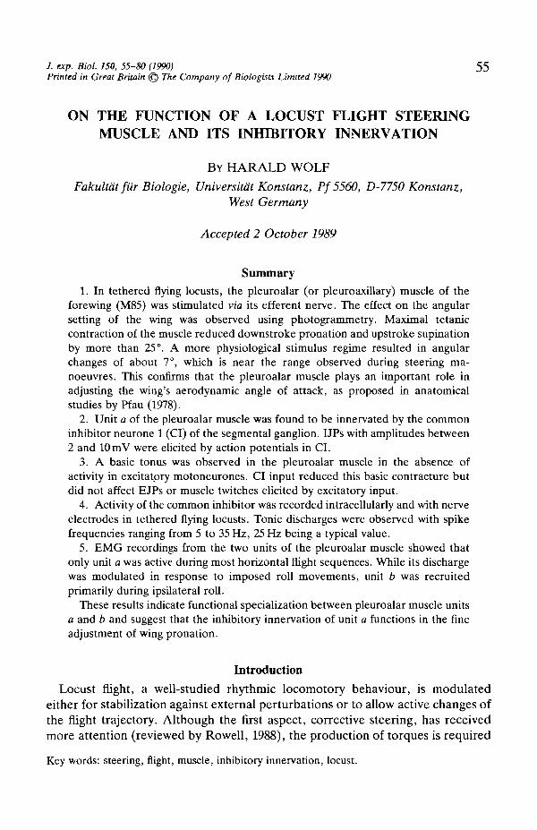
Fig. 1 is a diagram of the mesothoradc anatomy showing the muscles and nervesdescribed in this section. Bipolar stimulation electrodes (stainless-steel wire,30 pan in diameter, insulated except for the inner surfaces of the hooks) wereattached to mesothoracic nerve 4D4 (nomenclature after Campbell, 1961) whichsupplies the pleuroalar muscle of the forewing (M85 after Snodgrass, 1929).
The locusts were cooled and mounted on plasticine. A window was cut into the
M85 -PS3
EMG
Electrode
N3A3
N4D1
Mesothoracicganglion
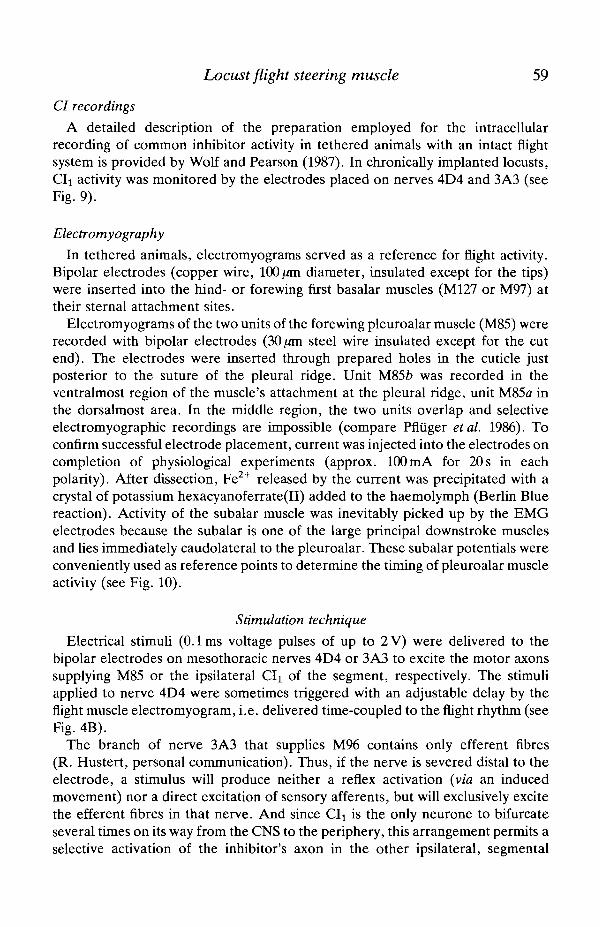
Fig. 1. Schematic inside view of the pleural region of the locust mesothorax. Anterioris to the left. Only cuticular structures, the muscles mentioned in the text and the nervebranches innervating them are shown. The sites of hook electrode placement on nerves4D4 and 3A3 are indicated, as is a bipolar EMG electrode inserted into pleuroalarmuscle unit a (insertion from the anterior is shown, although electrodes were usuallyinserted from the caudal direction). Numbering of the muscles is according toSnodgTass (1929): M85, pleuroalar; M96, abductor coxae; M99, subalar. N4D1, nerve4D1; PS3, third pleuroaxillary sclerite; PR, pleural ridge.
58 H. WOLF
mesothoracic pleura above the subalar muscle (M99). Without damaging tracheaor muscle, the subalar muscle was gently displaced caudally to expose nerve 4D4.The electrode was slid down along the pleural ridge, hooked onto the nerve, andsecured to the pleura with wax resin (beeswax/collophonium mixture: 1/2).Silicone grease was squeezed into the opening of the hooks for additionalinsulation. The subalar muscle was then released and the cuticle lid replaced andsealed with wax resin.
The mesothoracic common inhibitor neurone 1 (CIx, Hale and Burrows, 1985)was stimulated antidromically by placing a bipolar electrode on the branch ofnerve 3A3 which supplies muscle 96 (Fig. 1). The nerve was crushed distal to thestimulation site to avoid activation of the muscle by the stimulus. Experimentswere completed within 2 days of electrode implantation, i.e. before nervedegeneration occurred.
The animals were glued to a steel rod by their thoracic sternites and placed infront of a wind tunnel. The holder did not impair movements of wings orabdomen. In most experiments, the animals' legs were severed in the coxotrochan-teral joint to avoid the undesired interruption of flight sequences by tarsal contactwith the holder.
Acute preparations of M85
Animals were left intact and unanaesthetized, except for brief cooling. Theywere mounted on plasticine with the thoracic pleura of one side of the body facingupwards. Thorax and legs were immobilized with metal clasps glued to the cuticleto achieve stable muscle force recordings. Abdomen and stigmata were left free,however, to allow normal ventilation of the tracheal system. Bipolar hookelectrodes (non-insulated minuten steel pins) were placed on nerves 4D4 and 3A3,as described for the chronic implantation (Fig. 1). However, the subalar musclewas removed to gain easier access to the pleuroalar muscle and nerve 4D4.Recording sites were insulated against the haemolymph with silicone grease. Insome experiments, the electrode for stimulating CI was placed on nerve 4D1(instead of 3A3), which supplies muscle 90 (see Fig. 7A). The third axillary sclerite(attachment site of M85) was excised from the wing and the wing removed. Thesclerite was connected to a force transducer (force-displacement transducer, GrassFTO3C) and the pleuroalar muscle stretched to a length corresponding approxi-mately to the upstroke position of the wing. Intracellular recordings from singlemuscle fibres were made with 20-50MQ glass electrodes filled with 3moll"1
potassium chloride.
In one set of experiments (see Fig. 11) the animals were not mounted onplasticine but tethered on a steel holder as described above. This permitted therecording of pleuroalar muscle force and neural activity in locusts flying with threeintact wings. The wing of the muscle under investigation and all legs wereremoved. The wing hinge with the insertions'of the flight muscles, except thejsubalar and the pleuroalar, were left in place.
Locust flight steering muscle 59
CI recordings
A detailed description of the preparation employed for the intracellularrecording of common inhibitor activity in tethered animals with an intact flightsystem is provided by Wolf and Pearson (1987). In chronically implanted locusts,CIi activity was monitored by the electrodes placed on nerves 4D4 and 3A3 (seeFig. 9).
Electromyography
In tethered animals, electromyograms served as a reference for flight activity.Bipolar electrodes (copper wire, 100/an diameter, insulated except for the tips)were inserted into the hind- or forewing first basalar muscles (M127 or M97) attheir sternal attachment sites.
Electromyograms of the two units of the forewing pleuroalar muscle (M85) wererecorded with bipolar electrodes (30/an steel wire insulated except for the cutend). The electrodes were inserted through prepared holes in the cuticle justposterior to the suture of the pleural ridge. Unit M85& was recorded in theventralmost region of the muscle's attachment at the pleural ridge, unit M85a inthe dorsalmost area. In the middle region, the two units overlap and selectiveelectromyographic recordings are impossible (compare Pfliiger et al. 1986). Toconfirm successful electrode placement, current was injected into the electrodes oncompletion of physiological experiments (approx. 100 mA for 20 s in eachpolarity). After dissection, Fe2+ released by the current was precipitated with acrystal of potassium hexacyanoferrate(II) added to the haemolymph (Berlin Bluereaction). Activity of the subalar muscle was inevitably picked up by the EMGelectrodes because the subalar is one of the large principal downstroke musclesand lies immediately caudolateral to the pleuroalar. These subalar potentials wereconveniently used as reference points to determine the timing of pleuroalar muscleactivity (see Fig. 10).
Stimulation technique
Electrical stimuli (0.1ms voltage pulses of up to 2 V) were delivered to thebipolar electrodes on mesothoracic nerves 4D4 or 3A3 to excite the motor axonssupplying M85 or the ipsilateral CIi of the segment, respectively. The stimuliapplied to nerve 4D4 were sometimes triggered with an adjustable delay by theflight muscle electromyogram, i.e. delivered time-coupled to the flight rhythm (seeFig. 4B).
The branch of nerve 3A3 that supplies M96 contains only efferent fibres(R. Hustert, personal communication). Thus, if the nerve is severed distal to theelectrode, a stimulus will produce neither a reflex activation {via an inducedmovement) nor a direct excitation of sensory afferents, but will exclusively excitethe efferent fibres in that nerve. And since Cl! is the only neurone to bifurcateseveral times on its way from the CNS to the periphery, this arrangement permits aselective activation of the inhibitor's axon in the other ipsilateral, segmental
60 H. WOLF
nerves (compare Hale and Burrows, 1985). Successful activation of the inhibitorand spike transmission across the bifurcations was monitored with the electrode onnerve 4D4.
Nerve 4D4 also contains only efferent fibres, and the two excitatory motoraxons innervating the two units of M85 have much larger diameters than theremaining fibres (Pfliiger etal. 1986). Thus, through careful adjustment of thestimulus voltage, the excitatory axons were selectively activated without inter-fering with signal transmission in the smaller units, particularly CIi.
Stimulation of nerve 4D4 with the chronically implanted electrodes specificallyactivated M85 and did not spread to neighbouring muscles, even the closelyoverlying subalar (M99). This could easily be determined after moving thefore wing of the resting animal into the stroke position. A contraction of M85resulted in a clear supination of the wing, while activation of the subalar produceda pronounced downward twitch and coxal movement.
Experimental situation, data acquisition and evaluation
Flight sequences were initiated by removing a styrofoam ball from the animal'starsi or by directing a wind stream (2.5-3.0ms"1) onto its head. For photo-grammetry experiments and some intracellular CI recordings, the wind stimuluswas terminated after the initiation of flight because the pattern of wing movementsproved to be more stable without it (Wolf and Pearson, 1987). For photogram-metric recordings only, compound eyes and ocelli were covered to avoidorientation of the animal to the stroboscope light. In all other experiments, anilluminated artificial horizon was displayed in front of the animal, covering about110° of its visual field. Roll stimuli were applied by rotating the animal around itslongitudinal axis, i.e. under open-loop conditions. The holder was coupled to aposition-sensing transducer that monitored the degree of rotation. Stimuli weredelivered by hand.
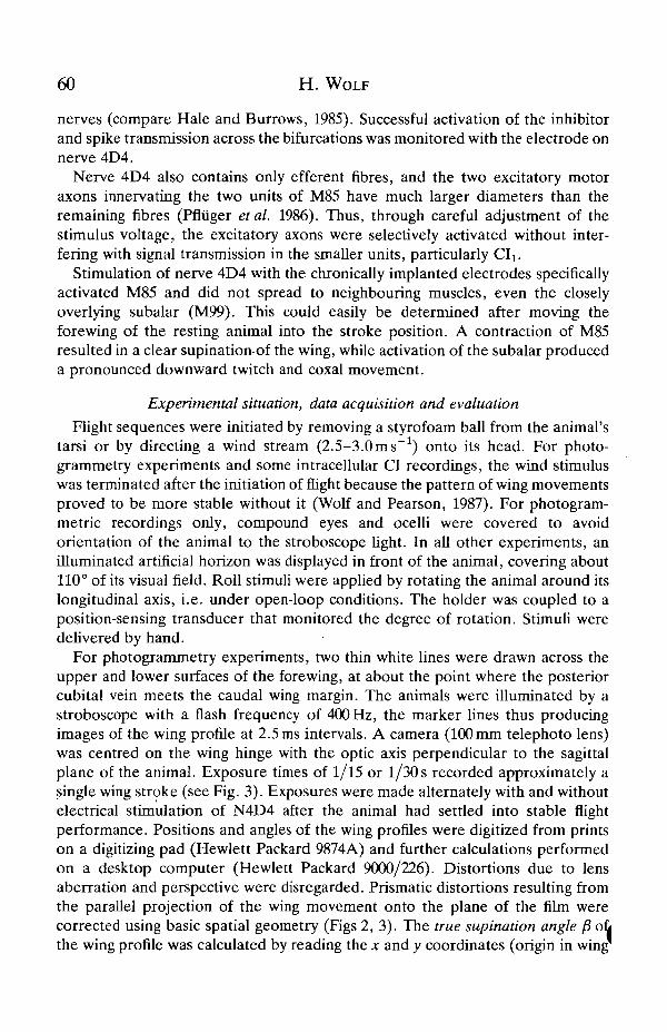
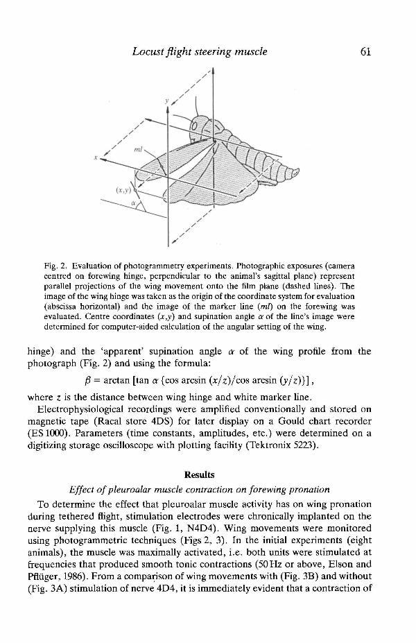
For photogTammetry experiments, two thin white lines were drawn across theupper and lower surfaces of the forewing, at about the point where the posteriorcubital vein meets the caudal wing margin. The animals were illuminated by astroboscope with a flash frequency of 400 Hz, the marker lines thus producingimages of the wing profile at 2.5 ms intervals. A camera (100 mm telephoto lens)was centred on the wing hinge with the optic axis perpendicular to the sagittalplane of the animal. Exposure times of 1/15 or 1/30s recorded approximately asingle wing stroke (see Fig. 3). Exposures were made alternately with and withoutelectrical stimulation of N4D4 after the animal had settled into stable flightperformance. Positions and angles of the wing profiles were digitized from printson a digitizing pad (Hewlett Packard 9874A) and further calculations performedon a desktop computer (Hewlett Packard 9000/226). Distortions due to lensaberration and perspective were disregarded. Prismatic distortions resulting fromthe parallel projection of the wing movement onto the plane of the film werecorrected using basic spatial geometry (Figs 2, 3). The true supination angle f3 ofrthe wing profile was calculated by reading the x and y coordinates (origin in wing
Locust flight steering muscle
Fig. 2. Evaluation of photogrammetry experiments. Photographic exposures (cameracentred on forewing hinge, perpendicular to the animal's sagittal plane) representparallel projections of the wing movement onto the film plane (dashed lines). Theimage of the wing hinge was taken as the origin of the coordinate system for evaluation(abscissa horizontal) and the image of the marker line {ml) on the forewing wasevaluated. Centre coordinates (x,y) and supination angle a of the line's image weredetermined for computer-aided calculation of the angular setting of the wing.
hinge) and the 'apparent' supination angle a of the wing profile from thephotograph (Fig. 2) and using the formula:
P = arctan [tan a {cos arcsin (x/z)/cos arcsin (y/z)}],
where z is the distance between wing hinge and white marker line.Electrophysiological recordings were amplified conventionally and stored on
magnetic tape (Racal store 4DS) for later display on a Gould chart recorder(ES1000). Parameters (time constants, amplitudes, etc.) were determined on adigitizing storage oscilloscope with plotting facility (Tektronix 5223).
ResultsEffect of pleuroalar muscle contraction on forewing pronation
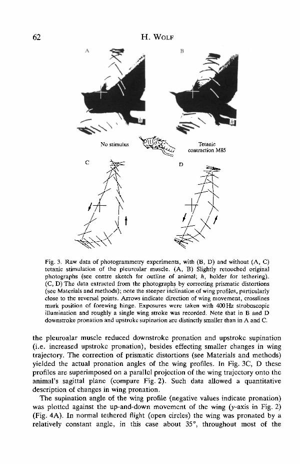
To determine the effect that pleuroalar muscle activity has on wing pronationduring tethered flight, stimulation electrodes were chronically implanted on thenerve supplying this muscle (Fig. 1, N4D4). Wing movements were monitoredusing photogrammetric techniques (Figs 2, 3). In the initial experiments (eightanimals), the muscle was maximally activated, i.e. both units were stimulated atfrequencies that produced smooth tome contractions (50 Hz or above, Elson andPfliiger, 1986). From a comparison of wing movements with (Fig. 3B) and without(Fig. 3A) stimulation of nerve 4D4, it is immediately evident that a contraction of
H. WOLF
B
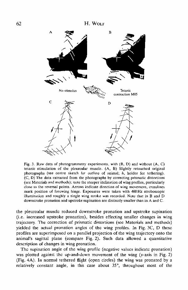
Fig. 3. Raw data of photogrammetry experiments, with (B, D) and without (A, C)tetanic stimulation of the pleuroalar muscle. (A, B) Slightly retouched originalphotographs (see centre sketch for outline of animal; h, holder for tethering).(C, D) The data extracted from the photographs by correcting prismatic distortions(see Materials and methods); note the steeper inclination of wing profiles, particularlyclose to the reversal points. Arrows indicate direction of wing movement, crosslinesmark position of forewing hinge. Exposures were taken with 400 Hz stroboscopicillumination and roughly a single wing stroke was recorded. Note that in B and Ddownstroke pronation and upstroke supination are distinctly smaller than in A and C.
the pleuroalar muscle reduced downstroke pronation and upstroke supination(i.e. increased upstroke pronation), besides effecting smaller changes in wingtrajectory. The correction of prismatic distortions (see Materials and methods)yielded the actual pronation angles of the wing profiles. In Fig. 3C, D theseprofiles are superimposed on a parallel projection of the wing trajectory onto theanimal's sagittal plane (compare Fig. 2). Such data allowed a quantitativedescription of changes in wing pronation.
The supination angle of the wing profile (negative values indicate pronation)was plotted against the up-and-down movement of the wing (y-axis in Fig. 2)(Fig. 4A). In normal tethered flight (open circles) the wing was pronated by arelatively constant angle, in this case about 35°, throughout most of the
62 H. WOLF
Fig. 3. Raw data of photogrammetry experiments, with (B, D) and without (A, C)tetanic stimulation of the pleuroalar muscle. (A, B) Slightly retouched originalphotographs (see centre sketch for outline of animal; h, holder for tethering).(C, D) The data extracted from the photographs by correcting prismatic distortions(see Materials and methods); note the steeper inclination of wing profiles, particularlyclose to the reversal points. Arrows indicate direction of wing movement, crosslinesmark position of forewing hinge. Exposures were taken with 400 Hz stroboscopicillumination and roughly a single wing stroke was recorded. Note that in B and Ddownstroke pronation and upstroke supination are distinctly smaller than in A and C.
the pleuroalar muscle reduced downstroke pronation and upstroke supination(i.e. increased upstroke pronation), besides effecting smaller changes in wingtrajectory. The correction of prismatic distortions (see Materials and methods)yielded the actual pronation angles of the wing profiles. In Fig. 3C, D theseprofiles are superimposed on a parallel projection of the wing trajectory onto theanimal's sagittal plane (compare Fig. 2). Such data allowed a quantitativedescription of changes in wing pronation.
The supination angle of the wing profile (negative values indicate pronation)was plotted against the up-and-down movement of the wing (y-axis in Fig. 2)(Fig. 4A). In normal tethered flight (open circles) the wing was pronated by arelatively constant angle, in this case about 35°, throughout most of the
Locust flight steering muscle 63
60
40
20
-20
-40- 6 0 - 4 0 - 2 0 20 40
- 4 0 - 2 0 20 40 60Upstroke angle (degrees)
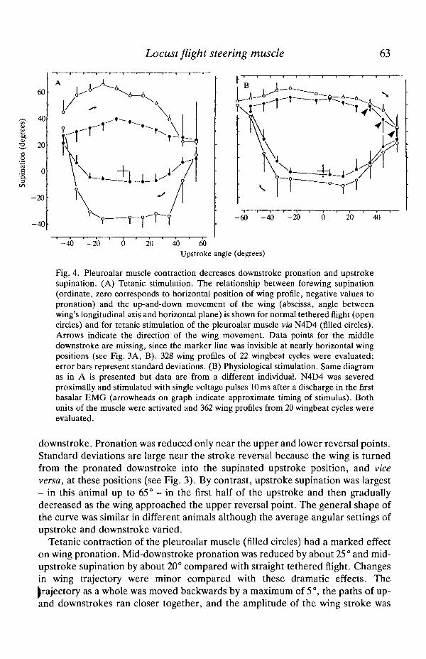
Fig. 4. Pleuroalar muscle contraction decreases downstroke pronation and upstrokesupination. (A) Tetanic stimulation. The relationship between forewing supination(ordinate, zero corresponds to horizontal position of wing profile, negative values topronation) and the up-and-down movement of the wing (abscissa, angle betweenwing's longitudinal axis and horizontal plane) is shown for normal tethered flight (opencircles) and for tetanic stimulation of the pleuroalar muscle via N4D4 (filled circles).Arrows indicate the direction of the wing movement. Data points for the middledownstroke are missing, since the marker line was invisible at nearly horizontal wingpositions (see Fig. 3A, B). 328 wing profiles of 22 wingbeat cycles were evaluated;error bars represent standard deviations. (B) Physiological stimulation. Same diagramas in A is presented but data are from a different individual. N4D4 was severedproximally and stimulated with single voltage pulses 10 ms after a discharge in the firstbasalar EMG (arrowheads on graph indicate approximate timing of stimulus). Bothunits of the muscle were activated and 362 wing profiles from 20 wingbeat cycles wereevaluated.
downstroke. Pronation was reduced only near the upper and lower reversal points.Standard deviations are large near the stroke reversal because the wing is turnedfrom the pronated downstroke into the supinated upstroke position, and viceversa, at these positions (see Fig. 3). By contrast, upstroke supination was largest- in this animal up to 65° - in the first half of the upstroke and then graduallydecreased as the wing approached the upper reversal point. The general shape ofthe curve was similar in different animals although the average angular settings ofupstroke and downstroke varied.
Tetanic contraction of the pleuroalar muscle (filled circles) had a marked effecton wing pronation. Mid-downstroke pronation was reduced by about 25° and mid-upstroke supination by about 20° compared with straight tethered flight. Changesin wing trajectory were minor compared with these dramatic effects. Thetrajectory as a whole was moved backwards by a maximum of 5°, the paths of up-and downstrokes ran closer together, and the amplitude of the wing stroke was
64 H . W O L F
slightly reduced as a result of tetanic pleuroalar muscle contraction (quantitativedata not shown, compare Fig. 3A,C with 3B,D).
A tetanic contraction of the pleuroalar muscle never occurs during flight inintact locusts. In tethered flying animals, about one muscle potential is generatedper wingbeat cycle, according to electromyographic records. The force producedby the muscle if activated at such a rate (about 20 Hz) ranges from about 15 to 25 %of the tetanic force output, depending on which of the muscle's units are actuallyactivated and considering individual variability (Elson and Pfliiger, 1986; see alsoFig. 11). Assuming a linear relationship between muscle force and effect on wingpronation, as a first approximation, a physiological activation of the muscle shouldreduce downstroke pronation and upstroke supination by about 3-6°.
In the experiment shown in Fig. 4B, both units of the pleuroalar muscle werestimulated with one impulse per wingbeat cycle, delivered at a physiologicallatency of 10 ms time-coupled to the spike in a depressor muscle EMG (firstbasalar 97). This stimulus regime mimics pleuroalar muscle activity duringmoderate steering action (Elson and Pfliiger, 1986, see also Fig. 10). Nerve 4D4was severed proximal to the stimulation site to avoid normal, efferent activation ofthe muscle. The stimulus reduced downstroke pronation and upstroke supinationby an average of 7° during about the last three-quarters of the downstroke and thefirst half of the upstroke. During the rest of the wingbeat, near the upper reversalpoint, stimulus-related differences in the angular setting of the wing were distinctlysmaller (average of 3°). Similar observations were made in the four other animalsexamined. Changes in wing stroke trajectory were negligible in this experiment.
In a quiescent animal with its forewings clicked into the stroke position,application of a 20 Hz stimulus train resulted in a supination of the respective wing(see also Heukamp, 1984). Angular changes of 5-6° (i.e. comparable to thoserecorded during flight) were observed.
Inhibitory innervation of M85
Electrophysiological analysis
Pfliiger etal. (1986) reported that backfill experiments with cobalt ions, whenperformed on mesothoracic nerve 4D4, sometimes stained a neurone with aventromedial cell body and axonal branches in the roots of nerves 3, 4 and 5. Thiswas recognized as an indication that the pleuroalar muscle may be innervated bythe mesothoracic common inhibitor 1 (CIx). In the present study, backfillexperiments confirmed that CIi is stained in about 50 % of the animals if the cobaltions (1.5% CoCl2) are allowed to diffuse for about 5 days (see also Kutsch andSchneider, 1987). However, Elson and Pfliiger (1986) reported that 'intracellularrecordings from single muscle fibres do not show inhibitory junction potentials'and Pfliiger etal. (1986) suggested in their detailed anatomical study that'innervation of the pleuroaxillary muscle by Cli may be variable or non-functional'. They proposed that the inhibitory innervation might be present irllarvae but retract in the course of the larval-adult transition.

Locust flight steering muscle 65
M85a
•N4D4
Fig. 5. Inhibitory innervation of M85 by CIi. Intracellular recordings from unit amuscle fibres are shown in the top traces, en passant recordings from N4D4 in thebottom traces. CIj was stimulated in N3A3, stimulus artefacts are marked byarrowheads in A. (A) Potentials recorded during three stimulus presentations aresuperimposed. CI spikes in N4D4 and resulting IJPs in the muscle fibre are clearlyvisible. Note the small spike about half-way along the N4D4 record. Comparing itsbiphasic potential to that of the CI spike shows that it is from an efferent fibre.(B) Stimulus trains (200 ms duration) were presented with repetition rates of 20, 30 and50Hz and summation of IJPs was observed. Scale bars, 20ms, 10mV in A, 500ms,15mVinB.
The consistent observation of inhibitory junction potentials (IJPs) in intracellu-lar pleuroalar muscle fibre recordings was therefore unexpected. Stimulation ofthe ipsilateral CIi with hook electrodes on mesothoracic nerve 3A3 (see Materialsand methods, Fig. 1 and Hale and Burrows, 1985) revealed that the IJPsrepresented input from this inhibitory neurone (Fig. 5). The IJPs occurred atlatencies of about 10 ms with regard to the stimulus and had amplitudes rangingfrom 2 to 10 mV, rarely 12 mV. En passant recordings from nerve 4D4 demon-strated that most of this latency (approx. 7 ms) was due to the conduction timerequired for spike propagation from nerve 3A3 to nerve 4D4 via the mesothoracicganglion.
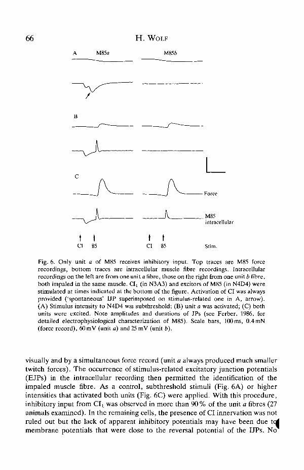
Only one of the two units of the pleuroalar muscle, unit a, was found to receiveinhibitory innervation. It was often clearly visible to which of the two units theimpaled muscle fibre belonged (for anatomy see Pfliiger et al. 1986 and Fig. 1).However, since the two units overlap, particularly in the middle region, theprocedure shown in Fig. 6 was applied for an unequivocal identification. Theexcitatory axons in nerve 4D4 were stimulated in addition to CI (which wasactivated in N3A3). At low stimulus intensities only one of the two motor axonswas excited, usually that of unit a (Fig. 6B). The activated unit could be identified
66
Force
t ta 85
t tCI 85
M85intracellular
Stim.
Fig. 6. Only unit a of M85 receives inhibitory input. Top traces are M85 forcerecordings, bottom traces are intracellular muscle fibre recordings. Intracellularrecordings on the left are from one unit a fibre, those on the right from one unit b fibre,both impaled in the same muscle, d^ (in N3A3) and excitors of M85 (in N4D4) werestimulated at times indicated at the bottom of the figure. Activation of CI was alwaysprovided ('spontaneous' UP superimposed on stimulus-related one in A, arrow).(A) Stimulus intensity to N4D4 was subthreshold; (B) unit a was activated; (C) bothunits were excited. Note amplitudes and durations of JPs (see Ferber, 1986, fordetailed electrophysiological characterization of M85). Scale bars, 100 ms, 0.4 mN(force record), 60mV (unit a) and 25 mV (unit b).
visually and by a simultaneous force record (unit a always produced much smallertwitch forces). The occurrence of stimulus-related excitatory junction potentials(EJPs) in the intracellular recording then permitted the identification of theimpaled muscle fibre. As a control, subthreshold stimuli (Fig. 6A) or higherintensities that activated both units (Fig. 6C) were applied. With this procedure,inhibitory input from Clt was observed in more than 90 % of the unit a fibres (27animals examined). In the remaining cells, the presence of CI innervation was notruled out but the lack of apparent inhibitory potentials may have been due tedmembrane potentials that were close to the reversal potential of the I JPs. No
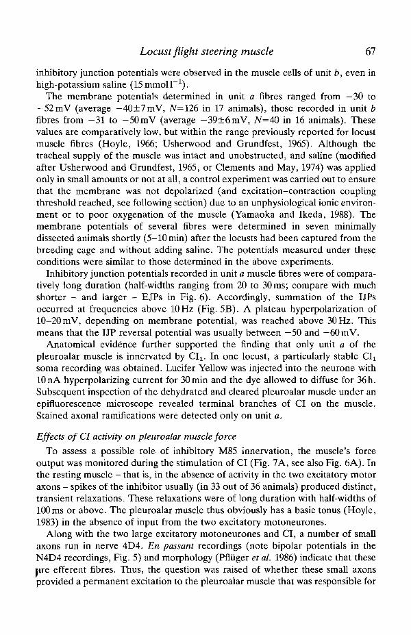
Locust flight steering muscle 67
inhibitory junction potentials were observed in the muscle cells of unit b, even inhigh-potassium saline (ISmmoir1) .
The membrane potentials determined in unit a fibres ranged from —30 to—52mV (average — 40±7mV, N=126 in 17 animals), those recorded in unit bfibres from -31 to -50mV (average -39±6mV, N=40 in 16 animals). Thesevalues are comparatively low, but within the range previously reported for locustmuscle fibres (Hoyle, 1966; Usherwood and Grundfest, 1965). Although thetracheal supply of the muscle was intact and unobstructed, and saline (modifiedafter Usherwood and Grundfest, 1965, or Clements and May, 1974) was appliedonly in small amounts or not at all, a control experiment was carried out to ensurethat the membrane was not depolarized (and excitation-contraction couplingthreshold reached, see following section) due to an unphysiological ionic environ-ment or to poor oxygenation of the muscle (Yamaoka and Ikeda, 1988). Themembrane potentials of several fibres were determined in seven minimallydissected animals shortly (5-10 min) after the locusts had been captured from thebreeding cage and without adding saline. The potentials measured under theseconditions were similar to those determined in the above experiments.
Inhibitory junction potentials recorded in unit a muscle fibres were of compara-tively long duration (half-widths ranging from 20 to 30 ms; compare with muchshorter - and larger - EJPs in Fig. 6). Accordingly, summation of the IJPsoccurred at frequencies above 10 Hz (Fig. 5B). A plateau hyperpolarization of10-20 mV, depending on membrane potential, was reached above 30 Hz. Thismeans that the UP reversal potential was usually between —50 and — 60 mV.
Anatomical evidence further supported the finding that only unit a of thepleuroalar muscle is innervated by CI^ In one locust, a particularly stable CIisoma recording was obtained. Lucifer Yellow was injected into the neurone with10 nA hyperpolarizing current for 30 min and the dye allowed to diffuse for 36 h.Subsequent inspection of the dehydrated and cleared pleuroalar muscle under anepifluorescence microscope revealed terminal branches of CI on the muscle.Stained axonal ramifications were detected only on unit a.
Effects of CI activity on pleuroalar muscle force
To assess a possible role of inhibitory M85 innervation, the muscle's forceoutput was monitored during the stimulation of CI (Fig. 7A, see also Fig. 6A). Inthe resting muscle - that is, in the absence of activity in the two excitatory motoraxons - spikes of the inhibitor usually (in 33 out of 36 animals) produced distinct,transient relaxations. These relaxations were of long duration with half-widths of100ms or above. The pleuroalar muscle thus obviously has a basic tonus (Hoyle,1983) in the absence of input from the two excitatory motoneurones.
Along with the two large excitatory motoneurones and CI, a number of smallaxons run in nerve 4D4. En passant recordings (note bipolar potentials in theN4D4 recordings, Fig. 5) and morphology (Pfliiger et al. 1986) indicate that theseare efferent fibres. Thus, the question was raised of whether these small axonsprovided a permanent excitation to the pleuroalar muscle that was responsible for

68 H. WOLF
Force M85
Stim. N4D1
Force M85
•Stim.
C
Superposition
M85a _intra-cellular
Fig. 7. CI activity reduces basic tonus. (A) Mesothoracic CIi was stimulated (inN4D1, bottom trace) and the force output of M85 recorded (top trace). Each stimulus,i.e. spike of CI, caused a transient relaxation of M85 (see also Fig. 6A). The effectpersisted after transection of N4 root (arrow and movement artefact in the middle).(B) Both excitors of M85 were stimulated in N4D4 (stimulus frequency about 1 Hz,bottom trace), CIi was activated in N3A3 (stimulus frequency 50Hz, black bar inbottom trace) and M85 force output was recorded (top trace). CI stimulation reducedbasic tonus but left muscle twitches almost unaffected. (C, inset) One muscle twitchelicited with simultaneous CI stimulation (smaller twitch) and one without (largertwitch) are superimposed to demonstrate the minute effect of CI activity on twitchamplitude (approx. 2 % decrease; time scale 10 times, amplitude two times expanded,arrow indicates level of basic tonus without CI stimulation). (D) During theexperiment described in Fig. 6 spontaneous struggling of the animal occurred,accompanied by strong Cl! discharges (two middle segments, IJP rate 25-35 Hz).Reductions in membrane potential and basic tonus and constancy of EJPs and twitchresponses are visible. Recordings from the quiescent animal are presented in the firstand last segment, only unit a was activated in the second and third muscle twitch shown(arrows). Scale bars, 500ms, 0.1 mN in A, 0.3 mN in B; 400 ms, 35 mV or 0.25 mN in D.
the observed basic contracture. This appeared not to be the case, however,because the relaxation in response to stimulation of CI persisted after long periodsof silence in en passant recordings of nerve 4D4 and even after transection of theroot of nerve 4 (Fig. 7A, CIx was stimulated in N4D1 in this case).
Repetitive stimulation of CI (compare Fig. 5) caused a considerable decrease inthe basic contracture. In response to high-frequency stimulation of the inhibitoilthe force declined by up to 0.2 mN, which is about one-third of the M85 twitch

Locust flight steering muscle 69
intracellular
Sum. CI
Fig. 8. Interaction of excitatory and inhibitory input to M85. CIi was stimulated inN3A3 at 50Hz (bars at bottom), the two excitatory motoneurones in N4D4 at 20 Hz(stimulus trace not shown). The effects were monitored in an intracellular unit a fibrerecording (lower trace, slightly unstable due to muscle contractions) and a muscle forcerecording (upper trace). (A) CIi was initially stimulated alone to demonstrate theresulting decline in basic tonus. (B) The start of the stimulus train to N4D4. Noteaccumulation of tetanic tension. (C) CI was activated during ongoing N4D4 stimu-lation. (D) The restoration of basic tonus after CI stimulation had been terminated.Broken reference line indicates force level without N3A3 or N4D4 stimulation. Scalebars, 100ms, 20mV or 0.25 mN.
force (Fig. 7B,D). Contractions elicited by excitatory input, in contrast, remainedalmost unaffected by CI discharges. Twitch amplitudes were reduced by less than5 % during CI activity and no changes were noted in twitch relaxation (Fig. 7C).Similar constancy was noted with regard to the EJPs recorded in unit a musclefibres (see Figs 7D, 8).
The same held true for twitch trains elicited in M85 (Fig. 8) which mimicked theactivity pattern observed during flight (Elson and Pfluger, 1986). Initially, CI wasactivated alone to show the resulting decreases in membrane potential and basictonus (Fig. 8A). When both units of M85 were subsequently stimulated at a rate of20FIz, summation of the individual muscle twitches occurred and a gradualaccumulation of residual tetanic tension was observed during the initial contrac-tions. Stimulation of CI at a frequency of about 50 Hz then resulted in a generaldecline of the force level. This decline was equal to the reduction in basic tonuscaused by the CI discharge alone. Simultaneous intracellular recording from a unita muscle fibre demonstrated that the EJPs remained unaffected by the hyperpolar-ization resulting from CI stimulation.
CI activity during flight
Considering both the function of the pleuroalar muscle in flight steering and theinfluence CI discharges have on the muscle's contraction, two questions areimmediately evident: is CIi active during flight and, if so, what is the pattern of itsactivity? Intracellular soma recordings were performed in intact, tethered flying|[ocusts using the method of Wolf and Pearson (1987). In these recordings, CIactivity was characterized by a steady depolarization that lasted throughout the
70 H. WOLF
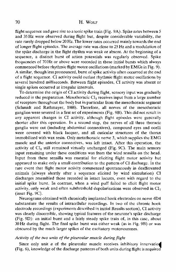
flight sequence and gave rise to a tonic spike train (Fig. 9A). Spike rates between 5and 35 Hz were observed during flight but, despite considerable variability, therate rarely dropped below 20 Hz. The lower rates occurred mainly towards the endof longer flight episodes. The average rate was close to 25 Hz and a modulation ofthe spike discharge in the flight rhythm was weak or absent. At the beginning of asequence, a distinct burst of action potentials was regularly observed. Spikefrequencies of 70 Hz or above were recorded in these initial bursts which alwayscommenced before rhythmic flight motor oscillations (marked by EMGs in Fig. 9).A similar, though less pronounced, burst of spike activity often occurred at the endof a flight sequence. CI activity could outlast rhythmic flight motor oscillations byseveral hundred milliseconds. Between flight episodes, CI activity was absent orsingle spikes occurred at irregular intervals.
To determine the origin of CI activity during flight, sensory input was graduallyreduced in the preparation. Mesothoracic CIx receives input from a large numberof receptors throughout the body but in particular from the mesothoracic segment(Schmidt and Rathmayer, 1988). Therefore, all nerves of the mesothoracicganglion were severed in a first set of experiments (Fig. 9B). This did not result inany apparent changes in CI activity, although flight episodes were generallyshorter after this operation. In a second step, the nerves of all three thoracicganglia were cut (including abdominal connectives), compound eyes and ocelliwere covered with black lacquer, and all cuticular structures of the thoraximmobilized with wax resin. Only metathoracic nerve 3, which supplies the EMGmuscle and the anterior connectives, was left intact. After this operation, theactivity of CIi still remained virtually unchanged (Fig. 9C). The main sensoryinput remaining under these conditions was from the wind sensilla on the head.Input from these sensilla was essential for eliciting flight motor activity butappeared to make only a small contribution to the pattern of CI discharge. In therare event that flight motor activity commenced spontaneously in deafferentedanimals (always shortly after a sequence elicited by wind stimulation) CIdischarges resembled those recorded in intact locusts, even with regard to theinitial spike burst. In contrast, when a wind puff failed to elicit flight motoractivity, only weak and often subthreshold depolarizations were observed in CIi(inset Fig. 9C).
Neurograms obtained with chronically implanted hook electrodes on nerve 4D4substantiate the results of intracellular recordings. In two of the chronic hookelectrode recordings (experiments described in initial Results section), CI activitywas clearly discernible, showing typical features of the neurone's spike discharge(Fig. 9D): an initial burst and a fairly steady spike train of, in this case, about30 Hz during flight. The final spike burst was either weak (as in Fig. 9B) or wasobscured by the much larger spikes of the excitatory motoneurones.
Activity of the two units of the pleuroalar muscle during flight
Since only unit a of the pleuroalar muscle receives inhibitory innervatio(Fig. 6), knowledge of the discharge patterns of both units during flight is require
Locust flight steering muscle 71A Intact
127- •fftt
B Deafferented mesothoracic roots
_J^^UKOUU^^^
Deafferented
I H» M» 1» l» M ' li I' I Wind
D Intact neurogram
\ 85a
Fig. 9. CIi activity during flight. Intracellular soma recordings (A-C) and a neuro-gram (D) demonstrate the patterns of CIi depolarization and spike discharge duringtethered flight (top traces; EMGs from hindwing first basalar, 127, monitor flightactivity in bottom traces). (A) Sequence from an intact, tethered flying animal(particularly short flight episode selected to match records in B and C). The sameindividual had all mesothoracic nerve roots severed in B and the thoracic nerve cordisolated from the periphery in C [only metathoracic nerve 3 (EMG) and neckconnectives intact]. In addition, the response to a wind stimulus that did not elicit flightmotor activity is shown (bar marks duration of stimulus). (D) Neurogram recorded onN4D4 in an intact, tethered animal. Small action potentials are from CIi, large onesfrom unit a motoneurone (85a) (final burst contains also unit b potentials). An unusualflight episode was selected, in which excitatory motoneurones of M85 were rarelyactive, to provide a clear picture of CI activity. Scale bars, 10 mV, 500 ms (A-C) or250ms (D).
72 H. WOLF
Horizontal Roll contralateral
M85a
M85b
5 ms
Fig. 10. Activity of motor units M85a (A) and M85b (B) during horizontal flight(middle) and imposed roll movements (approx. 45°, contralateral side down in rightand up in left diagram, see sketches). Recordings from five trials are superimposedtaking pick-up of M99 potentials as reference (arrowhead, top middle, 99). Duringhorizontal flight or ipsilateral roll the units sometimes fired doublets (arrows). Notepick-up of unit a potentials in B (arrowheads, 85a).
for a functional interpretation. Elson and Pfliiger (1986) and Reuse (1987) havealready presented detailed electromyographic studies of pleuroalar muscle activityduring horizontal flight and steering manoeuvres. However, selective recordingsof the two motor units were not made and, hence, an identification of theircontribution to muscle activity was not possible.
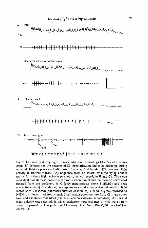
Selective electromyographic recordings from pleuroalar muscle units a and b (indifferent individuals, N=12) yielded the following results (Fig. 10). In accordancewith the report by Elson and Pfliiger (1986), usually one but sometimes both of themuscle's units were active after the animals had settled into stable flight. In thehorizontal position, unit a regularly discharged one (less often two) potential per
.wingbeat cycle. The potentials occurred with latencies of about 5-15 ms withregard to a spike in the ipsilateral subalar muscle (large pick-up potentials inFig. 10). Unit b was rarely active during horizontal flight and was usually recruitedonly by imposed roll movements towards the ipsilateral side. This presumablyreflected compensatory steering action - that is, the production of a countertorque(Elson and Pfliiger, 1986). An ipsilateral roll also decreased the latency of unit apotentials and increased the probability of the unit firing doublets. A rollmovement to the contralateral side increased the latency and decreased theprobability of a discharge in both units. Unit b was thus mainly recruited for theproduction of a presumed corrective roll torque to the contralateral side, whereasunit a was active during horizontal flight.
Additional evidence for this conclusion was provided by an experiment in whidlone forewing of the locust was removed and a force gauge attached to th?

Locust flight steering muscle 73
jflu
Force
Nerve4D4
EMG 127"(113)
Fig. 11. Activity of motor units M85a and M85b. In a tethered animal with oneforewing removed, pleuroalar muscle force (top trace), activity in N4D4 (middle trace)and a hindwing first basalar EMG (bottom trace; plus pick-up from M113, initial M127potentials marked by dots for identification) were recorded. The smallest spikes in theneurogram are from CI^ Unit a produced the middle-sized action potentials, unit b thelargest ones. The animal flew undisturbed at the beginning and end of the sequence.Arrows mark start and termination of a visual roll stimulus.
pleuroalar muscle (Fig. 11, upper trace). A bipolar hook electrode on nerve 4D4recorded neural input to the muscle (Fig. 11, middle trace) and an EMG electrodemonitored flight activity (Fig. 11, bottom trace). The animal was otherwise intact.Single motoneurone spikes elicited by visual stimuli in the quiescent animalindicated that the motoneurones of units b and a produced the largest and second-largest spikes in the neurogram, respectively, while the smallest class of actionpotentials was from CIi. As mentioned above, unit a produced smaller muscletwitches than unit b. At the beginning of the flight sequence, unit a showed a quitetypical pattern of activity, discharging about one action potential per wingbeatcycle, but after about 1 s its activity decreased. The open arrow marks the onset ofa visual roll stimulus, produced by switching on a light contralateral to theoperated side. This resulted in the' recruitment of unit b, discernible by theoccurrence of larger spikes in the neurogram and muscle twitches of more thandouble the amplitude of the unit a twitches in the force record. The second openarrow marks the dimming of the light source, which resulted in a return to theoriginal pattern of activity. The flight sequence terminated soon afterwards andthe typical burst of pleuroalar activity associated with wing backfolding wasfcbserved (Snodgrass, 1929; Pringle, 1968). It is notable that the neurogramprovides further support for the pattern of CI activity during flight reported in the
74 H. W O L F
previous section. The average rate of CI spikes during the flight sequence wasabout 20 Hz. A frequency of 75 Hz was measured in the initial spike burst (noteinitial, negative deflection in force record).
Discussion
The present study confirms through nerve stimulation in tethered flying locuststhat the pleuroalar muscle of the forewing (M85) reduces downstroke pronationand upstroke supination (Figs 3, 4), as proposed by Pfau (1978) in his anatomicalinvestigations. It was further demonstrated that the same common inhibitor (CIj)which supplies a large number of the locust's intrinsic and extrinsic leg muscles alsoinnervates M85 (Figs 5, 6). The effect this inhibitory innervation has on themuscle's contractile response was analysed (Figs 7, 8) and CI activity during flightwas recorded (Fig. 9).
Effect of pleuroalar muscle activity on forewing pronation
Pfau (1977, 1978, 1983) carried out a number of detailed investigations of thefunctional anatomy of the locust forewing hinge. From these studies he concludedthat the pleuroalar muscle serves the specific function of regulating the angularsetting of the wing without influencing the stroke trajectory. He proposed thatactivity of the muscle reduces downstroke pronation and upstroke supination, andpresumed the muscle to be tonically active during flight. The present data providethe first direct confirmation of these assumptions. A smooth tetanic contraction ofthe pleuroalar muscle was observed to reduce downstroke pronation and upstrokesupination by about 25°. By comparison, only small changes were recorded in wingtrajectory (Fig. 3), even with this strong, unphysiological activation of the muscle.A more physiological stimulus regime (Fig. 4B) produced no noticeable changes inwing trajectory. It should be noted that efferent nerve stimulation with chronicallyimplanted electrodes proved to be essential for these experiments. Muscleelectrodes inflict considerable damage to the small pleuroalar muscle and stimulieasily spread to neighbouring muscles.
The pleuroalar muscle is not the locust's only means of changing the aerody-namic angle of attack of the forewing: the muscle is one component of a synergismthat also involves a number of principal downstroke muscles, namely the first andsecond basalar and subalar muscles. With these muscles it is the timing of theircontractions, relative to each other and to the wing movement, that influences thecourse of wing pronation during the downstroke. Correlations between the activityof these muscles and several parameters of the wing stroke (Zarnack, 1988; Reuse,1987; Schmidt and Zarnack, 1987) or roll torques generated by the animal(Waldmann and Zarnack, 1988; Reuse, 1987) are relatively well studied. Changesin wing pronation in the range of 5° - sometimes of up to about 10° - wererecorded during steering activity (Zarnack, 1988; Reuse, 1987). However,correlations between forewing pleuroalar activity and wingstroke parameters o |roll torques have not yet been analysed (although correlations between stroke
Locust flight steering muscle 75
parameters and roll torque are well established by Waldmann and Zarnack, 1988;Reuse, 1987), nor have the relative contributions of the pleuroalar and theprincipal downstroke muscles to changes in wing pronation been assessed.
One experiment represented a first step towards determining the pleuroalar'scontribution to the adjustment of wing pronation, relative to that of the principalflight muscles. After severing nerve 4D4, the pleuroalar muscle was stimulatedwith a pattern similar to that recorded in electromyograms during moderatesteering action (Elson and Pfliiger, 1986). Under such conditions, decreases indownstroke pronation and upstroke supination of about 7° were observed as aresult of muscle activation. This indicates that the pleuroalar muscle contributessignificantly to the adjustment of wing pronation, since the observed changes areof the same magnitude as those recorded during steering manoeuvres in tetheredflight (Zarnack, 1988; Reuse, 1987). Changes of this magnitude were recordedonly during about the last three-quarters of the downstroke and the initial half ofthe upstroke. Near the upper reversal point, stimulus-related decreases in wingpronation were distinctly smaller (Fig. 4B). This may indicate that phasicactivation of the pleuroalar muscle has, in part, a phasic effect on the angularsetting of the wing - contradicting Pfau's assumption of purely tonic M85 activityduring flight.
Innervation of the pleuroalar muscle by CIj
Electrophysiology
The results presented in Figs 5 and 6 provide convincing evidence that most, ifnot all, of the unit a fibres in the pleuroalar muscle are innervated by themesothoracic CIx, while the fibres of unit b lack inhibitory innervation. Two pointsneed to be discussed, however. First, why did Elson and Pfliiger (1986) fail torecord IJPs in M85? This led to the assumption that there was no inhibitoryinnervation of M85 (Pfliiger et al. 1986; compare Kutsch and Schneider, 1987). Theprobable reason is that the muscle was studied after severing nerve 4D4 proximalto the stimulation site. When the nerve is stimulated, the excitatory motoneuronesare excited at lower intensities than CI owing to their much larger diameters(Pfliiger et al. 1986). This means that stimulation of CI is impossible withoutsynchronous activation of the excitors. Consequently, IJPs will be difficult toobserve in intracellular muscle fibre recordings, particularly since the IJPscommence slightly later than the EJPs owing to the lower conduction velocity ofthe small CI axon. In addition, CI activity has little or no effect on EJP size(Figs 7D, 8). Only separate stimulation of the CI axon via a different nerve (e.g.N3A3) or spontaneous activity clearly reveal the inhibitory input and, in bothcases, an intact connection of nerve 4D4 to the CNS is essential.
Second, it should be mentioned that the pleuroalar muscle is an unusual targetfor CIi. The locust's inhibitor, as a rule, innervates only leg muscles recruitedduring walking and only those fibres that also receive input from a 'slow'motoneurone (Hale and Burrows, 1985). M91 is the only exception known: it acts

76 H. WOLF
not only as coxa remotor but is also involved in wing elevation. In other insects,too, inhibitory innervation of flight muscles is uncommon (e.g. Ikeda andBoettiger, 1965).
Effects of CI activity on pleuroalar muscle force
The main effect of CI activity on the force developed by the pleuroalar muscle isa reduction in basic tonus (Figs 7, 8). The observation of a basic musclecontracture in the absence of discharges in excitatory motoneurones is unusual butnot unprecedented (Hoyle, 1968a,b). The membrane potentials determined inM85 were remarkably low (average near — 40 mV). Experimental conditions(proper oxygenation through intact tracheal supply, little or no saline added,muscle undamaged) and controls (membrane potentials determined in minimallydissected animals) suggest that these low membrane potentials are of functionalsignificance and not an artefact. Apparently, excitation-contraction coupling(EC) threshold is reached in the absence of excitatory motoneurone discharges,leading to the basic contracture of pleuroalar muscle fibres (see Hoyle, 1983).
Whether the low membrane potentials are due to tonic release of excitatorytransmitter (demonstrated to be effective at some inhibitory junctions in crus-taceans; Parnas et al. 1975) or are caused by differences in membrane permeabilityto sodium (as has recently been shown for crustacean tonic muscle fibres;Hammelsbeck and Rathmayer, 1989) remains to be investigated. Miniaturejunction potentials were not recorded in pleuroalar muscle fibres. The reduction ofbasic tonus caused by CI activity persisted after transection of the nerve 4 root, i.e.after the abolition of any possible efferent input (Fig. 7A), as well as after longperiods of silence in nerve 4D4. This establishes that spike activity in the group ofsmall N4D4 axons with unknown function (Pfliiger et al. 1986) is not necessary forthe maintenance of basic tonus in M85.
CI activity during flight
Cli is tonically active during flight with a discharge rate adequate to affect thecontraction of M85 (compare Figs 9, 5B). In intracellular and neurogramrecordings, discharge frequencies between 20 and 35 Hz were consistently ob-served. In view of the results discussed in the previous paragraphs, it appears clearthat under physiological conditions the level of basic contracture in unit a isregulated by CI activity (Figs 7B,D, 8). Contractions elicited by excitatory inputare superimposed on this basic force level. Deafferentation experiments furthersuggest that CI depolarizations are under strong central nervous control duringflight, despite the manifold and powerful sensory input demonstrated in quiescentlocusts by Schmidt and Rathmayer (1988). This may indicate that CI innervation isemployed in the fine control of flight performance. The initial, high-frequencyspike burst observed in intracellular CI recordings (Fig. 9A) is probably associatedwith the unfolding of the wings. This spike burst may serve to relax M85completely, which otherwise might counteract wing unfolding in its function as Aretractor of the wing (Snodgrass, 1929; Pringle, 1968).
Locust flight steering muscle 11
Activity of the two M85 units during flightThe above interpretation is substantiated by the finding that unit a of the
pleuroalar muscle - which receives CI innervation - is active during normal,horizontal flight, whereas unit b is recruited mainly for steering manoeuvres(Figs 10, 11). If a muscle with two units is activated below or within the frequencyrange where an accumulation of residual, tetanic tension begins, the force outputcan be regulated only coarsely - that is, in increments whose size is determined bythe twitch amplitudes of the two units. At flight frequency, i.e. in the workingrange of the pleuroalar muscle, only partial tetanic fusion of the individual muscletwitches was observed (Figs 8, 11; Elson and Pfliiger, 1986). Thus, regulation ofmuscle force via the two excitatory motoneurones might be too coarse to permit asufficiently subtle adjustment of wing pronation as required during 'idling'horizontal flight. CI activity could provide the more gradual regulation necessary.This task might also be accomplished, however, by steady changes in the timing ofthe principal flight muscles' activity (see above).
Another, though not alternative, interpretation is as follows. By reducing thebasic tonus of the pleuroalar muscle, CI activity regulates the force level uponwhich the contractions resulting from the excitors' activity are superimposed. Amarked decrease in basic contracture will enhance the relative effect of the phasiccomponent of M85 contractions and impart particular importance to the timing ofmuscle activity. The normal timing of excitatory motoneurone spikes in nerve 4D4is such that the resulting muscle twitch will peak during the first half of thedownstroke. A delay of the muscle contraction, as occurs during active rollmanoeuvres to the ipsilateral side, will lead to a delayed reduction of downstrokepronation and, thus, to a decrease in lift produced by the wing. Accordingly, anadvanced muscle contraction will increase lift production during the downstroke.Upstroke supination may be affected to a much smaller extent than downstrokepronation in the same wingbeat cycle if the M85 twitch force has decayed by thetime the wing has passed the lower reversal point (see also data in Fig. 4Bdiscussed above). This may reduce or eliminate generation of negative lift duringthe upstroke. In fact, variability of wing pronation was clearly more pronouncedduring the downstroke (Fig. 4A). This may indicate that downstroke pronationand upstroke supination are indeed regulated independently.
A selective stimulation of CIi after elimination of its normal activity (as shownin Fig. 4B for the excitatory motoneurones) would be necessary to test thesehypotheses. As yet, however, this experiment has not been possible. If CIi wereinvolved in corrective steering, its discharge during flight would be expected to bedependent on roll stimuli. Visual input to CIi has been reported by Burrows andRowell (1973) and Schmidt and Rathmayer (1988). This input is bilaterallysymmetrical, however. Visual stimulation of the locust when recording CI activityduring tethered flight (Fig. 9) revealed a marked phasic response of the inhibitorto a dimming of the illumination. The response to a brightening of the illuminationwas less pronounced. Bilateral asymmetrical responses, although sometimesobserved, were not reproducible. This may indicate that CI is mainly involved in
78 H . W O L F
the overall adjustment of lift and thrust; for instance, when switching fromclimbing to horizontal flight. It may also indicate that visual stimulation duringintracellular neural recording in tethered animals was inadequate.
Another question raised by the present study concerns the activity of the legmuscles during flight. Many of them are known to receive inhibitory input fromCIx (Hale and Burrows, 1985). The reported CI discharge should affect toniccontractions of the leg musculature, contractions one would expect to beresponsible for the maintenance of the locust's flight posture. The question is ofparticular significance with regard to the hindlegs, which are used as rudders inflight steering (Arbas, 1986).
The present study has demonstrated that the pleuroalar muscle plays animportant role in adjusting the angular setting of the forewing during flight.Inhibitory innervation of pleuroalar muscle unit a was determined and a functionin the fine adjustment of wing pronation has been proposed. Establishing whetherCI activity serves to regulate overall lift and thrust or is involved in compensatorysteering and whether the phasic component of M85 activity is actually ofimportance for flight steering requires experiments under closed-loop conditions,combined with more accurate methods of measuring wing pronation (Zarnack,1978). Only such experiments could account for the locust's ability to adapt toexternal and internal perturbations (e.g. Mohl, 1988) and may reveal how thepleuroalar muscle and CI activity are actually employed during flight.
I appreciate the interest that W. Rathmayer, J. Schmidt, M. Ferber,R. Kittmann and J. Tautz took in the present work, their readiness to discussproblems, and their comments on the initial version of the manuscript. I also thankK.G. Pearson for helpful criticism. Thanks are due to C. Dittrich for technicalassistance, to M. A. Cahill for correcting the English text, to W. Kutsch for lendinghis wind tunnel and to J. Tautz for supplying the stroboscope. Most of the presentwork was supported by the Deutsche Forschungsgemeinschaft, SFB 156.
ReferencesARBAS, E. A. (1986). Control of hindlimb posture by wind-sensitive hairs and antennae during
locust flight. /. comp. Physiol. A 159, 849-857.BAKER, P. S. (1979). The wing movements of flying locusts during steering behaviour. J. comp.
Physiol. 131, 49-58.BURROWS, M. AND ROWELL, C. H. F. (1973). Connections between descending visual
intemeurons and metathoracic motoneurons in the locust. J. comp. Physiol. 85, 221-234.CAMPBELL, J. I. (1961). The anatomy of the nervous system of the mesothorax of Locusta
migratoria migratoroides R. and F. Proc. R. Soc. 137, 403-432.CLEMENTS, A. N. AND MAY, T. E. (1974). Studies on locust neuromuscular physiology in relation
to glutamic acid. /. exp. Biol. 60, 673-705.ELSON, R. C. (1987). Flight motor neurone reflexes driven by strain-sensitive wing
mechanoreceptors in the locust. J. comp. Physiol. A 161, 747-760. .ELSON, R. C. AND PFLOGER, H.-J. (1986). The activity of a steering muscle in flying locusts. /.(
exp. Biol. 120, 421-441.
Locust flight steering muscle 79
FERBER, M. (1986). Charakterisierung eines Flugsteuermuskels bei der Wanderheuschrecke,Locusta migratoria. Diploma thesis, Fakultat Biologie, Universitat Konstanz, pp. 1-83.
HALE, J. P. AND BURROWS, M. (1985). Innervation patterns of inhibitory motor neurones in thethorax of the locust. J. exp. Biol. 117, 401-413.
HAMMELSBECK, M. AND RATHMAYER, W. (1989). Intracellular Na+, K+ and Cl~ activity in tonicand phasic muscle fibers of the crab Eriphia. Pfliigers Arch. ges. Physiol. 413, 487-492.
HEUKAMP, U. (1984). Sensory regulation of the pleuroalar muscles in the migratory locust.Naturwissenschaften 71, 481-482.
HOYLE, G. (1966). An isolated insect ganglion-nerve-muscle preparation. /. exp. Biol. 44,413-427.
HOYLE, G. (1968a). Correlated physiological and ultrastructural studies on specialized muscles.la. Neuromuscular physiology of the levator of the eyestalk of Podophthalmus vigil (Weber).J. exp. Zool. 167, 471-486.
HOYLE, G. (19686). Resting tension, "negative" contraction and "break" contraction inspecialized crustacean muscle fibres. J. exp. Zool. 167, 551-566.
HOYLE, G. (1983). Forms of modulatable tension in skeletal muscles. Comp. Biochem. Physiol.76A, 203-210.
IKEDA, K. AND BOETTIGER, E. G. (1965). Studies on the flight mechanism of insects. II. Theinnervation and electrical activity of the fibrillar muscles of the bumble bee, Bombus. J. InsectPhysiol. 11, 779-789.
KUTSCH, W. AND SCHNEIDER, H. (1987). Histological characterization of neurones innervatingfunctionally different muscles of Locusta. J. comp. Neurol. 261, 515-528.
MOHL, B. (1988). Short-term learning during flight control in Locusta migratoria. J. comp.Physiol. A 163, 803-812.
PARNAS, I., RAHAMIMOFF, R. AND SARNE, Y. (1975). Tonic release of transmitter at theneuromuscular junction of the crab. /. Physiol., Lond. 250, 275-286.
PFAU, H. K. (1977). Zur Morphologie und Funktion des Vorderfliigels und Vorderflugelgelenksvon Locusta migratoria L. Fortschr. Zool. 24, 341-346.
PFAU, H. K. (1978). Funktionsanatomische Aspekte des Insektenflugs (Aspects of functionalanatomy of insect flight). Zool. Jb. Anat. 99, 99-108.
PFAU, H. K. (1983). Mechanik und sensorische Kontrolle der Flugel - Pronation undSupination. In BIONA-Report 1 (ed. W. Nachtigall) pp. 61-77, Stuttgart, New York: Fischer-Verlag.
PFLOGER, H.-J., ELSON, R., BINKLE, U. AND SCHNEIDER, H. (1986). The central nervousorganization of the motor neurones to a steering muscle in locusts. /. exp. Biol. 120,403-420.
PRINGLE, J. W. S. (1968). Comparative physiology of the flight motor. Adv. Insect Physiol. 5,163-227.
REUSE, G. (1987). Der EinfluB der Fliigelsenker auf die FlOgelbewegungen bei Heuschrecken.Staatsarbeit, FB Biologie, Universitat Gottingen, pp. 1-68.
ROWELL, C. H. F. (1988). Mechanisms of flight steering in locusts. Experientia 44, 389-395.SCHMIDT, J. AND RATHMAYER, W. (1988). Sensory input on common inhibitory neurones in the
locust. In Sense Organs: Proceedings of the 16th Gottingen Neurobiology Conference (ed.N. Eisner and F. G. Barth). Stuttgart: Thieme, p. 97.
SCHMIDT, J. AND ZARNACK, W. (1987). The motor pattern of locusts during visually inducedrolling in long-term flight. Biol. Cybernetics 56, 397—410.
SNODGRASS, R. E. (1929). The thoracic mechanism of a grasshopper and its antecedents.Smithson. misc. Collns 82, 1-112.
THURING, D. A. (1986). Variability of motor output during flight steering in locusts. J. comp.Physiol. 156, 655-664.
USHERWOOD, P. N. R. AND GRUNDFEST, H. (1965). Peripheral inhibition in skeletal muscle ofinsects. J. Neurophysiol. 28, 497-518.
WALDMANN, B. AND ZARNACK, W. (1988). Forewing movements and motor activity during rollmanoeuvers in flying desert locusts. Biol. Cybernetics 59, 325-335.
WOLF, H. AND PEARSON,. K. G. (1987). Intracellular recordings from interneurones andmotoneurones in intact flying locusts. /. Neurosci. Meth. 21, 345-354.
YAMAOKA, K. AND IKEDA, K. (1988). Electrogenic responses elicited by transmembrane
80 H. WOLF
depolarizing current in aerated body wall muscles of Drosophila melanogaster larvae. J.comp. Physiol. A 163, 705-714.
ZARNACK, W. (1978). A transducer recording continuously 3-dimensional rotations of biologicalobjects. /. comp. Physiol. A 126, 161-168.
ZARNACK, W. (1988). The effect of forewing depressor activity on wing movement during locustflight. Biol. Cybernetics 59, 55-70.


![historic locust grove GROVE GAZETTE · historic locust grove GROVE GAZETTE [FAll/WINTER 2016] Savvy Costumed Interpreters Enliven Locust Grove’s ‘Christmastide’ C hristmastide](https://img.dokumen.tips/doc/110x75/5f665b0e862ec713605d20c5/historic-locust-grove-grove-gazette-historic-locust-grove-grove-gazette-fallwinter.jpg)


![[Colette Shortt, John O'Brien] Handbook of Functio(BookFi.org)](https://img.dokumen.tips/doc/110x75/545dff74af795937758b45ed/colette-shortt-john-obrien-handbook-of-functiobookfiorg.jpg)


