Embed Size (px)
Citation preview

On high heels and short muscles: A multiscale model for sarcomereloss in the gastrocnemius muscle
Alexander M. Zöllner a, Jacquelynn M. Pok a, Emily J. McWalter b,Garry E. Gold b,c,d, Ellen Kuhl a,d,e,n
a Department of Mechanical Engineering, Stanford University, Stanford, CA 94305, USAb Department of Radiology, Stanford University, Stanford, CA 94305, USAc Department of Orthopaedics, Stanford University, Stanford, CA 94305, USAd Department of Bioengineering, Stanford University, Stanford, CA 94305, USAe Department of Cardiothoracic Surgery, Stanford University, Stanford, CA 94305, USA
H I G H L I G H T S
� Skeletal muscle can change itslength through the addition andremoval of sarcomeres.
� Frequent high heel wear inducesmuscle shortening associated witha loss of sarcomeres.
� We create a multiscale model of thelower limb from magnetic resonanceimages.
� Wearing 13-cm-high heels shortensthe gastrocnemius by 5% with localextrema of 22%.
� Our model indicates that thisinduces a sarcomere loss of 9% withlocal extrema of 39%.
G R A P H I C A L A B S T R A C T
a r t i c l e i n f o
Article history:Received 29 May 2014Received in revised form28 October 2014Accepted 29 October 2014Available online 7 November 2014
Keywords:Skeletal muscleGrowthContractureFinite element analysis
a b s t r a c t
High heels are a major source of chronic lower limb pain. Yet, more than one third of all womencompromise health for looks and wear high heels on a daily basis. Changing from flat footwear to highheels induces chronic muscle shortening associated with discomfort, fatigue, reduced shock absorption,and increased injury risk. However, the long-term effects of high-heeled footwear on the musculoske-letal kinematics of the lower extremities remain poorly understood. Here we create a multiscalecomputational model for chronic muscle adaptation to characterize the acute and chronic effects ofglobal muscle shortening on local sarcomere lengths. We perform a case study of a healthy femalesubject and show that raising the heel by 13 cm shortens the gastrocnemius muscle by 5% while theAchilles tendon remains virtually unaffected. Our computational simulation indicates that muscleshortening displays significant regional variations with extreme values of 22% in the central gastro-cnemius. Our model suggests that the muscle gradually adjusts to its new functional length by a chronicloss of sarcomeres in series. Sarcomere loss varies significantly across the muscle with an average loss of9%, virtually no loss at the proximal and distal ends, and a maximum loss of 39% in the central region.These changes reposition the remaining sarcomeres back into their optimal operating regime.Computational modeling of chronic muscle shortening provides a valuable tool to shape our under-standing of the underlying mechanisms of muscle adaptation. Our study could open new avenues in
Contents lists available at ScienceDirect
journal homepage: www.elsevier.com/locate/yjtbi
Journal of Theoretical Biology
http://dx.doi.org/10.1016/j.jtbi.2014.10.0360022-5193/& 2014 Elsevier Ltd. All rights reserved.
n Corresponding author at: Department of Mechanical Engineering, Stanford University, Stanford, CA 94305, USA. Tel.: þ1 650 450 0855; fax: þ1 650 725 1587.E-mail address: [email protected] (E. Kuhl).URL: http://www.biomechanics.stanford.edu (E. Kuhl).
Journal of Theoretical Biology 365 (2015) 301–310
orthopedic surgery and enhance treatment for patients with muscle contracture caused by otherconditions than high heel wear such as paralysis, muscular atrophy, and muscular dystrophy.
& 2014 Elsevier Ltd. All rights reserved.
1. Motivation
More than two thirds of all American women frequently dressin high-heeled shoes (American Podiatric Medical Association,2003), 40% wear their high heels on a daily basis, 10% even morethan eight hours per day (Yoon et al., 2009). High heels are a majorcontributor to foot problems and lower limb pain, associated withchronic conditions such as hallux vagus, corns, callusses, metatar-salgia, Achilles tendon tightness, planar fasciitis, and Haglund'sdeformity (Cronin, 2014). In the United States alone, the annualhealth care cost attributed to high-fashion footwear is estimatedto exceed $3 billion (Thompson and Coughlin, 1994). High-heeledfootwear forces the foot into a plantarflexed position associatedwith shortening of the calf muscle–tendon unit (Cronin et al.,2012). Short-term, this position is energetically inefficient: itcauses excessive actin–myosin overlap and forces muscle fibersinto a non-optimal operating range (Ebbeling et al., 1994). Long-term, our calf muscles adapt to their new position: they shorten toreposition the actin–myosin overlap into back its optimal regime(Cronin, 2014). Fig. 1 summarizes the spatial scales involved inchronic muscle adaptation (Wisdom et al., 2014).
On the muscle level, frequent high heel wear affects primarilyin the gastrocnemuius muscle, while the lengths of the soleusmuscle and the Achilles tendon remain virtually unchanged (Kimet al., 2013). On the fascicle level, frequent high heel use shortensthe average fascicle length of the medial gastrocnemius muscle by12% (Csapo et al., 2010). Not surprisingly, these functional andstructural changes affect the active range of motion of the anklejoint and cause a noticeable shift towards the supinated position(Cronin, 2014). This reduced range of motion decreases efficientshock absorption and increases the risk of ligament sprains (Kimet al., 2013). In addition, habitual high heel wearers compromisemuscle efficiency, suffer from discomfort and muscle fatigue, andincrease the risk of strain injuries (Cronin et al., 2012). Yet,switching back to flat footwear can be extremely painful (Knight,2010); it overstretches the triceps surae and may trigger planarfasciitis (Opila et al., 1988), the most common cause of heel pain(Theodorou et al.).
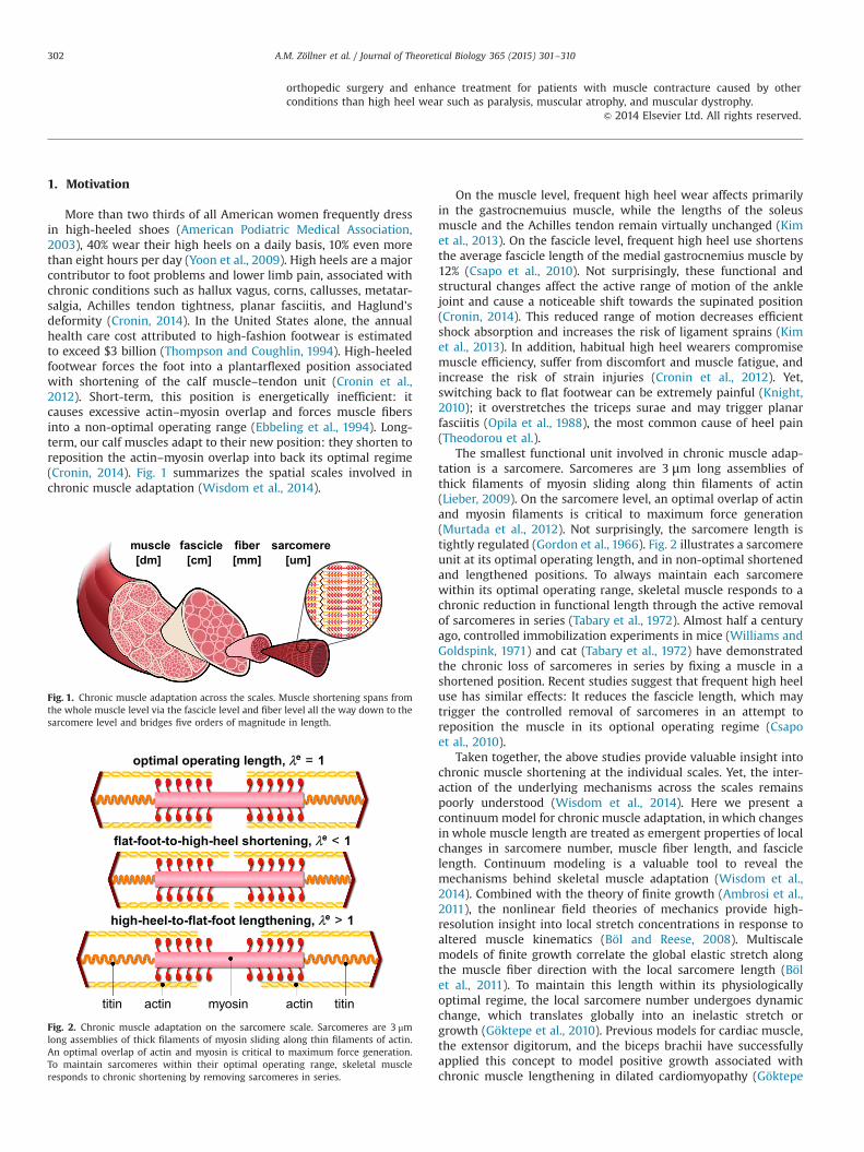
The smallest functional unit involved in chronic muscle adap-tation is a sarcomere. Sarcomeres are 3 μm long assemblies ofthick filaments of myosin sliding along thin filaments of actin(Lieber, 2009). On the sarcomere level, an optimal overlap of actinand myosin filaments is critical to maximum force generation(Murtada et al., 2012). Not surprisingly, the sarcomere length istightly regulated (Gordon et al., 1966). Fig. 2 illustrates a sarcomereunit at its optimal operating length, and in non-optimal shortenedand lengthened positions. To always maintain each sarcomerewithin its optimal operating range, skeletal muscle responds to achronic reduction in functional length through the active removalof sarcomeres in series (Tabary et al., 1972). Almost half a centuryago, controlled immobilization experiments in mice (Williams andGoldspink, 1971) and cat (Tabary et al., 1972) have demonstratedthe chronic loss of sarcomeres in series by fixing a muscle in ashortened position. Recent studies suggest that frequent high heeluse has similar effects: It reduces the fascicle length, which maytrigger the controlled removal of sarcomeres in an attempt toreposition the muscle in its optional operating regime (Csapoet al., 2010).
Taken together, the above studies provide valuable insight intochronic muscle shortening at the individual scales. Yet, the inter-action of the underlying mechanisms across the scales remainspoorly understood (Wisdom et al., 2014). Here we present acontinuum model for chronic muscle adaptation, in which changesin whole muscle length are treated as emergent properties of localchanges in sarcomere number, muscle fiber length, and fasciclelength. Continuum modeling is a valuable tool to reveal themechanisms behind skeletal muscle adaptation (Wisdom et al.,2014). Combined with the theory of finite growth (Ambrosi et al.,2011), the nonlinear field theories of mechanics provide high-resolution insight into local stretch concentrations in response toaltered muscle kinematics (Böl and Reese, 2008). Multiscalemodels of finite growth correlate the global elastic stretch alongthe muscle fiber direction with the local sarcomere length (Bölet al., 2011). To maintain this length within its physiologicallyoptimal regime, the local sarcomere number undergoes dynamicchange, which translates globally into an inelastic stretch orgrowth (Göktepe et al., 2010). Previous models for cardiac muscle,the extensor digitorum, and the biceps brachii have successfullyapplied this concept to model positive growth associated withchronic muscle lengthening in dilated cardiomyopathy (Göktepe
elcsum elcicsaf rebfi eremocras]md[ ]mc[ ]mm[ ]mu[
Fig. 1. Chronic muscle adaptation across the scales. Muscle shortening spans fromthe whole muscle level via the fascicle level and fiber level all the way down to thesarcomere level and bridges five orders of magnitude in length.
titin actin myosin actin titin
optimal operating length, λλe = 1
flat-foot-to-high-heel shortening, λe < 1
high-heel-to-flat-foot lengthening, λe > 1
Fig. 2. Chronic muscle adaptation on the sarcomere scale. Sarcomeres are 3 μmlong assemblies of thick filaments of myosin sliding along thin filaments of actin.An optimal overlap of actin and myosin is critical to maximum force generation.To maintain sarcomeres within their optimal operating range, skeletal muscleresponds to chronic shortening by removing sarcomeres in series.
A.M. Zöllner et al. / Journal of Theoretical Biology 365 (2015) 301–310302
et al., 2010), limb lengthening, and tendon tear (Zöllner et al.,2012). Here we adopt the same paradigm to model negativegrowth associated with chronic muscle shortening in frequenthigh heel wear.
2. Methods
To simulate the short- and long-term effects of high-heeled foot-wear, we create a subject-specific model of the lower limb usingmagnetic resonance images in flat foot and high heel positions. Weperform a finite element analysis of acute and chronic muscle short-ening using the continuum theory of finite growth.
2.1. Continuum model
To represent large muscle deformations, we adopt the kine-matics of finite growth, and introduce the deformation map φðX; tÞmapping particles X from the initial configuration to particlesx¼φðX; tÞ in the new configuration. We multiplicatively decom-pose its gradient F ¼∇Xφ into an elastic contribution Fe and aninelastic contribution Fg (Rodriguez et al., 1994), which weassociate with chronic muscle adaptation (Zöllner et al., 2012),
F ¼∇Xφ¼ Fe � Fg: ð1ÞThe Jacobian J ¼ detðFÞ defines the change in muscle volume,
J ¼ detðFÞ ¼ JeJg; ð2Þwhich we decompose into an elastic volume change Je ¼ detðFeÞand an inelastic volume change Jg ¼ detðFgÞ attributed to muscleadaptation. Similar to the deformation gradient and the Jacobian,we interpret the total stretch λ along the fiber direction of theinitial configuration n0 as the product of the elastic stretch λe andthe inelastic stretch λg associated with chronic changes in musclelength (Menzel and Kuhl, 2012),
λ¼ ½n0 � F t � F � n0�1=2 ¼ λeλg: ð3ÞWe model chronic muscle shortening as the removal of sarco-meres in series. This implies that we can express the muscleadaptation tensor Fg as rank-one update of the identity tensor I(Zöllner et al., 2012),
Fg ¼ Iþ½ϑ�1� n0 � n0; ð4Þwhere ϑ is the relative serial sarcomere number. Values of ϑo1represent the removal of sarcomeres in series associated withchronic muscle shortening; values of ϑ41 represent the additionof sarcomeres in series associated with chronic muscle lengthen-ing (Kuhl, 2014). As a consequence of the specific format of theadaptation tensor Fg, the serial sarcomere number ϑ representsnot only the local muscle fiber shortening λg, but also the volumechange in response to sarcomere loss Jg,
ϑ¼ λg ¼ detðFgÞ ¼ Jg: ð5ÞWe use the Sherman–Morrison formula to calculate the inverse ofthe adaptation tensor, Fg�1 ¼ Iþ½1�ϑ�=ϑ n0 � n0. With the mus-cle fiber direction in the current configuration n¼ F � n0 and theinverse Fg�1 we find an explicit expression for the elastic tensor,
Fe ¼ F � Fg�1 ¼ Fþ1�ϑϑ
n � n0: ð6Þ
We can then express the Finger tensor in terms of the serialsarcomere number ϑ,
be ¼ Fe � Fet ¼ F � F tþ1�ϑ2
ϑ2 n � n: ð7Þ
To focus on chronic muscle shortening, for simplicity, we assumehomogeneously distributed isotropic material properties for the
passive muscle tissue and adopt a strain energy function of Neo-Hookean type,
ψ ¼ 12L ln2ðJeÞþ1
2G½be : i�3�2lnðJeÞ�; ð8Þ
where L and G are the Lamé constants and i is the spatial identitytensor. We assume that the overall muscle microstructure, stiff-ness, and density are preserved upon adaptation (O'Dwyer et al.,1989), and derive the corresponding Kirchhoff stress tensor fromthe second law of thermodynamics,
τ ¼ 2∂ψ∂be � be ¼ ½LlnðJeÞ�G� iþGbe: ð9Þ
At the subcellular level, we model the evolution of the serialsarcomere number ϑ as a strain-driven process (Taber, 1998).We adopt the following evolution equation (Zöllner et al., 2012),
_ϑ ¼ kðϑÞ ϕðλeÞ; ð10Þwhere k is the adaptation function (Lubarda and Hoger),
k¼ �1τϑ�ϑmin
1�ϑmin
" #γ; ð11Þ
parameterized in terms of the adaptation speed τ, the shapeparameter for the adaptation function γ, and the minimum serialsarcomere number ϑmin, and ϕ is the adaptation criterion,
ϕ¼ ⟨λcrit�λe⟩: ð12ÞSimilar to the yield criterion in plasticity, the adaptation criterionϕ activates sarcomere removal only if the elastic stretch is lowerthan the critical stretch as ⟨λcrit�λe⟩¼ λcrit�λe and deactivatessarcomere changes for elastic stretches above the critical stretch as⟨λcrit�λe⟩¼ 0.
2.2. Computational model
To solve the nonlinear finite element equations for chronicmuscle adaptation, we implement our model as a user subroutineinto the implicit commercial finite element solver Abaqus/Stan-dard Version 6.13 (Simulia, Providence, RI) (Abaqus 6.13, 2013). Weintroduce the relative serial sarcomere number ϑ as an internalvariable, and solve its evolution equation (10) locally at theintegration point level (Göktepe et al., 2010). At each discrete timestep t, we determine the current sarcomere number ϑ for a givencurrent deformation state F and a given sarcomere number ϑn
from the previous time step tn using a finite difference approx-imation,
_ϑ ¼ϑ�ϑn
Δt; ð13Þ
where Δt ¼ t�tn denotes the current time increment. We adoptan implicit time integration scheme and reformulate the evolutionequation (10) with the help of Eq. (13), to introduce the discreteresidual R in terms of the unknown sarcomere number,
R¼ϑ�ϑnþk ϕΔt60: ð14ÞWe solve this nonlinear equation using a local Newton iteration(Göktepe et al., 2010). Within each iteration step, we calculate thelinearization of the residual R with respect to the serial sarcomerenumber ϑ,
K¼ ∂R∂ϑ
¼ 1� ∂k∂ϑ
ϕþk∂ϕ∂ϑ
� �Δt: ð15Þ
Here, ∂k=∂ϑ¼ �γ k=½ϑ�ϑmin� and ∂ϕ=∂ϑ¼ λ=ϑ2 denote the line-arizations of the adaptation function (11) and of the adaptationcriterion (12). Within each Newton iteration, we update the
A.M. Zöllner et al. / Journal of Theoretical Biology 365 (2015) 301–310 303

unknown sarcomere number,
ϑ’ϑ�R=K; ð16Þuntil we achieve local convergence, i.e., until the absolute value ofthe sarcomere update Δϑ¼ �R=K reaches a user-defined thresh-old value, here we choose 10�8. After determining the currentsarcomere number ϑ, we can successively determine the adapta-tion tensor Fg from Eq. (4), the elastic tensor Fe ¼ F � Fg�1 fromEq. (6), the elastic left Cauchy-Green tensor be ¼ Fe � Fe t from Eq.(7), the Kirchhoff stress τ from Eq. (9), and, finally, the fourth-order tensor of the Eulerian constitutive moduli,
c¼ 4 be � ∂2ψ∂be � be � be ¼ ceþcg: ð17Þ
The first term, the Hessian of the free energy function at constantgrowth Fg, defines the elastic constitutive moduli,
ce ¼ L i � iþ½G�L ln ðJeÞ�½i � iþ i � i�; ð18Þwith the common abbreviations, f��○gijkl ¼ f�gik f○gjl andf��○gijkl ¼ f�gil f○gjk, for the non-standard fourth order products.The second term, the Hessian of the free energy function atconstant deformation F , defines the correction of the constitutivemoduli due to muscle adaptation (Zöllner et al., 2012),
cg ¼ � kλϑ K
½L Iþ2G=ϑ2 n � n� � ½n � n�Δt: ð19Þ
Instead of the Kirchhoff stress (9) and the constitutive moduli (17),the user-defined subroutine in Abaqus/Standard utilizes theCauchy or true stress, σ ¼ τ=J,
σabaqus ¼ ½½L ln ðJeÞ�G� iþGbe�=J; ð20Þand the Jauman rate of the Kirchhoff stress divided by theJacobian, which requires the following modification of the tangentmoduli (Zöllner et al., 2013),
cabaqus ¼ cþ12
½τ � iþi � τþτ � iþ i � τ�� �
=J: ð21Þ
The local stress σabaqus of Eq. (20) and the local tangent modulicabaqus of Eq. (21) enter the righthand side vector and the iterationmatrix of the global Newton iteration. Upon its convergence, westore the relative serial sarcomere number ϑ locally at theintegration point level.
2.3. Magnetic resonance imaging
To create a finite element model of the lower limb, we collectmagnetic resonance images of a healthy 20-year old female subject(Blemker et al., 2007). We acquire two sets of axial images, in flat footand in high heel positions, using a product fat water separationgradient echo sequence with parallel imaging using IDEAL (GEHealthcare, Waukesha, WI) (Reeder et al., 2004) and a 3 T MRI systemusing Discovery MR750 (GE Healthcare, Waukesha, WI). To control thefoot position during the high heel scan, we build a cardboard shoemodel with a well-defined heel height of 13 cm.
Fig. 3 shows the resulting magnetic resonance images of theright lower limb in flat foot and high heel positions. Images havean in-plane resolution of 0.62 mm�0.62 mm and a slice thicknessof 3 mm. The magnetic resonance images provide the basis for thefinite element model and define the boundary conditions whenchanging from flat foot to high heel position. Fig. 4 illustrates theworkflow to generate a finite element model from magneticresonance images. Using the medical image viewer OsiriX(Rosset et al., 2004), we manually identify the individual muscles,bones, and tendons as regions of interest in a slice-by-slicemanner. We then export binary masks from these regions tocreate a mesh for every component using the ComputationalGeometry Algorithms Library CGAL (Fabri and Pion, 2009). Finally,
we filter the preliminary mesh to create a final mesh with smoothsurfaces (Taubin et al., 1996).
2.4. Finite element model
Fig. 5 shows the final finite element model of the lower limb,which contains 81,146 nodes and 377,034 linear tetrahedral andpyramid elements. We choose a combination of tetrahedral andpyramid elements because of their ease of use whenmeshing complexgeometries from clinical images. We note, however, that lineartetrahedral elements are known to be overly stiff, and that using
Fig. 3. Sagittal magnetic resonance images of the right lower limb of a healthy20-year old female subject. Images show the ankle joint in flat foot position, right,and in high heel position at a heel hight of 13 cm, left. The scans provide the basisfor the finite element model and define the boundary conditions when changingfrom flat foot to high heel position.
Fig. 4. Generation of finite element model from magnetic resonance images of thelower limb. We manually segment the image slices and identify the muscles, bones,and tendons as regions of interest in the medical image viewer OsiriX (Rosset et al.,2004), left. To create a mesh for each component, we convert the individual regionsinto binary masks and process them with CGAL (Fabri and Pion, 2009), middle.Finally, we filter the preliminary mesh to create smooth surfaces (Taubin et al.,1996), right.
Fig. 5. Finite element model of the lower limb. The model consists of 81,146 nodesand 377,034 linear tetrahedral and pyramid elements. The proximal end of thegastrocnemius muscle (165,820 elements, red) is fixed next to the femur while thedistal end is connected to the Achilles tendon (55,945 elements, yellow). The femur(22,838 elements, magenta), patella (8,987 elements, blue), tibia (48,369 elements,cyan), fibula (22,086 elements, green), and the tarsal bones (52,989 elements,orange) are modeled as rigid bodies. (For interpretation of the references to color inthis figure caption, the reader is referred to the web version of this paper.)
A.M. Zöllner et al. / Journal of Theoretical Biology 365 (2015) 301–310304

either quadratic tetrahedra or hexahedral elements would fix thisissue. Our model consists of the femur with 22,838 elements, shownin magenta, the patella with 8,987 elements, shown in blue, thegastrocnemius muscle with 165,820 elements, shown in red, the tibiawith 48,369 elements, shown in cyan, the fibulawith 22,086 elements,shown in green, the Achilles tendon with 55,945 elements, shown inyellow, and the tarsal bones with 52,989 elements, shown in orange.
We model the gastrocnemius muscle as Neo-Hookean elasticwith Lamé constants L¼0.714 N/mm2 and G¼0.179 N/mm2. Oncethe elastic fiber stretch falls below the threshold of λcrit¼1.0, wegradually remove sarcomeres towards a minimum sarcomerenumber of ϑmin ¼ 0:0 with an adaptation speed τ¼1.0 days and ashape parameter γ¼1.0. This implies that here, we drive muscleloss through non-tensile loading, while physiological muscle losscould generally occur in response to a variety of other environ-mental changes (Wisdom et al., 2014). For computational effi-ciency, we assume that the bones and the Achilles tendon aresignificantly stiffer than the gastrocnemius muscle and modelthem as rigid bodies (Maganaris and Paul, 2002; Zioupos andCurrey, 1998).
Table 1 summarizes the dimensions of the muscle tendon unitin flat foot and high heel positions extracted from our magneticresonance images. When changing from the flat footwear to highheels, the gastrocnemius medialis shortens by 12 mm while theAchilles tendon maintains its length. These measurements supportour model assumption of a rigid tendon. To fix the muscle–tendonunit in space, we apply homogeneous Dirichlet boundary condi-tions at the medial and lateral heads of the gastrocnemius musclewhere they attach to the condyle of the femur. To move the modelfrom flat foot to high heel position and vice versa, we applyinhomogeneous Dirichlet boundary conditions at the distal nodesof the Achilles tendon and prescribe a gradual upward anddownward displacement of 12 mm.
2.5. Muscle fiber model
As the foot is moved into the high heel position, we graduallyallow the gastrocnemius muscle to remove sarcomeres in series to
Fig. 7. Finite element model of muscle shortening in response to frequent high heel use. The left model shows the baseline state created frommagnetic resonance images in flatfoot position. The right model shows the shortened state created from images in high heel position. To move the flat foot into the high heel position, we apply inhomogeneousDirichlet boundary conditions by prescribing a 12 mm upward displacement of the distal end of the Achilles tendon. Then we maintain the foot in this position to allow themuscle to adapt to its new physiological length.
Table 2Muscle fiber orientations in the gastrocnemius muscle. The gastrocnemius muscleis a bipennate muscle. It consists of two rows of oblique muscle fibers, which facein opposite diagonal directions and converge in the Achilles tendon. Fig. 6illustrates six characteristic regions of interest with the six fiber orientations n0
(Rana et al., 2013).
Region Location n0x n0y n0z
I Medial–proximal 0.014 �0.197 0.980II Medial-central 0.025 �0.242 0.970III Medial–distal 0.075 �0.317 0.946IV Lateral–proximal 0.021 �0.193 0.981V Lateral–central 0.024 �0.208 0.978VI Lateral–distal 0.040 �0.247 0.968
Fig. 6. Muscle fiber orientation model of the gastrocnemius muscle. The gastro-cnemius muscle (red) is a bipennate muscle. It consists of two rows of obliquemuscle fibers, which face in opposite diagonal directions and converge in a joinedtendon (grey). Table 2 summarizes the six characteristic regions of interest withtheir six discrete fiber orientations n0 (Rana et al., 2013). (For interpretation of thereferences to color in this figure caption, the reader is referred to the web version ofthis paper.)
Table 1Dimensions of muscle tendon unit in flat foot and high heel positions. Wearingfootwear with a heel height of 13 cm shortens the gastrocnemius medialis by12 mm while the Achilles tendon remains unaffected. These changes in lengthdefine the inhomogeneous Dirichlet boundary conditions of our finite elementsimulation.
Muscle�tendonunit(mm)
Gastrocnemiusmedialis(mm)
Achillestendon(mm)
Flat foot 396 225 171High heel 384 213 171Difference �12 �12 0
A.M. Zöllner et al. / Journal of Theoretical Biology 365 (2015) 301–310 305

reposition the remaining sarcomeres at their optimal operatinglength. As a bipennate muscle, the gastrocnemius muscle consistsof two rows of oblique muscle fibers, which face in oppositediagonal directions and converge jointly in the Achilles tendon. Toaccount for these regionally varying fiber orientations, we dividethe muscle into six regions of interest and assign each region anindividual fiber orientation (Rana et al., 2013).
Fig. 6 illustrates our muscle fiber orientation model of thegastrocnemius muscle. Table 2 summarizes the fiber orientationsn0 associated with six discrete regions of interest (Rana et al., 2013).
2.6. Boundary conditions
To explore the acute and chronic effects of global muscleshortening on local sarcomere lengths, we simulate three scenar-ios: (i) acute effects when changing from flat foot to high heelposition, (ii) chronic effects when maintaining the high heelposition, and (iii) acute effects when changing back from highheel to flat foot position.
Fig. 7 illustrates our finite element model of the lower limb. Theleft model shows the baseline state created from magneticresonance images in flat foot position. The right model showsthe shortened state created from images in high heel position.First, we move the model from flat foot to high heel position byprescribing an upward displacement on the distal nodes of theAchilles tendon. Then, we keep the foot in this position and allowthe gastrocnemius to shorten and adapt to its new physiologicallength. Last, after the muscle has adapted, we move the foot backinto the flat foot position.
3. Results
All three simulations run robustly, converge quadratically, andgenerate conceptually feasible results, which we discuss in detailin the following three subsections.
3.1. Acute effects when switching from flat foot to high heel
Fig. 8 illustrates the acute change in sarcomere length whenswitching from flat footwear to high heels. Wearing shoes witha heel height of 13 cm induces a muscle shortening of 12 mm. Inresponse to acute muscle shortening, the sarcomere lengthdecreases. A decrease in length places the sarcomeres into anon-optimal operating regime at λeo1:0 and the whole muscleinto an energetically unfavorable working range. Sarcomerelengths display significant regional variations with an extremeshortening of ls=l0 ¼ 0:78 in the central gastrocnemius.
3.2. Chronic effects when switching from flat foot to high heel
Fig. 9 illustrates the chronic change in sarcomere length infrequent high heel wearers. In response to chronic muscle short-ening, the initially shortened sarcomere length increases graduallyas the muscle remains in its shortened position. This repositionsthe sarcomeres back into their optimal operating regime atλe ¼ 1:0. Initially, sarcomere lengths display significant regionalvariations with extreme values of ls=l0 ¼ 0:78 located in the centralgastrocnemius. Over time, these variations disappear. As themuscle adapts to its new physiological length, the sarcomerelengths converge towards a homogeneous distribution at theirinitial length l0.
Fig. 10 illustrates the chronic change in sarcomere number infrequent high heel wearers. In response to chronic muscle short-ening, the sarcomere number decreases gradually as the muscleremains in its shortened position. The average weighted sarco-mere number gradually decreases from ϑ¼ 1:00 to 0.96 after oneweek and 0.94 after two weeks until it converges to 0.91 after tenweeks of frequent high heel use. At this point, each sarcomere isrepositioned back in its optimal operating regime.
The side-by-side comparison of Figs. 9 and 10 illustrates theinterplay between the elastic and inelastic fiber stretches λe andλg: Initially, changing from flat footwear to high heels compressesthe muscle and the elastic fiber stretch drops significantly belowits baseline value of one. On the sarcomere level, this impliessignificant sarcomere shortening and an increase in actin andmyosin overlap. Over time, the relative serial sarcomere numberdecreases below its baseline value of one, while, at the same time,the sarcomere length returns to its initial value. The solutionconverges towards a state at which the elastic fiber stretch hasreturned to one throughout the entire muscle and the inelasticfiber stretch has taken up all the deformation.
Figs. 9 and 10 indicate that the sarcomere loss is highlyheterogeneous. At the proximal end of the gastrocnemius, wherethe medial and lateral heads attach to the rigid condyle of thefemur, the muscle does not sense the kinematic change associatedwith wearing high heels. At its distal end, where the compliantmuscle smoothly blends into the stiff Achilles tendon, relativekinematic changes are suppressed by the structural support of thetendon. Sarcomere loss is localized between these two regionswith extreme values of ϑ¼ 0:61 corresponding to a chronic localfiber shortening 39%.
Figs. 11 and 12 summarize the dynamic changes in sarcomerelength and sarcomere number. Both graphs reflect the interplaybetween elastic and inelastic fiber stretches with a gradualtransition from acute sarcomere shortening with λeo1 at λg ¼ 1to chronic sarcomere loss with λgo1 at λe ¼ 1. Since the graphscontain the averaged values across the entire muscle, the mini-mum average sarcomere length and average sarcomere number of0.94 and 0.91 are less pronounced than the local extreme values of0.78 and 0.61 of the contour plots in Figs. 9 and 10.
Fig. 8. Acute decrease in sarcomere length when switching from flat footwear tohigh heels. In response to acute muscle shortening, the sarcomere length decreases.Sarcomere lengths display significant regional variations with extreme values ofls=l0 ¼ 0:78 in the central gastrocnemius. The relative sarcomere length ls=l0 is ameasure for the elastic fiber stretch λe.
A.M. Zöllner et al. / Journal of Theoretical Biology 365 (2015) 301–310306
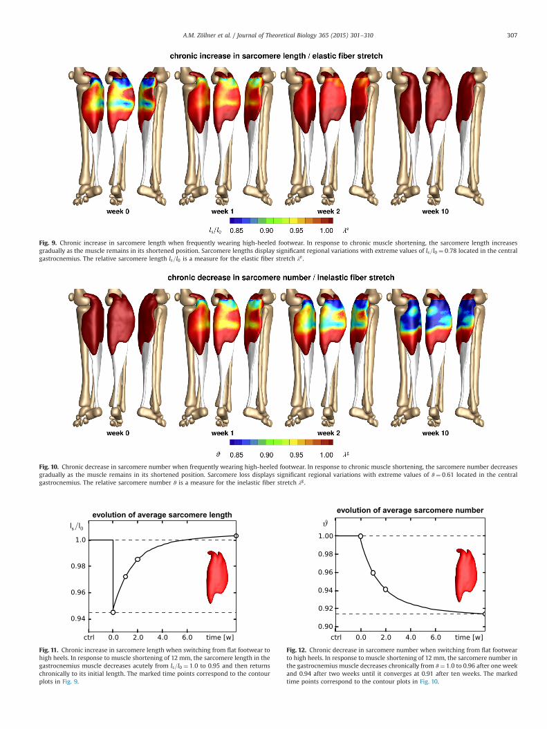
Fig. 9. Chronic increase in sarcomere length when frequently wearing high-heeled footwear. In response to chronic muscle shortening, the sarcomere length increasesgradually as the muscle remains in its shortened position. Sarcomere lengths display significant regional variations with extreme values of ls=l0 ¼ 0:78 located in the centralgastrocnemius. The relative sarcomere length ls=l0 is a measure for the elastic fiber stretch λe.
Fig. 10. Chronic decrease in sarcomere number when frequently wearing high-heeled footwear. In response to chronic muscle shortening, the sarcomere number decreasesgradually as the muscle remains in its shortened position. Sarcomere loss displays significant regional variations with extreme values of ϑ¼ 0:61 located in the centralgastrocnemius. The relative sarcomere number ϑ is a measure for the inelastic fiber stretch λg.
evolution of average sarcomere length
Fig. 11. Chronic increase in sarcomere length when switching from flat footwear tohigh heels. In response to muscle shortening of 12 mm, the sarcomere length in thegastrocnemius muscle decreases acutely from ls=l0 ¼ 1:0 to 0.95 and then returnschronically to its initial length. The marked time points correspond to the contourplots in Fig. 9.
evolution of average sarcomere number
Fig. 12. Chronic decrease in sarcomere number when switching from flat footwearto high heels. In response to muscle shortening of 12 mm, the sarcomere number inthe gastrocnemius muscle decreases chronically from ϑ¼ 1:0 to 0.96 after one weekand 0.94 after two weeks until it converges at 0.91 after ten weeks. The markedtime points correspond to the contour plots in Fig. 10.
A.M. Zöllner et al. / Journal of Theoretical Biology 365 (2015) 301–310 307

3.3. Acute effects when switching from high heel to flat foot
Fig. 13 illustrates the acute change in sarcomere length whenswitching back to flat foot position after a period of extended highheel wearing. In response to acute muscle lengthening, thesarcomere length increases. An increase in length places thesarcomeres into a non-optimal operating regime at λe41:0.Sarcomere lengths display significant regional variations with anextreme lengthening of ls=l0 ¼ 1:29 in the central gastrocnemius.
4. Discussion
Even though high heels are known to be a major source ofchronic lower limb pain, more than one third of American womenwear high heels on a daily basis. To characterize the effects ofhigh-heeled footwear on muscle adaptation in the lower limb, wecreated a multiscale computational model for chronic muscleshortening. To calibrate the model, we performed a case study ofa healthy female subject and created two lower limb models usingmagnetic resonance images, one in flat foot and one in high heelposition. Surprisingly, when moving the foot from flat position to aheel height of 13 cm, the length of the calf muscle–tendon unitchanges only marginally from 396 mm to 384 mm correspondingto a stretch of λ¼ 0:97. However, a closer look reveals twoessential characteristics: The Achilles tendon remains at a constantlength of 171 mm with λ¼ 1:00, while the gastrocnemius experi-ences the entire length change of 12 mm from 225 mm to 213 mmcorresponding to a stretch of λ¼ 0:95. More importantly, the finiteelement simulation suggests that this stretch is not distributedhomogeneously across the gastrocnemius muscle: In the proximaland distal regions, where the muscle is structurally supported bythe stiffer femur and Achilles tendon, the muscle experiencevirtually no stretch; in the central muscle region, local stretchestake extreme values of λ¼ 0:78.
Acutely, switching from flat footwear to high heels causesexcessive actin–myosin overlap and forces the muscle to operatein a non-optimal operating regime (Cronin et al., 2012), see Fig. 2.
When moving from the flat to the high heel position, oursimulations predict an acute average reduction in sarcomerelength by 5%, see Fig. 11, with local extrema of sarcomere short-ening of 22% in the central gastrocnemius, see Fig. 9. These valuesare in excellent agreement with recent studies, which havereported a fascicle length difference of 12% in the medial gastro-cnemius muscle, with lengths of 56.077.7 mm in the flat footgroup compared to 49.675.7 mm in the high heel group (Csapoet al., 2010). In the same study, in agreement with our measure-ments and simulations, the length of the Achilles tendon wasalmost identical in both groups with 18.69 cm in the flat footgroup and 18.91 cm in the high heel group (Csapo et al., 2010).
Chronically, skeletal muscle adapts to its new physiological lengthby removing sarcomeres in series to reposition its sarcomeres backinto their optimal regime (Williams and Goldspink, 1971), see Fig. 2.When maintaining the foot in the high heel position, our simulationspredict a chronic average reduction in serial sarcomere number by 9%,see Fig. 12, with local maxima of sarcomere loss of 31% in the centralgastrocnemius, see Fig. 10. These values lie within the range of thereported sarcomere loss during immobilization experiments in mice,where the serial sarcomere number decreased by 10% from 2,200 to1,975 in the soleus after four weeks of immobilization (Williams andGoldspink, 1971) and by 9% from 3,200 to 2,900 in the tibialis anteriorafter two weeks of immobilization (Burkholder and Lieber, 1998).These values are slightly lower than the reported sarcomere loss in cat,where the serial sarcomere number decreased by 40% from 13,844 to8,258 in the soleus after four weeks of immobilization (Tabary et al.,1972). Undoubtably, these immobilization experiments impose a moredrastic kinematic constraint than frequent high heel wearing (DupontSalter et al., 2011). Nonetheless, the chronic sarcomere loss reportedupon immobilization represents a valuable upper limit for thesarcomere loss we can expect in women who frequently wear high-heeled shoes.
Acutely, switching back from frequent high heel use to flatfootwear causes a drastic reduction in actin–myosin overlap andforces the muscle to operate in a non-optimal operating regime(Gordon et al., 1966), see Fig. 2. When moving from the foot fromhigh heel to flat, our simulations predict an acute sarcomerelengthening with local extrema of 29% in the central gastrocne-mius, see Fig. 13. Our large regional variations in sarcomere lengthare in agreement with previous studies, which have reportedsignificant regional variations in fascicle lengths and sarcomerelengths in the stretched cat biceps femoris muscle (Chanaud et al.,1991). Our sarcomere lengths agree with common observationthat switching back from high heels to flat footwear inducesmuscle overstretch associated with muscle pain (Knight, 2010)and increased risk of strain injuries (Cronin et al., 2012).
While our study provides valuable insight into the mechanisms ofchronic muscle shortening on the muscle and sarcomere levels, ourcurrent model has a few limitations: First, to illustrate the conceptualfeasibility of our method, we have only prototyped the study for asingle heel height and a single study subject. It would be interesting toexplore the dimensions of the muscle–tendon unit in Table 1 for flatfoot and high heel positions with varying heel heights in differentstudy subjects (Ebbeling et al., 1994). Second, for simplicity, in Eq. (8),we have modeled skeletal muscle as isotropic Neo-Hookean material.While an isotropic model might be a sufficient approximation undercompressive loading when switching from flat foot to high heel, amore sophisticated anisotropic model that takes into account strainstiffening might be more appropriate under tensile loading whenswitching from high heel to flat foot (Böl and Reese, 2008; Göktepeet al., 2014). Since we have only reported strains and stretches, here,sophisticated constitutive modeling might not be critical; yet, it wouldbe worth considering in future studies, especially when addressingchronic force alterations in muscle (Kim et al., 2013) and tendon(Eurviriyanukul and Askes, 2011; Magnusson et al., 2008). Third, the
Fig. 13. Acute increase in sarcomere length when switching from high heels to flatfootwear. In response to acute muscle lengthening, the sarcomere length increases.Sarcomere lengths display significant regional variations with extreme values ofls=l0 ¼ 1:29 located in the central gastrocnemius. The relative sarcomere length ls=l0is a measure for the elastic fiber stretch λe.
A.M. Zöllner et al. / Journal of Theoretical Biology 365 (2015) 301–310308

model presented here only accounts for sarcomere removal uponchronic understretch. To add sarcomeres in series upon chronicoverstretch (Wisdom et al., 2014), we would need to modify theadaptation function in Eq. (11) to k¼ ½½ϑmax�ϑ�=½ϑmax�1��γ=τ andactivate it through the modified adaptation criterion in Eq. (12) asϕ¼ ⟨λe�λcrit⟩ using the generic approach (Lubarda and Hoger)adopted for skeletal muscle (Zöllner et al., 2012). Last, until now, wehave only compared our model qualitatively against animal immobi-lization models in mice (Williams and Goldspink, 1971) and cat(Tabary et al., 1972). Recent developments in microendoscopy nowenable the non-invasive sarcomere imaging in vivo (Llewellyn et al.,2008). We have designed our model with these new technologies inmind (Cromie et al., 2013). We are currently in the process ofmeasuring sarcomere lengths in flat foot and high heel positions indifferent regions of the gastrocnemius muscle to validate our modelwith in vivo human data.
What can we learn from this study? First and foremost, we haveseen that we can interpret chronic changes in whole muscle length asemergent properties of local changes in sarcomere number, musclefiber length, and fascicle length. This has allowed us to create multi-scale computational models, which strongly support the hypothesisthat frequent high-heel use alters the natural position of the calfmuscle–tendon complex. Our results suggest that this change couldinitiate a chain reaction of negative effects (Cronin, 2014): Acutely, itcreates excessive actin–myosin overlap associated with energeticallyinefficient muscle use (Ebbeling et al., 1994). Chronically, it initiates anadaptation process associated with the loss of sarcomeres andsignificant muscle shortening (Csapo et al., 2010). A major concern isthat these changes are chronic. Changing back to flat footwear doesnot provide immediate cure; quite the contrary: For most frequenthigh heel wearers, switching to the flat foot position can be extremelypainful. It overstretched the triceps surae (Knight, 2010), which mightcause muscle pain and planar fasciitis (Opila et al., 1988). To ensurecomfort and reduce risk of injury (Cronin et al., 2012), recentrecommendations suggest limiting the heel hight to 5 cm or less(Ebbeling et al., 1994). To maintain muscle fiber lengths and anklerange of motion (Knight, 2010), recent studies recommend intensivepassive stretching exercise with long frequent stretching times indorsiflexion direction (Kim et al., 2013). Our study strongly supportsthese recommendations.
5. Concluding remarks
We have created a computational model of the lower limb to studythe acute and chronic effects of high-heeled footwear on the kine-matics of the calf muscle–tendon unit. Through the case study of ahealthy female subject, we have shown that raising the heel by 13 cmreduces the length of the muscle–tendon unit by 12 mm or 3%.Notably, this change affects almost exclusively the gastrocnemiusmuscle, which experiences an average shortening of 5%, while thelength of the Achilles tendon remains virtually unchanged. Ourcomputational simulations indicate that muscle shortening displayssignificant regional variations with extreme values of 22% in thecentral gastrocnemius. Our model suggests that the muscle graduallyadjusts to its new functional length by a chronic loss of sarcomeres inseries. Sarcomere loss varies regionally with virtually no loss at theproximal and distal ends and a maximum loss of 39% in the centralmuscle region. Collectively, these changes result in chronic muscleshortening associated with discomfort, compromised muscle effi-ciency, increased fatigue, reduced shock absorption, and increasedrisk of strain injuries. Computational modeling of chronic muscleshortening provides a valuable tool to shape our understanding of theinteracting mechanisms in skeletal muscle adaptation. Our study couldopen new avenues in orthopedic surgery and enhance treatmentin patients with muscle contracture caused by other conditions than
high heel wear such as paralysis, muscular atrophy, and musculardystrophy.
Acknowledgments
We thank Professor Gerald E. Loeb for stimulating discussionsthat initiated this study. This research was supported by theStanford Graduate Fellowship to Alexander Zöllner, by the Stan-ford Mechanical Engineering SURI support to Jacquelynn Pok, bythe NSERC PDF Fellowship to Emily McWalter, by GE Healthcare,by the NIH grant NIH EB002524, and by the NIH grant AR062068to Garry Gold, and by the NSF CAREER award CMMI 09520952021,by the NSF INSPIRE grant 1233054, and by the NIH grant U01HL119578 to Ellen Kuhl.
References
Abaqus 6.13. Analysis User's Manual. 2013. Simulia. Dassault Systèmes.Ambrosi, D., Ateshian, G.A., Arruda, E.M., Cowin, S.C., Dumais, J., Goriely, A.,
Holzapfel, G.A., Humphrey, J.D., Kemkemer, R., Kuhl, E., Olberding, J.E., Taber,L.A., Garikipati, K., 2011. Perspectives on biological growth and remodeling.J. Mech. Phys. Solids 59, 863–883.
American Podiatric Medical Association. 2003. High Heels Survey.Blemker, S.S., Asakawa, D.S., Gold, G.E., Delp, S.L., 2007. Image-based musculoske-
letal modeling: applications, advances, and future opportunities. J. Magn. Res.Imaging 25, 441–451.
Böl, M., Reese, S., 2008. Micromechanical modelling of skeletal muscles based onthe finite element method. Comput. Methods Biomech. Biomed. Eng. 11,489–504.
Böl, M., Stark, H., Schilling, N., 2011. On a phenomenological model for fatigueeffects in skeletal muscles. J.Theor. Biol. 281, 122–132.
Burkholder, T.J., Lieber, R.L., 1998. Sarcomere number adaptation after retinaculumtransection in adult mice. J. Exp. Biol. 201, 309–316.
Chanaud, C.M., Pratt, C.A., Loeb, G.E., 1991. Functionally complex muscles of the cathindlimb. II. Mechanical and architectural heterogenity within the bicepsfemoris. Exp. Brain Res. 85, 257–270.
Cromie, M.J., Sanchez, G.N., Schnitzer, M.J., Delp, S.L., 2013. Sarcomere lengths inhuman extensor carpi radials brevis measured by microendoscropy. MuscleNerve 48, 286–292.
Csapo, R., Maganaris, C.N., Seynnes, O.R., Narici, M.V., 2010. On muscle, tendon andhigh heels. J. Exp. Biol. 213, 2582–2588.
Cronin, N.J., Barrett, R.S., Carty, C.P., 2012. Long-term use of high-heeled shoes altersthe neuromechanics of human walking. J. Appl. Physiol. 112, 1054–1058.
Cronin, N.J., 2014. The effects of high heeled shoes on female gait: a review.J. Electromyogr. Kinesiol. 24, 258–263.
Dupont Salter, A.C., Richmond, F.J.R., Loeb, G.E., 2011. Effects of muscle immobiliza-tion at different lengths on tetrodotoxin-induced disuse atrophy. IEEE Trans.Neural Syst. Rehab. Eng. 11, 209–217.
Ebbeling, C.J., Hamill, J., Crussemeyer, J.A., 1994. Lower extremity mechanics andenergy cost of walking in high-heeled shoes. J. Orthop. Sports Phys. Therapy 19,190–196.
Eurviriyanukul, S., Askes, H., 2011. Tendon layout optimization through configura-tional force equilibration in plane stress analysis of prestressed concretestructures. Compos. Struct. 89, 1673–1680.
Fabri, A., Pion, S., 2009. CGAL: the Computational Geometry Algorithms Library. In:Seventeenth ACM SIGSPATIAL International Conference. ACM Press, New York,New York, USA, pp. 538–539.
Göktepe, S., Abilez, O.J., Kuhl, E., 2010. A generic approach towards finite growthwith examples of athlete's heart, cardiac dilation, and cardiac wall thickening. J.Mech. Phys. Solids 58, 1661–1680.
Göktepe, S., Abilez, O.J., Parker, K.K., Kuhl, E., 2010. A multiscale model for eccentricand concentric cardiac growth through sarcomerogenesis. J. Theor. Biol. 265,433–442.
Göktepe, S., Menzel, A., Kuhl, E., 2014. The generalized Hill model: a kinematicapproach towards active muscle contraction. J. Mech. Phys. Solids 72, 20–39.
Gordon, A.M., Huxley, A.F., Julian, F.J., 1966. The variation in isometric tension withsarcomere length in vertebrate muscle fibers. J. Physiol. 184, 170–192.
Kim, Y., Lim, J.M., Yoon, B.C., 2013. Changes in ankle range of motion and musclestrength in habitual wearers of high-heeled shoes. Foot Ankle Int. 34, 414–419.
Knight, K., 2010. Why walking flat-footed hurts after high heels. J. Exp. Biol. 213,Insight JEB i.
Kuhl, E., 2014. Growing matter—a review of growth in living systems. J. Mech.Behav. Biomed. Mater. 29, 529–543.
Llewellyn, M.E., Barretto, R.P.J., Delp, S.L., Schnitzer, M.J., 2008. Minimally invasivehigh-speed imaging of sarcomere contractile dynamics in mice and humans.Nature 454, 784–788.
Lieber, R.L., 2009. Skeletal Muscle Structure, Function, and Plasticity. LippincottWilliams& Wilkins, Baltimore.
A.M. Zöllner et al. / Journal of Theoretical Biology 365 (2015) 301–310 309

Lubarda, V.A, Hoger, A. On the mechanics of solids with a growing mass. Int. J.Solids Struct. 39, 4627–4664.
Maganaris, C.N., Paul, J.P., 2002. Tensile properties of the in vivo human gastro-cnemius tendon. J. Biomech. 35, 1639–1646.
Magnusson, S.P., Varici, M.V., Maganaris, C.N., Kjaer, M., 2008. Human tendonbehavior and adaptation, in vivo. J. Physiol 586, 71–81.
Menzel, A., Kuhl, E., 2012. Frontiers in growth and remodeling. Mech. Res. Commun.42, 1–14.
Murtada, S.C., Arner, A., Holzapfel, G.A., 2012. Experiments and mechanochemicalmodeling of smooth muscle contraction, Significance of filament overlap.J. Theor. Biol. 297, 176–186.
O'Dwyer, N.J., Neilson, P.D., Nash, J., 1989. Mechanisms of muscle growth related tomuscle contracture in cerebral palsy. Dev. Med. Child Neurol. 31, 543–547.
Opila, K.A., Wagner, S.S., Schiowitz, S., Chen, J., 1988. Postural alignment in barefootand high-heeled stance. Spine 13, 542–547.
Rana, M., Hamarneh, G., Wakeling, J.M., 2013. 3D fascicle orientations in tricepssurae. J. Appl. Physiol. 115, 116–125.
Reeder, S.B., Wen, Z., Yu, H., Pineda, A.R., Gold, G.E., Markl, M., Pelc, N.J., 2004.Multicoil Dixon chemical species separation with an iterative least-squaresestimation method. Magn. Reson. Med. 51, 35–45.
Rodriguez, E.K., Hoger, A., McCulloch, A.D., 1994. Stress-dependent finite growth insoft elastic tissues. J. Biomech. 27, 455–467.
Rosset, A., Spadola, L., Ratib, O., 2004. OsiriX: An open-source software fornavigating in multidimensional DICOM images. J. Digit Imaging 17, 205–216.
Tabary, J.C., Tabary, C., Tardieu, C., Tardieu, G., Goldspink, G., 1972. Physiological andstructural changes in the cat's soles muscle due to immobilization at differentlengths by plaster casts. J. Physiol. 224, 231–244.
Taber, L.A., 1998. Biomechanical growth laws for muscle tissue. J. Theor. Biol. 193,201–213.
Taubin, G., Zhang, T., Golub, G., 1996. Optimal surface smoothing as filter design. In:Buxton, B., Cipolla, R. (Eds.), Lecture Notes in Computer Science. Springer,Berlin, Heidelberg, pp. 283–292.
Theodorou, D.J., Theodorou, S.J., Kakitsubata, U., Lektrakul, N., Gold, G.E., Roger, B.,Resnick, D. Plantar fasciitis and fascial rupture: MR imaging findings in 26patients supplemented with anatomic data in cadavers. Radiograpics 20,S181–S197.
Thompson, F.M., Coughlin, M.J., 1994. The high price of high-fashion footwear.J. Bone Joint Surg. 76, 1586–1593.
Williams, P.E., Goldspink, G., 1971. Longitudinal growth of striated muscle fibres.J. Cell Sci. 9, 751–767.
Williams, P.E., Goldspink, G., 1971. The effect of immobilization on the longitudinalgrowth of striated muscle fibres. J. Anat. 116, 45–55.
Wisdom, K.M., Delp, S.L., Kuhl, E., 2014. Review. Use it or lose it: multiscale skeletalmuscle adaptation to mechanical stimuli. Biomech Mod Mechanobio. availableonline first. http://dx.doi.org/10.1007/s10237-014-0607-3.
Yoon, J.Y., An, D.H., Yoo, W.G., Kwon, Y.R., 2009. Differences in activities of the lowerextremity muscles with and without heel contact during stair ascent by youngwomen wearing high-heeled shoes. J. Orthop. Surg. 14, 418–422.
Zioupos, P., Currey, J.D., 1998. Changes in the stiffness, strength, and toughness ofhuman cortical bone with age. Bone 22, 57–66.
Zöllner, A.M., Abilez, O.J., Böl, M., Kuhl, E., 2012. Stretching skeletal muscle—chronicmuscle lengthening through sarcomerogenesis. PLoS ONE 7 (10), e45661.
Zöllner, A.M., Buganza Tepole, A., Kuhl, E., 2012. On the biomechanics andmechanobiology of growing skin. J. Theor. Biol. 297, 166–175.
Zöllner, A.M., Holland, M.A., Honda, K.S., Gosain, A.K., Kuhl, E., 2013. Growth ondemand—reviewing the mechanobiology of stretched skin. J. Mech. Behav.Biomed. Mater. 28, 495–509.
A.M. Zöllner et al. / Journal of Theoretical Biology 365 (2015) 301–310310