Embed Size (px)
Citation preview

ORIGINAL RESEARCH
Olive leaf extract modulates permethrin induced geneticand oxidative damage in rats
Hasan Turkez • Basak Togar • Elif Polat
Received: 20 October 2011 / Accepted: 23 December 2011 / Published online: 20 January 2012
� Springer Science+Business Media B.V. 2012
Abstract Permethrin is a common synthetic chemi-
cal, widely used as an insecticide in agriculture and other
domestic applications. The previous reports indicated
that permethrin is a highly toxic synthetic pyrethroid
pesticide to human and environmental health. There-
fore, the present experiment was undertaken to deter-
mine the effectiveness of olive leaf extract in
modulating the permethrin induced genotoxic and
oxidative damage in rats. The animals used were
broadly divided into four (A, B, C and D) experimental
groups. Group A rats served as control animals and
received distilled water intraperitoneally (n = 5).
Groups B and C rats received intraperitoneal injec-
tions of permethrin (60 mg kg-1 b.w) and olive leaf
extract (500 mg kg-1 b.w), respectively. Group D rats
received permethrin (60 mg kg-1 b.w) plus olive leaf
extract (500 mg kg-1 b.w). Rats were orally adminis-
tered their respective feed daily for 21 days. At the end
of the experiment rats were anesthetized and serum
and bone marrow cell samples were obtained. Geno-
toxic damage was assessed by micronucleus and
chromosomal aberration assays. Total antioxidant
capacity and total oxidant status were also measured
in serum samples to assess oxidative status. Treatment
of Group B with permethrin resulted in genotoxic
damage and increased total oxidant status levels.
Permethrin treatment also significantly decreased
(P \ 0.05) total antioxidant capacity level when com-
pared to Group A rats. Group C rats showed significant
increases (P \ 0.05) in total antioxidant capacity level
and no alterations in cytogenetic parameters. Moreover,
simultaneous treatments with olive leaf extract signif-
icantly modulated the toxic effects of permethrin in
Group D rats. It can be concluded that olive leaf
extract has beneficial influences and could be able to
antagonize permethrin toxicity. As a result, this inves-
tigation clearly revealed the protective role of olive leaf
extract against the genetic and oxidative damage by
permethrin in vivo for the first time.
Keywords Permethrin � Olive leaf extract �Chromosomal aberrations � Micronucleus � Total
antioxidant capacity � Total oxidant status
Introduction
Olea europea L. leaves contain polyphenolic com-
pounds (hydroxytyrosol, oleuropein, secoiridoids,
flavonoids and triterpenes) that may protect in tissue
H. Turkez
Department of Molecular Biology and Genetics,
Faculty of Science, Erzurum Technical University,
Erzurum, Turkey
B. Togar (&)
Department of Biology, Faculty of Science,
Ataturk University, 25240 Erzurum, Turkey
e-mail: [email protected]
E. Polat
Department of Biochemistry, Medical Faculty,
Ataturk University, 25240 Erzurum, Turkey
123
Cytotechnology (2012) 64:459–464
DOI 10.1007/s10616-011-9424-z

cells against oxidative stress (Kranz et al. 2010; Fares
et al. 2011; Bouallagui et al. 2011). European and
Mediterranean countries have been used in the human
diet as extract, herbal tea, and powder. Since these
leaves contain many potentially bioactive compounds,
they may have antioxidant properties (El and Kara-
kaya 2009; Moussaoui et al. 2010). At the same time
olive leaves have been heavily exploited for the
prevention or the treatment of hypertension, carcino-
genesis, diabetes, atherosclerosis, gingivitis and many
other traditional therapeutic uses (Han et al. 2009;
Haloui et al. 2010; Bouallagui et al. 2011). Poudyal
et al. (2010) reported that olive leaf extract (OLE)
containing polyphenols such as oleuropein and
hydroxytyrosol reversed the chronic inflammation
and oxidative stress by diet-induced obesity and
diabetes in cardiovascular, hepatic, and metabolic
symptoms in rats. In additon, OLE possessed gastro-
protective activity against cold restraint stress-induced
gastric lesions in rats, possibly related to its antiox-
idative properties (Dekanski et al. 2009). Interestingly
Bao et al. (2007) found that OLE has anti-HIV activity
by blocking the HIV virus entry to host cells.
Permethrin (C21H10Cl2O3), (PM) entered use in the
1970s as an insecticide in a wide range of applications,
including agriculture, horticultural, and forestry
(Turner et al. 2010). The previous reports showed that
PM was a highly toxic synthetic pyrethroid pesticide
widely used in agriculture and vector control programs.
About 60% of the PM produced is used on cotton
plants. Other crops to which PM is applied are maize,
soya beans, coffee, tobacco, rape seed oil, wheat,
barley, alfalfa, vegetables and fruit (WHO 1990). It
was found that PM is highly toxic to fish; but it is less
toxic to guppies (Baser et al. 2003). Likewise, Sutton
et al. (2007) reported that 96.9% of exposed cats
developed clinical effects, 87.8% developed increased
muscular activity and 10.5% of cases resulted in
fatalities. PM was shown to induce DNA damage on rat
heart cells (Vadhana et al. 2010). Furthermore,
potential carcinogenicity of PM was ascertained in
human nasal mucosal cells (Tisch et al. 2002).
So far, antioxidants have attracted much interest
with respect to their protective effect against damage
by free radical that may be the cause for many diseases
including cancer (Shon et al. 2004). Since the complete
avoidance of exposure to PM is very difficult, chemo-
prevention is an attractive strategy for protecting
humans and animals from the risk of cancer caused
by exposure to this insecticide. In fact, recent studies
focused on exploring protective agents, such as
vitamins C and E, coenzyme Q(10) and glutathione,
against PM toxicity (Vontas et al. 2001; Gabbianelli
et al. 2004; Falcioni et al. 2010). To our best
knowledge, the effects of OLE against the toxicity of
PM have not been investigated. Therefore, in this study
we evaluated the effects of OLE against PM-induced
DNA damages for improving its therapeutic gain.
Firstly, important oxidative parameters, TAS (Total
antioxidant capacity) and TOS (Total oxidant status)
were used to monitor the development and extent of
damage due to oxidative stress in rat blood serum
(Turkez et al. 2012). In addition, chromosomal aber-
rations (CA) and micronucleus (MN) test providing
sensitive and rapid monitoring of induced genetic
damage as primary DNA damage were performed on
bone marrow cells (de Souza et al. 2006).
Materials and methods
Plant materials and extracts
Olive leaves were collected from the Balıkesir-Edre-
mit region in Turkey in June 2010. Olive leaf samples
were handpicked randomly from the trees. They were
dried in the shade and crushed. After crushing, 100 g
of powdered leaves was extracted. The extract was
manufactured from the dried leaves of O. europaea
applying ethyl acetate extraction procedure previously
reported by Turkez and Togar (2011).
Animals and chemicals
Experiment was carried out on male Sprague–Dawley
rats, 8-weeks old, weighing 180–200 g. The animals
were kept on a 12-h light–dark cycle and allowed free
access to food and water. All experiments were per-
formed in accordance with the Guide for the Care and
Use of Laboratory Animals (National Research Coun-
cil 1996). PM (Cas No 52645-53-1; C21H20Cl2O3) was
obtained from Riedel–de Haen� company (Germany).
All other chemicals were purchased from Sigma� (USA).
Experimental design
The animals were randomly divided into equal four
groups (n = 5): group A: rats were given an equivalent
460 Cytotechnology (2012) 64:459–464
123
of distilled water, group B: Rats received OLE (500
mg kg-1), group C: rats received PM (60 mg kg-1)
and group D: rats received OLE plus PM for 21 days
intraperitoneally. The doses were selected according to
literature data (Van Haaren et al. 2000; Hussain et al.
2010). After the treatments of PM, OLE and PM plus
OLE, the animals were anesthetized with ether.
Biochemical and cytogenetic studies
Blood samples were collected into serum separator
tubes (Microtainer; Becton–Dickinson, Franklin
Lakes, NJ, USA). The samples were allowed to stand
(75–90 min) and centrifuged (860 g for 20 min) for
obtaining serum samples. The TAC and TOS levels
were determined spectrophotometrically on serum
samples using commercially available kits (Assay
Diagnostics, Turkey) (Erel 2004; Erel 2005).
Bone marrow cell preparations for the analysis of
CAs (gaps and breaks) were produced by the colchi-
cine–hypotonic citrate technique. Potassium chloride
(0.075 M) was used in this technique. Colchicine
(0.1%, 1 ml-1 100 g body wt) was injected i.p. 90 min
before killing the animals. Animals were killed and
then bone marrow cells were flushed from the femora
with 0.075 M potassium chloride. Slides were pre-
pared by an air drying procedure and stained with 5%
Giemsa stain (Assayed et al. 2010). Criteria to classify
the different types of aberrations were in accordance
with the recommendation of Environmental Health
Criteria (EHC) 46 for environmental monitoring of
human populations (IPCS 1985). Structural and
numerical chromosomal aberrations (CA) were scored
in 100 metaphases per animal (a total of 500 metapha-
ses for each group). For MN analysis, bone marrow was
isolated from the other femur bone with the help of
syringes and homogenized with fetal bovine serum
(FBS). The slides were coded, fixed with methanol and
stained with Giemsa solution. Two thousand poly-
chromatic erythrocytes (PCE) from each animal were
scored for MN presence (Sekeroglu et al. 2011). Slides
were scored in duplicates at a magnification of 1,0009
using a light microscope by one observer (B. Togar).
Statistical analysis
The statistical analysis of experimental values in the
CA, MN and TAC- TOS analysis was performed by
oneway analysis of variance (ANOVA) and Fisher’s
LSD test using the S.P.S.S. 13.0 software. The level of
0.05 was regarded as indicative of statistical signifi-
cance for all tests.
Results
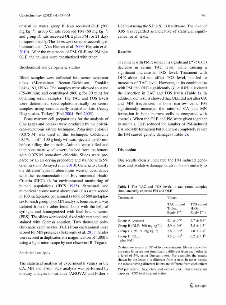
Treatment with PM resulted in a significant (P \ 0.05)
decrease in serum TAC level, while causing a
significant increase in TOS level. Treatment with
OLE alone did not affect TOS level, but led to
increases of TAC level. However, in its combination
with PM, the OLE significantly (P \ 0.05) alleviated
the distortion in TAC and TOS levels (Table 1). In
addition, our results showed that OLE did not alter CA
and MN frequencies in bone marrow cells. PM
significantly increased the rates of CA and MN
formation in bone marrow cells as compared with
controls. When the OLE and PM were given together
to animals, OLE reduced the number of PM-induced
CA and MN formation but it did not completely revert
the PM caused genetic damages (Table 2).
Discussion
Our results clearly indicated the PM induced geno-
toxic and oxidative damage in rats in vivo. Similarly to
Table 1 The TAC and TOS levels in rats serum samples
simultaneously exposed PM and OLE
Treatments Values
TAC (mmol
Trolox
Equiv l-1)
TOS (lmol
H2O2
Equiv l-1)
Group A (control) 4.1 ± 0.7c 5.7 ± 0.9a
Group B (OLE; 500 mg kg-1) 5.9 ± 0.6d 5.5 ± 1.3a
Group C (PM; 60 mg kg-1) 2.8 ± 0.5a 7.8 ± 1.4c
Group D (OLE
plus PM)
3.5 ± 0.5b 6.2 ± 1.1b
(Values are means ± SD of five experiments. Means shown by
the same letter are not significantly different from each other at
a level of 5%, using Duncan’s test. For example, the means
shown by the letter b is different from a or c. In other words,
the means having different letters are different from each other)
PM permethrin, OLE olive leaf extract, TAC total antioxidant
capacity, TOS total oxidant status
Cytotechnology (2012) 64:459–464 461
123

our findings, few reports indicated in vitro and in vivo
PM genotoxicity. Institoris et al. (1999) investigated
the genotoxic effects of PM by structural and numer-
ical CA in bone marrow cells. They showed that PM
increased the number of numerical CA. In another
report, PM significantly increased the DNA damage in
a concentration-dependent manner in healthy human
lymphocytes (Undeger and Basaran 2005). Possible
genotoxic effects in primary human nasal mucosal
cells were also investigated by Tisch et al. (2002).
Their findings indicated a significant genotoxic
response that was concentration dependent. Moreover,
findings provided evidence for the potential carcino-
genicity of PM to human nasal mucosal cells. PM gave
mostly negative results, although it increased the MN
frequency in human blood cultures (Surralles et al.
1995). In contrast to those results, Djelic and Djelic
(2000) reported that PM was non-genotoxic by using
MN assay in cultured human lymphocytes. The
present findings also indicated that PM caused oxida-
tive stress on blood cells in rats. Because, the PM
administration (group C) caused increases of TOS
level and decrease of TAC level as compared to
control group (group A). In accordance with our
findings, recent investigations clearly indicated that
PM-induced oxidative stress leads to biochemical and
functional changes in organisms (Nasuti et al. 2007;
Gabbianelli et al. 2009; Issam et al. 2011).
The present study also demonstrated that the
reduction of PM induced oxidative and DNA damages
was caused by the protective effect of OLE. O’Brien
et al. (2006) provided evidence that non-nutrient
dietary constituents could act as significant bioactive
compounds and that plant extracts, such as OLE,
strongly protect against oxidative stress. Moreover,
OLE demonstrated strong antioxidant potency and
inhibited cancer and endothelial cell proliferation at
low micro molar concentrations (Goulas et al. 2009).
Thus, OLE probably modulated the PM-induced
genetic and oxidative damage by preventing free
radical generation or by stimulating components of the
antioxidant defense system. In fact, OLE was reported
to have free oxygen radicals and lipoperoxyradicals
scavenging capacity and, anti-clastogenic activity due
to its polyphenolic contents, mainly catechol groups
(rutin, oleuropein, hydroxytyrosol, verbascoside, lute-
olin) (Benavente-Garcıa et al. 2002).
In conclusion the authors recommend the con-
sumption of plenty of OLE within the food especially
for humans that work in agriculture or are overtly
exposed to various pesticides or consume pesticide-
treated fruits and vegetables in order to protect
themselves and their offspring against any expected
harm. Bestowed with strong antioxidant and genopro-
tective potentials, the neutraceutical value of olive
leaves make them ideal candidates to protect against
pesticide-induced mutagenicity or carcinogenicity.
References
Assayed ME, Khalaf AA, Salem HA (2010) Protective effects of
garlic extract and vitamin C against in vivo cypermethrin-
induced cytogenetic damage in rat bone-marrow. Mutat
Res 702:1–7
Bao J, Zhang DW, Zhang JZ, Huang PL, Huang PL, Lee-Huang
S (2007) Computational study of bindings of olive leaf
extract (OLE) to HIV-1 fusion protein gp41. FEBS Lett
12:2737–2742
Baser S, Erkoc F, Selvi M, Kocak O (2003) Investigation of
acute toxicity of permethrin on guppies Poecilia reticulata.
Chemosphere 51:469–474
Benavente-Garcıa O, Castillo J, Lorente J, Alcaraz M (2002)
Radioprotective effects in vivo of phenolics extracted from
Olea europaea L. leaves against X-ray-induced chromo-
somal damage: comparative study versus several flavo-
noids and sulfur-containing compounds. J Med Food 5:
125–135
Table 2 Frequency of MN and CA in bone marrow cells
simultaneously exposed to PM and OLE
Treatments Values
Number of
MNPCE
per animal
Number of
CAs
per animal
Group A (control) 0.46 ± 0.22a 1.82 ± 0.46a
Group B (OLE;
500 mg kg-1)
0.48 ± 0.25a 1.76 ± 0.55a
Group C (PM;
60 mg kg-1)
3.85 ± 1.14c 4.47 ± 0.87c
Group D (OLE plus PM) 1.54 ± 0.73b 2.66 ± 0.81b
(Values are means ± SD of five experiments. Means shown by
the same letter are not significantly different from each other at
a level of 5%, using Duncan’s test. For example, the means
shown by the letter b is different from a or c. In other words,
the means having the different letters are different from each
other)
MNPCE micronucleated polychromatic erythrocytes, CAchromosomal aberrations, chromatid/chromosome gapschromatid/chromosome breaks and centric fusions or
fragments were considered as same in CA assay
462 Cytotechnology (2012) 64:459–464
123

Bouallagui Z, Han J, Isoda H, Sayadi S (2011) Hydroxytyrosol
rich extract from olive leaves modulates cell cycle pro-
gression in MCF-7 human breast cancer cells. Food Chem
Toxicol 49:179–184
de Souza AB, Souza LM, Carvalho JCT, Maistro EL (2006) No
clastogenic activity of Caesalpinia ferrea Mart. (Legumi-
nosae) extract on bone marrow cells of Wistar rats. Genet
Mol Biol 29:380–383
Dekanski D, Janicijevic-Hudomal S, Ristic S, Radonjic NV,
Petronijevic ND, Piperski V, Mitrovic DM (2009) Atten-
uation of cold restraint stress-induced gastric lesions by an
olive leaf extract. Gen Physiol Biophys 28:135–142
Djelic N, Djelic D (2000) Evaluation of cytotoxic and genotoxic
effects of permethrin using in vitro micronucleus test. Acta
Veterinaria-Beograd 50:263–269
El SN, Karakaya S (2009) Olive tree (Olea europaea) leaves:
potential beneficial effects on human health. Nutr Rev
67:632–638
Erel O (2004) A novel automated direct measurement method
for total antioxidant capacity using a new generation, more
stable ABTS radical cation. Clin Biochem 37:277–285
Erel O (2005) A new automated colorimetric method for mea-
suring total oxidant status. Clin Biochem 38:1103–1111
Falcioni ML, Nasuti C, Bergamini C, Fato R, Lenaz G, Gab-
bianelli R (2010) The prımary role of glutathione against
nuclear DNA damage of striatum induced by permethrine
in rats. Neurosci 168:2–10
Fares R, Bazzi S, Baydoun SE, Abdel-Massih RM (2011) The
antioxidant and anti-proliferative activity of the Lebanese
Olea europaea extract. Plant Foods Hum Nutr 66:58–63
Gabbianelli R, Nasuti C, Falcioni G, Cantalamessa F (2004)
Lymphocyte DNA damage in rats exposed to pyrethroids:
effect of supplementation with Vitamins E and C. Toxicol
203:17–26
Gabbianelli R, Falcioni ML, Nasuti C (2009) Effect of per-
methrin insecticide on rat polymorphonuclear neutrophils.
Chem-Biol Interact 182:245–252
Goulas V, Exarchou V, Troganis AN, Psomiadou E, Fotsis T,
Briasoulis E, Gerothanassis IP (2009) Phytochemicals in
olive-leaf extracts and their antiproliferative activity
against cancer and endothelial cells. Mol Nutr Food Res
53:600–608
Haloui E, Marzouk Z, Marzuk B, Bouftira I, Bouraoui A, Fenina
N (2010) Pharmacological activities and chemical com-
position of the Olea europaea L. leaf essential oils from
Tunisia. J Food Agricult Environ 8:204–208
Han J, Talorete TP, Yamada P, Isoda H (2009) Anti-prolifera-
tive and apoptotic effects of oleuropein and hydroxytyrosol
on human breast cancer MCF-7 cells. Cytotechnology 59:
45–53
Hussain K, Ismail Z, Sadikun A (2010) Evaluation of ethanol
extracts of leaves and fruit of piper sarmentosum for in vivo
hepatoprotective activity. Latin Am J Pharm 29:1215–
1220
Institoris L, Undeger U, Siroki O, Nehez M, Desi I (1999)
Comparison of detection sensitivity of immuno- and
genotoxicological effects of subacute cypermethrin and
permethrin exposure in rats. Toxicology 137:47–55
IPCS (International Program on Chemical Safety) (1985)
Environmental health criteria, 46. In: guidelines for the
study of genetic effects in human populations. Geneva:
World Health Organisation, pp 1–54
Issam C, Zohra H, Monia Z, Hassen BC (2011) Effects of dermal
sub-chronic exposure of pubescent male rats to permethrin
(PRMT) on the histological structures of genital tract,
testosterone and lipoperoxidation. Exp Toxicol Pathol
63:393–400
Kranz P, Braun N, Schulze N, Kunz B (2010) Sensory quality of
functional beverages: bitterness perception and bitter
masking of olive leaf extract fortified fruit smoothies.
J Food Sci 75:308–311
Moussaoui R, Siziani D, Youyou A, Sharrock P, Fiallo MML
(2010) Antioxidant effect of phenolic compounds recov-
ered from olive mill wastewater of Chemlal variety culti-
vated in Kabylia (Algeria) on the oxidative stability of
virgin olive oil. J Food Agricult Environ 8:86–89
Nasuti C, Gabbianelli R, Falcioni M, Di Stefano A, Sozio P,
Cantalamessa F (2007) Dopaminergic system modulation,
behavioral changes, and oxidative stress after neonatal
administration of pyrethroids. Toxicol 229:194–205
National Research Council (1996) Guide for the care and use of
laboratory animals. National Academy Press, Washington
O’Brien NM, Carpenter R, O’Grady MN, Kerry JPJ (2006)
Modulatory effects of resveratrol, citroflavan-3-ol, and
plant-derived extracts on oxidative stress in U937 cells.
Med Food 9:187–195
Poudyal H, Campbell F, Brown L (2010) Olive leaf extract
attenuates cardiac, hepatic, and metabolic changes in high
carbohydrate-, high fat-fed rats. J Nutr 140:946–953
Sekeroglu V, Sekeroglu ZA, Kefelioglu H (2011) Cytogenetic
effects of commercial formulations of deltamethrin and/or
thiacloprid on Wistar rat bone marrow cells. Environ
Toxicol. doi:10.1002/tox.20746
Shon MY, Choi SD, Kahng GG, Nam SH, Sung NJ (2004)
Antimutagenic, antioxidant and free radical scavenging
activity of ethyl acetate extracts from white, yellow and red
onions. Food Chem Toxicol 42:659–666
Surralles J, Xamena N, Creus A, Catalan J, Norppa H, Marcos R
(1995) Induction of micronuclei by five pyrethroid insec-
ticides in whole-blood and isolated human lymphocyte
cultures. Mutat Res 341:169–184
Sutton NM, Bates N, Campbell A (2007) Clinical effects and
outcome of feline permethrin spot-on poisonings reported
to the veterinary poisons information service (VPIS),
London. J Feline Med Surg 9:335–339
Tisch M, Schmezer P, Faulde M, Groh A, Maier H (2002)
Genotoxicity studies on permethrin, DEET and diazinon in
primary human nasal mucosal cells. Eur Arch Otorhino-
laryngol 259:150–153
Turkez H, Togar B (2011) Olive (Olea europaea L.) leaf extract
counteracts genotoxicity and oxidative stress of permethrin
in human lymphocytes. J Toxicol Sci 36:531–537
Turkez H, Geyikoglu F, Mokhtar YI, Togar B (2012) Eicosapen-
taenoic acid protects against 2,3,7,8-tetrachlorodibenzo-
p-dioxin-induced hepatic toxicity in cultured rat hepatocytes.
Cytotechnology 64:15–25
Turner T, Cartmell E, Lester JN, Casse F, Comber SD, Scrim-
shaw MD (2010) The pharmaceutical use of permethrin:
sources and behavior during municipal sewage treatment.
Arch Environ Contam Toxicol 61:193–201
Cytotechnology (2012) 64:459–464 463
123

Undeger U, Basaran N (2005) Effects of pesticides on human
peripheral lymphocytes in vitro: induction of DNA dam-
age. Arch Toxicol 79:169–176
Vadhana MS, Nasuti C, Gabbianelli R (2010) Purine bases
oxidation and repair following permethrin insecticide
treatment in rat heart cells. Cardiovasc Toxicol 3:199–207
Van Haaren F, Cody B, Hoy JB, Kalix JL, Schmidt C, Tebbett
IR, Wielbo D (2000) The effects of pyridostigmine bro-
mide and permethrin, alone or in combination, on response
acquisition in male and female rats. Pharmacol Bioche-
micahem Behav 66:739–746
Vontas JG, Small GJ, Hemingway J (2001) Glutathione
S-transferases as antioxidant defence agents confer pyre-
throid resistance in Nilaparvata lugens. Biochem J
357:65–72
WHO (1990) Permethrin (Environ mental Health Criteria 94),
Geneva
464 Cytotechnology (2012) 64:459–464
123