Embed Size (px)
Citation preview

OITEOITE
DR. ElfatoriDR. Elfatori
October / 11 / 2006October / 11 / 2006
A 15 year old boy is diagnosed with a high A 15 year old boy is diagnosed with a high grade osteogenic sarcoma, imaging studies grade osteogenic sarcoma, imaging studies reveal a 6cm lesion of the right humerus and reveal a 6cm lesion of the right humerus and no regional distant spread of the no regional distant spread of the disease .what is the current American joint disease .what is the current American joint commission on cancer stage of the disease ?commission on cancer stage of the disease ?
1) IA.1) IA.
2) I B.2) I B.
3) II A.3) II A.
4) II B.4) II B.
5) III .5) III .
A 15 year old boy is diagnosed with a high A 15 year old boy is diagnosed with a high grade osteogenic sarcoma, imaging studies grade osteogenic sarcoma, imaging studies reveal a 6cm lesion of the right humerus reveal a 6cm lesion of the right humerus and no regional distant spread of the and no regional distant spread of the disease .what is the current American joint disease .what is the current American joint commission on cancer stage of the disease ?commission on cancer stage of the disease ?
1) IA.1) IA.
2) IB.2) IB.
3) IIA.3) IIA.
4) IIB.4) IIB.
5) III.5) III.
Pathological fractures that occur secondary Pathological fractures that occur secondary to metastatic carcinoma most frequently to metastatic carcinoma most frequently involve what long bone location?involve what long bone location?
1) Distal femur.1) Distal femur.
2) proximal humerus.2) proximal humerus.
3) proximal femur.3) proximal femur.
4) humeral diaphysis.4) humeral diaphysis.
Pathological fractures that occur secondary Pathological fractures that occur secondary to metastatic carcinoma most frequently to metastatic carcinoma most frequently involve what long bone location?involve what long bone location?
1) Distal femur.1) Distal femur.
2) proximal humerus.2) proximal humerus.
33) proximal femur.) proximal femur.
4) humeral diaphysis4) humeral diaphysis..
The femur is the mostly likely long bone to The femur is the mostly likely long bone to be affected by metastasis .the proximal be affected by metastasis .the proximal third is involved in 50% of the caseses, with third is involved in 50% of the caseses, with introchonteric region accounting for 20% introchonteric region accounting for 20% because the developmental of bone because the developmental of bone metastasis is a dynamic process,it is metastasis is a dynamic process,it is important to stabilize as much of the bone important to stabilize as much of the bone as possible. Tip of the chosen fixation as possible. Tip of the chosen fixation device should pass the lesion by a least device should pass the lesion by a least twice the diameter of the femur.twice the diameter of the femur.
AAOS instructional course lectures ,volume AAOS instructional course lectures ,volume 53,2004.53,2004.
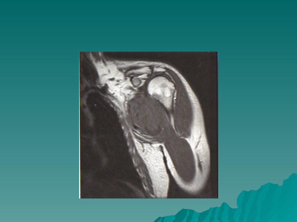
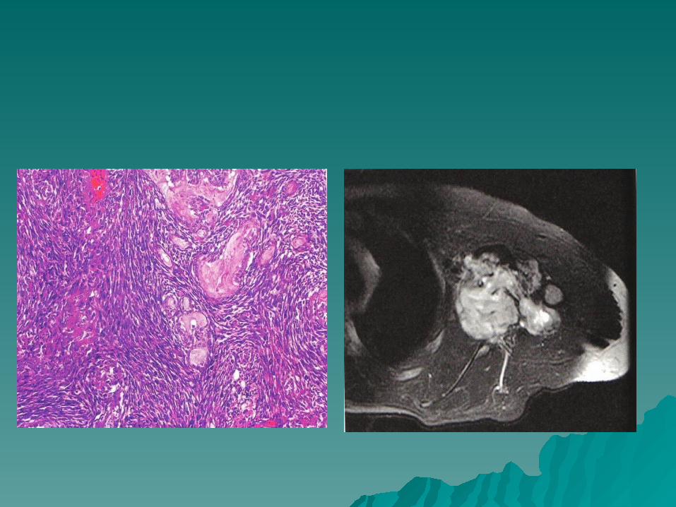
A 41 year old man report a lump in his A 41 year old man report a lump in his axilla .axilla .
Coronal T1-T2 weighted MRI scan are Coronal T1-T2 weighted MRI scan are shown in figures a and b. A biopsy specimen shown in figures a and b. A biopsy specimen is shown in figures c. is shown in figures c. immunohistochemistery reveals that the immunohistochemistery reveals that the tumor stains positive for vimentin and tumor stains positive for vimentin and epithelial membrane antigen .What is the epithelial membrane antigen .What is the most likely diagnosis?most likely diagnosis?
1) Liposarcoma.1) Liposarcoma.
2) Malignant fibrous histiocytoma.2) Malignant fibrous histiocytoma.
3) Synovial sarcoma.3) Synovial sarcoma.
4) Leiomyosarcoma4) Leiomyosarcoma..
A 41 year old man report a lump in his A 41 year old man report a lump in his axilla.axilla.
coronal T1-T2 weighted MRI scan are shown coronal T1-T2 weighted MRI scan are shown in figures a and b. A biopsy specimen is in figures a and b. A biopsy specimen is shown in figures c. immunohistochemistery shown in figures c. immunohistochemistery reveals that the tumor stains positive for reveals that the tumor stains positive for vimentin and epithelial membrane vimentin and epithelial membrane antigen .What is the most likely diagnosis?antigen .What is the most likely diagnosis?
1) Liposarcoma.1) Liposarcoma.
2) Malignant fibrous histiocytoma.2) Malignant fibrous histiocytoma.
3) Synovial sarcoma.3) Synovial sarcoma.
4) Leiomyosarcoma4) Leiomyosarcoma..
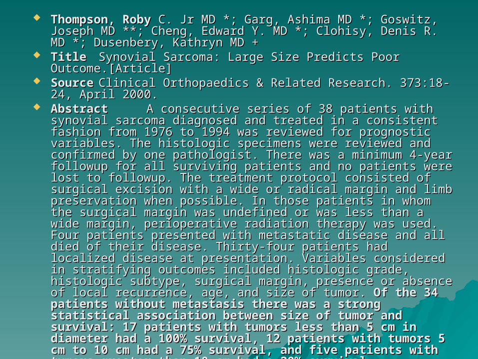
Thompson, RobyThompson, Roby C. Jr MD *; Garg, Ashima MD *; Goswitz, Joseph C. Jr MD *; Garg, Ashima MD *; Goswitz, Joseph MD **; Cheng, Edward Y. MD *; Clohisy, Denis R. MD *; Dusenbery, MD **; Cheng, Edward Y. MD *; Clohisy, Denis R. MD *; Dusenbery, Kathryn MD +Kathryn MD +
TitleTitle Synovial Sarcoma: Large Size Predicts Poor Outcome.Synovial Sarcoma: Large Size Predicts Poor Outcome.[Article][Article]
SourceSourceClinical Orthopaedics & Related Research. 373:18-24, April Clinical Orthopaedics & Related Research. 373:18-24, April 2000.2000.
AbstractAbstract A consecutive series of 38 patients with synovial A consecutive series of 38 patients with synovial sarcoma diagnosed and treated in a consistent fashion from 1976 sarcoma diagnosed and treated in a consistent fashion from 1976 to 1994 was reviewed for prognostic variables. The histologic to 1994 was reviewed for prognostic variables. The histologic specimens were reviewed and confirmed by one pathologist. specimens were reviewed and confirmed by one pathologist. There was a minimum 4-year followup for all surviving patients There was a minimum 4-year followup for all surviving patients and no patients were lost to followup. The treatment protocol and no patients were lost to followup. The treatment protocol consisted of surgical excision with a wide or radical margin and consisted of surgical excision with a wide or radical margin and limb preservation when possible. In those patients in whom the limb preservation when possible. In those patients in whom the surgical margin was undefined or was less than a wide margin, surgical margin was undefined or was less than a wide margin, perioperative radiation therapy was used. Four patients presented perioperative radiation therapy was used. Four patients presented with metastatic disease and all died of their disease. Thirty-four with metastatic disease and all died of their disease. Thirty-four patients had localized disease at presentation. Variables patients had localized disease at presentation. Variables considered in stratifying outcomes included histologic grade, considered in stratifying outcomes included histologic grade, histologic subtype, surgical margin, presence or absence of local histologic subtype, surgical margin, presence or absence of local recurrence, age, and size of tumor. recurrence, age, and size of tumor. Of the 34 patients without Of the 34 patients without metastasis there was a strong statistical association metastasis there was a strong statistical association between size of tumor and survival: 17 patients with between size of tumor and survival: 17 patients with tumors less than 5 cm in diameter had a 100% survival, 12 tumors less than 5 cm in diameter had a 100% survival, 12 patients with tumors 5 cm to 10 cm had a 75% survival, patients with tumors 5 cm to 10 cm had a 75% survival, and five patients with tumors greater than 10 cm had a and five patients with tumors greater than 10 cm had a 20% survival20% survival....
Which of the following sarcomas is the Which of the following sarcomas is the most commonly found in young adult ?most commonly found in young adult ?
1) Chondrosarcoma.1) Chondrosarcoma.
2) Malignant fibrous histiocytoma.2) Malignant fibrous histiocytoma.
3) Liposarcoma.3) Liposarcoma.
4) Synovial sarcoma.4) Synovial sarcoma.
5) Fibrosarcoma.5) Fibrosarcoma.
Which of the following sarcomas is the most Which of the following sarcomas is the most commonly found in young adult ?commonly found in young adult ?
1) Chondrosarcoma.1) Chondrosarcoma.
2) Malignant fibrous histiocytoma.2) Malignant fibrous histiocytoma.
3) Liposarcoma.3) Liposarcoma.
4) Synovial sarcoma.4) Synovial sarcoma.
5) Fibrosarcoma.5) Fibrosarcoma.
Soft tissue sarcomas occour at any age , Soft tissue sarcomas occour at any age , although the majority are seen in patients although the majority are seen in patients older than age 40 years.older than age 40 years.
Half of the soft tissue sarcomas occurring in Half of the soft tissue sarcomas occurring in children are rhabdomyosarcomas.children are rhabdomyosarcomas.
In young adult , synovial sarcomas are more In young adult , synovial sarcomas are more likely to be diagnosed than other types of likely to be diagnosed than other types of soft tissue sarcomas. Twice as many soft soft tissue sarcomas. Twice as many soft tissue sarcomas occour in the lower tissue sarcomas occour in the lower extremities than in upper extrimities.(40% - extremities than in upper extrimities.(40% - 20% ). 30% in trunk,10% in head and neck.20% ). 30% in trunk,10% in head and neck.
AAOS Instructional course lectures, volume AAOS Instructional course lectures, volume 53,2004.53,2004.

Which of the following is a true tumor? Which of the following is a true tumor?
1) Bone cyst 1) Bone cyst
2) Fibrous dysplasia 2) Fibrous dysplasia
3) Osteochondroma 3) Osteochondroma
4) Enchondroma 4) Enchondroma
5) Nonosteogenic fibroma 5) Nonosteogenic fibroma

Which of the following is a true tumor ? Which of the following is a true tumor ?
1) Bone cyst 1) Bone cyst
2) Fibrous dysplasia 2) Fibrous dysplasia
3) Osteochondroma 3) Osteochondroma
4) Enchondroma 4) Enchondroma
5) Nonosteogenic fibroma 5) Nonosteogenic fibroma
A simple bone cyst (Synonym: unicameral bone cyst, A simple bone cyst (Synonym: unicameral bone cyst, solitary cyst) is solitary cyst) is a solitary cavity containing clear fluida solitary cavity containing clear fluid originating in the metaphysis of growing children adjacent originating in the metaphysis of growing children adjacent to the metaphyseal aspect of the growth plate.to the metaphyseal aspect of the growth plate.
Fibrous dysplasia is a Fibrous dysplasia is a developmental hamartomadevelopmental hamartoma in which in which areas of the skeleton fail to mature normally and remain areas of the skeleton fail to mature normally and remain indefinitely as immature, poorly mineralized trabeculae.indefinitely as immature, poorly mineralized trabeculae.
Exostosis (osteochondroma) is a Exostosis (osteochondroma) is a hamartomatous outgrowth hamartomatous outgrowth of cartilageof cartilage from the peripheral aspect of the physeal plate from the peripheral aspect of the physeal plate that undergoes enchondral ossification. that undergoes enchondral ossification.
An enchondroma is a benign, cartilaginous lesion An enchondroma is a benign, cartilaginous lesion that arises in the interior of the bone.that arises in the interior of the bone.
Non-ossifying fibroma (Synonym: fibrous cortical defect, Non-ossifying fibroma (Synonym: fibrous cortical defect, fibrous metaphyseal defect, histiocytic fibroma) is a fibrous metaphyseal defect, histiocytic fibroma) is a hamartomatous defect in the metaphyseal cortexhamartomatous defect in the metaphyseal cortex of of skeletally-immature adolescents.skeletally-immature adolescents.
(Enneking CD)(Enneking CD)
Figures a ,b show the radiograph and MRI Figures a ,b show the radiograph and MRI scan of a 12 year old boy who has a scan of a 12 year old boy who has a pathological fracture through a right pathological fracture through a right femoral osteogenic sarcoma . The patient femoral osteogenic sarcoma . The patient undergoes an above knee amputation at undergoes an above knee amputation at the level of the line which of the following the level of the line which of the following terms best characterizes the type of terms best characterizes the type of resection performed.?resection performed.?
1) intralesional.1) intralesional. 2) Marginal.2) Marginal. 3) Wide.3) Wide. 4) Radical.4) Radical. 5) Complete.5) Complete.


Figures a ,b show the radiograph and MRI Figures a ,b show the radiograph and MRI scan of a 12 year old boy who has a scan of a 12 year old boy who has a pathological fracture through a right pathological fracture through a right femoral osteogenic sarcoma . The patient femoral osteogenic sarcoma . The patient undergoes an above knee amputation at the undergoes an above knee amputation at the level of the line which of the following level of the line which of the following terms best characterizes the type of terms best characterizes the type of resection performed.?resection performed.?
1) 1) intralesional.intralesional. 2) Marginal.2) Marginal. 3) Wide.3) Wide. 4) Radical.4) Radical. 5) Complete.5) Complete.
Malawer MM, sugarbaker PH:Musculoskeletal cancer Malawer MM, sugarbaker PH:Musculoskeletal cancer surgery: Treatment of sarcoms and allied disease.surgery: Treatment of sarcoms and allied disease.
Menendez LR :orthopaedic knowledge Menendez LR :orthopaedic knowledge update :musculoskeletal tumors. American academy update :musculoskeletal tumors. American academy of the orthopaedic surgons,2002.of the orthopaedic surgons,2002.

Which of the following disorders most Which of the following disorders most commonly places a patient at a risk for commonly places a patient at a risk for malignancy ?malignancy ?
1) McCune Albright syndrome1) McCune Albright syndrome
2) Ollier’s disease.2) Ollier’s disease.
3) Mafucci syndrome.3) Mafucci syndrome.
4) Neurofibromatosis.4) Neurofibromatosis.
5) Hereditary multiple exostoses.5) Hereditary multiple exostoses.
Which of the following disorders most Which of the following disorders most commonly places a patient at a risk for commonly places a patient at a risk for malignancy ?malignancy ?
1) McCune Albright syndrome1) McCune Albright syndrome
2) Ollier’s disease.2) Ollier’s disease.
3) 3) Mafucci syndromeMafucci syndrome..
4) Neurofibromatosis.4) Neurofibromatosis.
5) Hereditary multiple exostoses5) Hereditary multiple exostoses
– The malignant potential of enchondromatosisThe malignant potential of enchondromatosis HS Schwartz, NB Zimmerman, MA Simon, RR Wroble, EA HS Schwartz, NB Zimmerman, MA Simon, RR Wroble, EA
Millar and M Bonfiglio Millar and M Bonfiglio In a tri-institutional, retrospective study with long-term follow-up, In a tri-institutional, retrospective study with long-term follow-up,
forty-four patients who had multiple enchondromas were identified. forty-four patients who had multiple enchondromas were identified. Thirty-seven patients did not have hemangiomas (Ollier Thirty-seven patients did not have hemangiomas (Ollier disease) and seven did (Maffucci syndrome).disease) and seven did (Maffucci syndrome). Of the thirty-seven Of the thirty-seven patients who had Ollier disease, a low-grade chondrosarcoma patients who had Ollier disease, a low-grade chondrosarcoma developed in four; an astrocytoma, in one; and a granulosa-cell developed in four; an astrocytoma, in one; and a granulosa-cell ovarian tumor, in one. In four of the seven patients who had Maffucci ovarian tumor, in one. In four of the seven patients who had Maffucci syndrome, there were six low-grade chondrosarcomas, one high-syndrome, there were six low-grade chondrosarcomas, one high-grade osteosarcoma, one pancreatic adenocarcinoma, one biliary grade osteosarcoma, one pancreatic adenocarcinoma, one biliary adenocarcinoma, and one astrocytoma. None of the patients in adenocarcinoma, and one astrocytoma. None of the patients in either group died of the skeletal sarcoma, but four of five patients either group died of the skeletal sarcoma, but four of five patients who had a non-skeletal malignant lesion died. who had a non-skeletal malignant lesion died. From life-table From life-table analyses of these patients, we estimated that the incidence analyses of these patients, we estimated that the incidence of secondary chondrosarcoma in patients who have Ollier of secondary chondrosarcoma in patients who have Ollier disease is about 25 per cent at the age of forty years, and disease is about 25 per cent at the age of forty years, and that malignant degeneration is almost a certainty in patients that malignant degeneration is almost a certainty in patients who have Maffucci syndrome.who have Maffucci syndrome. We concluded that periodic We concluded that periodic surveillance of the brain and abdomen for occult malignant lesions is surveillance of the brain and abdomen for occult malignant lesions is indicated in patients who have enchondromatosis. indicated in patients who have enchondromatosis.

When a giant cell tumor of the bone arises in When a giant cell tumor of the bone arises in the axial skeleton what is the most common the axial skeleton what is the most common location?location?
1) Ribs.1) Ribs.
2) Thoracic vertebra.2) Thoracic vertebra.
3) cervical posterior elements.3) cervical posterior elements.
4) Lumber posterior elements.4) Lumber posterior elements.
5) Sacrum.5) Sacrum.
When a giant cell tumor of the bone arises When a giant cell tumor of the bone arises in the axial skeleton what is the most in the axial skeleton what is the most common location?common location?
1) Ribs.1) Ribs.
2) Thoracic vertebra.2) Thoracic vertebra.
3) cervical posterior elements.3) cervical posterior elements.
4) Lumber posterior elements.4) Lumber posterior elements.
55) Sacrum.) Sacrum.
Low grade chondrosarcoma can best Low grade chondrosarcoma can best distinguished radiographically from benign distinguished radiographically from benign enchondroma based on which of the enchondroma based on which of the following ?following ?
1) increased tracer uptake on bone scan.1) increased tracer uptake on bone scan.
2) Endosteal erosion of one fourth of the inner 2) Endosteal erosion of one fourth of the inner cortex.cortex.
3) Stippled calcification.3) Stippled calcification.
4) increased radiolucency on follow-up 4) increased radiolucency on follow-up radiographs.radiographs.
5) Size of more than 5cm.5) Size of more than 5cm.

Low grade chondrosarcoma can best Low grade chondrosarcoma can best distinguished radiographically from benign distinguished radiographically from benign enchondroma based on which of the enchondroma based on which of the following ?following ?
1) increased tracer uptake on bone scan.1) increased tracer uptake on bone scan. 2) Endosteal erosion of one fourth of the 2) Endosteal erosion of one fourth of the
inner cortex.inner cortex. 3) Stippled calcification.3) Stippled calcification. 4) increased radiolucency on follow-up 4) increased radiolucency on follow-up
radiographsradiographs.. 5) Size of more than 5cm.5) Size of more than 5cm.

Enchondromas are always completely intraosseous.Enchondromas are always completely intraosseous. They are benign, do not grow and should not change They are benign, do not grow and should not change
rdiographically in adult ,if a presumed enchondroma rdiographically in adult ,if a presumed enchondroma changes on follow up radiographs it consider changes on follow up radiographs it consider chondrosarcoma.chondrosarcoma.
radiographic changes is usually seen as decreasing radiographic changes is usually seen as decreasing calcification, or radiolucencies developing within the calcification, or radiolucencies developing within the calcified lesion. calcification occurs in long standing calcified lesion. calcification occurs in long standing resting cartilage. chondrosarcoma develops ,the resting cartilage. chondrosarcoma develops ,the malignant chondrocyte are actively dividing and do malignant chondrocyte are actively dividing and do not calcify because all cartilage grows in lobules ,the not calcify because all cartilage grows in lobules ,the radiolucencies are circular and often occour at the radiolucencies are circular and often occour at the edge of the calcification.edge of the calcification.
Weiner SD: Enchondroma and chondrosarcoma :clinical Weiner SD: Enchondroma and chondrosarcoma :clinical radiologic ,and histologic differentiation.radiologic ,and histologic differentiation.
AAOS instructional course ,volume53,204.AAOS instructional course ,volume53,204.
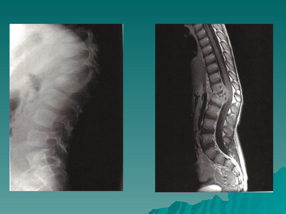
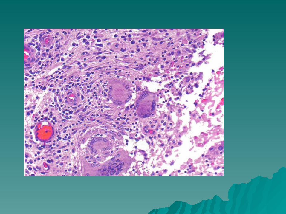
A 3 year old boy has back pain and is unable A 3 year old boy has back pain and is unable to walk A lateral radiograph is shown in to walk A lateral radiograph is shown in figure a, and sagital and axial MRI scan are figure a, and sagital and axial MRI scan are shown in figure b, c .low and high power shown in figure b, c .low and high power biopsy specimens are shown in figure d and biopsy specimens are shown in figure d and e, what is the most likely diagnosis?e, what is the most likely diagnosis?
1) Ewing’s sarcoma.1) Ewing’s sarcoma.
2) Langerhan’s cell histocytosis.2) Langerhan’s cell histocytosis.
3) Giant cell tumor.3) Giant cell tumor.
4) Bacterial osteomylitis.4) Bacterial osteomylitis.
5) Tuberculosis.5) Tuberculosis.

12) A 3 year old boy has back pain and is 12) A 3 year old boy has back pain and is unable to walk A lateral radiograph is unable to walk A lateral radiograph is shown in figure a, and sagittal and axial MRI shown in figure a, and sagittal and axial MRI scan are shown in figure b, c .low and high scan are shown in figure b, c .low and high power biopsy specimens are shown in figure power biopsy specimens are shown in figure d and e, what is the most likely diagnosis?d and e, what is the most likely diagnosis?
1) Ewing’s sarcoma.1) Ewing’s sarcoma.
2) Langerhan’s cell histocytosis.2) Langerhan’s cell histocytosis.
3) Giant cell tumor.3) Giant cell tumor.
4) Bacterial osteomylitis.4) Bacterial osteomylitis.
5) Tuberculosis.5) Tuberculosis.
CLASSIFICATION BASED ON THE SELECTION OF SURGICAL STRATEGIES CLASSIFICATION BASED ON THE SELECTION OF SURGICAL STRATEGIES
J. S. Mehta, MS Orth, D Orth, MCh Orth, Orthopaedic Specialist J. S. Mehta, MS Orth, D Orth, MCh Orth, Orthopaedic Specialist Registrar Registrar
Kent and Sussex Hospital, Mt Ephraim, Tunbridge Wells TNG 8AT, Kent BR2 Kent and Sussex Hospital, Mt Ephraim, Tunbridge Wells TNG 8AT, Kent BR2 0YA, UK. 0YA, UK.
In spinal tuberculosis MRI can clearly demonstrate combinations of anterior In spinal tuberculosis MRI can clearly demonstrate combinations of anterior and posterior lesions as well as pedicular involvement. We propose a and posterior lesions as well as pedicular involvement. We propose a classification system, using information provided by MRI, to help to plan the classification system, using information provided by MRI, to help to plan the appropriate surgical treatment for patients with thoracic spinal tuberculosis. appropriate surgical treatment for patients with thoracic spinal tuberculosis. We describe a series of We describe a series of 47 patients47 patients, , divided into four groups, based on divided into four groups, based on the surgical protocol used in the management. Group A consistedthe surgical protocol used in the management. Group A consisted of of patients with anterior lesions which were stable with no kyphotic deformity, patients with anterior lesions which were stable with no kyphotic deformity, and were treated with anterior debridement and strut grafting. and were treated with anterior debridement and strut grafting. Group BGroup B comprised patients with global lesions, kyphosis and instability who were comprised patients with global lesions, kyphosis and instability who were treated with posterior instrumentation using a closed-loop rectangle with treated with posterior instrumentation using a closed-loop rectangle with sublaminar wires, and by anterior strut grafting. sublaminar wires, and by anterior strut grafting. Group CGroup C were patients with were patients with anterior or global lesions as in the previous groups, but who were at a high anterior or global lesions as in the previous groups, but who were at a high risk for transthoracic surgery because of medical and possible anaesthetic risk for transthoracic surgery because of medical and possible anaesthetic complications. These patients had a global decompression of the cord complications. These patients had a global decompression of the cord posteriorly, the anterior portion of the cord being approached through a posteriorly, the anterior portion of the cord being approached through a transpedicular route. Posterior instrumentation was with a closed-loop transpedicular route. Posterior instrumentation was with a closed-loop rectangle held by sublaminar wires. rectangle held by sublaminar wires. Group D comprisedGroup D comprised patients with patients with isolated posterior lesions which required posterior decompression only. isolated posterior lesions which required posterior decompression only.
Figures a, b show the radiographs of a 34 Figures a, b show the radiographs of a 34 year old man who has an enlarging mass year old man who has an enlarging mass over the left iliac crest . A biopsy specimen over the left iliac crest . A biopsy specimen is shown in figure c, after the staging studies is shown in figure c, after the staging studies have been completed ,what is the next most have been completed ,what is the next most appropriate step in management.?appropriate step in management.?
1)observation with following in 3 months.1)observation with following in 3 months.
2) preoperative external beam radiation 2) preoperative external beam radiation therapy.therapy.
3)curettage and cement.3)curettage and cement.
5) wide excision.5) wide excision.

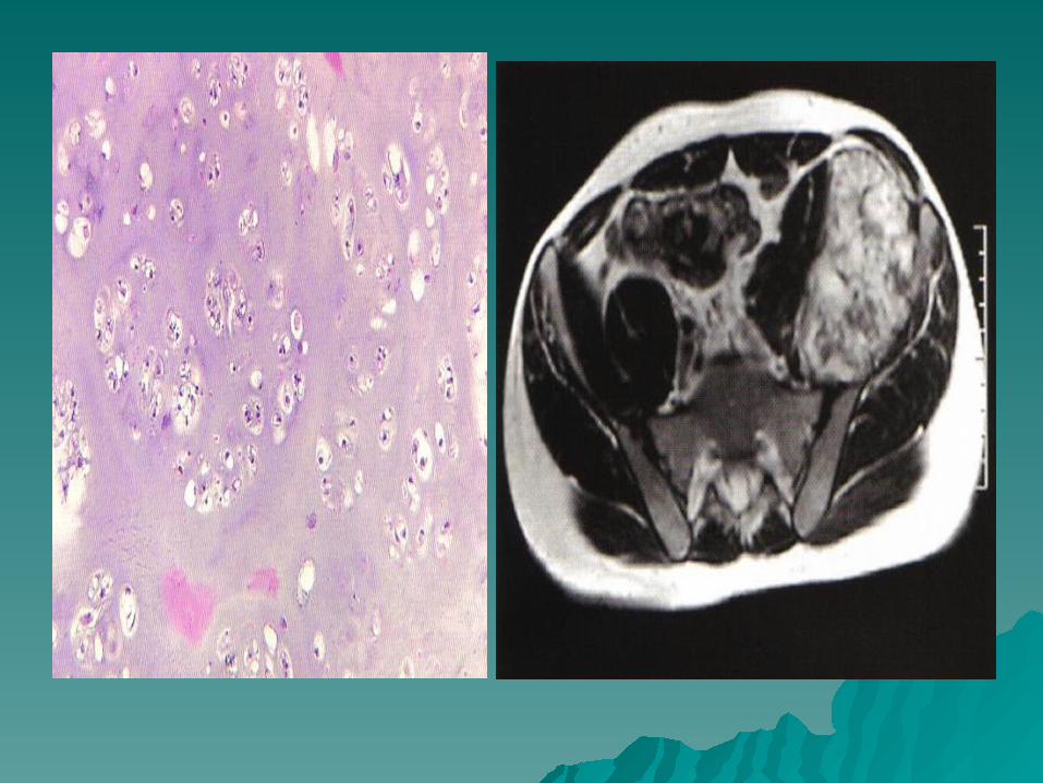

Figures a, b show the radiographs of a 34 Figures a, b show the radiographs of a 34 year old man who has an enlarging mass year old man who has an enlarging mass over the left iliac crest . A biopsy specimen over the left iliac crest . A biopsy specimen is shown in figure c, after the staging is shown in figure c, after the staging studies have been completed ,what is the studies have been completed ,what is the next most appropriate step in next most appropriate step in management.?management.?
1)observation with following in 3 months.1)observation with following in 3 months.
2) preoperative external beam radiation 2) preoperative external beam radiation therapy.therapy.
3)curettage and cement.3)curettage and cement.
5) wide excision.5) wide excision.
FRANCIS Y. LEE, M.D., HENRY J. MANKIN, M.D.FRANCIS Y. LEE, M.D., HENRY J. MANKIN, M.D., GERTRUDE FONDREN, , GERTRUDE FONDREN, B.S.B.S., MARK C. GEBHARDT, M.D., DEMPSEY S. SPRINGFIELD, M.D.§, , MARK C. GEBHARDT, M.D., DEMPSEY S. SPRINGFIELD, M.D.§, ANDREW E. ROSENBERG, M.D. and L. CANDACE JENNINGS, M.D., ANDREW E. ROSENBERG, M.D. and L. CANDACE JENNINGS, M.D., BOSTON, MASSACHUSETTS BOSTON, MASSACHUSETTS
Background:Background: The data on The data on 227 patients227 patients who had been managed for a who had been managed for a chondrosarcoma at one institution were reviewed to determine the nature of chondrosarcoma at one institution were reviewed to determine the nature of the lesions, the predictors of outcome, and whether there were any ways to the lesions, the predictors of outcome, and whether there were any ways to change the treatment approaches to improve the results. change the treatment approaches to improve the results. Methods:Methods: The The patients were patients were followed for a mean duration of six years (range, three followed for a mean duration of six years (range, three to twenty-five years). The mean age of the patients was forty-seven to twenty-five years). The mean age of the patients was forty-seven years (range, nine to eighty-four yearsyears (range, nine to eighty-four years). The most prevalent sites of the ). The most prevalent sites of the tumors were the femur (seventy-eight), the pelvis (fifty-one), and the tumors were the femur (seventy-eight), the pelvis (fifty-one), and the humerus (thirty-nine). The tumors were divided into two groups according to humerus (thirty-nine). The tumors were divided into two groups according to histological grade. Eighty-six tumors (sixteen atypical enchondromas and histological grade. Eighty-six tumors (sixteen atypical enchondromas and seventy grade-1 chondrosarcomas) that were locally destructive but were seventy grade-1 chondrosarcomas) that were locally destructive but were associated with a low likelihood of metastasis were considered to be low-associated with a low likelihood of metastasis were considered to be low-grade. The remaining 141 lesions, which were locally destructive, potentially grade. The remaining 141 lesions, which were locally destructive, potentially metastatic, and capable of causing death, were thought to be high-grade. metastatic, and capable of causing death, were thought to be high-grade. One hundred and three of these 141 lesions were grade 2, and thirty-eight One hundred and three of these 141 lesions were grade 2, and thirty-eight were grade 3 (eighteen of the thirty-eight were grade 3 only, and twenty were grade 3 (eighteen of the thirty-eight were grade 3 only, and twenty were both grade 3 and dedifferentiated). Two hundred and twenty-four were both grade 3 and dedifferentiated). Two hundred and twenty-four patients were managed with resection and a limb-sparing procedure; the patients were managed with resection and a limb-sparing procedure; the remaining three patients had an amputation. Postoperative adjuvant remaining three patients had an amputation. Postoperative adjuvant radiation was used for fifty-six patients; chemotherapy, for thirty-five; and radiation was used for fifty-six patients; chemotherapy, for thirty-five; and both radiation and chemotherapy, for nineteen. Flow cytometric patterns both radiation and chemotherapy, for nineteen. Flow cytometric patterns were analyzed for 105 patients. were analyzed for 105 patients. Results:Results: The patients who had a high-grade The patients who had a high-grade tumor were older than those who had a low-grade tumor (mean age [and tumor were older than those who had a low-grade tumor (mean age [and standard deviation], 50 standard deviation], 50 アア 17.0 years compared with 40 17.0 years compared with 40 アア 15.9 years; p < 15.9 years; p < 0.001). Pathological fracture, metastasis, local recurrence, and death were 0.001). Pathological fracture, metastasis, local recurrence, and death were more prevalent in the group that had a high-grade lesion (p < 0.001). more prevalent in the group that had a high-grade lesion (p < 0.001).
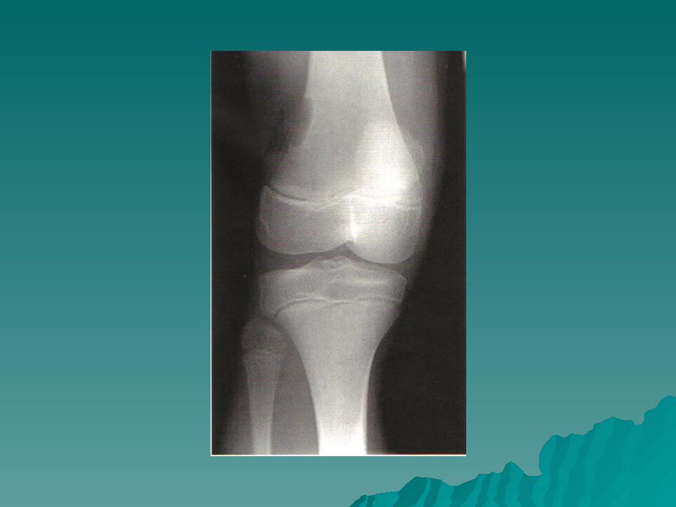
A12 year old girl sustains a knee injury A12 year old girl sustains a knee injury while playing soccer .Radiographs reveal while playing soccer .Radiographs reveal the lesion show in figures a, Examination the lesion show in figures a, Examination reveals mild effusion ,no tenderness, and a reveals mild effusion ,no tenderness, and a full knee range of motion. What is the next full knee range of motion. What is the next most appropriate step in management?most appropriate step in management?
1) Repeat radiographs in 3 months.1) Repeat radiographs in 3 months.
2) MRI of thee distal femur.2) MRI of thee distal femur.
3) Needle biopsy.3) Needle biopsy.
4) CT of the chest.4) CT of the chest.
Non -Ossifying Fibroma - Fibrous Cortical DefectNon -Ossifying Fibroma - Fibrous Cortical Defect Non-ossifying fibroma (NOF) is a well circumscribed, solitary fibrous proliferation. This Non-ossifying fibroma (NOF) is a well circumscribed, solitary fibrous proliferation. This
lesion is found mostly in children with 75% occurring in the second decade. The lesion lesion is found mostly in children with 75% occurring in the second decade. The lesion is found in males more commonly than in females and may occur in as many as 35% of is found in males more commonly than in females and may occur in as many as 35% of all children.' It is a non-neoplastic process that occurs in the juxtaepiphyseal region of all children.' It is a non-neoplastic process that occurs in the juxtaepiphyseal region of the long bones. The most common site is the femur followed by the tibia .the long bones. The most common site is the femur followed by the tibia .Clinically,Clinically, non-ossifying fibromas are asymptomatic and are usually discovered as an non-ossifying fibromas are asymptomatic and are usually discovered as an incidental finding on x-ray. Occasionally, a larger lesion presents as a pathologic incidental finding on x-ray. Occasionally, a larger lesion presents as a pathologic fracture. The classic scenario is a child who has a minor injury in a sports contest and a fracture. The classic scenario is a child who has a minor injury in a sports contest and a plain radiograph is taken in a local emergency room. A lesion is seen and the child is plain radiograph is taken in a local emergency room. A lesion is seen and the child is referred to an orthopaedic surgeon for evaluation. Generally, a careful history and referred to an orthopaedic surgeon for evaluation. Generally, a careful history and physical examination will show that the child's pain is related to the nearby joint rather physical examination will show that the child's pain is related to the nearby joint rather than the lesion, and that the pattern of the symptoms fits that which is expected in a than the lesion, and that the pattern of the symptoms fits that which is expected in a sprain or strain injury. If there was pain before the injury that seems to arise from the sprain or strain injury. If there was pain before the injury that seems to arise from the lesion, the diagnosis of NOF is highly suspect. Jaffe-Campanacci syndrome is a lesion, the diagnosis of NOF is highly suspect. Jaffe-Campanacci syndrome is a constellation of symptoms including multiple non-ossifying fibromas, cafe-au-lait spots, constellation of symptoms including multiple non-ossifying fibromas, cafe-au-lait spots, mental retardation, hypogonadism, ocular and cardiovascular abnormalities. mental retardation, hypogonadism, ocular and cardiovascular abnormalities. Non-ossifying fibromas have a very typical appearance on x-ray. They are eccentric, Non-ossifying fibromas have a very typical appearance on x-ray. They are eccentric, multi-loculated sub-cortical lesions with a central lucency and a scalloped sclerotic multi-loculated sub-cortical lesions with a central lucency and a scalloped sclerotic margin. There is sometimes cortical thinning but no periosteal reaction. Serial x-rays margin. There is sometimes cortical thinning but no periosteal reaction. Serial x-rays will show the lesion migrating away from the epiphyseal plate with time. will show the lesion migrating away from the epiphyseal plate with time. If the If the lesion is not eccentric, has no sclerotic rim, or is not multi-loculated, the lesion is not eccentric, has no sclerotic rim, or is not multi-loculated, the diagnosis of NOF is suspect and further evaluation is warranted. Since diagnosis of NOF is suspect and further evaluation is warranted. Since these lesions may be treated by observation only without biopsy, the these lesions may be treated by observation only without biopsy, the physician should be quite certain of the diagnosis before formulating a physician should be quite certain of the diagnosis before formulating a treatment plan. treatment plan. If any doubt exists, the advice of an orthopaedic oncology specialist If any doubt exists, the advice of an orthopaedic oncology specialist should be sought. . should be sought. . Marks, KE and TW Bauer, Marks, KE and TW Bauer, Fibrous Tumors of Bone, Fibrous Tumors of Bone, Orthopaedic Clinics of North Orthopaedic Clinics of North America,America, 20(3): July, 1989. 20(3): July, 1989.JX / gJX / gX,, ,X,, ,1 1/17/97 1 1/17/97

A 63year old man with a history of A 63year old man with a history of localized prostate cancer sustained a localized prostate cancer sustained a femoral subtrochanteric fracture in a femoral subtrochanteric fracture in a motor vehicle accident. Radiographs motor vehicle accident. Radiographs reveal a transverse fracture through a reveal a transverse fracture through a lytic lesion. What is the most lytic lesion. What is the most appropriate step in management?appropriate step in management?
1) Biopsy of the lesion.1) Biopsy of the lesion. 2) Antegrade femoral nailing.2) Antegrade femoral nailing. 3) Retrograde femoral nailing.3) Retrograde femoral nailing. 4) open reduction and internal fixation 4) open reduction and internal fixation
with a fixed angled plate.with a fixed angled plate. 5) Long-stemmed cemented 5) Long-stemmed cemented
hemiarthroplasty.hemiarthroplasty.
A 63year old man with a history of localized A 63year old man with a history of localized prostate cancer sustained a femoral prostate cancer sustained a femoral subtrochanteric fracture in a motor vehicle subtrochanteric fracture in a motor vehicle accident. Radiographs reveal a transverse accident. Radiographs reveal a transverse fracture through a lytic lesion. What is the fracture through a lytic lesion. What is the most appropriate step in management?most appropriate step in management?
1) Biopsy of the lesion.1) Biopsy of the lesion.
2) Antegrade femoral nailing.2) Antegrade femoral nailing.
3) Retrograde femoral nailing.3) Retrograde femoral nailing.
4) open reduction and internal fixation with 4) open reduction and internal fixation with a fixed angled plate.a fixed angled plate.
5) Long-stemmed cemented 5) Long-stemmed cemented hemiarthroplastyhemiarthroplasty
The following basic types of the formation The following basic types of the formation about musculoskeletal tumors need to be about musculoskeletal tumors need to be obtained prior to treatment:obtained prior to treatment:
The pathological diagnosis ,is obtained by The pathological diagnosis ,is obtained by biopsy.biopsy.
The local extent of the tumor, the extent of The local extent of the tumor, the extent of distant spread of the tumor ,is obtained by distant spread of the tumor ,is obtained by clinical staging. clinical staging.
Treatment of most musculoskeletal tumors in Treatment of most musculoskeletal tumors in extremities, the surgeon need to know the extremities, the surgeon need to know the interosseous and extra osseous (soft tissue) interosseous and extra osseous (soft tissue) extent of the tumor and if the tumor has a extent of the tumor and if the tumor has a metastasized ,because treatment of the metastasized ,because treatment of the primary tumor may need modified if primary tumor may need modified if disseminated disease is present.disseminated disease is present.
Diagnostic strategy of bone and soft tissue tumors Diagnostic strategy of bone and soft tissue tumors .Micheal A simon. Henry Finn..Micheal A simon. Henry Finn.
AAOS inst.course lecture volme43.1994.AAOS inst.course lecture volme43.1994.

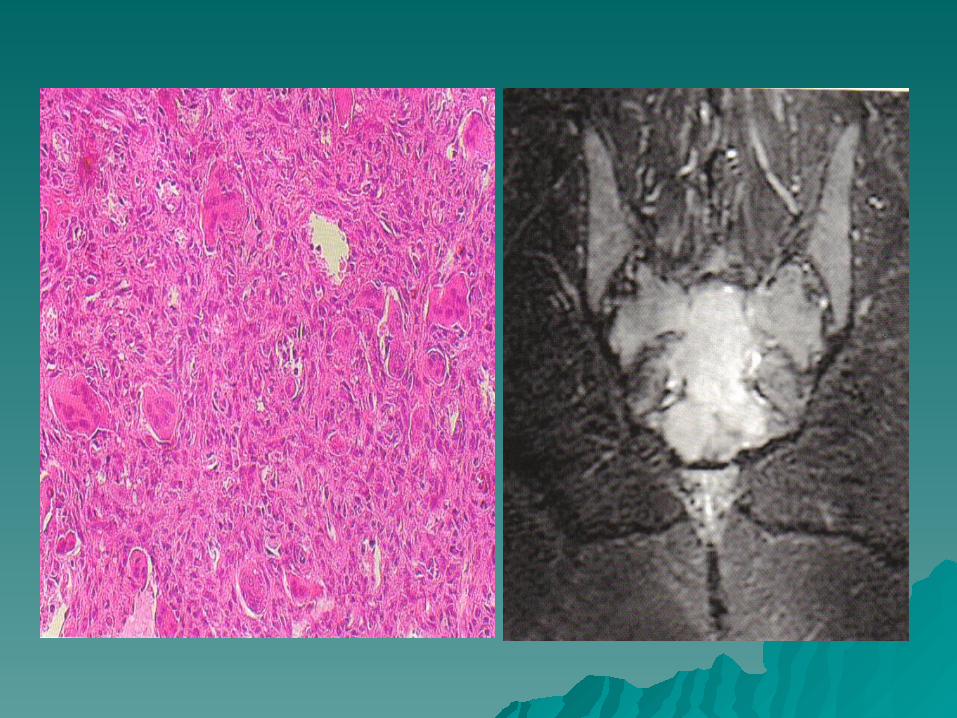
A 17 year old girl who noted low pain A 17 year old girl who noted low pain following a fall 1month ago now report following a fall 1month ago now report discomfort in the spine position and discomfort in the spine position and increasing difficulty initiating urination. An increasing difficulty initiating urination. An AP radiograph is shown in figure a ,and AP radiograph is shown in figure a ,and sagitall T1 and coronal T2 weight MRI scan sagitall T1 and coronal T2 weight MRI scan are shown in figures b ,c .A biopsy specimen are shown in figures b ,c .A biopsy specimen is shown in figure d, What is the most likely is shown in figure d, What is the most likely diagnosis?diagnosis?
1) Chondroma.1) Chondroma. 2) Giant cell tumor.2) Giant cell tumor. 3) Osteoblastoma.3) Osteoblastoma. 4) Chondrosarcoma.4) Chondrosarcoma. 5) Malignant fibrous histiocytoma of bone.5) Malignant fibrous histiocytoma of bone.
A 17 year old girl who noted low pain A 17 year old girl who noted low pain following a fall 1month ago now report following a fall 1month ago now report discomfort in the spine position and discomfort in the spine position and increasing difficulty initiating urination. An increasing difficulty initiating urination. An AP radiograph is shown in figure ,and AP radiograph is shown in figure ,and sagitall T1 and coronal T2 weight MRI scan sagitall T1 and coronal T2 weight MRI scan are shown in figures b ,c .A biopsy specimen are shown in figures b ,c .A biopsy specimen is shown in figure d, What is the most likely is shown in figure d, What is the most likely diagnosis?diagnosis?
1) Chondroma.1) Chondroma. 2) Giant cell tumor.2) Giant cell tumor. 3) Osteoblastoma.3) Osteoblastoma. 4) Chondrosarcoma.4) Chondrosarcoma. 5) Malignant fibrous histiocytoma of bone.5) Malignant fibrous histiocytoma of bone.
With respect to a 6 year old girl with an With respect to a 6 year old girl with an osteogenic sarcoma at the level of the knee osteogenic sarcoma at the level of the knee and 2 nodules in the inferior left lung ?and 2 nodules in the inferior left lung ?
1 ) she should be treated by amputation and 1 ) she should be treated by amputation and radiotherapy .radiotherapy .
2) should be treated by chemotherapy and 2) should be treated by chemotherapy and amputationamputation
3) should be treated by chemotherapy, lymph 3) should be treated by chemotherapy, lymph salvage procedure and thoractomy at a salvage procedure and thoractomy at a later stagelater stage
4) should be treated by radiotherapy alone 4) should be treated by radiotherapy alone 5) this patient has such a poor prognosis, no 5) this patient has such a poor prognosis, no
treatment should be o6ered treatment should be o6ered
With respect to a 6 year old girl with an With respect to a 6 year old girl with an osteogenic sarcoma at the level of the knee osteogenic sarcoma at the level of the knee and 2 nodules in the inferior left lung :and 2 nodules in the inferior left lung :
1 ) she should be treated by amputation and 1 ) she should be treated by amputation and radiotherapy .radiotherapy .
2) should be treated by chemotherapy and 2) should be treated by chemotherapy and amputationamputation
33) should be treated by chemotherapy, lymph salvage ) should be treated by chemotherapy, lymph salvage procedure and thoractomy at a later stageprocedure and thoractomy at a later stage
4) should be treated by radiotherapy alone 4) should be treated by radiotherapy alone 5) this patient has such a poor prognosis, no 5) this patient has such a poor prognosis, no
treatment should be o6ered treatment should be o6ered
Dee’s p.245Dee’s p.245 Localized Extremity OS with Synchronous Pulmonary MetastasesLocalized Extremity OS with Synchronous Pulmonary Metastases Metastatic pulmonary disease detected at initial diagnosis (approximately Metastatic pulmonary disease detected at initial diagnosis (approximately
10% of patients) does not preclude a curative treatment strategy, although 10% of patients) does not preclude a curative treatment strategy, although the presence of extrathoracic metastases makes cure extremely unlikely. the presence of extrathoracic metastases makes cure extremely unlikely. Newly diagnosed patients have not been exposed to chemotherapy and Newly diagnosed patients have not been exposed to chemotherapy and are thus less likely to have drug-resistant tumors. For the pateint are thus less likely to have drug-resistant tumors. For the pateint presenting withpresenting with resectable disease (ie. usually less than 15 resectable disease (ie. usually less than 15 pulmonary nodules and a primary tumor of the extremitypulmonary nodules and a primary tumor of the extremity), the ), the traditional approach has been resection of all macroscopic diesease by traditional approach has been resection of all macroscopic diesease by median sternotomy and limb amputation or resection, followed by median sternotomy and limb amputation or resection, followed by intensive adjuvant chemotherapy. The tumor burden is thereby reduced intensive adjuvant chemotherapy. The tumor burden is thereby reduced ato a minimum before adjuvant therapy begins. Altough the timing of ato a minimum before adjuvant therapy begins. Altough the timing of surgery for the primary tumor and metastatic sites has been variable,surgery for the primary tumor and metastatic sites has been variable, most modern approaches entail alternating chemotherapy and most modern approaches entail alternating chemotherapy and surgery. Treatment usually begins with a course of surgery. Treatment usually begins with a course of chemotherapy, followed by resection of the primary tumor, chemotherapy, followed by resection of the primary tumor, followed by a second course of chemotherapy, surgical ablation of followed by a second course of chemotherapy, surgical ablation of metastatic sites, and the remaining courses of chemotherapy.metastatic sites, and the remaining courses of chemotherapy. In In patients with inoperable metastases, primary treatment with patients with inoperable metastases, primary treatment with chemotherapy is probably appropriate; metastases may respond chemotherapy is probably appropriate; metastases may respond sufficiently to allow complete resection. Becasue these patients usually sufficiently to allow complete resection. Becasue these patients usually require surgery for the primary tumor as a palliative procedure, erarly require surgery for the primary tumor as a palliative procedure, erarly surgery may be recommended, depite the presence of unresectable surgery may be recommended, depite the presence of unresectable pulmonary disease. Although improving, the outlook for patients pulmonary disease. Although improving, the outlook for patients presenting with metastatic disease remains poor.presenting with metastatic disease remains poor.
Giant cell tumors in the spine is more Giant cell tumors in the spine is more commonly found in ?commonly found in ?
1) spinous process .1) spinous process .
2) disc. 2) disc.
3) body .3) body .
4) pedicle .4) pedicle .
5) lamina.5) lamina.

Giant cell tumor in the spine is more Giant cell tumor in the spine is more commonly found in?commonly found in?
1) spinous process 1) spinous process
2) disc 2) disc
3) body 3) body
4) pedicle 4) pedicle
5) lamina 5) lamina
OKU 5 p.659OKU 5 p.659 ““GCT’s are relatively common in the GCT’s are relatively common in the
appendicular skeleton, but rarely occur in appendicular skeleton, but rarely occur in the spine. Most common during the fourth the spine. Most common during the fourth or fifth decades of life, these lesions tend to or fifth decades of life, these lesions tend to affect the vertebral body...”affect the vertebral body...”
The most common malignant tumor of The most common malignant tumor of bone is ?bone is ?
1) Ewing’s1) Ewing’s
2) Multiple Myeloma2) Multiple Myeloma
3) Reticulum cell sarcoma3) Reticulum cell sarcoma
4) Hodgkins disease4) Hodgkins disease
5) Metastatic tumor5) Metastatic tumor
The most common malignant tumor of The most common malignant tumor of bone is ?bone is ?
1) Ewing’s1) Ewing’s
2) Multiple Myeloma2) Multiple Myeloma
3) Reticulum cell sarcoma3) Reticulum cell sarcoma
4) Hodgkins disease4) Hodgkins disease
5) Metastatic tumor5) Metastatic tumor

Bullough’s p.431 “Metastatic cancer Bullough’s p.431 “Metastatic cancer is the most frequent malignant tumor is the most frequent malignant tumor found in the bone and it usually found in the bone and it usually causes pain”causes pain”
14 yr female has a 3 week history of 14 yr female has a 3 week history of pain in her thigh. She has a pain in her thigh. She has a temperature of 38.2°, a WBC of 9600, temperature of 38.2°, a WBC of 9600, and an ESR of 42. X-rays reveal a and an ESR of 42. X-rays reveal a lytic lesion in the midshaft of her lytic lesion in the midshaft of her femur with new bone formation. The femur with new bone formation. The most likely diagnosis is?most likely diagnosis is?
1) Osteomyelitis1) Osteomyelitis 2) Ewing’s Sarcoma.2) Ewing’s Sarcoma. 3) Osteosarcoma3) Osteosarcoma 4) Eosinophilic granuloma4) Eosinophilic granuloma 5) Fibrosarcoma5) Fibrosarcoma
14 yr female has a 3 week history of 14 yr female has a 3 week history of pain in her thigh. She has a pain in her thigh. She has a temperature of 38.2°, a WBC of 9600, temperature of 38.2°, a WBC of 9600, and an ESR of 42. X-rays reveal a and an ESR of 42. X-rays reveal a lytic lesion in the midshaft of her lytic lesion in the midshaft of her femur with new bone formation. The femur with new bone formation. The most likely diagnosis is?most likely diagnosis is?
1) Osteomyelitis.1) Osteomyelitis. 2) Ewing’s Sarcoma.2) Ewing’s Sarcoma. 3) Osteosarcoma.3) Osteosarcoma. 4) Eosinophilic granuloma.4) Eosinophilic granuloma. 5) Fibrosarcoma.5) Fibrosarcoma.

Bullough’s p.70 & 108 states acute osteomyelitis in children Bullough’s p.70 & 108 states acute osteomyelitis in children is usually metaphyseal. This child is already a bit old for is usually metaphyseal. This child is already a bit old for hematogenous OM. Acute OM is very rare in healthy adults hematogenous OM. Acute OM is very rare in healthy adults (which she is closer to), but when it occurs, the diaphysis of (which she is closer to), but when it occurs, the diaphysis of femur & the spine are most common. WBC would likely be femur & the spine are most common. WBC would likely be higher.higher.
Bullough’s p.417 “...Bullough’s p.417 “...Ewing’sEwing’s is a small cell malignant is a small cell malignant neoplasm of bone which develops in the neoplasm of bone which develops in the diaphysisdiaphysis or or metaphyssi of long bones, most often the metaphyssi of long bones, most often the femurfemur, tibia, and , tibia, and humerus, as well as in the pelvis, scapulae, ribs, and other humerus, as well as in the pelvis, scapulae, ribs, and other bones. It is essentially a tumor of childhood, with bones. It is essentially a tumor of childhood, with most most patients being under 20 yearspatients being under 20 years of age...Patients usually of age...Patients usually complain of complain of painpain or tenderness in the affected bone or tenderness in the affected bone of of several weeks’several weeks’ or months’ or months’ durationduration. Physical exam may . Physical exam may reveal swelling and tenderness. reveal swelling and tenderness. FeverFever, anemia, , anemia, leukocytosisleukocytosis,a nd ,a nd elevated ESRelevated ESR often suggest a often suggest a diagnosis of osteomyelitisdiagnosis of osteomyelitis and, becasue of the histologic and, becasue of the histologic appearance of the tumor, appearance of the tumor, osteomyelitis is the most osteomyelitis is the most important microscopic differential diagnosis of important microscopic differential diagnosis of Ewing’s tumor.”Ewing’s tumor.”
Best treatment for chondrosarcoma ? Best treatment for chondrosarcoma ?
1) chemotherapy and radiation. 1) chemotherapy and radiation.
2) preop. radiation excision. 2) preop. radiation excision.
3) excision.3) excision.
4) excision and post-op radiation .4) excision and post-op radiation .

Best treatment for chondrosarcoma ? Best treatment for chondrosarcoma ?
1) chemotherapy and radiation. 1) chemotherapy and radiation.
2) preop. radiation excision. 2) preop. radiation excision.
3) excision3) excision..
4) excision and post-op radiation .4) excision and post-op radiation .
B&J 217B&J 217 best Rx is wide surgical resectionbest Rx is wide surgical resection XRT may be used before or after XRT may be used before or after
microscopically incomplete resections in microscopically incomplete resections in spine/pelvisspine/pelvis
XRT and chemo not effective for primary XRT and chemo not effective for primary method of Rxmethod of Rx
Disease free survival – 30% @ 5 years for Disease free survival – 30% @ 5 years for high grade, 90% low gradehigh grade, 90% low grade
Wide resection followed by XRT if margins Wide resection followed by XRT if margins positivepositive
A sixteen year old female has A sixteen year old female has achondroblastona of the proximal humerus achondroblastona of the proximal humerus which is according to Enneking staging a which is according to Enneking staging a stage 2. Treatment would be?stage 2. Treatment would be?
1) en bloc resection and fusion of the 1) en bloc resection and fusion of the glenohumeral jointglenohumeral joint
2) en bloc resection and arthroplasty of the 2) en bloc resection and arthroplasty of the shouldershoulder
3) progressive curettage and bone grafting3) progressive curettage and bone grafting
4)shoulder disarticulation4)shoulder disarticulation
5)Tinker-Lyndhoff suspension procedure5)Tinker-Lyndhoff suspension procedure

A chondroblastoma is a benign neoplasm arising from immature, A chondroblastoma is a benign neoplasm arising from immature, cartilaginous cells (chondroblasts). cartilaginous cells (chondroblasts).
Age: 10 to 20 Sex: M > FAge: 10 to 20 Sex: M > F Site: This lesion arises almost universally in the secondary Site: This lesion arises almost universally in the secondary
ossification centers of the major, long bones and has a ossification centers of the major, long bones and has a predilection for the proximal humeral head, femoral condyles, and predilection for the proximal humeral head, femoral condyles, and the tibial plateau. It may, occasionally, arise in small bones the tibial plateau. It may, occasionally, arise in small bones without secondary ossification centers (most commonly the without secondary ossification centers (most commonly the tarsals) or in the apophyses of flat bones (most often the iliac tarsals) or in the apophyses of flat bones (most often the iliac crest).crest).
CurettageCurettage for Stage 2 lesions has a for Stage 2 lesions has a recurrencerecurrence rate of rate of approximately approximately 10 percent10 percent, which is somewhat lower than for , which is somewhat lower than for most other benign, Stage 2 lesions. Bone grafting of the defect is most other benign, Stage 2 lesions. Bone grafting of the defect is needed when the lesion is large; however, this is often not needed when the lesion is large; however, this is often not required in smaller lesions because of the patient's young age. required in smaller lesions because of the patient's young age. Interposition of fat or synthetic substances, after a transphyseal Interposition of fat or synthetic substances, after a transphyseal approach, may be indicated to minimize the risk of growth approach, may be indicated to minimize the risk of growth deformity in a very young patient. Because of its propensity for deformity in a very young patient. Because of its propensity for recurrence after contamination, it is desirable to approach the recurrence after contamination, it is desirable to approach the lesion without entering the cavity of the adjacent joint. This often lesion without entering the cavity of the adjacent joint. This often necessitates a transphyseal-plate approach with the potential for necessitates a transphyseal-plate approach with the potential for subsequent growth deformity. When the potential for growth subsequent growth deformity. When the potential for growth deformation is modest, because of the area involved or the age of deformation is modest, because of the area involved or the age of the patient, this approach is preferable to the risk of intra-articular the patient, this approach is preferable to the risk of intra-articular recurrence.recurrence.
![Y'] / — http ://urokonohandmade. seesaa. net/ 6cm 1 9cm...Y'] / — http ://urokonohandmade. seesaa. net/ 6cm 1 9cm](https://img.dokumen.tips/doc/110x75/613c80da4c23507cb6356cd8/y-a-http-urokonohandmade-seesaa-net-6cm-1-9cm-y-a-http-urokonohandmade.jpg)