Embed Size (px)
Citation preview

Molecular determinants of differential pore blockingof kidney CLC-K chloride channelsAlessandra Picollo1*, Antonella Liantonio1,2*, Maria Paola Didonna1,2, Laura Elia1, Diana Conte Camerino2
& Michael Pusch1+1Istituto di Biofisica, Consiglio Nazionale delle Ricerche, Genova, Italy, and 2Sezione di Farmacologia,
Dipartimento Farmacobiologico, Universita di Bari, Bari, Italy
The highly homologous Cl� channels CLC-Ka and CLC-Kb areimportant for water and salt conservation in the kidney and forthe production of endolymph in the inner ear. Mutations in CLC-Kblead to Bartter’s syndrome and mutations in the small CLC-Ksubunit barttin lead to Bartter’s syndrome and deafness. Here weshow that CLC-Ka is blocked by the recently identified blocker2-(p-chlorophenoxy)-3-phenylpropionic acid of the rat channelCLC-K1 with an apparent KDB80lM. We also found that DIDS(4,40-diisothiocyanatostilbene-2,20-disulphonic acid), a genericCl� channel blocker, inhibits CLC-Ka (KDB90lM). Surprisingly,the highly homologous channel CLC-Kb is fivefold to sixfold lesssensitive to both compounds. Guided by the crystal structure ofbacterial CLC proteins, we identify two amino acids, N68/D68and G72/E72, in CLC-Ka and CLC-Kb, respectively, that areresponsible for the differential drug sensitivity. Both residuesexpose their side chains in the extracellular pore mouth,delineating the probable drug binding site. These novel CLC-Kchannel blockers are promising lead compounds for the devel-opment of new diuretic drugs.Keywords: CLC-Ka; CLC-Kb; DIDS; wild type; block; mutationsEMBO reports (2004) 5, 584–589. doi:10.1038/sj.embor.7400169
INTRODUCTIONChloride channels allow the passive flux of anions acrossbiological membranes. CLC Cl� channels are important forseveral cellular and physiological processes, including trans-epithelial salt transport, electrical excitability, cell-volume regula-tion and acidification of internal and external compartments(Jentsch et al, 2002). CLC channels have been found in organismsas diverse as mammals, plants and yeast. Mammals have ninedifferent CLC genes. On the basis of sequence homology, they
can be grouped into three branches. The two CLC-K channels(CLC-Ka/CLC-K1 and CLC-Kb/CLC-K2; Uchida et al, 1993; Adachiet al, 1994; Kieferle et al, 1994) are plasma membrane channelsthat are predominantly expressed in the kidney and the inner ear.They are essential for water and salt conservation in the kidneyand for the production of endolymph in the inner ear. Mutations inCLC-Kb lead to Bartter’s syndrome (Simon et al, 1997) andmutations in the small, essential CLC-K channel subunit barttinlead to Bartter’s syndrome and deafness (Birkenhager et al, 2001).Rodent CLC-K1 (the orthologue of CLC-Ka) is important fortransepithelial transport in the thin ascending limb, and CLC-K1knockout mice show nephrogenic diabetes insipidus (Matsumuraet al, 1999). Recently, CLC-K channels were shown to require thesmall transmembrane protein barttin as a b-subunit for efficientfunctional expression (Estevez et al, 2001). Coexpression of CLC-Kchannels with barttin leads to greatly enhanced surface expression(Estevez et al, 2001) and maybe also to a change of biophysicalproperties (Waldegger et al, 2002). Thus, only recently has itbecome possible to study the biophysical and pharmacologicalproperties of CLC-K channels in detail. Small organic ligands thatinteract with ion channels have been extremely useful tools forunderstanding the function of these channels (Hille, 2001). Only afew specific blockers exist for CLC channels. Blockage of CLC-0and CLC-1 by small organic inhibitors that act from theintracellular side has been recently studied in detail (Pusch et al,2000, 2001, 2002; Liantonio et al, 2002, 2003; Accardi & Pusch,2003; Estevez et al, 2003; Traverso et al, 2003). A different type ofinhibition involving a separate binding site has been recentlydescribed for CLC-K channels (Liantonio et al, 2002). A group ofderivatives of 2-(p-chlorophenoxy)propionic acid (CPP) contain-ing two chlorophenoxy moieties that inhibit CLC-K1 withmicromolar affinity from the extracellular side was identified(Liantonio et al, 2002). In a subsequent structure–activity study,the 2-(p-chlorophenoxy)-3-phenylpropionic acid (3-phenyl-CPP)(Fig 1C) was identified as the minimal structure required to blockCLC-K1 (Liantonio et al, 2004). The block was reversibleand competitive with extracellular Cl�, suggesting a pore locationof the binding site. Among ‘classical’ Cl� channel inhibitors,only DIDS (4,40-diisothiocyanatostilbene-2,20-disulphonic acid;Fig 1C) blocked CLC-K1 with an affinity comparable with that of
Received 6 November 2003; revised 20 April 2004; accepted 20 April 2004;published online 28 May 2004
*These authors contributed equally to this work+Corresponding author. Tel: þ 39 0106475 561/522; Fax þ 39 0106475 500;E-mail: [email protected]
1Istituto di Biofisica, Consiglio Nazionale delle Ricerche, Via de Marini 6,16149 Genova, Italy2Sezione di Farmacologia, Dipartimento Farmacobiologico, Universita di Bari,70125 Bari, Italy
EMBO reports VOL 5 | NO 6 | 2004 &2004 EUROPEAN MOLECULAR BIOLOGY ORGANIZATION
scientificreportscientific report
584

3-phenyl-CPP, albeit in an apparently irreversible manner(Liantonio et al, 2004). We sought to extend our previous studiesto the human channels CLC-Ka and CLC-Kb. We found that CLC-Ka, like CLC-K1, is sensitive to 3-phenyl-CPP and DIDS, and thatCLC-Kb is much less sensitive to both drugs. This is surprisingbecause the two channels are about 80% identical at the proteinsequence level. We then used the crystal structure of bacterialCLC proteins as a guide and identified two amino acids as beingresponsible for the difference in drug sensitivity.
RESULTSWe examined blockage of the human homologues CLC-Ka andCLC-Kb. CLC-Ka currents activate at negative voltages and partlydeactivate at positive voltages (Fig 1A), similarly to CLC-K1(Liantonio et al, 2004). 3-Phenyl-CPP (Fig 1C) inhibited CLC-Ka(Fig 1A, middle) in a quickly reversible manner (Fig 1A, right).
Also DIDS blocked CLC-Ka with a similar affinity; however, theinhibition was not immediately reversible (Fig 1B). Washing for aprolonged time was necessary to recover from DIDS blockage(Fig 1D). This slow recovery rendered difficult a detailed analysisof the blocking mechanism. Surprisingly, CLC-Kb was much lesssensitive to both compounds (Fig 2). The voltage dependence andkinetics of CLC-Kb are also inverted with respect to CLC-Ka(Figs 1,2; Estevez et al, 2001). Furthermore, the gating kinetics ofCLC-Kb are slower than those of CLC-Ka (compare time scalesin Figs 1,2). A quantitative dose–response analysis of inhibition by3-phenyl-CPP and DIDS of CLC-Ka and CLC-Kb revealed thatblockage by both substances is practically complete at highconcentrations (Fig 3). Inhibition can be described well by asimple 1:1 binding (solid and dashed lines in Fig 3), resulting inthe apparent dissociation constants reported in Table 1. As forCLC-K1 (Liantonio et al, 2004), blockage by 3-phenyl-CPP andDIDS is slightly voltage dependent, being stronger at positivevoltages. We therefore measured inhibition at two voltages: �140and 60mV. At 60mV, 3-phenyl-CPP has a fivefold larger potencyon CLC-Ka compared with CLC-Kb, whereas the potency of DIDSis 6.5-fold larger (see Table 1).
The quick onset of blockage indicates that the binding site for3-phenyl-CPP and DIDS is accessible from the extracellular side.Competition with extracellular chloride suggests that it is locatedclose to the ion-conducting pore (Liantonio et al, 2004). Toidentify the amino acids that are responsible for the different drugsensitivities of CLC-Ka and CLC-Kb, we used the crystal structureof the bacterial StCLC (Dutzler et al, 2002) as a guide to identifyamino acids that possibly expose their side chains in theextracellular pore entrance (Fig 4A, amino acids shown in darkgrey, blue, red, orange and magenta). As both CLC-K1 and CLC-Kaare sensitive to 3-phenyl-CPP and DIDS, whereas CLC-Kb iscomparably insensitive, we selected for mutation those residuesthat were different in CLC-Ka and CLC-Kb and that were identicalin CLC-K1 and CLC-Ka. Only four residues fulfilled all criteria.They are all located in helix B (Fig 4). We mutated all four aminoacids of CLC-Ka singly to the corresponding residue of CLC-Kband vice versa. In addition, we combined the two charge-changing mutants (see Table 1 for all mutations). With theexception of CLC-KaY65C and CLC-KbD68N/E72G, all mutants were
A
B
Cl
O
CH2
COOH
SCN
SO3H
SO3H
NCS
3-Phenyl-CPP DIDSC
200 μM 3-phenyl-CPPControl
Control
Pulseprotocol
Wash
Wash200 μM DIDS
+ 80
−100
D
0 1,000 2,000 3,0000
2
4
6
8
10 200 μM DIDS
Wash
Time (s)
I (μA
)
Fig 1 | Effects of 3-phenyl-CPP and DIDS on CLC-Ka. (A,B) Voltage-
clamp traces of CLC-Ka currents before and during application of
200mM 3-phenyl-CPP (A) and 200mM DIDS (B), and immediately after
wash-out. The pulse protocol is shown in the inset. Horizontal scale bars
indicate 50ms (A,B). Vertical scale bars indicate 5 mA (A,B).
(C) Chemical structures of 3-phenyl-CPP and DIDS. (D) Slow recovery
from DIDS block for a representative oocyte. Arrows indicate application
of DIDS and wash-out. A pulse to 60mV was applied every 2 s and the
current is shown as a function of the elapsed time.
A 200 μM 3-phenyl-CPPControl Wash
B Control 200 μM DIDS Wash
Fig 2 | Effects of 3-phenyl-CPP and DIDS on CLC-Kb. Voltage-clamp
traces of CLC-Kb currents before and during application of 200mM3-phenyl-CPP (A) and 200mM DIDS (B), and immediately after wash-out.
Horizontal scale bars indicate 100ms. Vertical scale bars indicate 2mA(A) and 1mA (B).
Molecular determinants of CLC-K pore block
A. Picollo et al
&2004 EUROPEAN MOLECULAR BIOLOGY ORGANIZATION EMBO reports VOL 5 | NO 6 | 2004
scientificreport
585
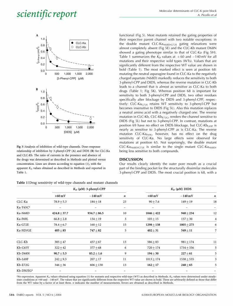
functional (Fig 5). Most mutants retained the gating properties oftheir respective parent channel with two notable exceptions: inthe double mutant CLC-KaN68D/G72E gating relaxations werealmost completely absent (Fig 5E) and the CLC-Kb mutant D68Nshowed a gating phenotype similar to that of CLC-Ka (Fig 5H).Table 1 summarizes the KD values at þ 60 and �140mV for allmutations and their respective wild types (WTs). Values that aresignificantly different from the respective WT value are shown inbold (Table 1). The most marked effect is seen at position 68:mutating the neutral asparagine found in CLC-Ka to the negativelycharged aspartate (N68D) markedly reduces the sensitivity to both3-phenyl-CPP and DIDS, whereas the reverse mutation in CLC-Kbleads to a channel that is almost as sensitive as CLC-Ka to bothdrugs (Table 1; Fig 5K). Whereas position 68 is important forsensitivity to both 3-phenyl-CPP and DIDS, two other residuesspecifically alter blockage by DIDS and 3-phenyl-CPP, respec-tively: CLC-KaG72E retains WT sensitivity to 3-phenyl-CPP butbecomes insensitive to DIDS (Fig 5L). Also this mutation replacesa neutral amino acid with a negatively charged one. The reversemutation in CLC-Kb, CLC-KbE72G, renders the channel sensitive toDIDS (Fig 5L) but not to 3-phenyl-CPP. In contrast, mutations atposition 69 have no effect on DIDS blockage, but CLC-KbL69F isnearly as sensitive to 3-phenyl-CPP as is CLC-Ka. The reversemutation CLC-KaF69L, however, has no effect on the drugsensitivity of CLC-Ka. No large effects were observed formutations at position 65. Not surprisingly, the double mutantCLC-KaN68D/G72E is similar to the single mutant CLC-KaN68D,being less sensitive to both compounds.
DISCUSSIONOur results clearly identify the outer pore mouth as a crucialpart of the binding pocket for the structurally dissimilar molecules3-phenyl-CPP and DIDS. The most crucial position is 68, with a
[DIDS] (μM)
0 500 1,000 1,500 2,0000.0
0.2
0.4
0.6
0.8
1.0
[3-Phenyl-CPP] (μM)
I [3-P
heny
l-C
PP
]/I0
CLC-Ka
CLC-Kb
0 500 1,000 1,500 2,0000.0
0.2
0.4
0.6
0.8
1.0I [D
IDS
]/I0
A
B
Fig 3 | Analysis of inhibition of wild-type channels. Dose–response
relationship of inhibition by 3-phenyl-CPP (A) and DIDS (B) for CLC-Ka
and CLC-Kb. The ratio of currents in the presence and absence of
the drugs was determined as described in Methods and plotted versus
concentration. Lines are drawn according to equation (1), with the
apparent KD values obtained as described in Methods and reported in
Table 1.
Table 1 |Drug sensitivity of wild-type channels and mutant channels
KD (lM) 3-phenyl-CPP KD (lM) DIDS
+60mV �140mV n +60mV �140mV n
CLC-Ka 78.975.3 184718 23 9077.6 149719 18
Ka-Y65Ca – – – – – –
Ka-N68D 424.8737.7 934.7786.5 10 10467422 9407234 12
Ka-F69L 66.872.8 134719 3 105715 157730 4
Ka-G72E 78.476.7 160712 15 12987138 10057275 6
Ka-ND/GE 405785 747782 5 402731 549711 7
CLC-Kb 385747 457767 13 586783 9817174 11
Kb-C65Y 322742 377768 6 7207174 17347336 5
Kb-D68N 90.775.5 85.271.6 9 194730 227761 5
Kb-L69F 24279.5 207717 11 10137174 15387535 3
Kb-E72G 346736 6067130 13 162717 248765 5
Kb-DN/EGa – – – – – –
aNo expression. Apparent KD values obtained using equation (1) for mutants and respective wild-type (WT) as described in Methods. KD values were determined under steady-state conditions at +60 and �140mV. The values that are significantly different from the respective WT value are shown in bold. These are arbitrarily defined as those that differfrom the WT value by a factor of at least three. n indicates the number of measurements. Errors are obtained as described in Methods.
Molecular determinants of CLC-K pore block
A. Picollo et al
EMBO reports VOL 5 | NO 6 | 2004 &2004 EUROPEAN MOLECULAR BIOLOGY ORGANIZATION
scientificreport
586

neutral asparagine in CLC-Ka and a negatively charged aspartatein CLC-Kb. A negatively charged residue is found in most CLCproteins including the bacterial homologues StCLC and eriC. Theside chain of the corresponding E54 in StCLC protrudes intothe putative permeation pathway and is close to the positivelycharged R147 of StCLC (Dutzler et al, 2002, 2003). Thecharge effect on drug affinity is probably due to electrostaticinteractions with the negatively charged blockers. N68 of CLC-Kais important for both 3-phenyl-CPP and DIDS blockage, and CLC-Kb becomes as sensitive as CLC-Ka to both drugs by changingthe aspartate to an asparagine. In contrast, a negativelycharged residue at position 72 of CLC-Ka or CLC-Kb appears toinfluence mainly DIDS binding. Position 72 is located moreextracellularly than position 68 in the rather wide extracellularvestibule. In this respect, it is important to note that 3-phenyl-CPPcarries one, whereas DIDS carries two, negative charges atphysiological pH values. Thus, we speculate that the carboxylategroup of 3-phenyl-CPP and one of the sulphonate groups of DIDSbind to a deep site, whereas the second sulphonate group of DIDSremains more superficial, where it is able to be influencedelectrostatically by a charge at position 72. Inhibition of CLC-K1
and CLC-Ka by DIDS is very slowly reversible. However, anirreversible covalent modification, as observed in the band 3anion exchanger (Bartel et al, 1989), is unlikely because thiswould predict steady-state inhibition that is almost independent ofthe applied concentration, in contrast to our measurements.
In addition to the effects on inhibitor binding, mutations atposition 68 have a profound effect on the gating of CLC-Kb: gatingof mutant D68 strongly resembles gating of CLC-Ka. Thus, asingle-point mutation converts the pharmacological as well as thebiophysical properties of CLC-Kb to those of CLC-Ka. The inversemutation of CLC-Ka, however, is not sufficient to change its gatingproperties markedly. Interestingly, the corresponding CLC-1mutant (D136G), which is responsible for recessive myotonia,and similar mutations in CLC-0 also markedly alter gating (Fahlkeet al, 1995; Ludewig et al, 1997). It is possible that the charge atposition 68 has a marked effect on the local Cl� concentration,which is known to be important for channel gating (Pusch et al,1995; Chen & Miller, 1996).
The different inhibitor sensitivities of CLC-Ka and CLC-Kbare interesting from a pharmacological viewpoint. In thisrespect, the pharmacokinetic properties of any drug have to beconsidered. For example, several commonly used loop diureticsare extensively bound to plasma proteins, reaching their target(the apical Na/K/2Cl co-transporter) by means of the organic-acidtransport system of the proximal tubule (Shankar & Brater,2003). Clofibrate (which is chemically similar to 3-phenyl-CPP)and other drugs used in the treatment of hyperlipoproteinaemiasare also probably largely secreted into the tubular lumen, asfinal excretion of these molecules occurs primarily through thekidneys (Wierzbicki et al, 2003). A similar pharmacokineticprofile is predicted for 3-phenyl-CPP. In addition, it must betaken into account that CLC-Ka is localized at both the apical andbasolateral levels of the renal epithelium; thus, a CLC-Ka-blockingdrug will reach its target by the basolateral fluid, as well asthe luminal fluid. CLC-Ka is expressed in the thin ascendinglimb of Henle’s loop such that its inhibition would lead towater diuresis, a situation that might be favourable in certainpathological conditions. Furthermore, application of a CLC-Ka-specific drug would prevent an osmotic diuresis becausethe function of CLC-Kb in the thick ascending limb ofHenle’s loop would be preserved. In addition, the importantfunction of CLC-K channels in the inner ear (Birkenhageret al, 2001; Estevez et al, 2001) would not be compromised ifCLC-Kb is not blocked. Thus, a diuretic drug that specificallyinhibits CLC-Ka but not CLC-Kb could have significant advantagescompared with a general CLC-K channel blocker.
METHODSMolecular biology and heterologous expression. Mutations wereintroduced by recombinant PCR as described previously (Accardi& Pusch, 2003). All constructs were in the PTLN vector (Lorenzet al, 1996). Human WT CLC-Ka and CLC-Kb, and their mutants,were coexpressed with mutant Y98A of barttin, leading to anenhancement of expression (Estevez et al, 2001).Electrophysiology. Oocyte expression and electrophysiologicalmeasurements were performed as described previously (Puschet al, 2000). Voltage-clamp data were acquired at 21–25 1Cusing the Pulse program (HEKA, Lambrecht, Germany) and acustom-built amplifier. Currents were recorded in a solution
Fig 4 | Selection of candidate residues involved in drug binding. (A) View
of the bacterial channel StCLC (Dutzler et al, 2002) from the extracellular
side. One subunit of the dimer is shown in wireframe (right-hand side),
whereas the other is shown in spacefill (left-hand side). Highlighted in dark
grey are all amino acids that possibly expose their side chains in the
extracellular pore entrance. Among these, we selected those residues that
were different in CLC-Ka and CLC-Kb and that were identical in CLC-K1
and CLC-Ka. Four residues fulfilled this criterion, as shown in (B). The
corresponding residues of StCLC are shown in colour in (A) (dark blue:
V51, corresponding to CLC-Ka-Y65; red: E54, CLC-Ka-N68; orange: K55,
CLC-Ka-F69; magenta: S58, CLC-Ka-G72). All four residues lie in helix B
(yellow). E148 (green: V166 in CLC-Ka and CLC-Kb) indicates the location
of the selectivity filter (Dutzler et al, 2002, 2003). (B) Alignment of the short
stretch of helix B. Mutated amino acids are shown in bold. Corresponding
residues in CLC-K1 are shown in bold and italic.
Molecular determinants of CLC-K pore block
A. Picollo et al
&2004 EUROPEAN MOLECULAR BIOLOGY ORGANIZATION EMBO reports VOL 5 | NO 6 | 2004
scientificreport
587

containing 90mM NaCl, 1mM MgCl2, 10mM CaCl2 and 10mMHEPES at pH 7.3. For the voltage-clamp measures, we used similarvoltage-clamp pulse protocols for CLC-Ka and CLC-Kb with a longerpulse duration for CLC-Kb. Pulses were elicited from a holdingpotential corresponding to the resting membrane potential (�30 to�50mV) using a prepulse to þ 60mV for 100 or 200ms (for CLC-Ka and CLC-Kb, respectively). This was followed by steps to varioustest values (from �140 to þ 80mV with 20mV increments) for 200or 500ms (for CLC-Ka and CLC-Kb, respectively) and a final tailpulse to �100mV. As a control, we routinely applied a solutioncontaining 100mM I� that blocks currents carried by CLC-Kchannels but not endogenous currents (Pusch et al, 2000).
Because CLC-Kb and several mutants had relatively lowexpression levels, we used the following strategy to estimate thecontribution of endogenous currents. The current amplitude atþ 60mV remaining in the presence of 100mM iodide was takenas being purely endogenous. The leak at �140mV was estimatedby linear extrapolation of this leak conductance. These estimatedleak currents were subtracted from the control currents in theabsence and presence of an inhibitor to calculate the ratio I(c)/I(0),where c is the concentration of the inhibitor. Apparent dissocia-tion constants, KD, were determined from the ratio of the steady-
state current in the presence and in the absence of the drug usingthe equation
IðcÞ=Ið0Þ ¼ 1=ð1þ c=KDÞ: ð1Þ
For the determination of KD, all data points, I(c)/I(0), for a givensubstance, channel type and voltage were simultaneously fitted byequation (1) without previous averaging of the data points. Thus,all data points count equally in the fit. From this fit (usingSigmaPlot, SPSS Inc.), an estimate of the confidence interval of theKD value was obtained (reported in Table 1). For display, the I(c)/I(0) values at a given concentration were averaged and plottedversus concentration (Fig 3).
DIDS was purchased from Sigma, and racemate 3-phenyl-CPPwas synthesized in-house (Liantonio et al, 2004).
ACKNOWLEDGEMENTSWe thank T. Jentsch and R. Estevez for providing the CLC-Ka, CLC-Kband barttin cDNA constructs; F. Loiodice, P. Tortorella andG. Fracchiolla for kindly providing 3-phenyl-CPP; E. Gaggero for theconstruction of the voltage-clamp amplifier and G. Gaggero for technicalsupport in the construction of the voltage-clamp set-up. This work wassupported by the Italian Ministero dell’Istruzione, dell’Universita e della
CLC-Ka Ka N68D Ka ND/GEKa G72EKa F69L
CLC-Kb Kb C65Y Kb E72GKb L69FKb D68N
A B C D E
F G H I J
[3-Phenyl-CPP] (μM)
0 500 1,000 1,500 2,0000.0
0.2
0.4
0.6
0.8
1.0 Ka-N68DKb-D68N
[DIDS] (μM)
0 500 1,000 1,500 2,000
I [DID
S]/I
0
0.0
0.2
0.4
0.6
0.8
1.0 Ka-G72EKb-E72G
K L
I [3-p
heny
l-C
PP
]/I0
Fig 5 | Phenotype of mutants. Typical voltage-clamp traces obtained with the pulse protocol described in Methods are shown for the indicated
constructs. Horizontal scale bars indicate 50ms (A–E) and 100ms (F–J). Vertical scale bars indicate 5 mA (A,C), 1 mA (B,I), 3mA (D,E), 2 mA (F),
4 mA (G), 0.5mA (H) and 0.3mA (J). Expression levels varied significantly between mutants. In particular, CLC-Kb and its mutants had lower
expression levels than those of CLC-Ka. The reason for this difference is unknown. (K,L) Dose–response relationships for 3-phenyl-CPP (K) and DIDS
(L) for the indicated constructs. Lines are drawn according to equation (1) with the apparent KD values reported in Table 1.
Molecular determinants of CLC-K pore block
A. Picollo et al
EMBO reports VOL 5 | NO 6 | 2004 &2004 EUROPEAN MOLECULAR BIOLOGY ORGANIZATION
scientificreport
588
Ricerca (MIUR COFIN 2003051709002 to D.C.C. and FIRBRBAU01PJMS to M.P.).
REFERENCESAccardi A, Pusch M (2003) Conformational changes in the pore of CLC-0.
J Gen Physiol 122: 277–293Adachi S, Uchida S, Ito H, Hata M, Hiroe M, Marumo F, Sasaki S (1994) Two
isoforms of a chloride channel predominantly expressed in thickascending limb of Henle’s loop and collecting ducts of rat kidney. J BiolChem 269: 17677–17683
Bartel D, Hans H, Passow H (1989) Identification by site-directedmutagenesis of Lys-558 as the covalent attachment site of H2DIDS in themouse erythroid band 3 protein. Biochim Biophys Acta 985: 355–358
Birkenhager R et al (2001) Mutation of BSND causes Bartter syndrome withsensorineural deafness and kidney failure. Nat Genet 29: 310–314
Chen TY, Miller C (1996) Nonequilibrium gating and voltage dependence ofthe ClC-0 Cl� channel. J Gen Physiol 108: 237–250
Dutzler R, Campbell EB, Cadene M, Chait BT, MacKinnon R (2002) X-raystructure of a ClC chloride channel at 3.0 A reveals the molecular basis ofanion selectivity. Nature 415: 287–294
Dutzler R, Campbell EB, MacKinnon R (2003) Gating the selectivity filter inClC chloride channels. Science 300: 108–112
Estevez R, Boettger T, Stein V, Birkenhager R, Otto E, Hildebrandt F, Jentsch TJ(2001) Barttin is a Cl� channel b-subunit crucial for renal Cl�
reabsorption and inner ear K+ secretion. Nature 414: 558–561Estevez R, Schroeder BC, Accardi A, Jentsch TJ, Pusch M (2003) Conservation
of chloride channel structure revealed by an inhibitor binding site inClC-1. Neuron 38: 47–59
Fahlke C, Rudel R, Mitrovic N, Zhou M, George Jr AL (1995) An aspartic acidresidue important for voltage-dependent gating of human musclechloride channels. Neuron 15: 463–472
Hille B (2001) Ion Channels of Excitable Membranes. Sunderland, MA: SinauerJentsch TJ, Stein V, Weinreich F, Zdebik AA (2002) Molecular structure and
physiological function of chloride channels. Physiol Rev 82: 503–568Kieferle S, Fong P, Bens M, Vandewalle A, Jentsch TJ (1994) Two highly
homologous members of the ClC chloride channel family in both rat andhuman kidney. Proc Natl Acad Sci USA 91: 6943–6947
Liantonio A et al (2002) Molecular requisites for drug binding to muscle CLC-1and renal CLC-K channel revealed by the use of phenoxy-alkylderivatives of 2-(p-chlorophenoxy)propionic acid. Mol Pharmacol 62:265–271
Liantonio A et al (2003) Structural requisites of 2-(p-chlorophenoxy)propionicacid analogues for activity on native rat skeletal muscle chlorideconductance and on heterologously expressed CLC-1. Br J Pharmacol139: 1255–1264
Liantonio A, Pusch M, Picollo A, Guida P, De Luca A, Pierno S, Fracchiolla G,Loiodice F, Tortorella P, Conte Camerino D (2004) Investigations ofpharmacologic properties of the renal CLC-K1 chloride channel co-expressed with barttin by the use of 2-(p-chlorophenoxy)propionic acidderivatives and other structurally unrelated chloride channels blockers.J Am Soc Nephrol 15: 13–20
Lorenz C, Pusch M, Jentsch TJ (1996) Heteromultimeric CLC chloridechannels with novel properties. Proc Natl Acad Sci USA 93:13362–13366
Ludewig U, Jentsch TJ, Pusch M (1997) Inward rectification in ClC-0 chloridechannels caused by mutations in several protein regions. J Gen Physiol110: 165–171
Matsumura Y et al (1999) Overt nephrogenic diabetes insipidus in micelacking the CLC-K1 chloride channel. Nat Genet 21: 95–98
Pusch M, Ludewig U, Rehfeldt A, Jentsch TJ (1995) Gating of the voltage-dependent chloride channel CIC-0 by the permeant anion. Nature 373:527–531
Pusch M, Liantonio A, Bertorello L, Accardi A, De Luca A, Pierno S, Tortorella V,Camerino DC (2000) Pharmacological characterization of chloridechannels belonging to the ClC family by the use of chiral clofibric acidderivatives. Mol Pharmacol 58: 498–507
Pusch M, Accardi A, Liantonio A, Ferrera L, De Luca A, Camerino DC, Conti F(2001) Mechanism of block of single protopores of the Torpedo chloridechannel ClC-0 by 2-(p-chlorophenoxy)butyric acid (CPB). J Gen Physiol118: 45–62
Pusch M, Accardi A, Liantonio A, Guida P, Traverso S, Camerino DC, Conti F(2002) Mechanisms of block of muscle type CLC chloride channels. MolMembr Biol 19: 285–292
Shankar SS, Brater DC (2003) Loop diuretics: from the Na–K–2Cl transporterto clinical use. Am J Physiol Renal Physiol 284: F11–F21
Simon DB et al (1997) Mutations in the chloride channel gene, CLCNKB,cause Bartter’s syndrome type III. Nat Genet 17: 171–178
Traverso S, Elia L, Pusch M (2003) Gating competence of constitutively openCLC-0 mutants revealed by the interaction with a small organic inhibitor.J Gen Physiol 122: 295–306
Uchida S, Sasaki S, Furukawa T, Hiraoka M, Imai T, Hirata Y, Marumo F(1993) Molecular cloning of a chloride channel that is regulated bydehydration and expressed predominantly in kidney medulla. J BiolChem 268: 3821–3824
Waldegger S, Jeck N, Barth P, Peters M, Vitzthum H, Wolf K, Kurtz A, KonradM,Seyberth HW (2002) Barttin increases surface expression andchanges current properties of ClC-K channels. Pflugers Arch 444:411–418
Wierzbicki AS, Mikhailidis DP, Wray R, Schacter M, Cramb R, Simpson WG,Byrne CB (2003) Statin–fibrate combination: therapy for hyperlipidemia:a review. Curr Med Res Opin 19: 155–168
Molecular determinants of CLC-K pore block
A. Picollo et al
&2004 EUROPEAN MOLECULAR BIOLOGY ORGANIZATION EMBO reports VOL 5 | NO 6 | 2004
scientificreport
589


![Genome-wide identification and expression analysis of the CLC … · 2020. 12. 11. · [23], etc. All of the CLC proteins have a highly con-served voltage-gated chloride channel (Voltage-gate](https://img.dokumen.tips/doc/110x75/6106dd3e9ccfce08576786e6/genome-wide-identification-and-expression-analysis-of-the-clc-2020-12-11-23.jpg)