Embed Size (px)
Citation preview

Approved IFCC Reference Method for the Measurement
of HbA1c in Human Blood
Clin Chem Lab Med 2002; 40(1):78–89 © 2002 by Walter de Gruyter · Berlin · New York
International Federation of Clinical Chemistry and
Laboratory Medicine (IFCC)1)2)
Scientific DivisionWorking Group on HbA1c Standardisation3)andNetwork of Reference Laboratories for HbA1c
4)
Prepared for publication5),6) by
Jan-Olof Jeppsson1,7), Uwe Kobold2, John Barr3, Andreas
Finke2, Wieland Hoelzel2, Tadao Hoshino4, Kor Miedema5,
Andrea Mosca6, Pierluigi Mauri7, Rita Paroni8, Linda
Thienpont9, Masao Umemoto10 and Cas Weykamp11
1 Department of Clinical Chemistry, Malmö UniversityHospital, Lund University, Malmö, Sweden2 Roche Diagnostics GmbH, Penzberg, Germany3 Center of Disease Control, Atlanta, USA4 Institute of Biopathological Medicine, Ono, Japan5 Department of Clinical Chemistry, Isala Klinieken, Zwolle,The Netherlands6 Department of Science and Biomedical Technology, University of Milan, Milan, Italy7 Institute of Biomedical Technology, Consiglio NazionaleDelle Ricerche, Milan, Italy8 IRCCS Hospital San Raffaele, Milan, Italy9 Laboratory of Analytical Chemistry, Faculty of Pharma-ceutical Sciences, University of Gent, Gent, Belgium
10 Standard Reference Center, Kawasaki, Japan.11 SKZL Queen Beatrix Hospital, Winterswijk, The Netherlands
HbA1c is the stable glucose adduct to the N-terminal
group of the �-chain of HbA0. The measurement of
HbA1c in human blood is most important for the long-
term control of the glycaemic state in diabetic pa-
tients. Because there was no internationally agreed
reference method the IFCC Working Group on HbA1c
Standardization developed a reference method which
is here described. In a first step haemoglobin is cleaved
into peptides by the enzyme endoproteinase Glu-C,
and in a second step the glycated and non-glycated N-
terminal hexapeptides of the �-chain obtained are sep-
arated and quantified by HPLC and electrospray ioni-
sation mass spectrometry or in a two-dimensional
approach using HPLC and capillary electrophoresis
with UV-detection. Both principles give identical re-
sults. HbA1c is measured as ratio between the glycated
and non-glycated hexapeptides. Calibrators consisting
of mixtures of highly purified HbA1c and HbA0 are used.
The analytical performance of the reference method
has been evaluated by an international network of ref-
erence laboratories comprising laboratories from Eu-
rope, Japan and the USA. The intercomparison studies
1) The excluxive for all languages and countries is vested inthe International Federation of Clinical Chemistry and Labo-ratory Medicine.2) IFCC Sections reprinted in J. Clin. Chem. Clin. Biochem. arelisted in the Cumulative Index, which appeared in connectionwith the contents of this journal Volume 27, 1989./Since 1998have been printed in (Eur.) J. Clin. Chem. Clin. Biochem./Since1998 have been printed in Clin. Chem. Lab. Med.IFCC 1991/1 Vol. 29, 435–457IFCC 1991/2 Vol. 29, 531–535IFCC 1991/3 Vol. 29, 577–586IFCC 1991/4 Vol. 29, 767–772IFCC 1992/1 Vol. 30, 901–905IFCC 1994/1 Vol. 32, 639–655IFCC 1995/1 Vol. 33, 247–253IFCC 1995/2 Vol. 33, 399–404IFCC 1995/3 Vol. 33, 623–625IFCC 1995/4 Vol. 33, 627–636IFCC 1995/5 Vol. 33, 637–660IFCC 1997/1 Vol. 35, 317–344IFCC 1997/2 Vol. 35, 345–349IFCC 1997/3 Vol. 35, 805–831IFCC 1997/4 Vol. 35, 833–843IFCC 1998/1 Vol. 36, 57–65IFCC 1998/2 Vol. 36, 185–203IFCC 1998/3 Vol. 36, 887–893IFCC 2000/1 Vol. 38, 363–370IFCC 2000/2 Vol. 38, 1301–1314
IFCC 2001/1 Vol. 39, 175–179IFCC 2001/3 Vol. 39, 283–289IFCC 2001/2 Vol. 39, 875–8893) Members are: K. Miedema, Zwolle (Netherlands, chair-man), DE. Goldstein, Columbia (USA), W. Hoelzel, Penzberg(Germany), T. Hoshino, Ono (Japan), J.O. Jeppsson, Malmo(Sweden), RR. Little, Columbia (USA), A. Mosca, Milano(Italy), G. Myers, Atlanta (USA), C. Weykamp, Winterswijk(Netherlands), G. John, Norwich (UK).4) Members are: J. Barr, Atlanta (USA), W. Hoelzel, Penzberg(Germany), T. Hoshino, Ono (Japan), J.O. Jeppsson, Malmo(Sweden), U. Kobold, Penzberg (Germany), P. Mauri, Milano(Italy), K. Miedema, Zwolle (Netherlands), A. Mosca, Milano(Italy), R. Paroni, Milano (Italy), L. Thienpont, Gent (Belgium),M. Umemoto, Kawasaki (Japan), C. Weykamp, Winterswijk(Netherlands).5) Received for publication 2001–11–206) The development of the reference method and the estab-lishment of a European Network of reference laboratorieswas supported by the R&D project CT 98-2248 of the S, M&TProgram of the European Commission. The followingauthors are partners of the project: JOJ, UK, AF, WH, KM,PM, RP, LT, CW.7) Reprint requests and inquiries should be addressed to: Jan-Olof Jeppsson M.D., Ph.D., Department of Clinical Chemistry,Malmö University Hospital, Lund University, 205 02 Malmö,Sweden, E-mail: [email protected]
IFCC 2002/1

Jeppsson et al.: IFCC Reference method HbA1c 79
of the network showed excellent results with intra-
laboratory CVs of 0.5 to 2% and inter-laboratory CVs
of 1.4 to 2.3%. Possible interferences have been care-
fully investigated. Due to the higher specificity of the
reference method the results are lower than those
generated with most of the present commercial meth-
ods which currently are calibrated with unspecific
designated comparison methods. The new reference
method has been approved by the member societies
of the International Federation of Clinical Chemistry
and Laboratory Medicine and will be the basis for the
future uniform standardization of HbA1c routine assays
worldwide. Clin Chem Lab Med 2002; 40(1):78–89
Key words: HbA1c; Glycohaemoglobin; Diabetes; Inter-national Standardization; Reference method; Electro-spray ionisation mass spectrometry; Capillary elec-trophoresis.
Abbreviations: amu, atomic mass unit; HPLC-CE,HPLC/capillary electrophoresis; HPLC-ESI/MS, HPLC/electrospray mass spectrometry; KCN, potassiumcyanide; MES, β-Morpholino-ethanesulfonic acid; SOP,standard operation procedure; TFA, trifluoroacetic acid.
Contents
1. Introduction
2. Principle
3. Instrumentation and Equipment
3.1 HPLC-ESI/MS approach3.2 HPLC-CE approach3.3 Data processing4. Reagents
5. Preparation of Solutions
5.1 Buffer solutions5.2 Calibrator solutions6. Specimen procurement
6.1 Sample preparation6.2 Enzymatic cleavage7. Measurement
7.1 The measuring sequence for samples and cali-brators
7.2 HPLC-ESI/MS7.3 HPLC-CE8. Calibration and Calculation
9. Analytical Variability
10. The Uncertainty of Measurements
11. Interference
11.1 Carbamylated and acetylated haemoglobin11.2 Haemoglobin variants11.3 Interference of reagents12. Reference Ranges
Appendix A: Description of Pertinent Factors for Opti-
mal Conditions for the Measurement
1. Introduction2. Enzymatic cleavage of haemoglobin3. HPLC separation and on-line ESI-MS detectionAppendix B: Reagent and Equipment Specifications
1. Introduction2. Sample preparation
3. Equipment for the HPLC-ESI/MS4. Equipment for the HPLC-CE13. References
1. Introduction
The measurement of HbA1c/glycohaemoglobin in bloodhas become the gold standard for the long-term controlof the glycaemic state of diabetic patients as presentedin the DCCT and UKPDS studies (1, 2). The optimal ther-apy of diabetic patients requires carefully validated,method-independent therapeutic target values for theglycohaemoglobin levels of diabetic patients in order toreduce the long-term risk of the late complications suchas retinopathy, nephropathy and neuropathy as well asthe short-term risk of life-threatening hypoglycaemia.More than 15 different analytical methods are currentlyused by the clinical laboratories, which are based on ionexchange or affinity chromatography, electrophoresisand immunological principles (3). There is no interna-tionally agreed reference method or reference materialto which routine assays could be traced back (4). How-ever, there are a several national initiatives for harmon-isation and certification such as the National Glyco-haemoglobin Standardisation Program (NGSP) in theUSA based on the DCCT HbA1c work (5–7) and stan-dardisation schemes in Japan (8) and Sweden (9) whichare based on different designated comparison meth-ods. As a consequence of the lack of international stan-dardisation, values vary considerably between meth-ods, as many surveys have shown (10–12).
HPLC methods often used as “reference method” forthe standardisation of routine tests provide good preci-sion and long-term stability but they are rather unspe-cific. Different values for HbA1c can be obtained whenthe same blood samples are measured, depending onthe chromatographic system, e.g. the kind of resin, lot-to-lot variation of resins, column size, buffer composi-tion and elution times. The peak considered to be HbA1c
may contain variable proportion of substances whichhave the same elution behaviour as HbA1c but are notHbA1c according to definition, since they lack immuno-logical activity or do not bind during affinity chromato-graphy. The differences between some methods can berather large. The comparison between the Bio-Rex 70HPLC method used as designated comparison methodin the DCCT study and in the NGSP programme in theUSA, and the Mono S HPLC method recommended forstandardisation in Sweden, shows about 20% differ-ences in values at the cut-off level in spite of the fact thatboth HPLC methods claim to measure HbA1c . It is obvi-ous that none of the methods meet the requirements ofa reference method in a metrological sense.
There are a few glycohaemoglobin species present inhuman blood due to several potential glycation sites ofthe haemoglobin molecule (N-terminal ends of the β-and α- chains and lysine residues) (13–16). HbA1c is themajor form of all glycohaemoglobin species in humanblood and is defined as the stable adduct of glucose andthe N-terminal amino group of the β-chain of haemoglo-

80 Jeppsson et al.: IFCC Reference method HbA1c
lowed by capillary electrophoresis with UV detection.The percentage of HbA1c is determined as a ratio of theglycated to non-glycated β-N-terminal hexapeptides ofhaemoglobin. There are three steps:
2.1 Isolation of erythrocytes and haemolysis
Human erythrocytes are separated, washed and haemol-ysed in water and kept in a storage buffer at pH 6.2.
2.2 Enzymatic cleavage of the haemoglobin
The haemoglobin solution is treated with endoproteinaseGlu-C in order to hydrolyse the protein into several pep-tides, among them the glycated (HbA1c) and the non-gly-cated (HbA0) N-terminal hexapeptides of the β-chains.
2.3 Analysis and calibration
The peptide mixture is analysed to measure the ratio ofglycated to non-glycated hexapeptide. These analysesare done either by HPLC-ESI/MS (approach A) or HPLC-CE (approach B). In both cases the areas containing theN-terminal hexapeptides of haemoglobin are mea-sured and ratios of the glycated and non-glycated pep-tides are calculated. Calibrators consisting of the mix-ture of pure HbA1c and HbA0 are used for calibration.
3. Instrumentation and Equipment
3.1 HPLC-ESI/MS approach
An analytical HPLC system suitable for microborecolumns is necessary. A slow gradient of water-trifluo-roacetic acid (TFA)/acetonitrile-TFA is used on the equip-ment with a switching valve between the HPLC-columnand the mass spectrometric detector, together with atemperature-controlled autosampler and autoinjector.
The HPLC column consists of cyanopropyl stationaryphase. The electrospray ionisation mass spectrometeris directly coupled to the microbore HPLC system(HPLC-ESI/MS).
3.2 HPLC-CE approach
An analytical HPLC system with a C18 column is used inthe first dimension along with a temperature controlledfraction collector. An analytical capillary electrophore-sis system with 214 nm photometric detector (or DAD)and a temperature controlled fused-silica capillary isused for the final separation in the second dimension.
3.3 Data processing
For both approaches chromatography data systemsare necessary which are capable of precise automaticor manual integration
4. Reagents
Acetic acidAcetonitrileAmmonium acetate, CH3COONH4
bin A0 [N-(1-deoxyfructosyl)haemoglobin]. Most of thecommercial tests already claim to measure this form ofglycohaemoglobin and some assays, which measure thesum of glycohaemoglobin species in blood, are alreadyinternally standardised against a HbA1c designated com-parison method. This is possible since the total glyco-haemoglobin and HbA1c values correlate very well (17).
To overcome the problem of poor standardisation theIFCC has created a working group to establish a uniform,scientifically well founded international standardisation.The group decided to develop a reference system towhich routine methods can be traced (3, 18–20). A majorcomponent of the reference system is the referencemethod, which specifically measures HbA1c and is neces-sary for assigning values to reference materials. Due tothe fact that all currently used designated comparisonmethods are rather unspecific, a new reference methodwhich specifically measures glycated N-terminal residueof the β-chain of haemoglobin had to be developed.Searching for an appropriate analytical technique, twoanalytical approaches have been proven to be ade-quately suited – namely HPLC /electrospray mass spec-trometry (HPLC-ESI/MS) and HPLC/capillary elec-trophoresis (HPLC-CE). Both principles work very welland generate identical results. Since flexibility with re-gard to equipment in the reference laboratories is an ad-vantage both principles have been developed (Figure 1).
2. Principle (Figure 2)
In the first step haemoglobin is cleaved into peptidesby the proteolytic enzyme endoproteinase Glu-C.Thereafter the resulting glycated and non-glycated N-terminal hexapeptides of the β-chains are separatedfrom the crude peptide mixture and quantified by HPLCand electrospray mass spectrometry or by HPLC fol-
Figure 2 Principle of the proteolytic digestion of haemoglo-bin chains.
Figure 1 Flow chart of the reference method procedure.

Jeppsson et al.: IFCC Reference method HbA1c 81
β-Morpholino-ethanesulfonic acid, MESEndoproteinase Glu-C: sequencing gradeEthylenediamine-tetraacetic acid disodium salt, Na2-EDTAHbA1c: human haemoglobin with glycated N-terminalβ-chain, purity ≥ 95% assigned by standard addition ap-proach.HbA0: human haemoglobin with non-glycated N-termi-nal β-chain, purity ≥ 99%Water, HPLC gradeOrtho-phosphoric acidPotassium cyanide, KCNSodium chloride, NaClSodium hydroxide, NaOHTrifluoroacetic acid, TFA
The purity of chemicals having no specified purity isof analytical grade.
5. Preparation of Solutions
5.1 Buffer solutions
Incubation solution: saline, NaCl 0.15 mol /lDigestion buffer: NH4-acetate, 50 mmol/l, pH 4.3Storage buffer: MES, 50 mmol/l, KCN, 10 mmol/l,Na2EDTA, 1 mmol/l, pH 6.2The enzyme endoproteinase Glu-C is freshly dissolvedin HPLC grade water, 200 µg/ml, and stored at 4 °C. Thesolution should be used within 8 hours.
5.2 Calibrator solutions
Calibrator solutions are prepared by mixing pure HbA0
and HbA1c solutions (19). A set of calibrators with sixlevels of HbA1c (0, 3, 6, 9, 12 and 15%) is prepared.
HbA0 solution in storage buffers approximately 120mg/ml and HbA1c solution approximately 20 mg/ml inthe same buffer is used. The exact haemoglobin contentof both solutions is determined by the ICSH referencemethod for haemoglobin. For the HbA1c solution thecontent of residual HbA0 is determined as specified inappendix B. Both solutions are mixed by weighing thevolumes from calibrated pipettes. For the calculation ofvolumes to be mixed, the haemoglobin concentrationsof both solutions and the amount of HbA0 contributionby the HbA1c sample are considered. The pipetting vol-umes are adjusted, to get a haemoglobin content of 1mg for each vial of calibrator within a total volume of 30µl, by filling up with storage buffer. In order to reducepipetting errors, for each calibrator level a bulk solutionof several hundred millilitres is prepared, which is splitinto 30 µl aliquots. The calibrator solutions are stable forat least 8 hours at room temperature and can be storedat –20 °C or at –80 °C for at least 1 year.
6. Specimen Procurement
6.1 Sample preparation
6.1.1 Patient samples
Fresh blood is collected in the presence of EDTA. Thecells are washed and incubated with saline solution for
4 hours at 37 °C to remove the pre-HbA1c (Schiff base).Haemolysates are prepared by mixing cells with waterand the storage solution. The clear haemolysates arestored at −20 °C and are stable for at least 1 year.
6.1.2 Calibrators
One vial of each concentration (1 mg total Hb in 30µlstorage buffer) is used.Enzyme solution and digestion buffer are added as forpatient samples.
6.1.3 Controls
Previous haemolysates of patient pools stored at – 70°C, with HbA1c values established by the Network, wereused as controls in each experiment.
6.2 Enzymatic cleavage
For the measurement of patient samples and calibra-tors an aliquot containing approximately 1 mg of totalHb is taken, 50 µl enzyme solution is added and themixture is made up with digestion buffer to a total vol-ume of 500 µl.
The vials are carefully closed with crimp caps and in-cubated under gentle shaking or rotating at 37 °C (± 2°C) for 18 hours. The digestion is stopped by freezingthe material at −20 °C. The digests of calibrators andsamples are stable and may be stored for at least 3months at –70 °C until analysis. After thawing the di-gests can be stored up to three days at 4 °C – 8° C.
7. Measurement
7.1 The measuring sequence for samples andcalibrators
Measurements are done in following sequence: calibra-tor set- controls- sample 1 to sample n –controls- cali-brator set (n= up to maximum 20–30 samples). Calibra-tors and samples are taken from the same digest andthe same lot of enzyme. For the HPLC-CE system two tothree injections per vial are suitable; for the HPLC-ESI/MS system two to four injections per vial may beneeded, depending on the stability of the ion source
7.2 HPLC–ESI/MS
An analytical reversed phase HPLC column withcyanopropyl stationary phase, 2.1 mm in diameter, di-rectly connected to an electrospray ionisation massspectrometer is used. Flow rate is set to 300 µl/min andcolumn temperature to 50 °C. Injection volume is 10 –25 µl of the digest. A gradient elution is performed witheluent A (0.025% TFA acid in water) and eluent B(0.023% TFA in acetonitrile). A column-switching valveis positioned after the HPLC column and, to avoid con-tamination of the electrospray ion source, only the frac-tion between 0 min and 12 min elution time is allowedto enter the detection system.
The mass spectrometer is tuned and calibrated ac-cording to the instructions of the manufacturer; resolu-

82 Jeppsson et al.: IFCC Reference method HbA1c
tion is set to 0.7 amu peak half width. The acquisitionmode is set at centroid and multiple ion detection atm/z 348.2 and 429.2 for the double protonated ions ofnon-glycated and glycated N-terminal hexapeptides ofthe haemoglobin β-chain.
7.3 HPLC-CE
7.3.1 HPLC-UV
An analytical HPLC system with a reversed phase C18
column is used together with a fraction collector able tocollect 0.5 min fractions at 4 °C – 8 °C. The flow rate is0.8 ml/min at a temperature of 20–25 °C; 200 µl of thedigest solution is injected after centrifugation at 14000g for 2 min. The gradient consists of a mobile phase A:0.1% TFA in water and phase B: 0.1% TFA in acetoni-trile. The major peak around 15–18 minutes, depend-ing on chromatography system, is collected and freeze-dried. The freeze-dried material from the C18 fraction isdissolved in 50 µl 0.01% TFA/water immediately beforeCE analysis.
7.3.2 Capillary electrophoresis – UV
A capillary electrophoresis system containing an effi-cient cooling system and a fused capillary 77 cm × 75µm ID is used. The absorbance is monitored at 214 nm,the temperature is 25 °C and the separation is per-formed in a H3PO4/NaH2PO4 buffer 0.1 mmol/l, pH 2.50at 18 kV during 43 min.
8. Calibration and Calculation
The peaks for the hexapeptides β 1–6 and glc-β 1–6 arecarefully integrated. The calculation algorithms areidentical for HPLC-CE and HPLC-ESI/MS. From the peakarea raw data the ratio glc-β 1–6 to β 1–6 is calculated.The ratio data of repetitive injections (two to four) areaveraged.
The following notation is used:
x:= [HbA1c] = concentration of HbA1c in the sampley:= [HbA0] = concentration of HbA0 in the sampleHbtot := total concentration [HbA1c] + [HbA0]zconc := percent HbA1c in the sample
100 · x 100 · xzconc = ––––––– = –––––––, Hbtot = x + y.
x + y Hbtot
Sx := signal obtained for [HbA1c] = area (glc ß1–6)Sy := signal obtained for [HbA0] = area (ß1–6)a := response coefficient of HbA1c of the measure-ment devicesb := response coefficient of HbA0 of the measure-ment devicesrsig := ratio of signals
Sx area(glcβ1 – 6)rsig = –––– = ––––––––––––––––
Sy area(β1 – 6)
Sx = α ⋅ x, Sy = β ⋅ y
For calibration of the method we simply plot the ratioof the signals rsig = Sx/Sy versus the ratio of the concen-
trations rconc = x/y of the calibrators prepared by mixingpure HbA1c and HbA0.
αrsig = –– ⋅ rconcβ
Since the calibration function is always linear, irrespec-tive of the response coefficients, a linear regression rsig
on rconc is performed. From this calibration functionrconc data for unknown patient samples are obtained. Toobtain the wanted quantity zconc (% HbA1c) an additionalcalculation is needed.
x 100 · rsig 100 · rconczconc = ––––– = ––––––––– = ––––––––––Hbtot α 1 + rconc–– + rsigβ
9. Analytical Variability
The transferability, robustness and the analytical per-formance of the reference method have been evalu-ated several times by a network of reference laborato-ries in Europe, the USA and Japan (publication inpreparation). All reference laboratories were able toimplement the candidate reference method accordingto the standard operation procedure (SOP) based on anearlier publication (18). Through the collaboration ofthe reference laboratories the SOP has been optimisedand finally fixed. Excellent calibration curves wereachieved for both approaches. In order to check the in-tra-laboratory and the inter-laboratory analytical per-formance intercomparison studies were organised.The results of the 4th Comparison Study, in which 11reference laboratories participated, are listed in Table1. The intra-laboraotry CV varied from 0.47 to 2.07%and the inter-laboratory CV from 1.35 to 2.27%. The cri-teria for acceptance were intra-laboratory CV<3% andthe deviation from the mean <2%.
Table 1 Mean value, inter-laboratory CV and mean intra-lab-oratory CV in the 4th Comparison Study of the Network ofHbA1c Reference Laboratories (1999). Participants: 11 refer-ence laboratories from Europe, USA and Japan.
Sample Mean value Inter-laboratory Mean intra-(% HbA1c) CV (%) laboratory CV
(%)
1 3.94 1.71 1.232 4.62 2.27 0.653 5.26 1.46 2.074 6.41 1.87 0.585 7.24 1.70 1.546 7.83 1.45 0.667 8.57 2.01 0.478 9.08 1.35 1.349 9.96 1.80 0.90
10 11.08 2.00 0.82
Mean CV 1.76 1.05

Jeppsson et al.: IFCC Reference method HbA1c 83
10. The Uncertainty of Measurements
The uncertainty of target values assigned with the ref-erence methods to secondary reference materials, cal-ibrators or trueness control materials depends on theuncertainty of the target value of the primary calibra-tor used for the calibration of the reference methods,and the uncertainty which results from the value as-signment procedure. The uncertainty of the primarycalibrator is the combined standard uncertainty (ucal)of the uncertainties which are related to the purity ofthe HbA0 and HbA1c preparations, the uncertainties ofthe target values for the haemoglobin concentration ofthese two materials, and the uncertainty resultingfrom mixing these materials in order to get calibratorswith a defined concentration of HbA1c. The combinedstandard uncertainty ucal for the target values of thecalibrators calculated according to the GUM (21) is0.63%. In the comparison studies of the Network ofHbA1c Reference Laboratories the average inter-labo-ratory CVs were about 2.0% (each sample was mea-sured in duplicate on two occasions by each referencelaboratory). If four reference laboratories participate ina value assignment exercise the uncertainty of thevalue assignment procedure uVA is 1.0% ( for 10 labo-ratories uVA = 0.63%), and a combined standard uncer-tainty utotal (including the uncertainty of the primarycalibrator) of about 1.2% can be attained for the targetvalue assigned to a secondary reference material, cal-ibrator or control material.
11. Interference
We have investigated the possible interference of N-terminal haemoglobin adducts, genetic variants andsome of the added chemicals.
11.1 Carbamylated and acetylated haemoglobin
Carbamylation and acetylation can also modify the N-terminal valine that is the major glycation site. Clinically,carbamylation occurs in normal individuals but to amuch higher degree in patients with reduced kidneyfunction and elevated serum urea levels. These com-pounds interfere with many ion exchange chromatogra-phy methods (14, 22). A normal sample was carbamy-lated in vitro by potassium cyanate (1 mmol/l, for 60 minat 37 °C). HbA1c increased from 5.2 to 5.9 measured byMono S ion exchange chromatography but the HPLC-CEprocedure gave identical results.
Some patients on high dose of aspirin show smallamount of acetylated haemoglobin. Normal HbA1c sam-ples were therefore incubated with acetylsalicylic acid, 5mmol/l, for 30 min at 37 °C. The HbA1c increased from 5.2to 5.5%. The HPLC-CE results were the same before andafter acetylation. Thus no interference was shown.
11.2 Haemoglobin variants
More than 750 different haemoglobin variants exist.The most common world-wide are Hb S and Hb C with
the amino acid substitutions of glutamic acid to valineand glutamic acid to lysine, respectively at position 6from the N-terminal end. Hb S and C were purified bystandard chromatography on a DEAE Sepharose A50column and treated as normal haemoglobin in bothprocedures. No signal at the position of the N-terminalhexapeptide was obtained in the HPLC-MS and in theHPLC-CE method.
Corresponding procedure for purified HbA2 (2.0–2.8%of total Hb) gave a normal signal for glycated and non-glycated hexapeptide since the first part of N-terminalamino acid sequence is identical to Hb0. The experimentwith the Hb S and C also demonstrate the specificity ofthe proteolytic enzyme used in the reference method.
11.3 Interference of reagents
Possible interference from potassium cyanide andsodium azide was evaluated by running sets with andwithout these chemicals. Potassium cyanide is origi-nally added to avoid methaemoglobin formation dur-ing the purification and sodium azide as a preservativeduring storage of calibrators (0.02%,w/v). There wasno interference of potassium cyanide but sodiumazide gave an unexpected interference. Sodium azidehas therefore been eliminated from the mixed calibra-tors.
12. Reference Ranges
A preliminary reference range study has been carried outby utilising EDTA-washed red cells collected from a Dan-ish population study (DiaRisk, Steno Diabetes Centre,Copenhagen, Denmark). one hundred and twenty non-diabetic subjects (aged 35–55 years) were selected ac-cording to the current WHO-ADA criteria (fasting plasmaglucose <7.0 mmol/l and 2 h glucose during a standardOGTT <11.1 mmol/l). The preliminary reference range forHbA1c as measured in this study using the referencemethods was 3.33 + 0.48% (mean + 2 SD). There was noinfluence of sex. As expected, the reference range for thenew reference method is lower and narrower than theranges for many routine methods, which vary consider-ably. The preliminary 97.5 percentile is 3.8% compared to5.0% or 6.2% for many routine methods.
Appendix A: Description of Pertinent Factors for
Optimal Conditions for the Measurement
1. Introduction
In order to obtain reliable results, the following factorshad to be optimised:• the cleavage of haemoglobin into peptides by a pro-
teolytic enzyme generating representative N-termi-nal peptides of the β-chain (glycated and non-gly-cated), so that the ratio of the glycated andnon-glycated peptides is representative for theHbA1c content of the haemoglobin sample and canreliably be quantified.
84 Jeppsson et al.: IFCC Reference method HbA1c
HPLC-ESI/MS approach• the optimal separation of the β-chain N-terminal
peptides by an appropriate HPLC system,• the detection and quantification of the β-chain N-ter-
minal peptides by electrospray mass spectrometry.
HPLC-CE approach• the optimal separation of the two β-chain N-terminal
peptides from other peptides and the quantitativeco-elution of this peptide fraction by an appropriateHPLC system (HPLC-CE method),
• the separation of the two β-chain N-terminal pep-tides in an appropriate capillary electrophoresis (CE)system and the quantitative detection of the pep-tides in the chosen system.
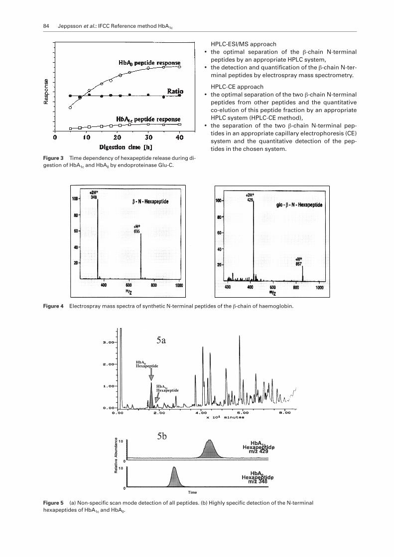
Figure 3 Time dependency of hexapeptide release during di-gestion of HbA1c and HbA0 by endoproteinase Glu-C.
Figure 4 Electrospray mass spectra of synthetic N-terminal peptides of the β-chain of haemoglobin.
Figure 5 (a) Non-specific scan mode detection of all peptides. (b) Highly specific detection of the N-terminalhexapeptides of HbA1c and HbA0.

Jeppsson et al.: IFCC Reference method HbA1c 85
2. Enzymatic cleavage of haemoglobin
The enzyme endoproteinase Glu-C cleaves the haemo-globin molecule so that the N-terminal hexapeptides C4H9O4-CO-CH2-NH-Val-His-Leu-Thr-Pro-Glu-COOHfrom HbA1c and NH2-Val-His-Leu-Thr-Pro-Glu-COOHfrom HbA0 are released from the two β-chains. We se-lected the Glu-C enzyme instead of the more commontrypsin, which cleaves at lysine and arginine residues.The first lysine residue is in position 8 and an octapep-tide could easily be obtained after trypsin digestion butthis lysine residue is a potential glycation site whichcould interfere in samples with a higher extent ofhaemoglobin glycation.
The digestion conditions were optimised to get ahigh reproducibility and equal digestion kinetics forboth HbA0 and HbA1c β- chains. To study the digestionkinetics a patient sample was digested over a period of4 to 40 hours. Aliquots were taken every 4 hours. Thereaction was stopped by freezing. The ratio of releasedglycated to non-glycated hexapeptides was constant
Figure 6 Calibration function for HPLC-ESI/MS. Figure 7 Calibration function for the HPLC-CE approach.
Figure 8 HPLC-C18 chromatogram of a mixture of peptides from proteolytic digestion of haemoglobinchains. Fractions 16–18 min are collected for capillary electrophoresis.
over a period of 40 hours (Figure 3) under the chosendigestion conditions. For the final methods we used 18hours digestion at 37 °C.
3. HPLC separation and on-line ESI-MS detection
For the HPLC-ESI/MS system the HPLC separation hasbeen optimised to get good resolution between gly-cated and non-glycated β-N-terminal hexapeptides anda good separation from all other peptide fragments.Mass spectra of synthetic glycated and non-glycated β-N-terminal hexapeptide standards are shown in Figure4. As it is typical for ESI-MS spectra of small peptides,single and doubly protonated ions are produced by theionisation process and no fragmentation is observed.For quantitative measurements the doubly protonatedions were chosen, because of their better response.Resolution of the mass analyser was set to 0.7 amupeak half width. The very high specificity of the massspectrometric detection is shown in Figure 5.
A chromatogram recorded in a scan mode (Figure
86 Jeppsson et al.: IFCC Reference method HbA1c
5a), which is similar to a photometric detection at 215nm, is compared to the multiple ion detection mode forthe doubly protonated ions at m/z 348.2 and 429.2 (Fig-ure 5b), which represent the hexapeptides releasedfrom HbA1c and HbA0. This comparison clearly showsthe superior specificity of the MS detection. The sensi-tivity of the analytical system at multiple ion detectionmodes is sufficient to get superior signal-to-noise ra-tios. By simple least-squares regression a linear cali-bration function was generated (Figure 6). Linearity has
been proven by measuring a set of six primary calibra-tors covering the range from 0 to 15% HbA1c.
4. HPLC separation and capillary electrophoresis
We have also employed a second independent two-di-mensional approach to quantify the ratio of glycated tonon-glycated β-N-terminal hexapeptides. This wasdone by reverse-phase HPLC combined with off linecapillary electrophoresis and photometric detection.The electrophoresis system has been optimised to ob-tain sufficient separation and to avoid interfering cont-amination. The cleaning between runs has been care-fully worked out to yield reproducible results overlonger periods.
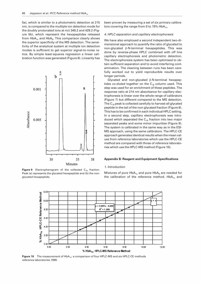
Glycated and non-glycated β-N-terminal hexapep-tides co-eluted together on the C18 column used. Thisstep was used for an enrichment of these peptides. Theresponse ratio at 214 nm absorbance for capillary elec-trophoresis is linear over the whole range of calibrators(Figure 7) but different compared to the MS detection.The C18 peak is collected carefully to harvest all glycatedpeptide in the tail of the non-glycated fraction (Figure 8).This has to be confirmed in each individual HPLC setting.In a second step, capillary electrophoresis was intro-duced which separated the C18 fraction into two majorseparated peaks and some minor impurities (Figure 9).The system is calibrated in the same way as in the ESI-MS approach, using the same calibrators. The HPLC-CEapproach generates identical results when the mean val-ues from reference laboratories which use the HPLC-CEmethod are compared with those of reference laborato-ries which use the HPLC-MS method (Figure 10).
Appendix B: Reagent and Equipment Specifications
1. Introduction
Mixtures of pure HbA1c and pure HbA0 are needed forthe calibration of the reference method. HbA1c and
Figure 9 Electropherogram of the collected C18 fraction.Peak (a) represents the glycated hexapeptide and (b) the non-glycated hexapeptide.
Figure 10 The measurement of HbA1c: a comparison of four HPLC-MS and six HPLC-CE methodsreference laboratories 1999.

Jeppsson et al.: IFCC Reference method HbA1c 87
mined by ion exchange chromatography (Mono S) of >99.5% HbA0 and > 98.5% HbA1c.
However, results from peptide mapping indicate thatthe HbA1c preparations still contain about 5% non-β-N-terminally glycated haemoglobin co-eluting with HbA1c
on Mono S. The exact amount of this fraction was deter-mined by a procedure consisting of standard addition,enzymatic cleavage and quantification of the resultingβ-N-terminal peptides (Figure 13). The total haemoglo-bin concentration in the HbA1c and HbA0 preparationswas determined by the ICSH haemoglobin referencemethod. Mixtures of both components could be suc-cessfully used to calibrate the reference methods.
2. Sample preparation
Fresh blood is collected in EDTA-containing tubes (Bec-ton Dickinson, Rutherford, NJ); 1.5 ml blood is cen-trifuged (10 min at 3000 g at +8 °C), the plasma is re-moved and the sedimented cells are washed twice witha 10 ml saline solution. The cells are incubated in 10 mlsaline solution at +37 °C for 4 hours in order to eliminatepre-HbA1c. The supernatant is discarded and he-molysate is prepared by mixing the cells with 1.0 ml wa-ter. The concentration of total haemoglobin is deter-mined. The haemolysates are diluted to 50 mg/ml totalhaemoglobin by mixing with equal amounts of storagesolution MES 50 mmol/l, EDTA 1 mmol/l, and potassiumcyanide 10 mmol/l. The pH is adjusted to 6.2 by addingsodium hydroxide, 4 mol/l. Cell debris is removed bycentrifugation for 20 min at 3000 g. Total haemoglobinconcentration is determined in an aliquot. Thehaemolysates are stable at –70 °C for at least 3 years.
3. Equipment for the HPLC-ESI/MS
A combined HPLC-ESI-MS system suitable for micro-bore columns is used. Essential is the possibility to runa slow gradient of water-TFA/acetonitrile-TFA on equip-ment with a switching valve between HPLC-columnand the detector. A temperature-controlled autosam-pler and autoinjector are mandatory. The electrospraymass spectrometric equipment coupled to the micro-bore HPLC system (HPLC-ESI/MS) must give stable andsensitive signals with good reproducibility, in order toallow quantitative data evaluation. The HPLC column isof cyanopropyl stationary phase.
An example of the appropriate equipment is as fol-lows: The HPLC system consisting of an HP 1090 liquidchromatograph (Hewlett-Packard, Waldbronn, Ger-many) with a DR 5 solvent delivery system, a thermo-stat-equipped autosampler and an autoinjector. A col-umn-switching valve Rheodyne No. 7010P (ERC,Regensburg, Germany), a T-piece 0.159 cm SwagelokNo. SS-100-3 (B.E.S.T., Munich, Germany), a relay boxfor control of pneumatic valves (Festo, Murnau, Ger-many). Programmable Absorbance Detector Spec-troflow 783, Kratos (GmbH Bioanalytische Instru-mente, Bensheim, Germany) with 2.4 µl cell volume.Analytical HPLC column ZORBAX SB-CN, 5 µ, 2.1 mm *150 mm, No.AS-RT-1245; P/N:883700.905 (Axel Sem-rau, Sprockhövel, Germany).
HbA0 were isolated, purified to homogeneity, and char-acterised (19). The techniques used for the characteri-sation were cation exchange and affinity chromatogra-phy for the purification as well as HPLC, capillaryisoelectric focusing, electrospray ionization mass spec-trometry (Figures 11 and 12) and peptide mapping.With these methods haemoglobin from healthy, non-diabetic volunteers was obtained with a purity deter-
Figure 11 ESI/MS of purified HbA0. 15125 Da non-glycatedα-chain, 15866 Da non-glycated β-chain.
Figure 12 ESI/MS of purified HbA1c. 15126 Da non-glycatedα-chain, 16028 Da mono-glycated β-chain.
Figure 13 Determination of the amount of HbA1c preparationby standard addition procedure.

88 Jeppsson et al.: IFCC Reference method HbA1c
Group. The effect of intensive treatment of diabetes on thedevelopment and progression of long term complicationsin insulin-dependent diabetes mellitus. N Engl J Med 1993;329:977–86.
2. Stratton IM, Adler AI, Neil HA, Matthews DR, Manley SE,Cull CA, et al. Association of glycaemia with macrovascularand microvascular complications of type 2 diabetes(UKPDS 35): prospective observational study. Br Med J2000; 321:405–11.
3. Hoelzel W, Miedema K. Development of a reference systemfor the international standardisation of HbA1c/glycohemo-globin determinations. JIFCC 1996; 9:62–7.
4. Bruns DE. Standardization, calibration, the care of diabeticpatients [editorial]. Clin Chem 1992; 38:2363–4.
5. Goldstein DE, Little RR, England JD, Wiedmeyer HM,McKenzie EM. Methods for quantitating glycosylated he-moglobins: high performance liquid chromatography andthiobarbituric acid colorometry: In Clarke WL, Larner, PohlSl, editors. Methods in diabetic research. Vol. 2. Clinicalmethods. New York: John Wiley, 1986:475–504.
6. Little RR, Goldstein DE. Endocrine standardization of glyco-hemoglobin measurement. Anal Chem 1995; 67:393R-7R.
7. Little RR. Recent progress in glycohemoglobin (HbA1c) test-ing. Diabetes Care 2000; 23:265–6.
8. Hoshino T, Okahashi M, Arai H. Survey and assessment ofthe actual state of routine measurement of glycohae-moglobin/GHb by commercial methods: warning to theusers and the providers. J Pharm Biomed Anal 1997;15:1551–62.
9. Jeppsson JO, Jerntorp P, Sundkvist G, Englund H, NylundV. Measurement of hemoglobin A1c by a new liquid chro-matographic assay: methodology, clinical utility and rela-tion to glucose tolerance evaluated. Clin Chem 1986;32:1867–72.
10. Little RR, Wiedmeyer HM, England JD,Naito HK, GoldsteinDE. Interlaboratory comparison of glycohemoglobin re-sults: College of American Pathologists Survay data. ClinChem 1991; 37:1725–9.
11. Weykamp CW, Penders TJ, Muskiet FA, van der Slik W.Glycohemoglobin: comparison of 12 analytical methods,applied to lyophilized haemolysates by 101 laboratories inan external quality assurance programme. Ann ClinBiochem 1993; 30:169–74.
12. Mosca A, Paleari R, Trapolino A, Capani F, Pagano G, Ple-bani M. A re-evaluation of glycohemoglobin standardiza-tion: the Italian experience with 119 laboratories and 12methods. Eur J Clin Chem Clin Biochem 1997; 35:243–8.
13. Garlick RL, Mazer JS, Higgins PJ, Bunn HF. Characteriza-tion of glycated hemoglobin: relevance to monitoring ofdiabetic control and analysis of other proteins. J Clin In-vest 1983; 71:1062–71.
14. Reinauer H. Biochemistry of protein glycation in diabetesmellitus. Klin Lab 1993; 39:984–7.
15. Peterson KP, Pavlovich JG, Goldstein D, Little R, England J,Peterson CM. What is hemoglobin A1c? An analysis of gly-cated hemoglobin by electrospay ionization mass spec-trometry. Clin Chem 1998; 44:1951–8.
16. Roberts NB, Amara AB, Morris M, Green BN. Long term eval-uation of electrospray ionization mass spectrometric analy-sis of glycated hemoglobin. Clin Chem 2001; 47:316–21.
17. Weykamp CW, Penders TJ, Miedema K, Muskiet FAJ, vander Slik W. Standardization of glycohemoglobin resultsand reference values in whole blood studied in 103 labora-tories using 20 methods. Clin Chem 1995; 41:82–6.
18. Kobold U, Jeppsson JO, Dülffer Th, Finke A, Hoelzel W,Miedema K. Candidate Reference Methods for HbA1c
based on Peptide Mapping. Clin Chem 1997; 43: 1944–51.
The mass spectrometric system is an SSQ700 single-stage quadrupole mass spectrometer with an electro-spray ion source (Finnigan MAT, Bremen, Germany).The HPLC system is connected on-line with the photo-metric detector and the electrospray ion source by 0.12mm ID steel capillaries. The electrospray ion source issupplied with 60 psi nitrogen sheath gas and nitrogenauxiliary gas at a HPLC flow rate of 300 µl/min. Sprayvoltage is 4.5 kV, transfer capillary temperature 200 °C.The mass spectrometer is tuned and calibrated withMRFA, myoglobin mixture; resolution is set to 0.7 amupeak half width and the electron multiplier set to 13 kV.Acquisition mode is set to centroid, multiple ion detec-tion at m/z 348.2 and 429.2 for the double protonatedions of non-glycated and glycated N-terminal hexapep-tides of the haemoglobin β-chain.
Gradient:Time (min) 0 3 9 13.5 13.6 17 17.1 23% B 0 0 5 5 100 100 0 stop run
Between-run time 14.5 to 18.0 min the ion source isswitched out of the HPLC flow. Expected retention timefor hexapeptides is 8–12 min.
4. Equipment for the HPLC-CE
Analytical HPLC system with a 214 nm photometric de-tector can be used together with a fraction collectorwith a collection device and a C18 Supelco TPR-100 col-umn (Cat no. 59154, Supelco, Bellafonte, PA, USA) to-gether with a guard column, Super guard TPR-100 (Catno. 5-9570). As an alternative a µRPC C2/C18 ST 4.6/100column (Cat no. 17-5057-01, Amersham PharmaciaBiotech, Uppsala, Sweden) can also be used. The deadvolume in the system between UV-detector and collec-tor must be taken into consideration. The third domi-nating peak with retention time at approximately 17min using the Pharmacia Biotech column (Figure 8) iscollected and freeze-dried and later dissolved in 50 µl0.01% TFA/water immediately before capillary elec-trophoresis.
A typical gradient is as follows:Time (min) 0 3 19 19.1 23.0 23.1 30% B 7 7 18.5 100 100 7 7
Analytical Capillary Electrophoresis System with 214nm photometric detector was used with fused-silicacapillary, 75 µm ID, 77 cm in length (Beckman-CoulterInc., Fullerton, USA, No: 33 84 72). The temperaturewas 25 °C and samples were injected during 10 s usinglow hydrodynamic flow. The separation was optimisedat 18 kV during 43 min. The instruments operate for 24hours. The rinsing of the capillary between runs is im-portant to obtain reproducible results over longer peri-ods. The following 1 min scheme per solvent is recom-mended: HCl, 100 mmol/l; water; NaOH, 100 mmol/l;H3PO4/H2NaPO4 buffer, 1.0 mol/l, pH 2.5.
13. References
1. The Diabetes Control and Complications Trial Research

Jeppsson et al.: IFCC Reference method HbA1c 89
19. Finke A., Kobold U, Hoelzel W, Weycamp C, Jeppsson JO,Miedema K. Preparation of a candidate primary referencematerial for the international standardisation of HbA1c de-terminations. Clin Chem Lab Med 1998; 36:299–308.
20. Jeppsson JO, Hoelzel W, Kobold U, Finke A, Mauri P,Miedema K, et al. International Network of reference labo-ratories for the determination of HbA1c [abstract]. ClinChem 1998; 44 (S6):A22.
21. Eurachem. Quantifying uncertainty in analytical measure-ment. 1st ed, 1995.
22. Bry L, Chen PC, Sacks DB. Effects of hemoglobin variantsand chemically modified derivatives on assays for glyco-hemoglobin. Clin Chem 2001; 47:153–63.