Embed Size (px)
Citation preview
3/17/21
1
Ocular Surface Oncology and Updates
Dr David SiaMB ChB FRANZCO
Vitreoretinal Surgeon & Ocular Oncologist
1
Overview
• Ocular surface anatomy • Classification of ocular surface tumours• Focus on 2 most common ocular surface malignancies• OSSN• Conjunctival melanoma
• Uveal melanoma updates
2
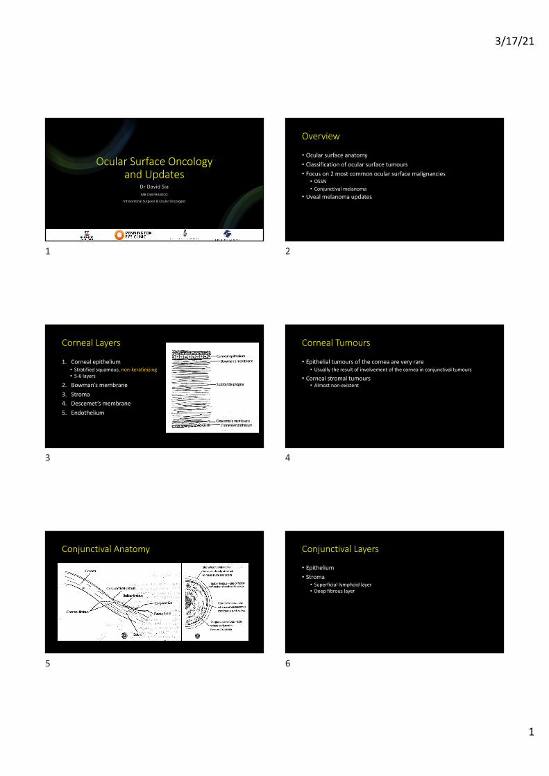
Corneal Layers
1. Corneal epithelium• Stratified squamous, non-keratinizing• 5-6 layers
2. Bowman’s membrane3. Stroma4. Descemet’s membrane5. Endothelium
3
Corneal Tumours
• Epithelial tumours of the cornea are very rare• Usually the result of involvement of the cornea in conjunctival tumours
• Corneal stromal tumours• Almost non-existent
4
Conjunctival Anatomy
5
Conjunctival Layers
• Epithelium• Stroma• Superficial lymphoid layer• Deep fibrous layer
6

3/17/21
2
Conjunctival Layers
• Epithelium• 2-5 layers• Stratified squamous, non-keratinizing epithelium
• Marginal and limbal zones• Stratified columnar epithelium
• Fornix• Cuboidal epithelium
• Bulbar and tarsal conjunctiva• Can secrete mucin
• Goblet cells• Specialised cells to secrete mucin• Present in the middle and superficial layers of epithelium• Most numerous in the lower fornix and close to plica
• Melanocytes• Scattered in the basal layer of the epithelium
7
Conjunctival Layers
• Stroma• Superficial lymphoid layer• Deep fibrous layer
8
Conjunctival Layers
• Stroma• Superficial lymphoid layer
• Thicker over the fornix • Thinner over palpebral conjunctiva and bulbar conjunctiva• Contains:
• Collagenous and elastic tissue• Mucosal associated lymphoid tissue (lymphocytes, plasma cells, mast cells, neutrophils)
• Deep fibrous layer
9
Conjunctival Layers
• Stroma• Superficial lymphoid layer
• Thicker over the fornix • Thinner over palpebral conjunctiva and bulbar conjunctiva• Contains:
• Collagenous and elastic tissue• Mucosal associated lymphoid tissue (lymphocytes, plasma cells, mast cells, neutrophils)
• Deep fibrous layer• Vessels (arteries, veins, lymphatics)• Nerves• Accessory lacrimal glands of Krause and Wolfring
10
Specialized Regions
• Plica Semilunaris• Vertical fold of conjunctiva lateral to caruncle• Contains many goblet cells• May contain nonstriated muscle fibers and fatty tissue
• Caruncle• Fleshy prominence located in the medial canthus• Contains both conjunctival and cutaneous structures• Non-keratinized stratified squamous epithelium• Numerous goblet cells, sebaceous glands, sweat glands, accessory lacrimal
glands, hair follicles• Tumours of the caruncle can be both mucosal and cutaneous origin
11
Lymphatic Drainage
• Lateral conjunctiva• Superficial parotid lymph nodes
• Medial conjunctiva• Submandibular lymph nodes
12

3/17/21
3
Classification of Conjunctival Tumours
Location Type of conjunctival tumours
Epidermal Non-melanocytic
Melanocytic
Stromal Vascular
Neural
Myxoid
Lipomatous
Melanocytic
Fibrous tissue
Histiocytic
Myogenic
Lymphoproliferative
13
Classification of Conjunctival and Corneal Tumours
Location Types Subtypes
Epiderm al Non-m elanocytic Benign Squam ous papillom a
Keratotic plaque
Keratoacanthom a
Inverted follicular keratosis
Oncocytom a
Dacryoadenom a
Prem alignant and m alignant Actinic (solar) keratosis
Conjunctival intraepithelial neoplasia (CIN)
Squam ous cell carcinom a
M ucoepiderm oid carcinom a
Sebaceous gland carcinom a (pagetoid spread)
Basal cell carcinom a
M elanocytic Benign Naevus (junctional, com pound, Spitz, Blue)
PAM without atypia
Congenital m elanosis
Com plexion associated m elanosis
Prem alignant and m alignant PAM with atypia
Conjunctival m elanom a
14
Location Types Subtypes
Strom a Vascular Capillary haem angiom a
Varix
Haem angiopericytom a
Kaposi’s sarcom a
Cavernous haem angiom a
Racem ose angiom a
Lym phangiectasia
Lym phangiom a
Pyogenic granulom a
Fibrous Nodular fasciitis
Benign fibrous histiocytom a
Fibrom a
M alignant fibrous histocytom a
Neural Neurofibrom a
Schwannom a (neurilem m om a)
Granular cell tum our
Histiocytic Xanthom a
Reticulohistiocytom a
Juvenile xanthogranulom a
Location Types Subtypes
Strom a M yxoid M yxom a
Lipom atous Lipom a
Liposarcom a
Herniated orbital fat
Lym phoproliferative Benign reactive lym phoid hyperplasia
Leukem ic infiltrates
Lym phom a
15Am J Ophthalmol 2017;173:106–133. © 2016 Elsevier Inc. All rights reserved
Tumour type %
Naevus 23
OSSN (SCC, CIN) 14
Melanoma 12
PAM 12
Lymphoma 7
16
Focus on• OSSN
• Conjunctival Melanoma
17
OSSN
• Ocular Surface Squamous Neoplasia (OSSN)• Spectrum of pre-malignant and malignant epithelial lesions of the
conjunctiva and cornea• Includes:• Conjunctival intraepithelial neoplasia (CIN)
• Mild• Moderate• Severe (carcinoma in-situ)
• Invasive squamous cell carcinoma (SCC)• Only used clinically where invasive nature on histology cannot be
determined
18

3/17/21
4
OSSN
• Incidence• 0.3 per million (US)• 1.3 per million (Uganda)• 19 per million (Australia)
• 5x higher in males and whites• Two main patterns of presentation:• Older white male population – developed countries (UVB as primary risk)• Younger female – developing countries (HIV and HPV more prevalent)
19
OSSN – Risk Factors• Most important:
• UV light exposure
• Other risk factors:• Pale skin, blue iris, propensity to sunburn• Significant sun exposure as a young child (>50% of time outdoors in first 6 years of life)• Proximity to equator (habitation within 30˚ of the equator)• Cigarette smoking• Human papilloma virus (HPV) infection (16 and 18 most common)• Immunosuppression
• HIV• Xeroderma pigmentosum• Atopic disease• Iatrogenic (organ transplant, autoimmune diseases treatment)
• Vitamin A deficiency• Exposure to petroleum products• Ocular surface injury
20
OSSN – Clinical Features
• Usually in interpalpebral fissure zone• Often begins at limbus• Appearance:• Papilliform• Nodular or sessile• Gelatinous• Leukoplakic (keratinization caused by dyskeratosis)• Foamy infiltration of adjacent corneal epithelium• Epithelial thickening (frosted glass appearance)• Prominent nutrient feeder vessels
21 22
23 24

3/17/21
5
25 26
27
Work-up of ocular surface tumours
• Establish risk factors• Full slit lamp examination of ocular surface• Bulbar, palpebral, forniceal conjunctiva – evert eyelids!!• Caruncle• Fluorescein staining• ± gonio, dilated fundus exam
• Photograph – phone photography• Palpate pre-auricular and submandibular lymph nodes
28
Is Biopsy Necessary?
• Past 15 years – progressive shift to using topical chemotherapy as primary therapy for OSSN• Rationale that SCC is locally invasive disease
• Does histopathological diagnosis alter management of patients?
29
Is Biopsy Necessary?• Treatment of pre-malignant disease is different to invasive disease
• Invasive SCC may require adjuvant radiotherapy
• Distinguishing premalignant from invasive disease based on clinical features alone by experienced clinicians can have an accuracy of only 40%.1,2
• There may be risk of misdiagnosis based on clinical features alone:• Misdiagnosis as OSSN as compared to other non-epithelial lesions can be as high as
10%.3
• Amelanotic or minimally pigmented conjunctival melanoma – up to 30% of cases in some series
• Certain types of ocular surface carcinomas are more aggressive (mucoepidermoid, spindle cell, Merkel cell, sebaceous ca)
1. Lee GA, Hirst LW. Ocular surface squamous neoplasia. Surv Ophthalmol. 1995;39(6):429–50. 2. Kao AA, Galor A, Karp CL, Abdelaziz A, Feuer WJ, Dubovy SR. Clinicopathologic correlation of ocular surface squamous neoplasms at Bascom Palmer Eye Institute: 2001 to 2010. Ophthalmology. 2012;119(9):1773–6. https://doi.org/10.1016/j. ophtha.2012.02.049. 3. Rudkin AK, Dodd T, Muecke JS. The differential diagnosis of localised amelanotic limbal lesions: a review of 162 consecutive excisions. Br J Ophthal- mol. 2011;95(3):350–4. https://doi.org/10.1136/ bjo.2009.172189.
30

3/17/21
6
Is Biopsy Necessary?
• Full-thickness biopsy is still required for differentiating premalignant from invasive disease, aggressive variants and masquerades• Differentiation of these types affects management and patient
outcome
31
Management of OSSN
• Full-thickness tissue biopsy• Excision biopsy if less than 5 clock hours limbal involvement• ‘No-touch’ technique• Wide margin (2-3mm)• Alcohol epitheliectomy if corneal involvement• Double-freeze thaw cryotherapy
• Closure• Direct closure• Amniotic membrane graft
32
Management of OSSN• Topical chemotherapy
Mitomycin C 5-Fluorouracil Interferon α2b
Dose 0.02% - 0.04% QID 1 weekThen 1-3 weeks off 3-4 cyclesWeek off (flarex and poly gel)
1% QID for 1 weekThen 3 weeks off4 cycles
1 million IU/ml QID 3 million IU/0.5ml SC twice/week3-6 months
Storage Keep refrigerated Keep room temperature Keep refrigerated
Side-effects Pain, limbal stem cell failure, punctal stenosis
Pain, epitheliopathy, hyperemia
Irritation, conjunctivitis, flu-like symptoms
Efficacy as primary treatment
76-100% resolution (primary)0-35% recurrence
82-100% resolution7-43% recurrence
82-100% resolution0-9% recurrence
33
Management of OSSN
• Recurrent or refractory OSSN• Cidofovir 0.25% 3x/day for 4-9 weeks• 83% resolution of refractory OSSN
• Novel treatments• EGFR inhibitors• Checkpoint inhibitors
34
Management of OSSNExcision biopsy,
alcohol epitheliectomy,
cryotherapy
CIN
CIN
Margins clear
No further treatment
CIN, Lateral margins positive
Topical chemotherapy, observation if cryo
and low grade
SCC
Margins clear
Topical chemotherapy
Deep margins positive
Plaque brachytherapy + topical
chemotherapy
35
Conjunctival Melanoma
36
3/17/21
7
Conjunctival Melanoma
• Annual incidence• 0.3 – 0.5 cases per million in Western populations• 300% increase over 3 decades
• Origin:• 70% arise from pre-existing PAM• 20% from naevi• 10% de novo
• No sex predilection• Presentation in 60s
37
Conjunctival Melanoma
• Risk factors:• Whites, rare in Asian/Pacific Islanders• Older age• Pre-existing lesion (most well-established risk factor)
• PAM and conjunctival naevi• UV radiation is suggested but not conclusively linked (unlike cutaneous
melanoma)• No significant association with cutaneous melanoma, dysplastic naevus
syndrome, or ocular/oculodermal melanocytosis
38
Conjunctival Melanoma
• Clinical Features:• Vascularized lesion• Nodular, diffuse or mixed• Can be deeply pigmented, grey or amelanotic (fish flesh)• Common in the limbus, also palpebral, fornix, caruncle• Feeder vessel• Does not have surface keratinization• Haemorrhagic areas• Usually only unilateral
39
Conjunctival Melanoma
• Key points for differentiating:• Conjunctival naevi
• Pigmentation at the palpebral conjunctiva and fornix = excision• Most conjunctival naevi noticed in childhood and adolescence
• Newly-elevated pigmented conjunctival lesion in adulthood = excise• Enlarging pigmented conjunctival lesion from childhood = suspect
• Conjunctival naevi commonly associated with cysts• PAM
• Placoid thickening within area of PAM• Nodules in area of PAM
40
Conjunctival Melanoma
• Estimated mortality • 13-38% at 10 years in adult studies• Overall mortality 25% (Danish study)
• Local recurrence• 19% at 5 years• 37% at 10 years• Factors associated with lower recurrence rate:
• Smaller extent of initial disease• No touch surgical technique• Adjunctive radiotherapy
Ja in P , F in g e r P T , F ilì M , e t a l. C o n ju n ctiva l m e lan o m a tre atm e n t o u tco m e s in 2 8 8 p atie n ts: a m u ltice n tre in te rn atio n a l d ata -sh arin g stu d y. B r J O p h th a lm o l. Se p te m b e r 2 0 2 0 . d o i:1 0 .1 1 3 6 /b jo p h th a lm o l-2 0 2 0 -3 1 6 2 9 3 .
41
Conjunctival Melanoma
• Staging examinations:• Full slit lamp examination• Lymph node examination (pre-auricular, submandibular, cervical)• US/CT head and neck lymph nodes• Refer medical oncologist
• Management:• Body of tumour
• Excision biopsy with 4mm margins, alcohol epitheliectomy, cryotherapy• No touch technique
• Roots of tumour• Adjunctive radiotherapy (plaque, strontium, proton beam)
• Seeds of tumour• Topical mitomycin C 0.04% for 3 cycles• IFN-a2b (limited data)
42

3/17/21
8
Conjunctival Melanoma
• Surveillance:• Q4/12 follow-ups lifelong• Q6/12 US head and neck LNs for first 5 years, then annually for life
43
Conjunctival Melanoma
• Topical chemotherapy• MMC 0.04%
• QID 1 week on 1 week off for 3 cycles is the most widely used• Interferon a2b
• 1 million IU/ml, 5x/day for 6 weeks x 6 cycles = also used with good efficacy• 5-FU
• Has been used but less effective and less well studied
44
Management of Conjunctival MelanomaWide excision biopsy,
alcohol epitheliectomy,
cryotherapy
Plaque brachytherapy (100gy to 1mm)
MMC 0.04% QID (3 cycles)
45
Choroidal Melanoma
46
Choroidal Melanoma
• Suspicious choroidal naevi (TFSOMDim and MOLES)• Light activated therapy (AU-011)• Prognostication• Metastatic uveal melanoma treatment (IMCgp100/Tebentafusp)
47
• TFSOM “To Find Small Ocular Melanoma”• Shields CL, MD JAS, MD HK, De Potter MD P, PhD JRC. Risk Factors for Growth
and Metastasis of Small Choroidal Melanocytic Lesions. Ophthalmology. 1995;102(9):1351-1361.
• TFSOMUHHD “To Find Small Ocular Melanoma Using Helpful Hints Daily”• Shields CL, Furuta M, Berman EL, et al. Choroidal nevus transformation into
melanoma: analysis of 2514 consecutive cases. Arch Ophthalmol. 2009;127(8):981-987.
0 risk factors = monitor yearly1-2 risk factors = monitor 4-6 monthly≥3 risk factors = refer ocular oncology for discussion of treatment
48

3/17/21
9
• TFSOMDim• To Find Small Ocular Melanoma Doing IMaging
• Thickness = >2mm on US• Fluid = SRF on OCT• Symptoms = Snellen acuity ≤6/15• Orange = lipofuscin on AF imaging• Melanoma hollowness = internal acoustic hollowness on US• Diameter = >5mm on photography
49
MOLES scoring criteria
50
MOLES scoring criteria
51
Uveal Melanoma – Targeted Treatments• AU-011 (Aura Biosciences)
• Synthetic recombinantly derived viral-like particles (VLP)• Binds selectively to uveal melanoma cells• Upon activation with 689nm diode laser, selectively destroys the cell membranes of
malignant cells• 2 year interim Phase 2 results – small to medium choroidal melanoma
• Tumour control rate 92%• Vision preserved in all patients up to 24 months
52
Uveal Melanoma - Prognostication
• Evolution• Tumour clinical and histopathologic features
• Tumour location, dimensions, histopathologic cell type, vascular mimicry, infiltrating lymphocytes etc.
• AJCC classification• Age, CB involvement, extraocular extension, tumour size
• Cytogenetic and gene expression profiling• Ch 3, Ch 6, Ch 8q, Ch 8p• GEP Class 1a, 1b, 2
• The Cancer Genome Atlas (TCGA) classification
53
Uveal Melanoma - Prognostication• The Cancer Genome Atlas (TCGA)• Initiated in 2005 to comprehensively explore genetic mutations found in
human cancer• Uveal melanoma cohort found 4 molecularly distinct and clinically relevant
subgroups based on alterations in chromosome 3 and 8
54

3/17/21
10
55
Uveal Melanoma – Targeted Treatments
• IMCgp100 or Tebentafusp• Bispecific antibody
• Part 1 = modified T-cell receptor that targets gp100 in melanoma cells• Part 2 = antibody fragment that targets CD3, protein found on surface of T-cells
• Mechanism:• Binds to gp100 on melanoma cells, the anti-CD3 portion redirects T-cells to kill
melanoma cells
• Phase 3 trial:• Investigator’s choice: dacarbazine, ipilimumab, pembrolizumab• In previously untreated metastatic uveal melanoma• 378 patients randomized to 2:1 to receive either tebentafusp or investigator’s choice• Results:
• 1 year OS rate 73% vs 58%
56
Thank you!
57