Embed Size (px)
Citation preview
Aquatic Mammals 1997, 23.1, 17–28
Ocular anatomy, retinal ganglion cell distribution, and visualresolution in the gray whale, Eschrichtius gibbosus
Alla M. Mass and Alexander Ya. Supin
Institute of Ecology and Evolution, Russian Academy of Sciences, 33 Leninsky Prosp., 117071 Moscow, Russia
Abstract
The eye optics, size and distribution of ganglioncells in the retina of the gray whale were studied.The hemispheric retina is centered on the quasi-spherical lens which makes it equally possible tocreate visual images at any part of the retina.Ganglion cell size varied from 14 to 74 �m, mostly20 to 40 �m, mean 31 �m. Ganglion cells concen-trated at two spots of the highest density in thenasal and temporal quadrants, 26–28 mm (65–70�)from the optic disk. Mean peak cell densities were130 and 183 cells/mm2 in the nasal and temporalareas respectively. With a posterior nodal distanceof 23 mm (under water) this corresponds to 21 and29 cells/deg2, which provides retinal resolution ofabout 13� in the latero-caudal visual field (nasalretinal area) and 11� in the rostral visual field(temporal retinal area).
Introduction
Investigation of sensory systems of cetaceans isimportant to understand mechanisms of theirbehavior and orientation. To date, studies ofcetacean sensory systems are especially importantbecause of threatened state of this animal group.Apart from that, data on organization of cetaceansensory systems are of interest for comparativeanatomy and physiology.
However, data on cetacean sensory systems areinsufficient. Particularly, data for the mysticetevisual system are limited and for many species arecompletely absent. Some data on whale eyeanatomy and optics were collected during the yearsof whaling. These data resulted in some ideasconcerning functional properties of the whale visualsystem. An opinion was adopted for many yearsthat whales have poor vision and plays a minor rolein their lives. To date, this opinion does not seem tobe true.
The idea of immobility of the cetacean’s eyeball isan example of a wrong conclusion based on earlyanatomical findings. Weber (1886) first supposed
that the oculomotor functions of cetaceans isreduced. Later Pütter (1903) carried out a com-parative investigation of the ocular anatomy ofmysticetes and also supposed an immobile ocularbulbus in cetaceans. Walls (1963) supported thisidea. He supposed that for whales, the most import-ant gaze direction is downward, and the eyeball iscanted ventrally or nasoventrally, which helps outin tilting the visual axis. However, the idea ofeyeball immobility in cetaceans was not confirmedby numerous later investigations of Jansen &Jansen (1969) and Hosokawa (1951). It was shownthat whales have a whole set of completely de-veloped oculomotor muscles and nerves. Thesemuscles provide the mobility of the cetacean eyeballcomparable to that in other mammals. Apart fromthat, direct behavioral observations indicatedclearly that eyes of dolphins and killer whales aremobile (Slijper, 1962; Madsen & Herman, 1980;Dawson, 1980).
Some other early conclusions concerning thevisual abilities of mysticetes were not confirmedeither. Walls (1963) hypothesized regression of thevisual function in mysticetes due to their ‘trawling’method of feeding. However, morphological andoptical adaptations of the cetacean visual systemto the underwater environment were found, indi-cating the importance of the visual system forthese animals. Mysticetes have the quasi-sphericallens with a high refractive index. The hemisphericeyecup, wide pupil and well developed tapetumwere interpreted as adaptations to conditionsof low illumination under water (Waller, 1980,1984).
Contrary to early hypotheses, experimentalstudies and field observations of cetaceans duringthe last decade resulted in a re-estimation of therole of their vision. Reviewing experimental andbehavioral observations, Madsen & Herman (1980)summarized that cetaceans use their vision fororientation and navigation, coordination of groupmovements, identification of conspecifics and indi-viduals, communication, etc. These observationsconcern both odontocetes and mysticetes.
� 1997 EAAM
Histological investigations of the mysticete retinaare very limited. Mann (1946) was the first toinvestigate the retina of the fin whale Balaenopteraphysalus. He described two types of receptors andsupposed that the mysticete retina is capable ofperceiving visual images. The existence of cones androds has been reported by Pilleri & Wandeler (1964)in the retina of the fin whale. Apart from the tworeceptor types, these authors have describedganglion cell characteristics in this species. Somefeatures of the ganglion cells were also describedin retinal wholemounts of the minke whaleBalaenoptera acutorostrata (Murayama et al.,1992).
As to the visual acuity of mysticetes, the datawere absent until recently because behavioralmeasurements in large whales are very difficult toobtain. The problem may be solved using a mor-phological method, i.e., estimation of the visualacuity basing on the ganglion cell density in theretina. Ganglion cell spacing may be used as anindicator of the retinal resolution and thus predictsthe visual acuity. Such estimations based on theganglion cell density have been carried out for someterrestrial mammals (rev. Pettigrew et al., 1988) andsome cetaceans (odontocetes): Delphinus delphis(Dral, 1983), Phocoena phocoena (Mass & Supin,1986), Inia geoffrensis (Mass & Supin, 1989),Tursiops truncatus (Mass & Supin, 1995), andPhocoenoides dalli (Murayama et al., 1995).
The purpose of the present study was to investi-gate the characteristic features and dimensions ofthe eye of the gray whale, Eschrichtius gibbosus; andto assess the ganglion cell distribution in retinalwholemounts to estimate visual acuity; and to studyganglion cell sizes. Preliminary data of this studywere published earlier (Mass & Supin, 1990). Apartfrom that preliminary publication, there was onlyone study of such type carried out in anothermysticete species, Balaenoptera acutorostrata(Murayama et al., 1992). This paper presentsmore extensive data obtained from the eyes of thegray whale.
Material and methods
The material, four eyes, was collected in 1989 fromtwo adult gray whales that died near the Lorinosettlement in Chukotka, Russia. Three eyes wereused to prepare retinal wholemounts and one eyewas used to measure optic dimensions.
The eyes were fixed in 10% formalin a few hoursafter death. The wholemounts were prepared bymethod of Stone (1965) with our modification(Mass, 1992). Before the retina was excised, itsorientation was noted. Then the cornea, iris, lens,and vitreous body of the eye were removed and theretina was excised from the eyecup. The retina was
flattened on a slide, with the ganglion cell layerupward, covered with filter paper, and kept under aweight for several hours in 10% formalin solution.Radial cuts allowed flattening the retina on theslide. After that, the retina was dried and stainedby the Pishinger method in 0.06% metilene bluesolution under visual control. Shrinkage of largethick wholemounts was avoided by clearing withoutdehydration in the Apathy’s gumsyrup.
The ganglion cells were counted in 0.15 mm2
square samples on a 1 mm step grid over the wholeretina. The results of counting were converted intonumber of cells per mm2. These data were used formapping the distribution of ganglion cell density inthe retina, as well as for calculating the totalnumber of ganglion cells in the retina. Smoothingof the maps was carried out by averaging thenumber of cells in blocks of 3�3 samples.
To estimate the retinal resolution, the originalwholemount maps were transformed to continuousspherical maps of ganglion cell density in thespheric coordinates. For this purpose, wholemountmaps were transformed by a computer program insuch a way as to remove radial cuts and restore ahemisphere approximating the entire retina. Inthese spherical maps, the cell density was specifiedin cells/deg2:
D=d(�R/180�)2
where: D is the density in cells/deg2, d is the densityin cells/mm2, and R is the retinal radius in mm.
In order to estimate the position of optic pointsand the posterior nodal distance, one eye wasfrozen and sectioned horizontally. Then half of theeye had been removed, photographs were taken,and measurements were made from these photo-graphs. Apart from that, external dimensions andeyecup dimensions were measured in the eyes usedto prepare wholemounts.
Cell size was obtained by measuring two perpen-dicular diameters, i.e., the long and short ones, andcalculating the mean value.
Results
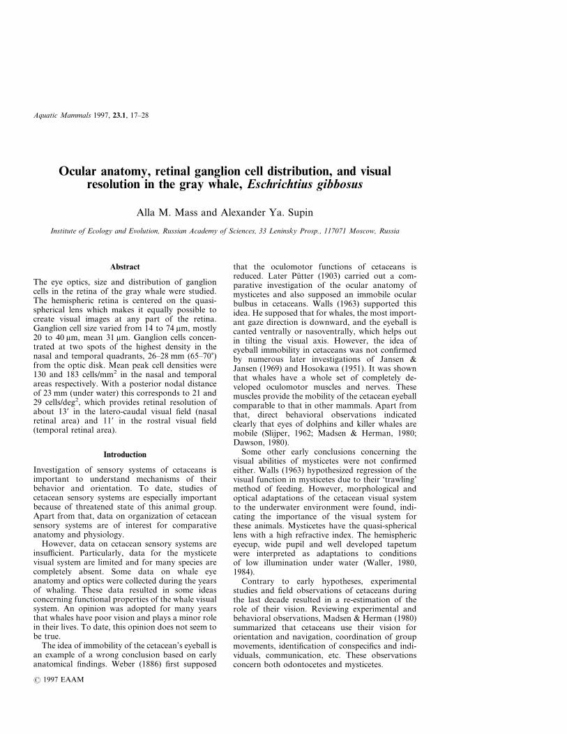
Eye dimensionsFigure 1 demonstrates a horizontal section of thefrozen eye. The eyecup of the gray whale, as well asthe eye of other cetaceans, is of hemispheric shape.It is slightly oval: the naso-temporal width (64–66 mm) longer than the dorso-ventral one (60–62 mm). Axial length between the external surfacesof the cornea and sclera was 55–60 mm. Internalnaso-temporal eyecup diameter was 46–47 mm andthe dorso-ventral one was 44–45 mm.
The lens was very convex but not truly spherical.Its transverse diameter was about 13 mm and axialdiameter about 10 mm. The distance from the
18 A. M. Mass and A. Ya. Supin
center of the lens to the retina was estimated as23 mm.
The cornea was elongated in the naso-temporaldirection. Its naso-temporal diameter was 28–30 mm and the dorso-ventral one was 20–21 mm.The peripheral rim of the cornea was much thicker(2–2.5 mm) than the central part (around 1 mm).The pupil was horizontally elongated and had theoperculum.
The shape of the retina was complex. Its majorpart, except the far periphery, had a shape ofan incomplete hemisphere, about 150� across. Aperipheral rim of the retina was bent inward. Theradius of the retinal hemisphere was assessed to be23 mm.
The tapetum was blue–gray and well developed.It covered a major part of the eyecup except theventral region and the region adjacent to theoptic disk.
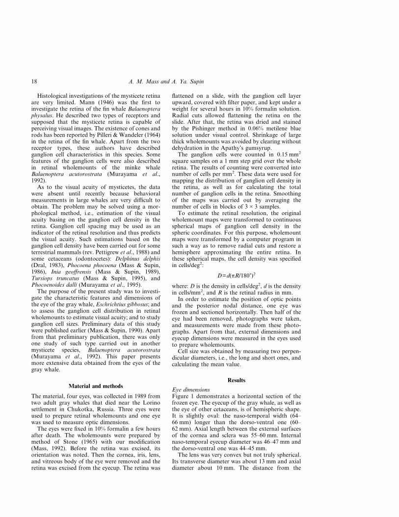
Ganglion cell characteristicsSimilarly to other cetaceans, the retina of the graywhale contained mostly large neurons. The mosttypical were cells 20 to 40 �m in size, although cellsup to 74 �m were found as well. The cells were of
various shapes. Most of them were polygonal inshape with clearly visible sites of originating of 3 to6 dendrites (Fig. 2). Oval cells were rare, and evenmore rare were round cells. The cells were charac-terized by a broad rim of cytoplasm with wellstained Nissl granules. Clearly visible light nucleuswith dark nucleolus could be disposed both in thesoma center and eccentrically.
Distribution of ganglion cellsThe mean total area of the three investigated retinalwholemounts was found to be 2520 mm2. Ganglioncell totals in the three wholemounts varied from165 000 to 184 000 with a mean of 174 000. Themean ganglion cell density averaged over the wholearea of the retina and among three wholemountswas 70 cells/mm2.
Counting ganglion cells throughout the retina at1 mm steps revealed that cell distribution variedin different parts of the retina. A representativepattern of ganglion cell distribution is shown by awholemount map in Fig. 3. A characteristic featureof the map is the presence of two areas of cellconcentration: one area was located in the nasalpart of the retina and another one in the temporal
Figure 1. Horizontal section of a whale’s eye (redrawn from a photograph). Right eye, thenasal pole leftward. C–cornea, Ir–iris, L–lens, R–retina, ON–optic nerve, S–sclera. Arrowsdelimit a part of the retina that can be approximated by an incomplete hemisphere (approx.150� relative to the lens center).
19Gray whale eye anatomy
part. Both areas of cell concentration were locatednear the equator of the retina.
The peak cell densities in the wholemount ofFig. 3 were 142 and 200 cells/mm2 in the nasal andtemporal areas respectively. In all wholemounts,peak cell density in the nasal area was lower than inthe temporal one. The averaged peak cell densitiesfor the three wholemounts were 130 and 183 cells/mm2 in the nasal and temporal areas respectively.
The high density areas were distinguished clearlyagainst the background. Outside these areas, celldensity declined markedly. In both central andperipheral parts of the retina, cell density was lessthan 50 cells/mm2.
Distribution of cell density with two areas of cellconcentration can be illustrated by profiles of celldensity across the naso-temporal equator (Fig. 4).This figure combines data obtained in all whole-mounts. The plots show two distinct peaks ofcell density in the nasal and temporal areas,
steep decrease of cell density towards the retinalperiphery and very low density in the central partadjacent to the optic disk.
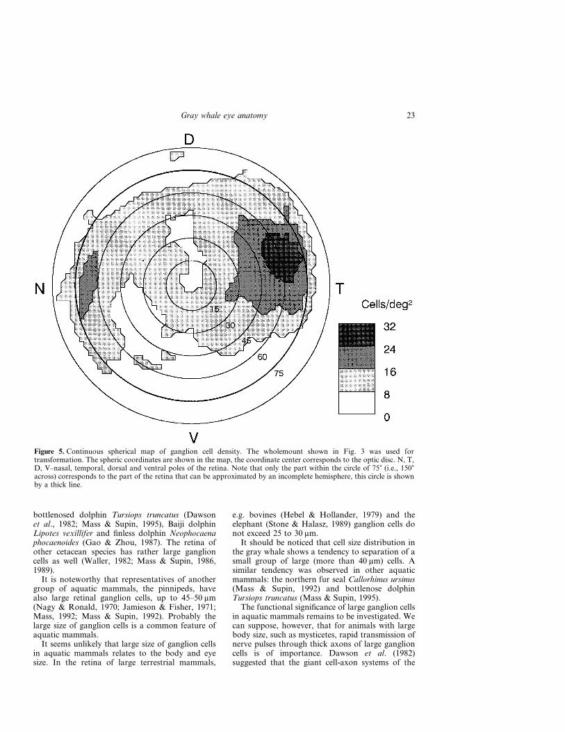
Figure 5 shows a spherical map of ganglion celldensity obtained by transformation of the whole-mount map shown in Fig. 3. Upon constructingcontinuous spherical maps of the retina, it wassupposed that the mean radius of the retinal hemi-sphere was 23 mm. At this radius, the arch of 1�corresponds to the distance of 0.4 mm alongthe retinal surface, and 1 deg2 corresponds to0.16 mm2. Note that according to Fig. 1, only thepart of the retina within the circle of 75� representsthe incomplete hemisphere; the periphery outsidethis circle corresponds to the retinal rim bentinward.
The map shows that the areas of high cell densityare situated in the nasal and temporal parts of theretina, near its naso-temporal equator. The celldensity peaks were 65 to 70� from the geometric
Figure 2. Light micrograph from the ganglion cell layer of a Nissl-stained retinal wholemount of agray whale. Cell density about 70 cells/mm2.
20 A. M. Mass and A. Ya. Supin

center. The peak cell densities were 23 cells/deg2 inthe nasal area and 32 cells/deg2 in the temporalarea.
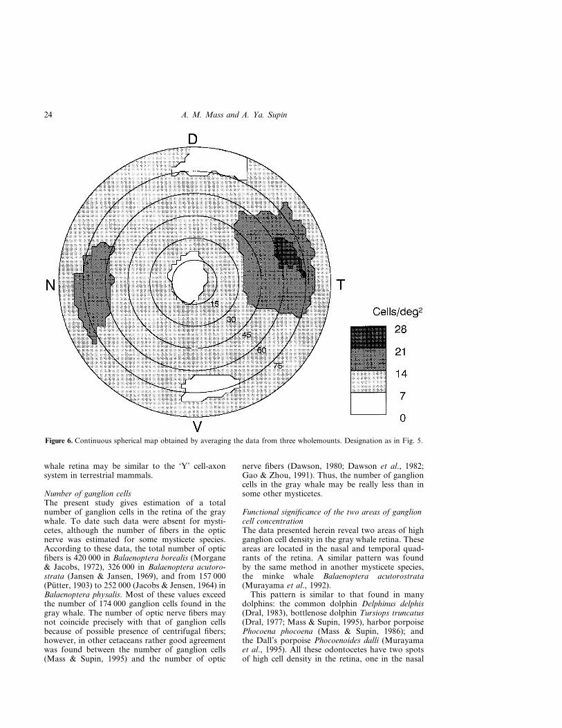
Such maps were construction for all three whole-mounts. The averaged peak cell density among thethree wholemounts were 21 and 29 cells/mm2 in thenasal and temporal areas respectively. Location ofthe high density areas was similar in all the whole-mounts. Therefore it was possible to average thedata among all the wholemounts. Figure 6 presentsthe averaged continuous spherical map. Similarly tothe map of an individual wholemount (Fig. 5), theareas of high cell density were situated near thenaso-temporal equator, with cell density peakslocated 65 to 70� from the center of the retina. Themaximum cell density in the averaged map was20 cells/deg2 in the nasal area and 28 cells/deg2 inthe temporal one. This is somewhat less than themeans of peak values indicated above (21 and 29cells/deg2). The difference arose since spatialpositions of the density peaks did not coincideprecisely in different wholemounts. In the retinalperiphery and near the optic disc, the cell densitywas 6 to 10 cells/deg2.
Both wholemount maps (Fig. 3) and sphericalmaps (Figs 5, 6) show that we did not find a streakconnecting the two high density areas, clearlyvisible in dolphins and described in the minke whaleby Murayama et al. (1992).
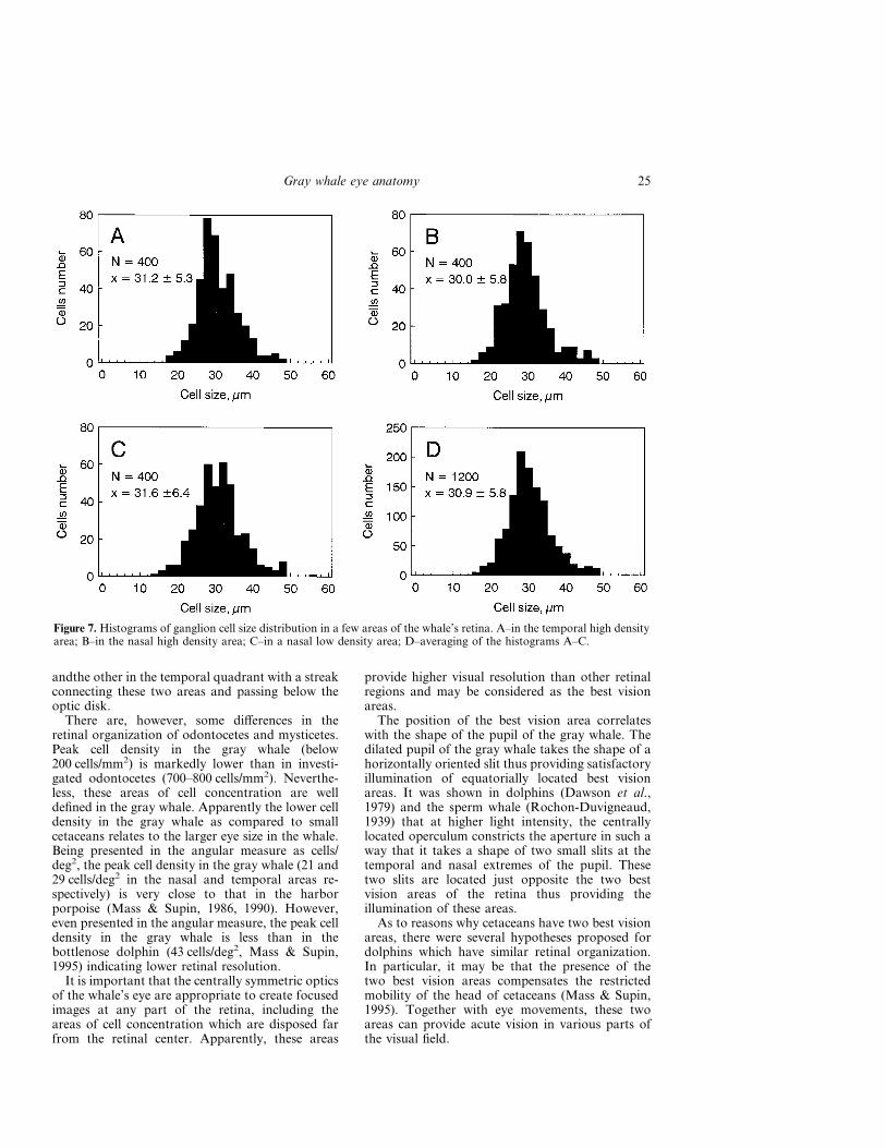
Ganglion cell sizeA total of 1200 cells were measured in three retinalareas with different cell densities (400 cells in eachsample). Most of the cells were of large size.Although maximal cell size observed in the whole-mounts was 74 �m, in the selected samples, cell sizevaried only from 14 to 56 �m since cells larger than50 �m were very rare. Cells smaller than 14 �m werenot found. The majority of cells were within therange of 20 to 40 �m.
Figure 7 presents frequency vs cell size histo-grams for ganglion cells in three areas (A–C).The cell size distributions were monomodal in allsamples, however, there was a slight tendency ofseparation of large cells (larger than 40 �m) in to adistinct group.
There was little difference between cell size distri-butions and mean sizes in areas of high and low cell
Figure 3. Map of ganglion cell density in a retinal wholemount. Cell density is designated according to the scale on theright. N, T, D, V–nasal, temporal, dorsal and ventral poles of the retina; OD–optic disk.
21Gray whale eye anatomy

densities. In all the investigated samples, mean cellsize was within a range of 30 to 31.6 �m (i.e.,differed within 5.3%) with a standard deviation ofdistributions of 5.3 to 6.4 �m. Similar cell size in allthe samples makes it reasonable to average all thedata. It results in the distribution with the mean of30.9 �m with SD of 5.8 �m.
Discussion
Eye opticsThe optic structure of the eye of the gray whaleis similar in general to that described in othercetaceans, both odontocetes and mysticetes (Mayer,1852; Pütter, 1903; Rochon-Duvigneaud, 1939,1943; Pilleri & Wandeler, 1964; Waller, 1980;Dawson et al., 1972; Vasilyevskaya, 1988; Mass &Supin, 1995). A distinctive feature of the eye of thegray whale and other large mysticetes is very thicksclera. This feature, however, does not influencedirectly the eye optics.
A common feature of the eye optics of bothodontocetes and mysticetes is a hemispheric retinacentered on the quasi-spheric lens. The corneaapparently plays a minor role under water becauseof small difference of its inner and outer curvaturesand small difference of refractive indices of themedia in front of and behind the cornea. Althoughrefraction at the cornea cannot be neglected com-pletely in cetaceans (Kröger & Kirschfeld, 1994),
the thick lens is obviously the main refractivestructure of the cetacean eye. In these conditions,the nodal point of the eye coincides with the centerof the lens. Since the hemispheric retina is centeredon the same point, the overall optic structure of theeye is symmetric relative to the lens center, and lightrays of any direction can be equally focused atcorresponding parts of the retina. It is noteworthy,however, that the cornea of the gray whale is muchthicker at its periphery than at the center, thusfunctioning as a weak dissipating refractive struc-ture. According to Kröger & Kirschfeld (1994) thisfunction of the cornea is important for refractioncorrection.
Ganglion cell sizeA characteristic feature of the retina of the graywhale is the very large size of ganglion cells. In thepresent study, we found ganglion cells as large as14 to 74 �m (means 30.9 �m). Large (mean 42.9 �m)and giant (up to 80 �m) ganglion cells were alsoobserved in the retina of the minke whaleBalaenoptera acutorostrata (Murayama et al.,1992). Very large (50–80 �m) and giant (up to160 �m) cells were observed in the retina of the finwhale Balaenoptera physalus (Pilleri & Wandeler,1964). The large size of ganglion cells is a commonfeature of many cetacean species. Ganglion cells upto 60–75 �m were described in the retina of thecommon dolphin Delphinus delphis (Dral, 1983),
Figure 4. Ganglion cell density distribution along the horizontal equator in the whale’s retina.Nas–nasal, Temp–temporal direction. Plots obtained from three wholemounts are designated bydifferent symbols.
22 A. M. Mass and A. Ya. Supin
bottlenosed dolphin Tursiops truncatus (Dawsonet al., 1982; Mass & Supin, 1995), Baiji dolphinLipotes vexillifer and finless dolphin Neophocaenaphocaenoides (Gao & Zhou, 1987). The retina ofother cetacean species has rather large ganglioncells as well (Waller, 1982; Mass & Supin, 1986,1989).
It is noteworthy that representatives of anothergroup of aquatic mammals, the pinnipeds, havealso large retinal ganglion cells, up to 45–50 �m(Nagy & Ronald, 1970; Jamieson & Fisher, 1971;Mass, 1992; Mass & Supin, 1992). Probably thelarge size of ganglion cells is a common feature ofaquatic mammals.
It seems unlikely that large size of ganglion cellsin aquatic mammals relates to the body and eyesize. In the retina of large terrestrial mammals,
e.g. bovines (Hebel & Hollander, 1979) and theelephant (Stone & Halasz, 1989) ganglion cells donot exceed 25 to 30 �m.
It should be noticed that cell size distribution inthe gray whale shows a tendency to separation of asmall group of large (more than 40 �m) cells. Asimilar tendency was observed in other aquaticmammals: the northern fur seal Callorhinus ursinus(Mass & Supin, 1992) and bottlenose dolphinTursiops truncatus (Mass & Supin, 1995).
The functional significance of large ganglion cellsin aquatic mammals remains to be investigated. Wecan suppose, however, that for animals with largebody size, such as mysticetes, rapid transmission ofnerve pulses through thick axons of large ganglioncells is of importance. Dawson et al. (1982)suggested that the giant cell-axon systems of the
Figure 5. Continuous spherical map of ganglion cell density. The wholemount shown in Fig. 3 was used fortransformation. The spheric coordinates are shown in the map, the coordinate center corresponds to the optic disc. N, T,D, V–nasal, temporal, dorsal and ventral poles of the retina. Note that only the part within the circle of 75� (i.e., 150�across) corresponds to the part of the retina that can be approximated by an incomplete hemisphere, this circle is shownby a thick line.
23Gray whale eye anatomy
whale retina may be similar to the ‘Y’ cell-axonsystem in terrestrial mammals.
Number of ganglion cellsThe present study gives estimation of a totalnumber of ganglion cells in the retina of the graywhale. To date such data were absent for mysti-cetes, although the number of fibers in the opticnerve was estimated for some mysticete species.According to these data, the total number of opticfibers is 420 000 in Balaenoptera borealis (Morgane& Jacobs, 1972), 326 000 in Balaenoptera acutoro-strata (Jansen & Jansen, 1969), and from 157 000(Pütter, 1903) to 252 000 (Jacobs & Jensen, 1964) inBalaenoptera physalis. Most of these values exceedthe number of 174 000 ganglion cells found in thegray whale. The number of optic nerve fibers maynot coincide precisely with that of ganglion cellsbecause of possible presence of centrifugal fibers;however, in other cetaceans rather good agreementwas found between the number of ganglion cells(Mass & Supin, 1995) and the number of optic
nerve fibers (Dawson, 1980; Dawson et al., 1982;Gao & Zhou, 1991). Thus, the number of ganglioncells in the gray whale may be really less than insome other mysticetes.
Functional significance of the two areas of ganglioncell concentrationThe data presented herein reveal two areas of highganglion cell density in the gray whale retina. Theseareas are located in the nasal and temporal quad-rants of the retina. A similar pattern was foundby the same method in another mysticete species,the minke whale Balaenoptera acutorostrata(Murayama et al., 1992).
This pattern is similar to that found in manydolphins: the common dolphin Delphinus delphis(Dral, 1983), bottlenose dolphin Tursiops truncatus(Dral, 1977; Mass & Supin, 1995), harbor porpoisePhocoena phocoena (Mass & Supin, 1986); andthe Dall’s porpoise Phocoenoides dalli (Murayamaet al., 1995). All these odontocetes have two spotsof high cell density in the retina, one in the nasal
Figure 6. Continuous spherical map obtained by averaging the data from three wholemounts. Designation as in Fig. 5.
24 A. M. Mass and A. Ya. Supin
andthe other in the temporal quadrant with a streakconnecting these two areas and passing below theoptic disk.
There are, however, some differences in theretinal organization of odontocetes and mysticetes.Peak cell density in the gray whale (below200 cells/mm2) is markedly lower than in investi-gated odontocetes (700–800 cells/mm2). Neverthe-less, these areas of cell concentration are welldefined in the gray whale. Apparently the lower celldensity in the gray whale as compared to smallcetaceans relates to the larger eye size in the whale.Being presented in the angular measure as cells/deg2, the peak cell density in the gray whale (21 and29 cells/deg2 in the nasal and temporal areas re-spectively) is very close to that in the harborporpoise (Mass & Supin, 1986, 1990). However,even presented in the angular measure, the peak celldensity in the gray whale is less than in thebottlenose dolphin (43 cells/deg2, Mass & Supin,1995) indicating lower retinal resolution.
It is important that the centrally symmetric opticsof the whale’s eye are appropriate to create focusedimages at any part of the retina, including theareas of cell concentration which are disposed farfrom the retinal center. Apparently, these areas
provide higher visual resolution than other retinalregions and may be considered as the best visionareas.
The position of the best vision area correlateswith the shape of the pupil of the gray whale. Thedilated pupil of the gray whale takes the shape of ahorizontally oriented slit thus providing satisfactoryillumination of equatorially located best visionareas. It was shown in dolphins (Dawson et al.,1979) and the sperm whale (Rochon-Duvigneaud,1939) that at higher light intensity, the centrallylocated operculum constricts the aperture in such away that it takes a shape of two small slits at thetemporal and nasal extremes of the pupil. Thesetwo slits are located just opposite the two bestvision areas of the retina thus providing theillumination of these areas.
As to reasons why cetaceans have two best visionareas, there were several hypotheses proposed fordolphins which have similar retinal organization.In particular, it may be that the presence of thetwo best vision areas compensates the restrictedmobility of the head of cetaceans (Mass & Supin,1995). Together with eye movements, these twoareas can provide acute vision in various parts ofthe visual field.
Figure 7. Histograms of ganglion cell size distribution in a few areas of the whale’s retina. A–in the temporal high densityarea; B–in the nasal high density area; C–in a nasal low density area; D–averaging of the histograms A–C.
25Gray whale eye anatomy
Another hypothesis suggests that one of the bestvisual areas provides satisfactory visual acuity inair. It was shown that dolphins have good vision inair (Herman et al., 1975). It had to be explainedhow the eye optics of dolphins prevents aerialmyopia which derives from the refractive power ofthe cornea surface in air added to that of the lens.Some of the hypotheses were discussed earlier(Mass & Supin, 1995); among them the followingare noteworthy: (1) closer position of the temporalfundus to the lens (Waller, 1980); (2) less refractivepower of the cornea surface in its frontal part(Dawson, 1987); (3) strong pupillary constructionin air results in double-slit shape of the pupilrendering light to pass through the margin of thelens, which is optically weaker than its central core(Rivamonte, 1976); and (4) the pin-hole aperturesof the constricted pupil improve visual acuity. Allthe suggested mechanisms work for oblique rayspassing through the nasal part of the pupil tothe temporal best vision area of the retina. Thus,the specific position of the best vision areas in thedolphin retina may be connected with theircombined underwater and aerial vision.
However, it remains unclear whether mysticeteshave satisfactory aerial vision. Matthiessen (1893)investigated in detail the mysticete eye optics inBalaenoptera physalus and showed that the mysti-cete eye is emmetropic in water and very myopic inair. He concluded that whales are unable to seeclearly objects in air and can only see some move-ments and the horizon line. Rochon-Duvigneaud(1939, 1943) also suggested that cetaceans had nocapacity for aerial vision and that a clear retinalimage could not be formed.
Experimental investigations of mysticete visualbehavior are absent since these animals have neverbeen kept in captivity for investigations. However,there were some observations made from ships thatmysticete whales may ‘spy-hop’ by raising the headvertically out of the water (rev. Madsen & Herman,1980). It suggests that mysticetes may use theirvision in air.
It is unknown yet whether mysticetes have eyeoptics which make it possible to prevent aerialmyopia. However, based on the similarity of themysticete eye optics with that of dolphins, it isreasonable to suppose that mysticetes have mech-anisms of preventing aerial myopia similar to thosehypothesized for dolphins. Direct investigations arenecessary to decide whether these hypotheses can beapplied to mysticetes.
Retinal resolution and visual acuity estimation inthe gray whaleData on ganglion cell density in the best visionareas and other parts of the retina make it possible
to calculate the retinal resolution of the gray whale.The retinal resolution can be estimated as meanangular spacing of ganglion cells; i.e.:
s=1/DY,
where s is angular spacing and D is cell density perdeg2 of visual angle. The latter depends on celldensity and posterior nodal distance (PND). Asshown above, the cetacean eye optics make itpossible to adopt PND equal to the radius of theretinal hemisphere. In this case, cell density indegrees of the visual field is the same as in termsof the retinal hemisphere; i.e., in the nasal retinalarea, peak cell density is 21 cells/deg2 and in thetemporal area, it is 29 cells/deg2. These valuescorrespond to retinal resolution of 0.22�=13� in thenasal area and 0.19�=11� in the temporal one. Atthe retinal periphery and around the optic disk,retinal resolution is worse: cell density of about10 cells/deg2 corresponds to the resolution of0.32�=19�.
The optical system of the eye and retinal resol-ution are two main factors determining visualacuity. Supposing that these two factors are incorrespondence, the values of retinal resolutionmentioned above can be adopted as a first approxi-mation of visual acuity of the gray whale. Thus, thebest visual acuity of the gray whale can be estimatedas about 11� in the frontal part of the visual field(corresponding to the temporal best vision area ofthe retina) and about 13� in the latero-caudal part ofthe visual field (corresponding to the nasal retinalarea).
These estimations indicate that the visual acuityof the gray whale is a little worse than, but compar-able to, that in some other cetaceans; e.g. about 7�in Balaenoptera acutorostrata (Murayama et al.,1992), 8–12� in Tursiops truncatus (Herman et al.,1975; Mass & Supin, 1995); and close to that(11–14�) in Phocoena phocoena (Mass & Supin,1986). It suggests that visual abilities of the graywhale (perhaps, of other mysticetes as well) arecomparable with those of dolphins which activelyuse their vision and demonstrate fine imagerecognition.
The estimations presented above concern visualacuity of mysticetes in water since their eye opticsare obviously adapted to the underwater vision. Werefrain from discussion of visual acuity of mysti-cetes in air since very little is known of their aerialrefraction.
Acknowledgement
This study was supported by the Russian Foun-dation for Basic Research, Grant #95-04-11127.
26 A. M. Mass and A. Ya. Supin
References
Dawson, W. W., Brindorf, L. A. & Perez, J. M. (1972)Gross anatomy and optics of the dolphin eye (Tursiopstruncatus). Cetology 10, 2–12.
Dawson, W. W., Adams, C. K., Barris, M. C. & Litzkov,C. A. (1979) Static and kinetic properties of the dolphinpupil. Am. J. Physiol. 237, R301–R305.
Dawson, W. W. (1980) The cetacean eye. In CetaceanBehavior: Mechanisms and Functions (ed. L. M.Herman) pp. 53–100. Wiley Interscience, New York.
Dawson, W. W., Hawthorne, M. N., Jenkins, R. L. &Goldston, R. T. (1982) Giant neural system in the innerretina and optic nerve of small whales. J. Comp. Neurol.205, 1–7.
Dawson, W. W. (1987) Corneal surface properties oftwo marine mammal species. Marine Mammal Sci. 3,186–197.
Dral, A. D. G. (1977) On the retinal anatomy of Cetacea(mainly Tursiops truncatus). In Functional Anatomy ofMarine Mammals (ed. J. Harrison), III, pp. 81–134.Academic Press, London.
Dral, A. D. G. (1983) The retinal ganglion cells ofDelphinus delphis and their distribution. AquaticMammals 10, 57–68.
Gao, A. & Zhou, K. (1987) On the retinal ganglion cells ofNeophocaena and Lipotes. Acta Zool. Sin. 33, 316–332.
Gao, G. & Zhou, K. (1991) Fiber analysis of the optic andcochlear nerves of small cetaceans. In Marine MammalSensory Systems (eds J. A. Thomas, R. A. Kasteleinand A. Ya. Supin) pp. 39–52. Plenum, New York.
Hebel, B. & Hollander, H. (1979) Size and distribution ofganglion cells in the bovine retina. Vision Res. 19,667–674.
Herman, L. M., Peacock, M. F., Yunker, M. P. &Madsen, C. J. (1975) Bottlenosed dolphin: Double-slitpupils yields equivalent aerial and underwater diurnalacuity. Science 189, 650–652.
Hosokawa, H. (1951) On the extrinsic eye muscles of thewhale with special remarks upon the innervation andfunction of the musculus retractor bulbi. Sci. Rep.Whales Res. Inst. 6, 1–31.
Jacobs, M. S. & Jensen, A. V. (1964) Gross aspects of thebrain and fiber analysis of cranial nerves in the greatwhale. J. Comp. Neurol. 123, 55–72.
Jansen, J. & Jansen, J. K. S. (1969) The nervous system ofCetacea. In The Biology of Marine Mammals (ed. H. T.Andersen) pp. 175–252. Academic Press, New York.
Jamieson, G. S. & Fisher, H. D. (1971) The retina of theharbor seal Phoca vitulina. Can. J. Zool. 49, 19–23.
Kröger, R. H. H. & Kirschfeld, K. (1994) Refractive indexin the cornea of a harbor porpoise (Phocoena phocoena)measured by two-wavelengths laser-interferometry.Aquatic Mammals 20, 99–107.
Madsen, C. J. & Herman, L. M. (1980) Social andecological correlates of cetacean vision and visualappearance. In Cetacean Behavior: Mechanisms andFunction (ed. L. M. Herman) pp. 101–147. WileyInterscience, New York.
Mann, G. (1946) Oyo y vision de las ballenas. Biologica 4,23–71.
Mass, A. M. (1992) Retinal topography in the walrus(Odobenus rosmarus divergence) and fur seal (Callo-
rhinus ursinus). In Marine Mammal Sensory Systems(eds. J. A. Thomas, R. A. Kastelein & A. Y. Supin)pp. 119–135. Plenum, New York.
Mass, A. M. & Supin, A. Ya. (1986) Topographic distri-bution of size and density of ganglion cells in the retinaof a porpoise, Phocoena phocoena. Aquatic Mammals12, 95–102.
Mass, A. M. & Supin, A. Ya. (1989) Distribution ofganglion cells in the retina of an Amazon river dolphinInia geoffrensis. Aquatic Mammals 15, 49–56.
Mass, A. M. & Supin, A. Ya. (1990) Best vision zones onthe retinae of some cetaceans. In Sensory Abilities ofCetaceans (eds J. A. Thomas & R. A. Kastelein)pp. 505–517. Plenum, New York.
Mass, A. M. & Supin, A. Ya. (1992) Peak density, size andregional distribution of ganglion cells in the retina ofthe fur seal Callorhinus ursinus. Brain, Behav. Evol. 39,69–76.
Mass, A. M. & Supin, A. Ya. (1995) Retinal cell topogra-phy of the retina in the bottlenosed dolphin Tursiopstruncatus. Brain, Behav. Evol. 45, 257–265.
Matthiessen, L. (1893) Ûber den physikalisch-optischenBau der Augen vom Knolwal (Megaptera boops, Fabr.)und Finwal (Balaenoptera musculus, Comp). Z. vergl.Augenheilk 7, 77–101.
Mayer, A. (1852) Anatomische Untersuchungen über dasAuge der Cetaceen. Henry and Cohen, Bonn.
Morgane, P. J. & Jacobs, M. S. (1972) Comparativeanatomy of the cetacean nervous system. In FunctionalAnatomy of Marine Mammals (ed. R. J. Harrison)pp. 117–244. Academic Press, New York.
Murayama, T., Somiya, H., Aoki, I. & Ishii, T. (1992) Thedistribution of ganglion cells in the retina and visualacuity of minke whale. Nippon Suissan Gakkaishi 58,1057–1061.
Murayama, T., Somiya, H., Aoki, I. & Ishii, T. (1995)Retinal ganglion cell size and distribution predict visualcapabilities of Dall’s porpoise. Marine Mammal Science11, 136–149.
Nagy, A. R. & Ronald, K. (1970) The harp seal,Pagophilus groenlandicus (Erxleben 1777). Canad. J.Zool. 48, 367–370.
Pettigrew, J. D., Dreher, B., Hopkins, C. S., McCall, M. J.& Brown, M. (1988) Peak density and distribution ofganglion cells in the retina of microchiropteran bats:implication for visual acuity. Brain, Behav. Evol. 32,39–56.
Pilleri, G. & Wandeler, A. (1964) Ontogenese undfunktionelle Morphologie des Auges des FinnwalsBalaenoptera Physalus L. (Cetacea, Mysticeti,Balaenopteridae). Acta Anat. Suppl. 50 ad. vol. 57,1–74.
Pütter, A. (1903) Die Augen der Wassersaugetiere. Zool.Jahrb. Abth. Anat. Ontog. Thiere 17, 99–402.
Rivamonte, L. A. (1976) Eye model to account for com-parable aerial and underwater acuities of the bottlenosedolphin. Neth. J. Sea Res. 10, 91–498.
Rochon-Duvigneaud, A. (1939) L’oeil des cétacés.Archives Museum National Histoire Naturella Parisv. 58.
Rochon-Duvigneaud, A. (1943) Les Yeux et la Vision desVertebres. Masson, Paris.
Slijper, E. J. (1962) Whales. Hutchinson, London.
27Gray whale eye anatomy
Stone, J. (1965) A quantitative analysis of the distributionof ganglion cells in the cat’s retina. J. Comp. Neurol.124, 337–352.
Stone, J. & Halasz, P. (1989) Topography of the retina inthe elephant Loxodonta africana. Brain, Behav. Evol.34, 84–95.
Vasilyevskaya, G. I. (1988) The eye of the minke whale. I.General structure and the sclera (in Russian). VestnikZool No. 5, 67–72.
Waller, G. (1980) The visual system of toothedwhales (Mammalia, Cetacea, Odontoceti). PhD thesis,University of Cambridge.
Waller, G. (1982) Retinal ultrastructure of the Amazonriver dolphin (Inia geoffrensis). Aquatic Mammals 9,17–28.
Waller, G. (1984) The ocular anatomy of cetacea—anhistorical perspective. Investigation of Cetaceae (ed.G. V. Pilleri) XVI, 138–148.
Walls, G. L. (1963) The Vertebrate Eye and its AdaptiveRadiation. Hafner, New York.
Weber, M. (1886) Studien über Säugethiere. Ein Beitragzur Frage nach den Ursprung der Cetaceen. Jena.
28 A. M. Mass and A. Ya. Supin


![Diversity of Retinal Ganglion Cells Identified by ... · of retinal ganglion cells [3,4,5,6]. Even in the monkey retina, Dacey and other researchers showed morphological diversity](https://img.dokumen.tips/doc/110x75/60fabf5bff27e94d36249fb0/diversity-of-retinal-ganglion-cells-identified-by-of-retinal-ganglion-cells.jpg)