Embed Size (px)
Citation preview
OCR AS Biology Unit 1: Cells, Exchange and Transport

To understand and discuss the basis of cell theory based upon milestones in cell biology
To carry out practical work on cell size and magnifications using a light microscope
To be able to draw a plant cell and an animal cell using a light microscope
To prepare slides using stains to identify cell structure.

Read through handout
Group discussion Discuss the impact of the microscope on
cell biology List things you think are the most
important
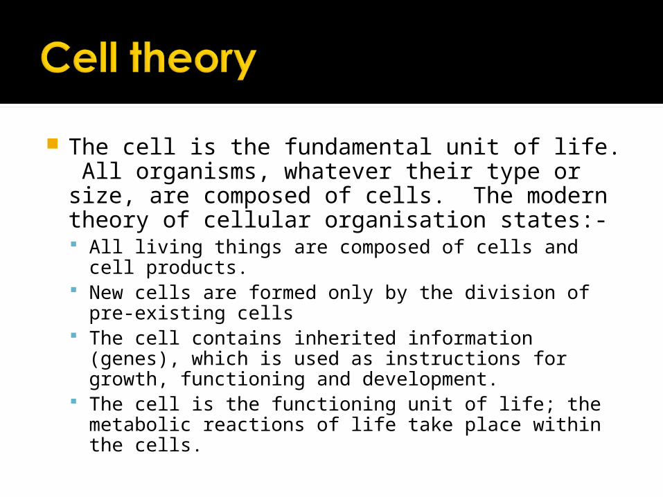
The cell is the fundamental unit of life. All organisms, whatever their type or size, are composed of cells. The modern theory of cellular organisation states:- All living things are composed of cells and cell
products. New cells are formed only by the division of pre-
existing cells The cell contains inherited information (genes), which
is used as instructions for growth, functioning and development.
The cell is the functioning unit of life; the metabolic reactions of life take place within the cells.
Before the development of cell theory, it was commonly believe that living organisms could arise by spontaneous generation.
Explain what this term means and why it has been discredited as a theory.


When Scientists began to observe cells, they started with simple microscopes
There are two different types of microscope both use a form of radiation to create an image of the specimen: Light microscope – uses light Electron microscope – uses electrons
Magnification Number of times larger an image is
compared with the real size of the object
Resolution The ability to distinguish between two
separate points


For the microscope in front of you, work out The magnification of each lens The field of view for each lens ▪ Using a graticule / stage micrometer
Collect a prepared slide of Squamous epithelium, draw what you can see. Your diagram should include title, labels,
magnification and a scale bar.
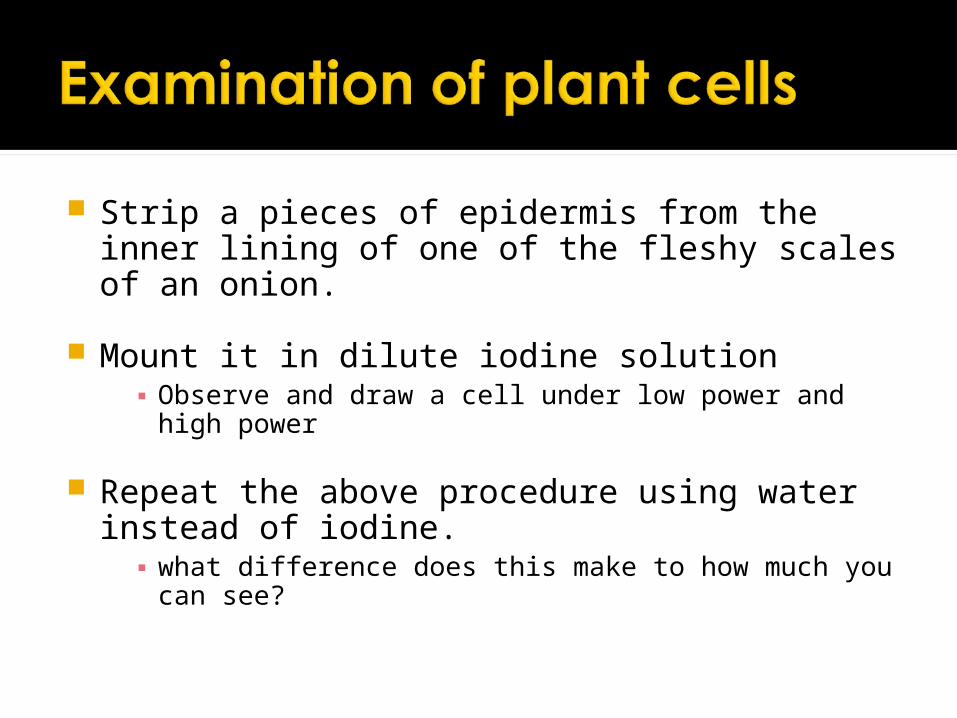
Strip a pieces of epidermis from the inner lining of one of the fleshy scales of an onion.
Mount it in dilute iodine solution▪ Observe and draw a cell under low power and high
power
Repeat the above procedure using water instead of iodine.▪ what difference does this make to how much you can
see?

What does this tell you about the value of staining cells before you look at them under the microscope?
Make sure all diagrams have a title, label, magnification and scale bar.

OCR AS Biology
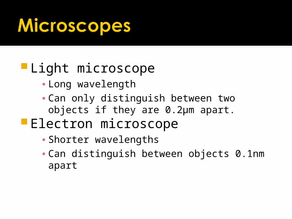
Light microscope▪ Long wavelength▪ Can only distinguish between two objects if
they are 0.2µm apart.Electron microscope
▪ Shorter wavelengths▪ Can distinguish between objects 0.1nm apart
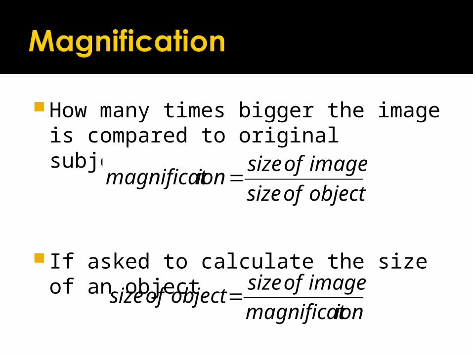
ionmagnificat
imageofsizeobjectofsize
How many times bigger the image is compared to original subject.
If asked to calculate the size of an object
objectofsize
imageofsizeionmagnificat
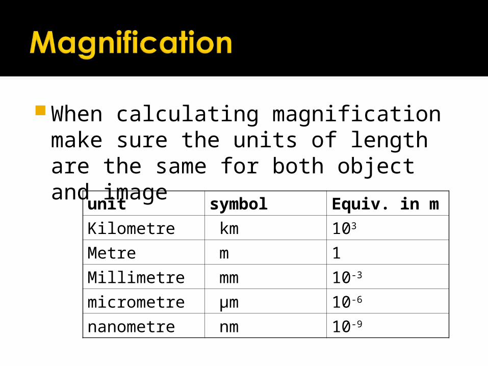
When calculating magnification make sure the units of length are the same for both object and image
unit symbol Equiv. in m
Kilometre km 103
Metre m 1
Millimetre mm 10-3
micrometre µm 10-6
nanometre nm 10-9
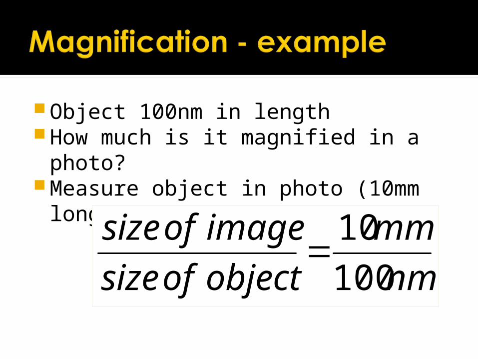
Object 100nm in lengthHow much is it magnified in a photo?Measure object in photo (10mm
long)
nm
mm
objectofsize
imageofsize
100
10
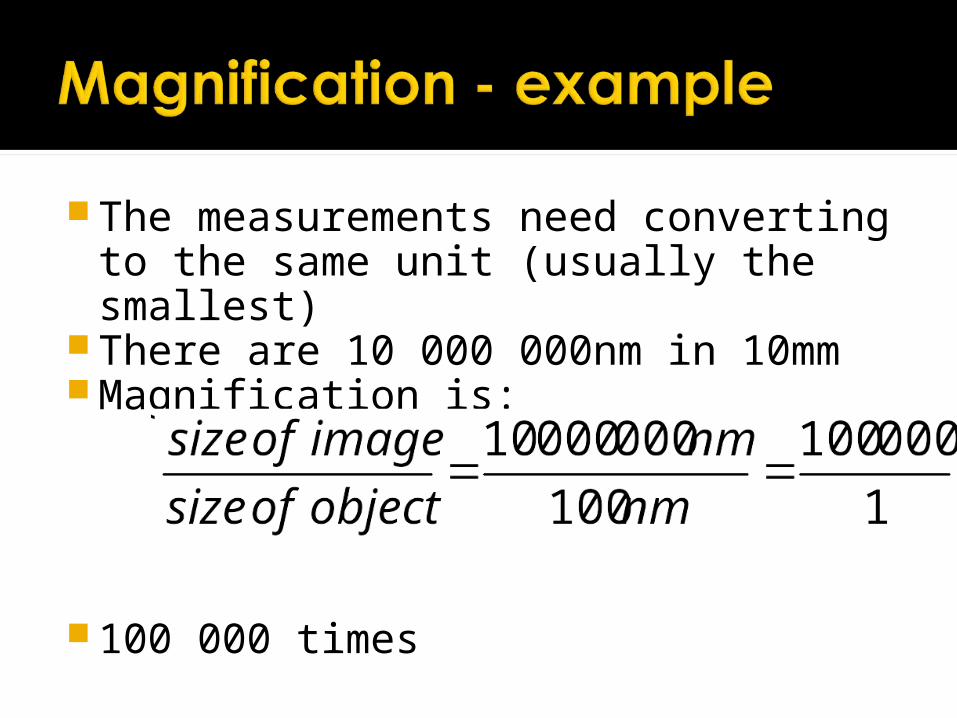
The measurements need converting to the same unit (usually the smallest)
There are 10 000 000nm in 10mmMagnification is:
100 000 times

OCR AS Biology
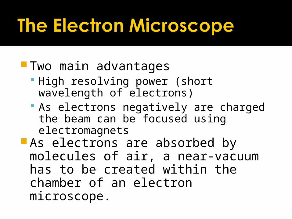
Two main advantages High resolving power (short wavelength
of electrons) As electrons negatively are charged the
beam can be focused using electromagnets
As electrons are absorbed by molecules of air, a near-vacuum has to be created within the chamber of an electron microscope.
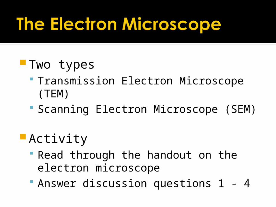
Two types Transmission Electron Microscope (TEM) Scanning Electron Microscope (SEM)
Activity Read through the handout on the
electron microscope Answer discussion questions 1 - 4

Light MicroscopeAdvantages
Electron Microscope Disadvantages
Small and portable very largeOperated in special rooms
Unaffected by magnetic fields Affected by magnetic fields
Preparation of material is quick and simple
Preparation of material is lengthyRequires expertise
Material rarely distorted by preparation
Preparation may distort material
Natural colour of object observed
Images are in black and white
Cheap to purchase and operate
Expensive to purchase and operate

Light MicroscopeDisadvantages
Electron MicroscopeAdvantages
Magnifies objects up to 1500x only
Magnifies objects more than 500 000X
Depth of field is restricted
Possible to investigate a greater field of depth
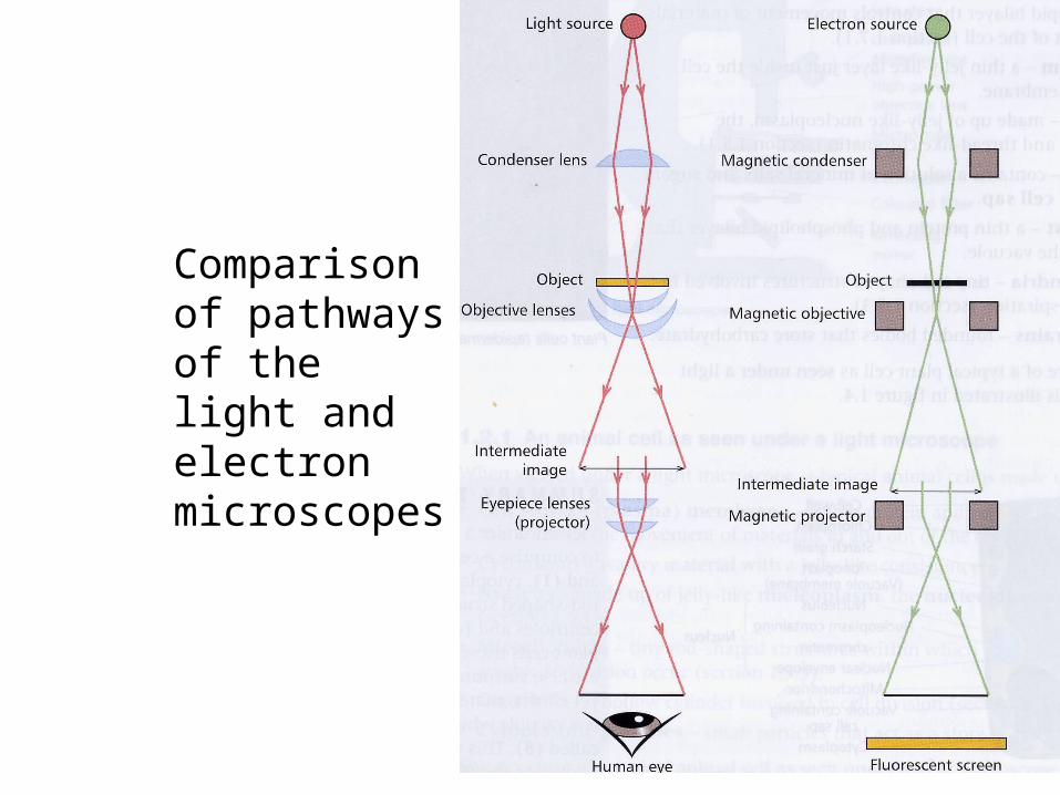
Comparison of pathways of the light and electron microscopes

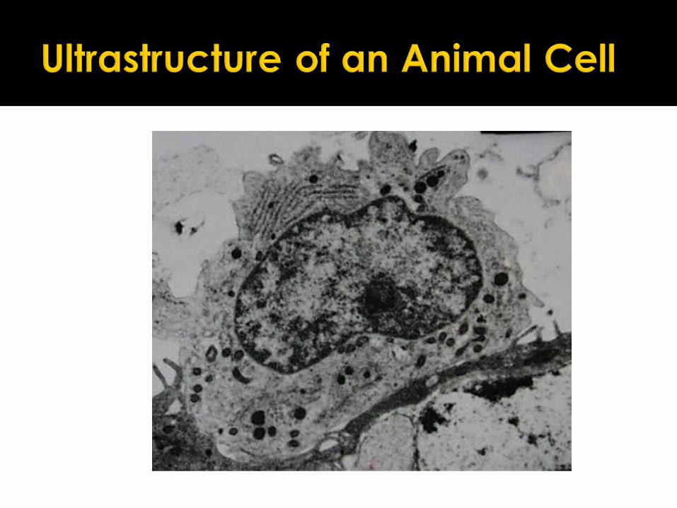
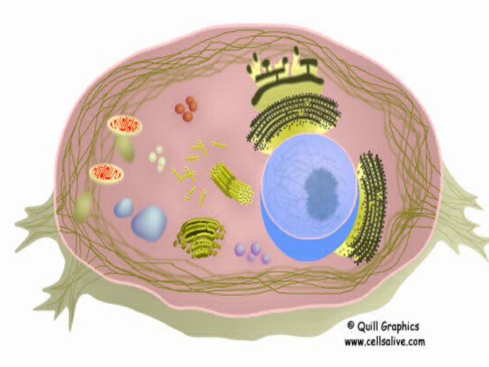
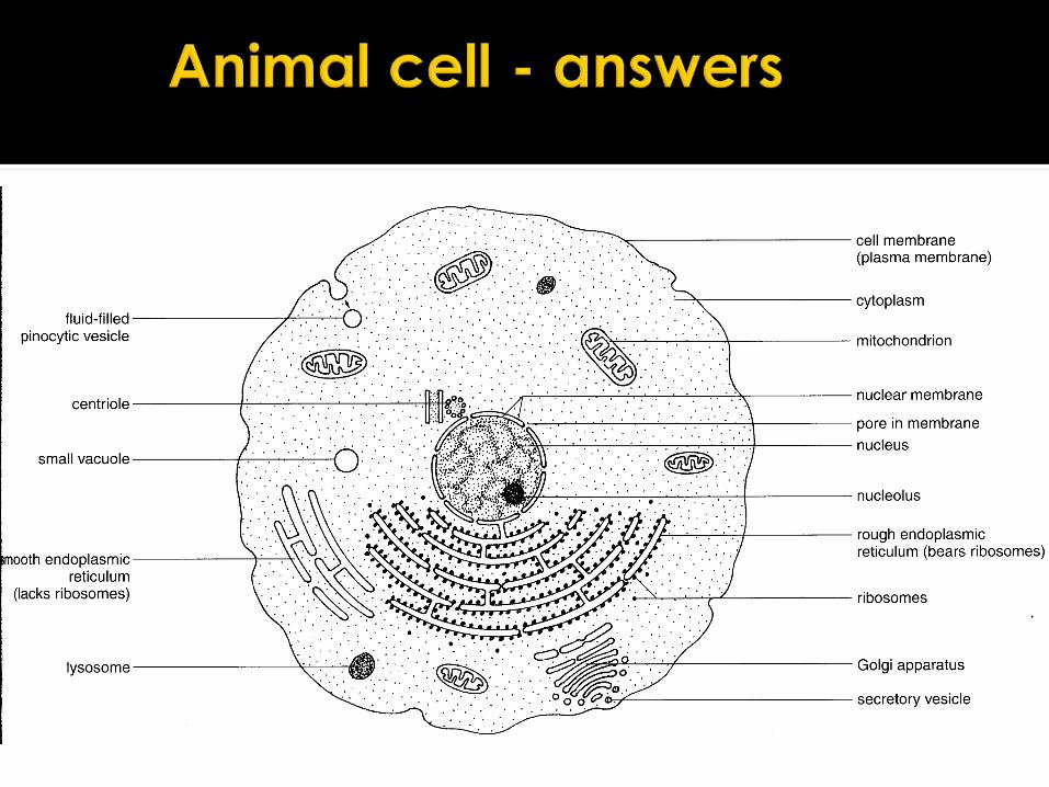
Ultrastructure of an animal cell as seen through an electron microscope

Cell structure Read through the information on each of
the organelles as you colour them in Follow the guidance on colouring them
in given at the bottom of the page
This works on the theory that whilst you are colouring in, you have time to consider and think about the structure and function of the organelles
OCR AS BiologyFOUNDATION
In pairs label the diagram of the animal cell given. How many structures can you identify? Look at the cells alive animation – how
many have you correctly identified?
Label the paper copy of the diagram of an animal cell


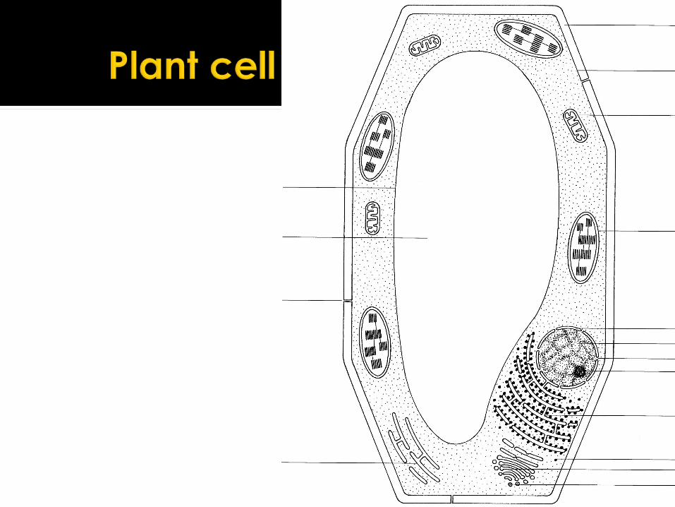
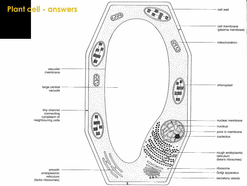
In pairs label the diagram of the plant cell given. How many structures can you identify? Look at the cells alive animation – how
many have you correctly identified?
Label the paper copy of a diagram of a plant cell

Using the cards, match up organelle, position and function.
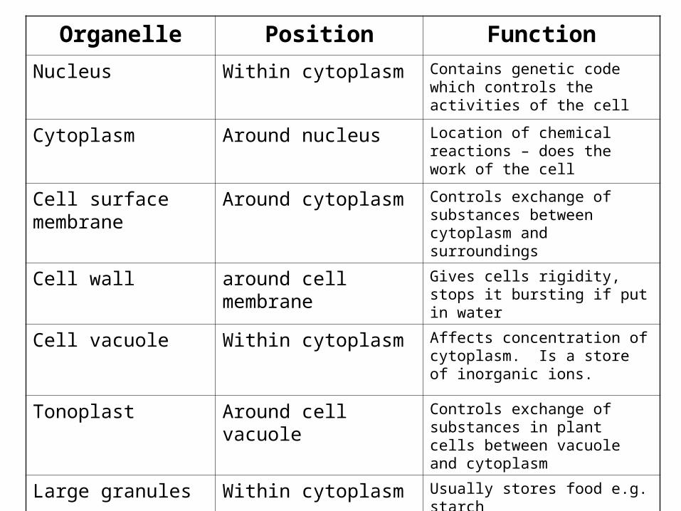
Organelle Position Function
Nucleus Within cytoplasm Contains genetic code which controls the activities of the cell
Cytoplasm Around nucleus Location of chemical reactions – does the work of the cell
Cell surface membrane
Around cytoplasm Controls exchange of substances between cytoplasm and surroundings
Cell wall around cell membrane
Gives cells rigidity, stops it bursting if put in water
Cell vacuole Within cytoplasm Affects concentration of cytoplasm. Is a store of inorganic ions.
Tonoplast Around cell vacuole Controls exchange of substances in plant cells between vacuole and cytoplasm
Large granules Within cytoplasm Usually stores food e.g. starch

OCR AS BiologyFOUNDATION
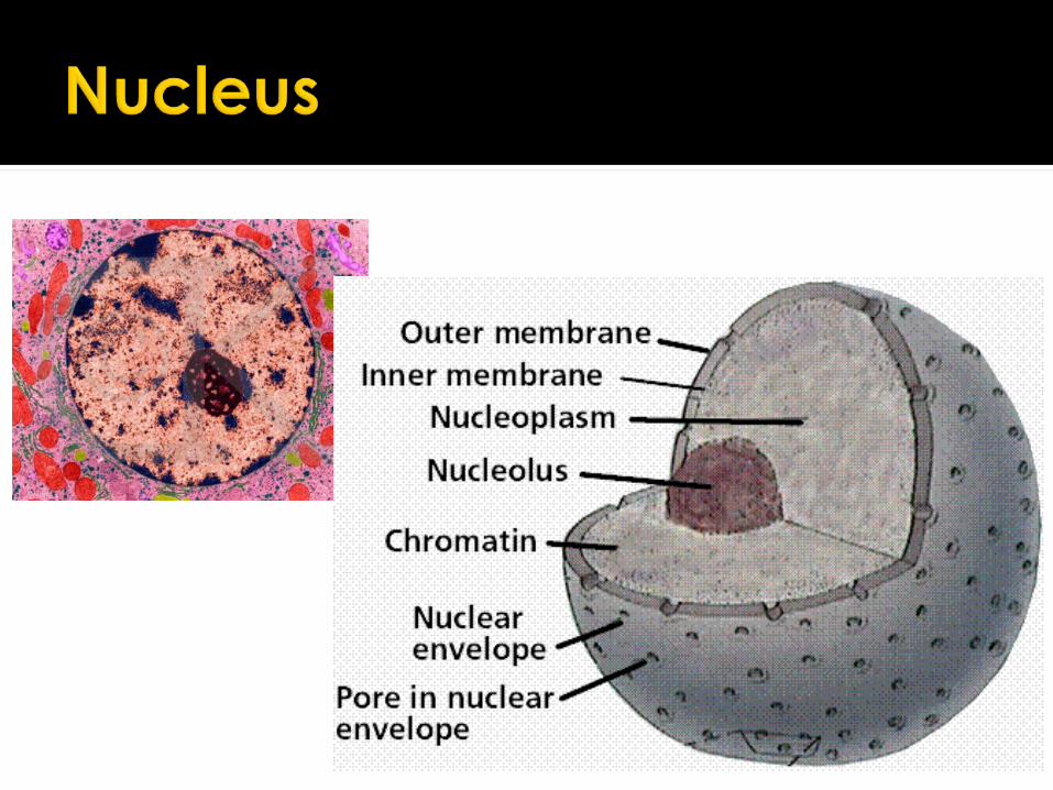

Nuclear envelopeNuclear poresNucleoplasmChromatinnuceolus
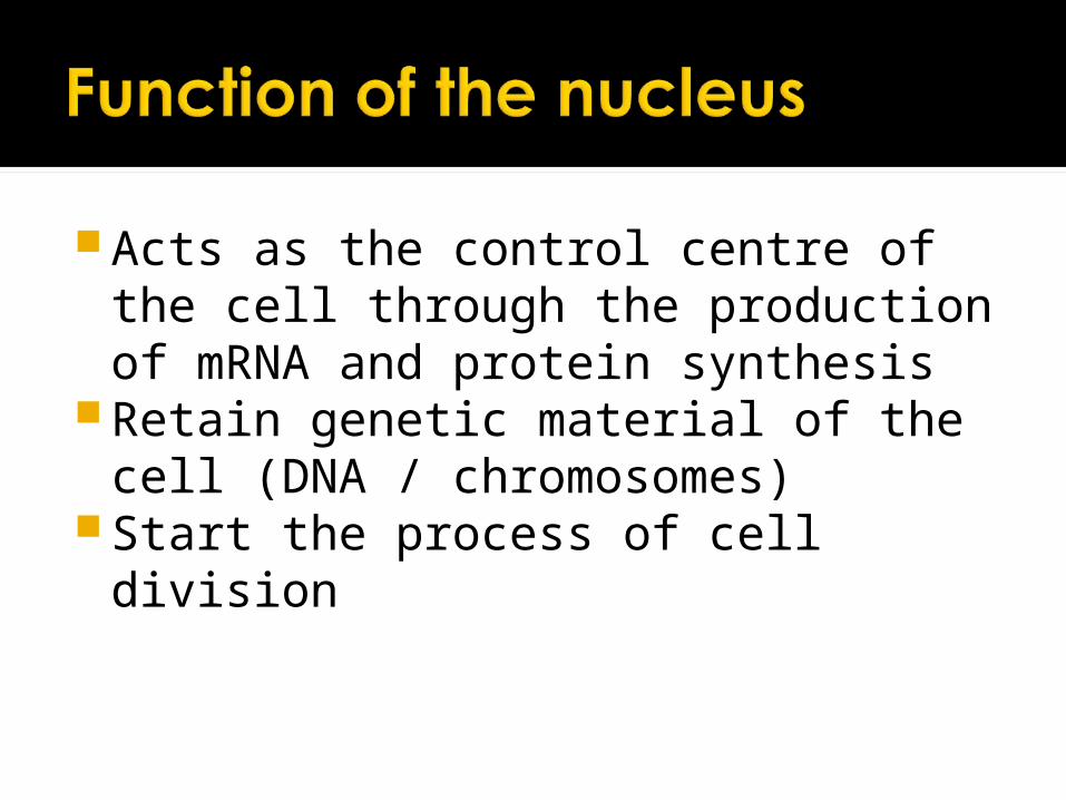
Acts as the control centre of the cell through the production of mRNA and protein synthesis
Retain genetic material of the cell (DNA / chromosomes)
Start the process of cell division
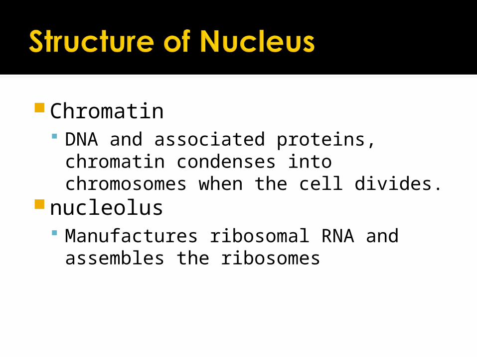
Chromatin DNA and associated proteins, chromatin
condenses into chromosomes when the cell divides.
nucleolus Manufactures ribosomal RNA and
assembles the ribosomes

Nuclear envelope Controls entry and exit of materials Outer membrane continuous with
endoplasmic reticulumNuclear pores
Passage of large molecules (mRNA) out of nucleus
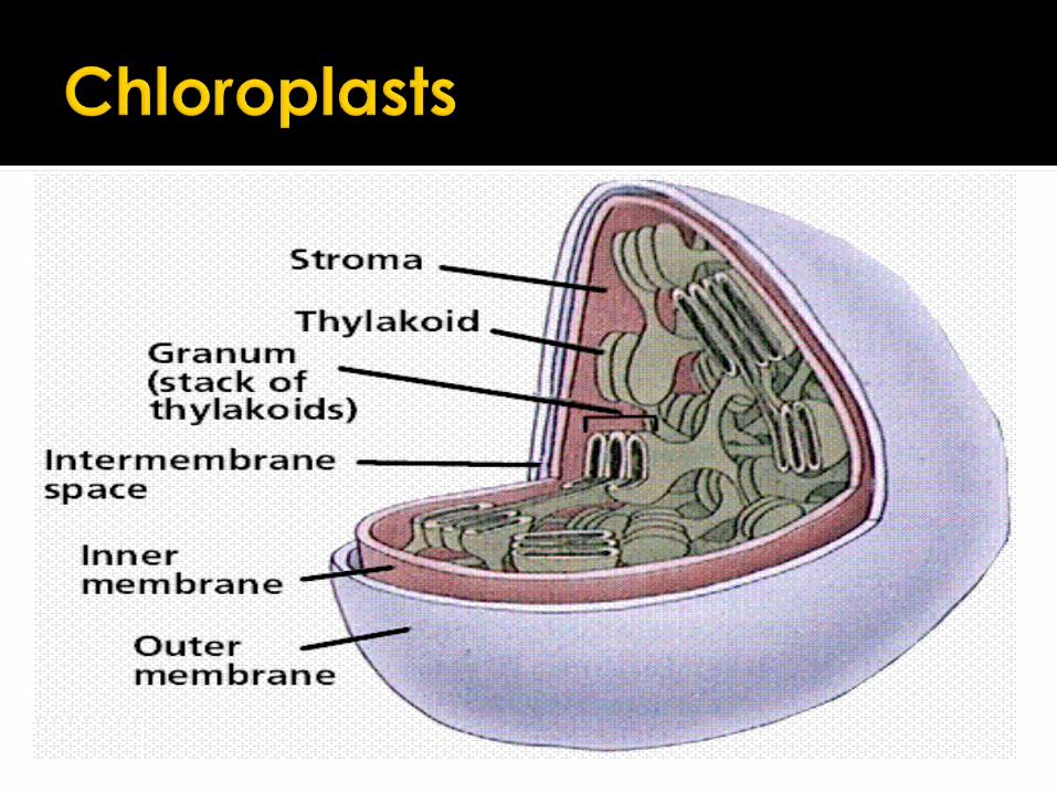

Chloroplast envelope Entry and exit of substances
Stroma Enzymes for the light independent
stages of photosynthesisGrana (thylakoids/lamellae)
Light dependent stage of photosynthesisStarch grains
Temporary stores of carbohydrates
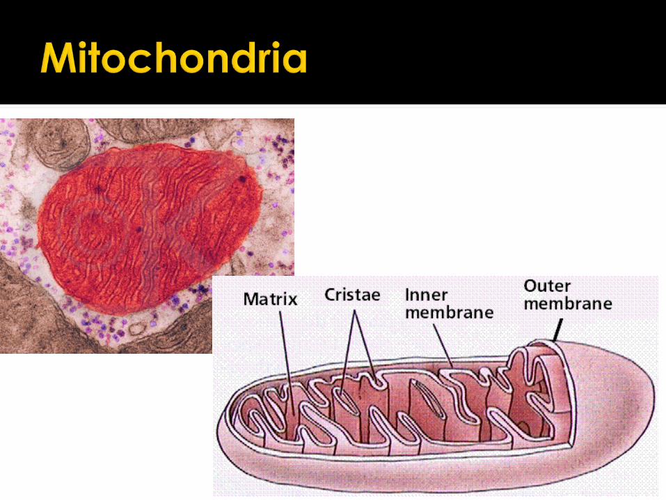

Double membrane Inner membrane folded into cristae
which provide a large surface areaMatrix
DNA, enzymes and ribosomes

Site of Krebs cycle and oxidative phosphorylation in aerobic respiration
Production of energy rich ATP molecules from carbohydrates
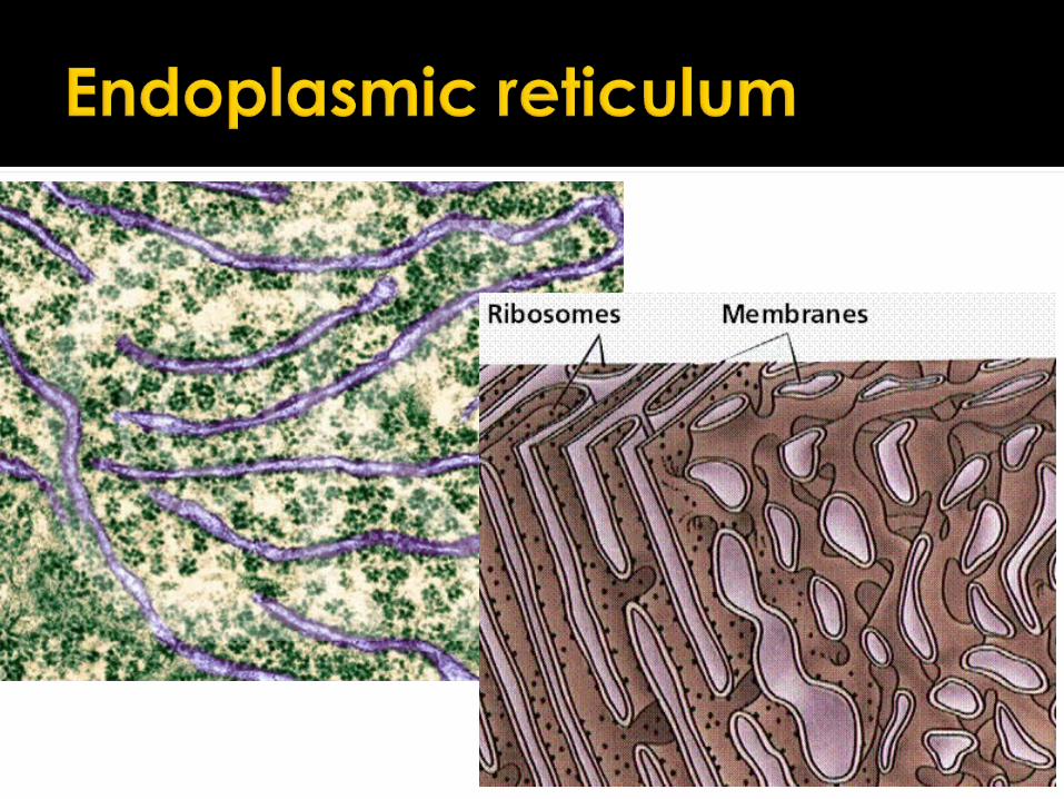
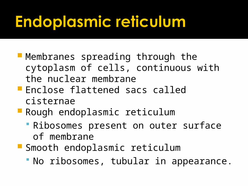
Membranes spreading through the cytoplasm of cells, continuous with the nuclear membrane
Enclose flattened sacs called cisternae Rough endoplasmic reticulum
Ribosomes present on outer surface of membrane
Smooth endoplasmic reticulum No ribosomes, tubular in appearance.

RER Provide LSA for synthesis of proteins Provides a pathway for the transport of
materials (esp. proteins) throughout the cell.
SER Synthesis, stores and transports lipids
and carbohydrates Contains lytic enzymes (liver cells)


Two types 80S – eukaryotic cells 70S – prokaryotic cells
Make up 25% of dry mass of cell
Important in protein synthesis

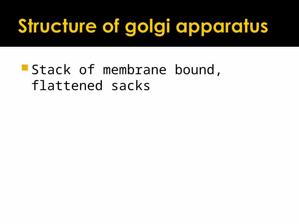
Stack of membrane bound, flattened sacks
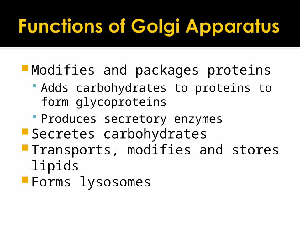
Modifies and packages proteins Adds carbohydrates to proteins to form
glycoproteins Produces secretory enzymes
Secretes carbohydrates Transports, modifies and stores lipidsForms lysosomes

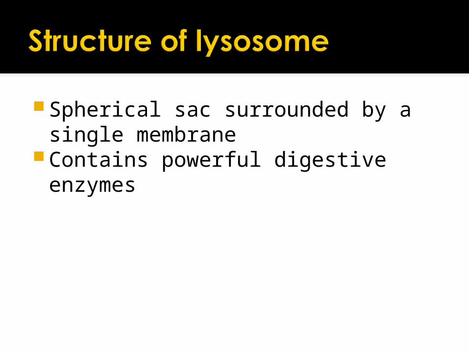
Spherical sac surrounded by a single membrane
Contains powerful digestive enzymes

Destroy foreign material inside or outside the cell. Breakdown material ingested by
phagocytic cells Release enzymes outside the cell Digest worn out organelles (autophagy) Autolysis break down cells after they
have died.
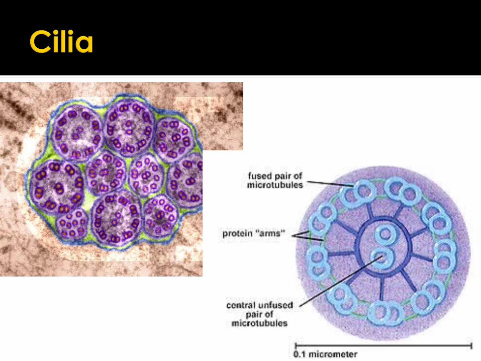
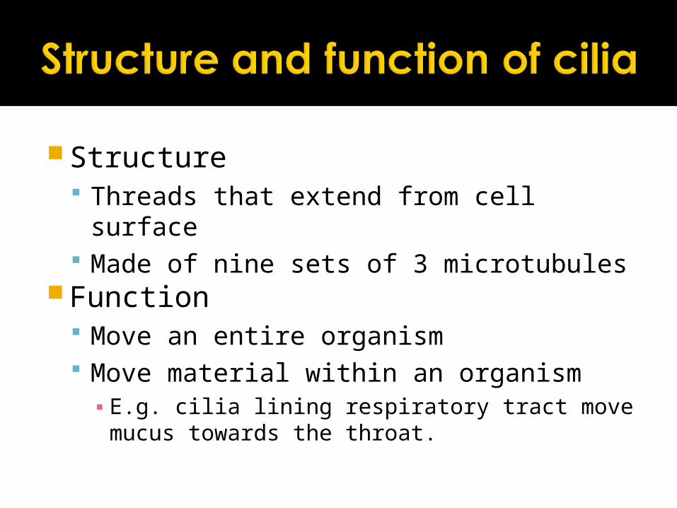
Structure Threads that extend from cell surface Made of nine sets of 3 microtubules
Function Move an entire organism Move material within an organism▪ E.g. cilia lining respiratory tract move mucus
towards the throat.
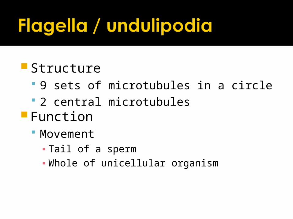
Structure 9 sets of microtubules in a circle 2 central microtubules
Function Movement▪ Tail of a sperm▪ Whole of unicellular organism
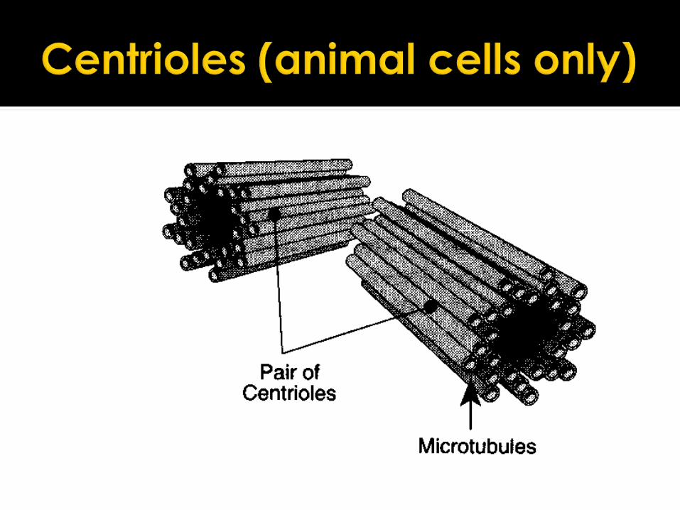
Hollow cylinders of microtubulesMicrotubules form spindle fibres for
nuclear divisionMaybe involved in formation of
microtubules that make up cells cytoskeleton
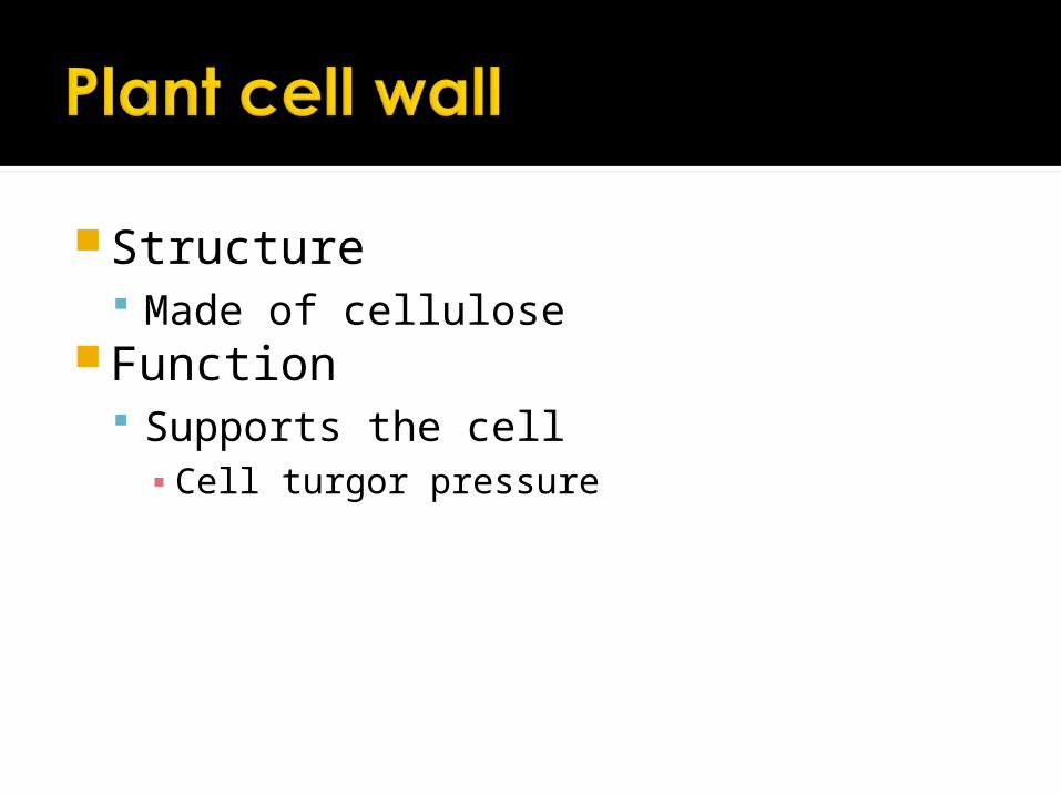
Structure Made of cellulose
Function Supports the cell▪ Cell turgor pressure

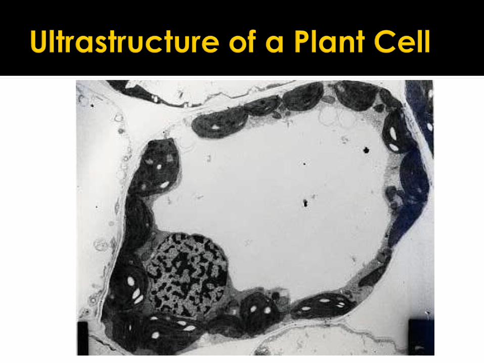
Similarities between plant and animal cells Make a list of the structures plant and
animal cells have in common Make a table of the differences between
plant and animal cells
Include all structures in plant and animal cells not just the ones observed through a Light microscope
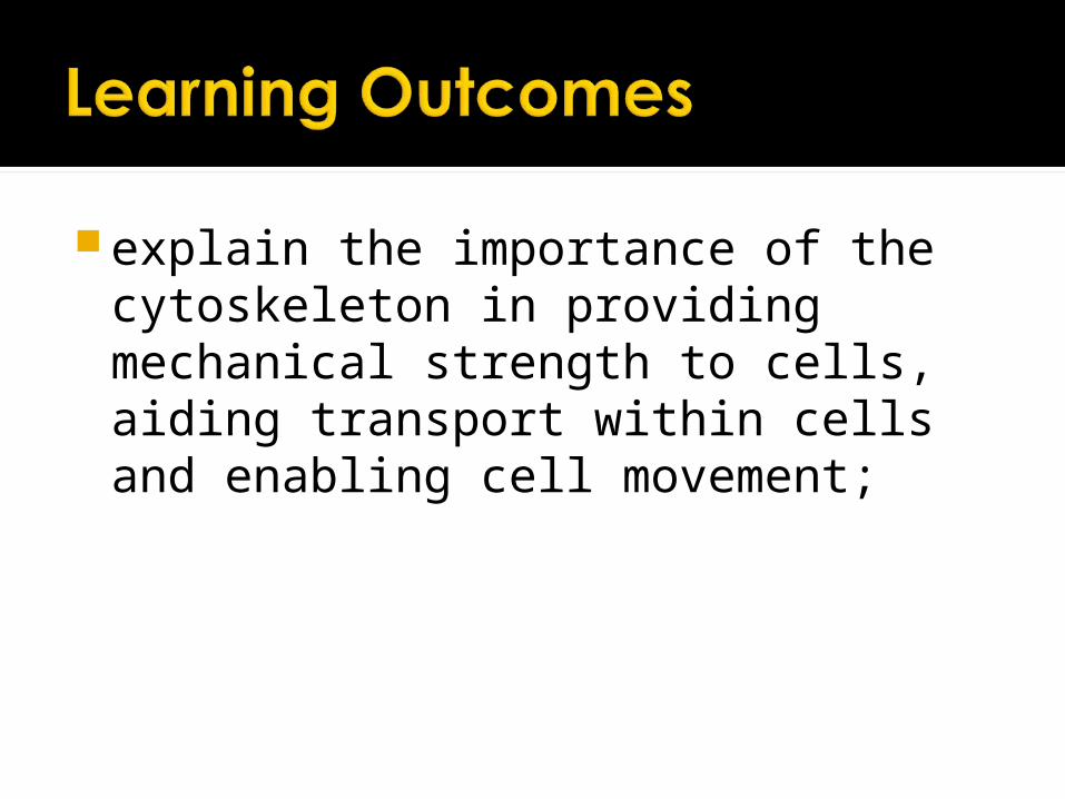
explain the importance of the cytoskeleton in providing mechanical strength to cells, aiding transport within cells and enabling cell movement;

Cells contain a network of fibres made of protein, providing an internal framework.
Fibres can move organelles round within a cell. Microtubules
Move chromosomes around in cell division Move vesicles from endoplasmic reticulum to Golgi
apparatus ATP is used to drive some of these movements


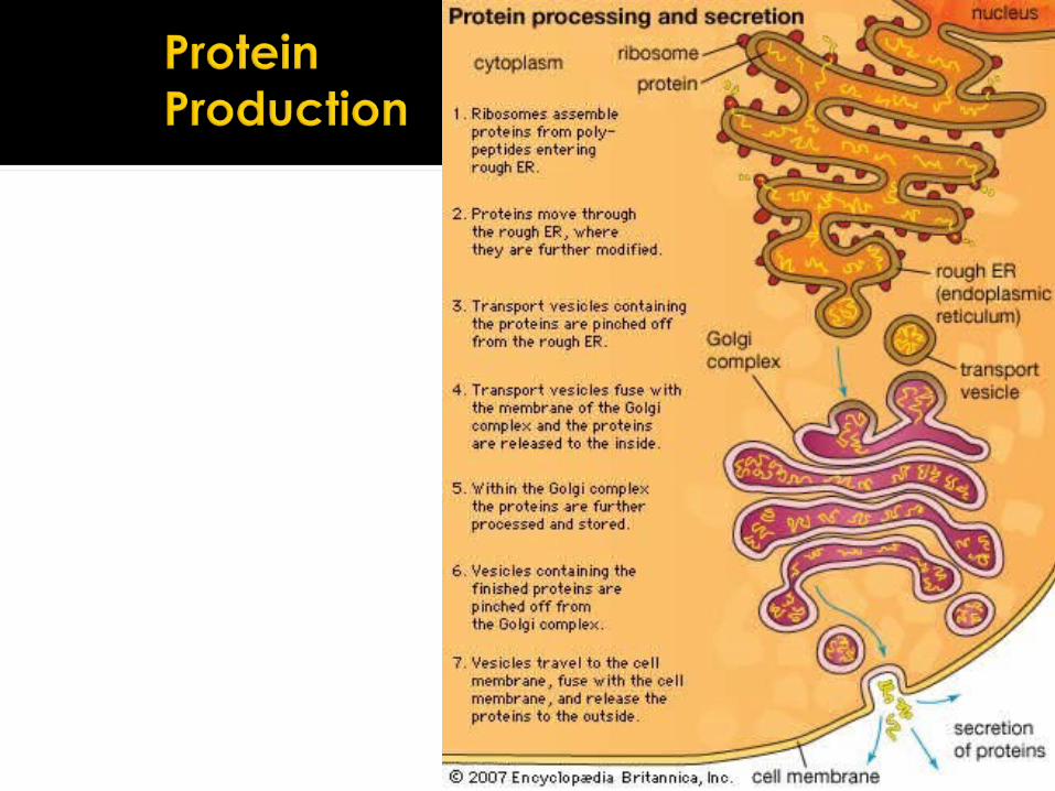
outline the interrelationship between the organelles involved in the production and secretion of proteins
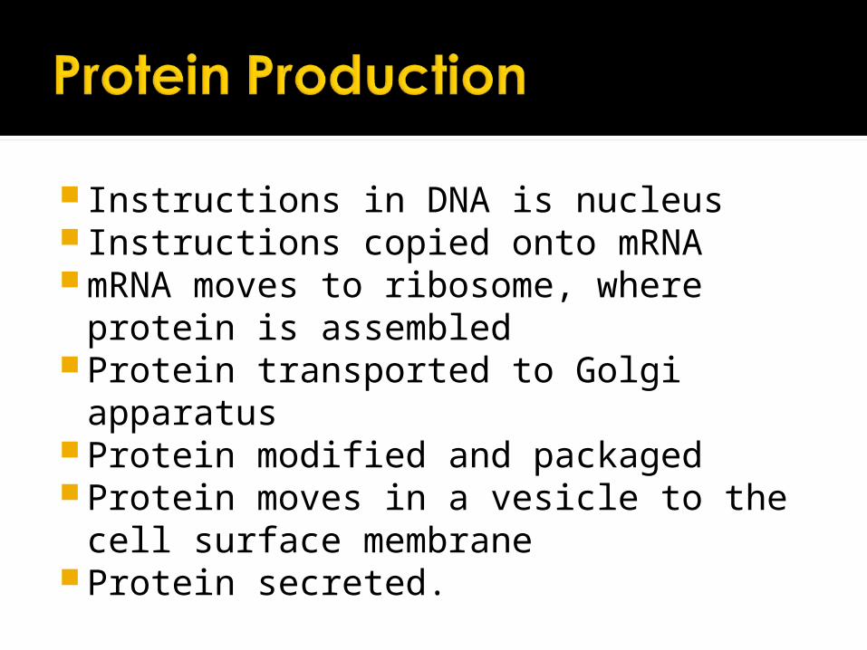
Instructions in DNA is nucleus Instructions copied onto mRNA mRNA moves to ribosome, where
protein is assembled Protein transported to Golgi apparatus Protein modified and packaged Protein moves in a vesicle to the cell
surface membrane Protein secreted.
OCR AS BiologyFOUNDATION

Prokaryotes were probably the first forms of life on earth. Their heredity material (DNA) is not enclosed within a nuclear membrane. There are no membrane bound organelles within a prokaryotic cell.
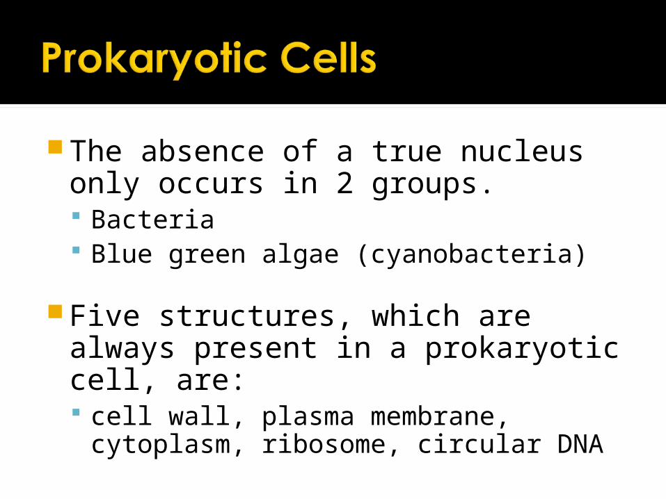
The absence of a true nucleus only occurs in 2 groups. Bacteria Blue green algae (cyanobacteria)
Five structures, which are always present in a prokaryotic cell, are: cell wall, plasma membrane, cytoplasm,
ribosome, circular DNA

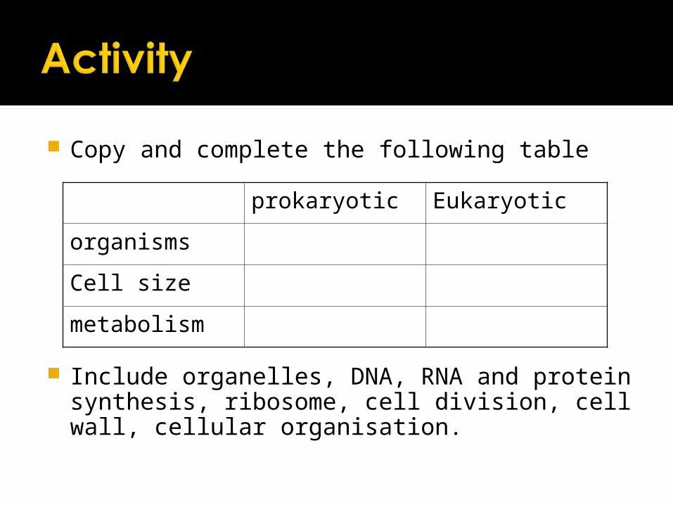
Copy and complete the following table
Include organelles, DNA, RNA and protein synthesis, ribosome, cell division, cell wall, cellular organisation.
prokaryotic Eukaryotic
organisms
Cell size
metabolism
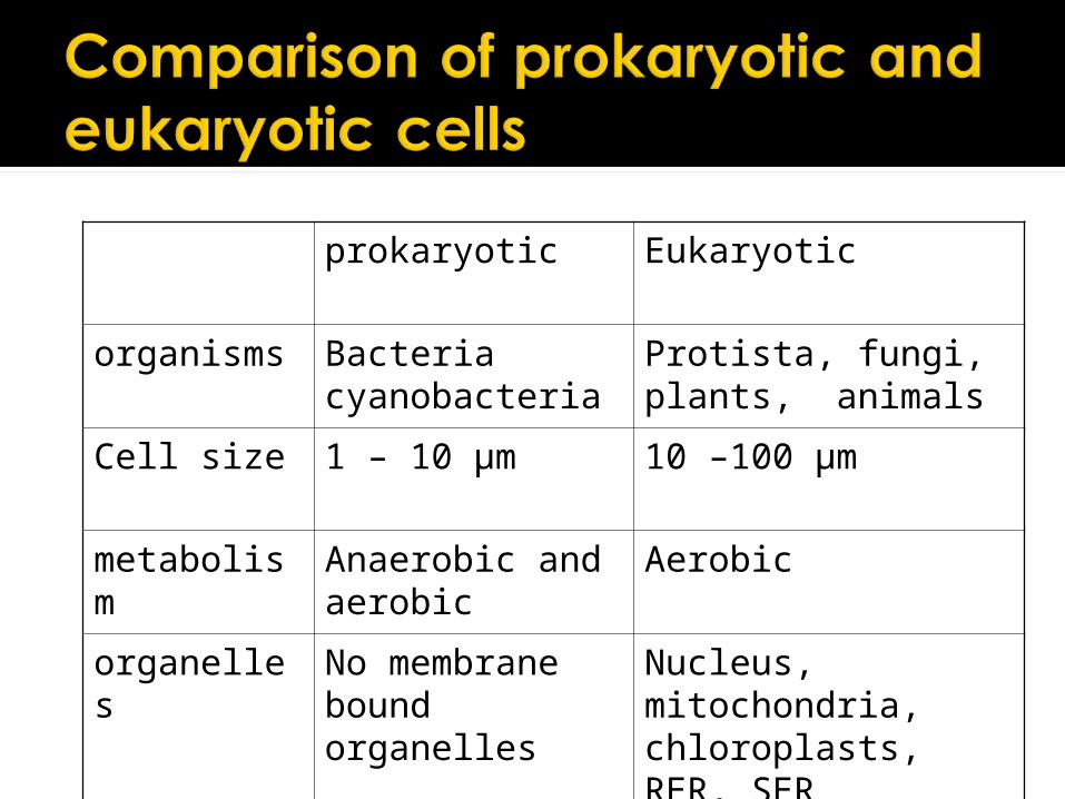
prokaryotic Eukaryotic
organisms Bacteriacyanobacteria
Protista, fungi, plants, animals
Cell size 1 – 10 µm 10 –100 µm
metabolism Anaerobic and aerobic
Aerobic
organelles No membrane bound organelles
Nucleus, mitochondria, chloroplasts, RER, SER
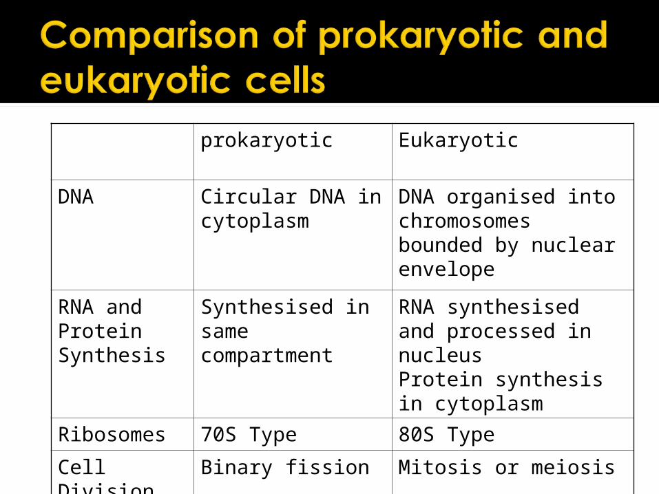
prokaryotic Eukaryotic
DNA Circular DNA in cytoplasm
DNA organised into chromosomes bounded by nuclear envelope
RNA and Protein Synthesis
Synthesised in same compartment
RNA synthesised and processed in nucleusProtein synthesis in cytoplasm
Ribosomes 70S Type 80S Type
Cell Division Binary fission Mitosis or meiosis
Cell Membrane
Learning Objectives
outline the roles of membranes within cells and at the surface of cells
state that plasma (cell surface) membranes are partially permeable barriers
describe, with the aid of diagrams, the fluid mosaic model of membrane structure
describe the roles of the components of the cell membrane; phospholipids, cholesterol, glycolipids, proteins and glycoproteins;
outline the effect of changing temperature on membrane structure and permeability
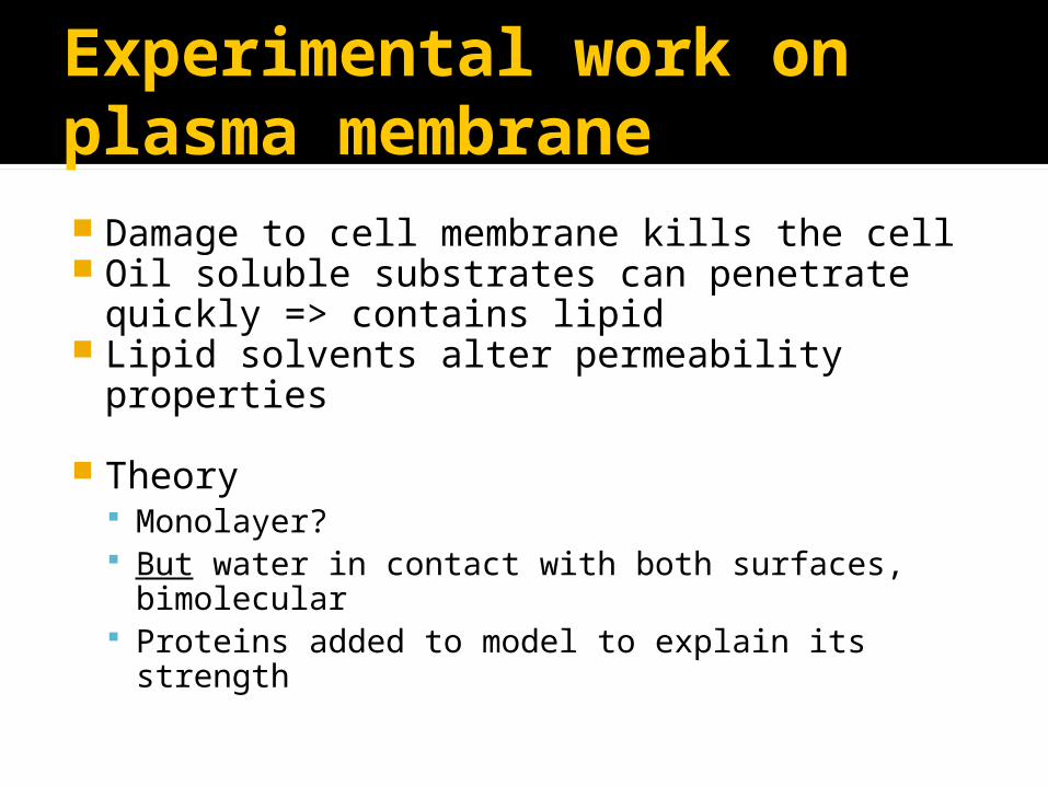
Experimental work on plasma membrane Damage to cell membrane kills the cell Oil soluble substrates can penetrate
quickly => contains lipid Lipid solvents alter permeability
properties
Theory Monolayer? But water in contact with both surfaces,
bimolecular Proteins added to model to explain its strength

Davson and Danielli1935
Lipid bilayer coated on both sides with a layer of protein molecules
Calculated that thickness of membrane was about 7.5nm.
Phospholipid bilayer
Protein molecule

Singer and Nicholson 1972Fluid mosaic modelCell membranes have a consistency
like oil, with sideways movement of molecules or membrane. Fluid ▪ individual phospholipid and protein molecules
move around within their layer Mosaic▪ pattern produced by scattered protein
molecules when surface membrane is viewed from above.

Fluid Mosaic Model
A phospholipid bilayerTransmembrane and peripheral
proteinsCholesterolGlycoproteins Glycolipid
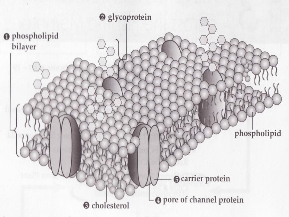

Cell Surface Membrane

Functions of the plasma membrane
Providing a partially permeable barrier
CompartmentalisationLocalising reactions in a cellTransport of solutesSignal transductionCell-cell recognition
Applying your knowledge questions
why can phospholipid molecules in a bilayer move only in the plane of the bilayer? Phosphate head can not pass through
the hydrophobic region in the centre of the bilayer
Applying your knowledge questions
Why do we describe cell membranes as partially permeable rather than semi-permeable? Different membranes are permeable to a
variety of substances and impermeable to a variety of others.
Semi-permeable suggests “half-permeable” which is unlikely to be the case in any membrane.

Roles of Components of Membrane
Phospholipid Can form sheets (bilayer) Form membrane bound compartments. Act as a barrier to most water soluble
substances

Roles of Components of Membrane
Cholesterol Helps regulate fluidity of membrane Stabilises phospholipid bilayer Prevent ions/polar molecules passing
through, important in myelin sheath around nerve cells.

Roles of Components of Membrane
Proteins Intrinsic proteins – span membrane Extrinsic proteins – embedded in one
half of membrane Channel forming proteins Carrier protein molecules
Roles of Components of Membrane
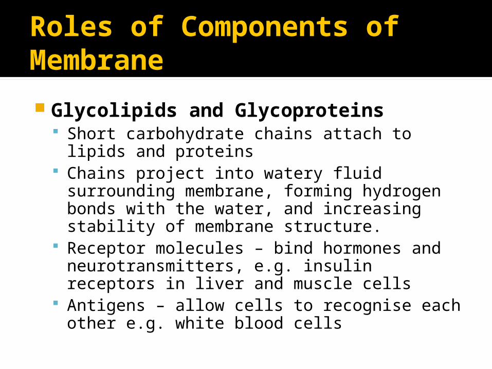
Glycolipids and Glycoproteins Short carbohydrate chains attach to lipids and
proteins Chains project into watery fluid surrounding
membrane, forming hydrogen bonds with the water, and increasing stability of membrane structure.
Receptor molecules – bind hormones and neurotransmitters, e.g. insulin receptors in liver and muscle cells
Antigens – allow cells to recognise each other e.g. white blood cells

Investigating Cell Membranes
Analysis of pigment leakage from beetroot cells
Two investigations Investigating the effects of temperature
on the cell membrane Investigating the effects of ethanol on
the cell membrane
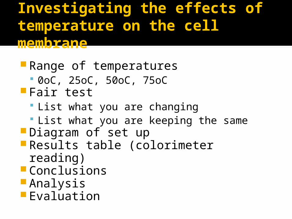
Investigating the effects of temperature on the cell membraneRange of temperatures
0oC, 25oC, 50oC, 75oCFair test
List what you are changing List what you are keeping the same
Diagram of set upResults table (colorimeter reading)ConclusionsAnalysisEvaluation
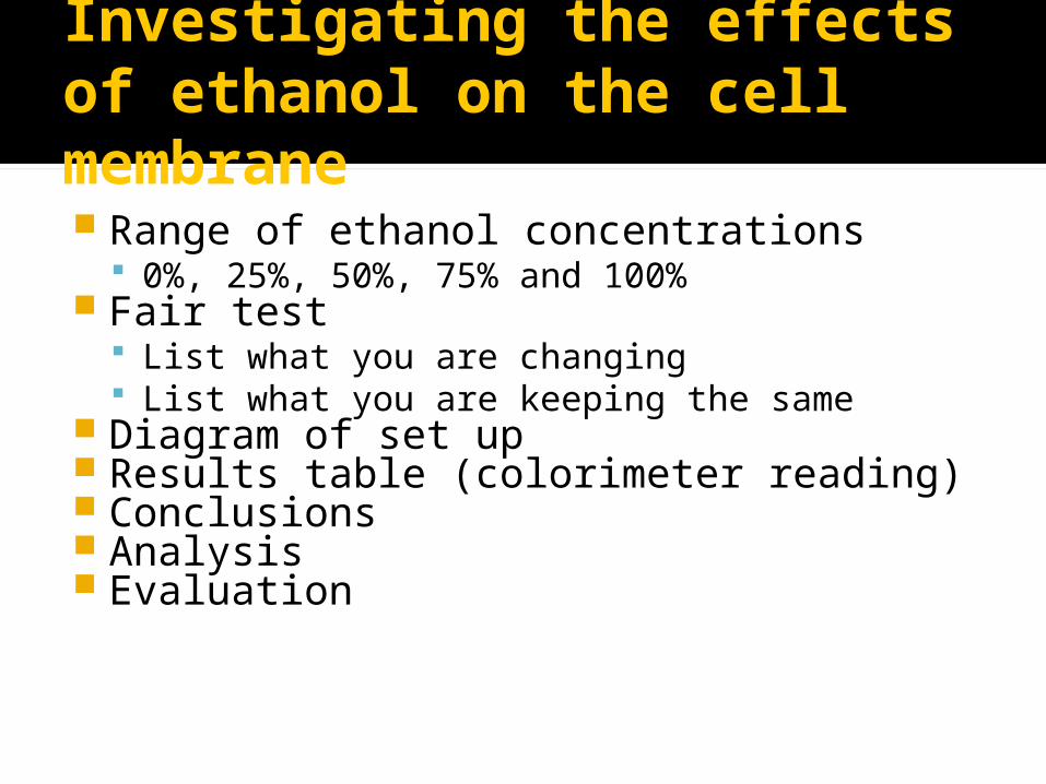
Investigating the effects of ethanol on the cell membrane Range of ethanol concentrations
0%, 25%, 50%, 75% and 100% Fair test
List what you are changing List what you are keeping the same
Diagram of set up Results table (colorimeter reading) Conclusions Analysis Evaluation
Cell Signalling
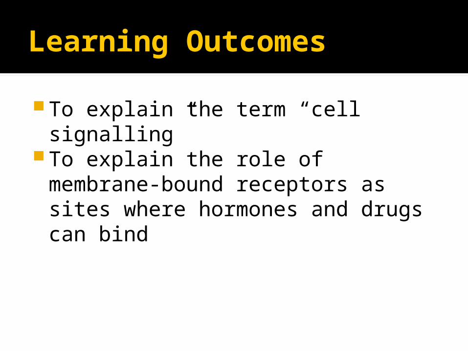
Learning Outcomes
To explain the term “cell signalling”To explain the role of membrane-
bound receptors as sites where hormones and drugs can bind

Cell Signalling
Cells communicate with each other by signals
Cells must be able to detect various internal and external signals in order to co-ordinate the life processes of growth, development, movement and excretion.
Receptors can be Internal – e.g. steroid receptors External – e.g. insulin receptors
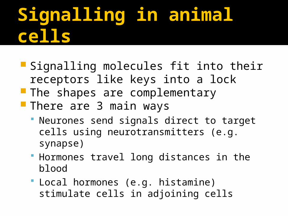
Signalling in animal cells
Signalling molecules fit into their receptors like keys into a lock
The shapes are complementary There are 3 main ways
Neurones send signals direct to target cells using neurotransmitters (e.g. synapse)
Hormones travel long distances in the blood Local hormones (e.g. histamine) stimulate cells
in adjoining cells

Hormones
Endocrine cells secrete hormones into the blood stream
Target cells have receptors for the hormone molecule
When the hormone binds to a receptor the cell responds

Insulin
The pancreas secretes insulin, which is large and water soluble
Insulin binds to receptor molecules on the cell surface membrane of liver and muscle cells
This increases glucose channels in the cell membrane
Cells uptake more glucose, which lowers the blood glucose levels

Local hormones
These only travel short distances to adjoining cells e.g. Histamine Cytokines▪ Stimulate lymphocytes to divide by mitosis
and produce antibodies▪ Stimulates phagocytes to become more
active
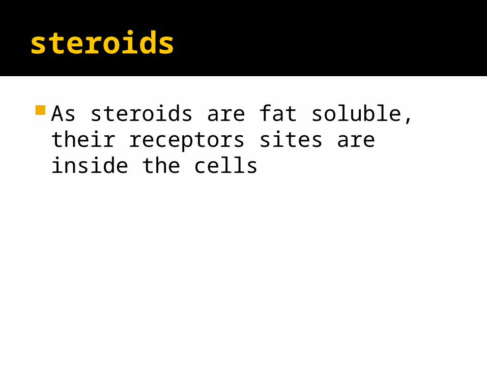
steroids
As steroids are fat soluble, their receptors sites are inside the cells
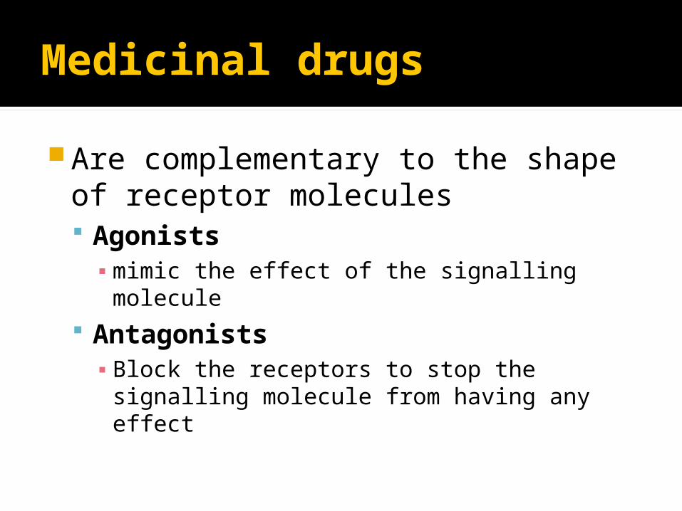
Medicinal drugs
Are complementary to the shape of receptor molecules Agonists ▪ mimic the effect of the signalling molecule
Antagonists▪ Block the receptors to stop the signalling
molecule from having any effect
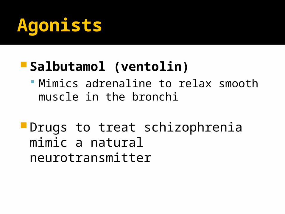
Agonists
Salbutamol (ventolin) Mimics adrenaline to relax smooth
muscle in the bronchi
Drugs to treat schizophrenia mimic a natural neurotransmitter
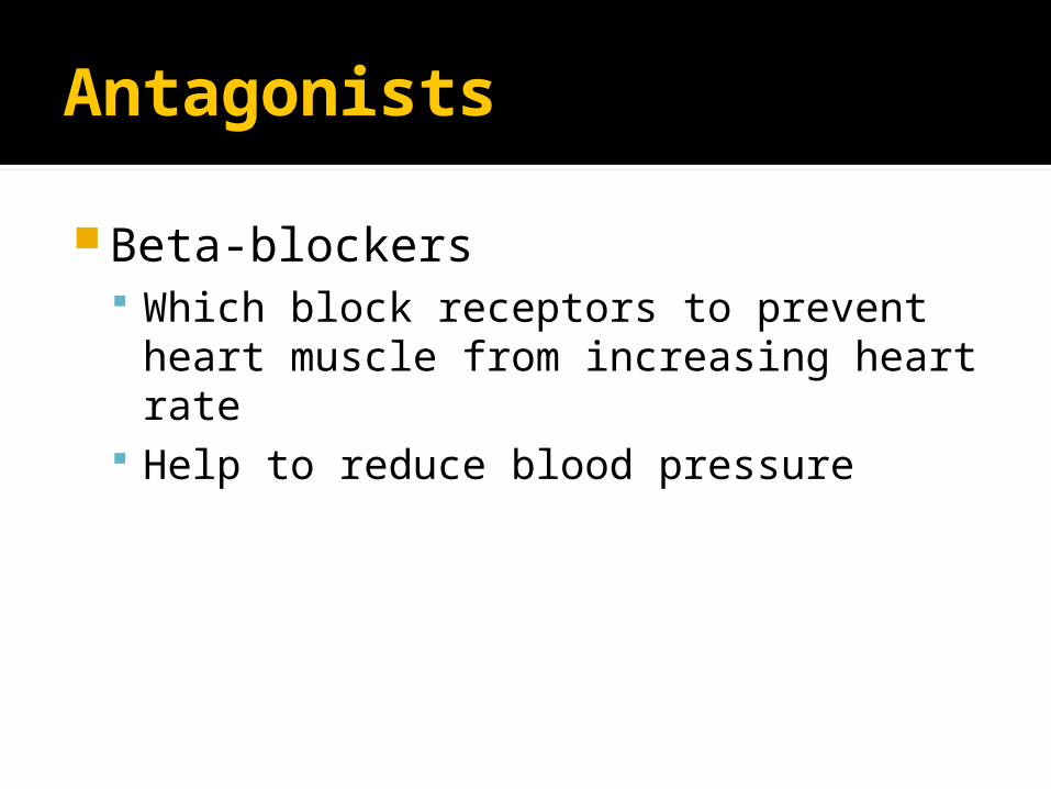
Antagonists
Beta-blockers Which block receptors to prevent heart
muscle from increasing heart rate Help to reduce blood pressure

Hijacking cells
Some viruses can bind with receptors on the cell surface membrane HIV and helper T lymphocytes
Some poison can bind with receptors BOTOX – toxin binds with receptors on
muscle fibres and prevents them from working causing paralysis

Transport across boundaries

Learning Outcomes
explain what is meant by passive transport (diffusion and facilitated diffusion including the role of membrane proteins), active transport, endocytosis and exocytosis;
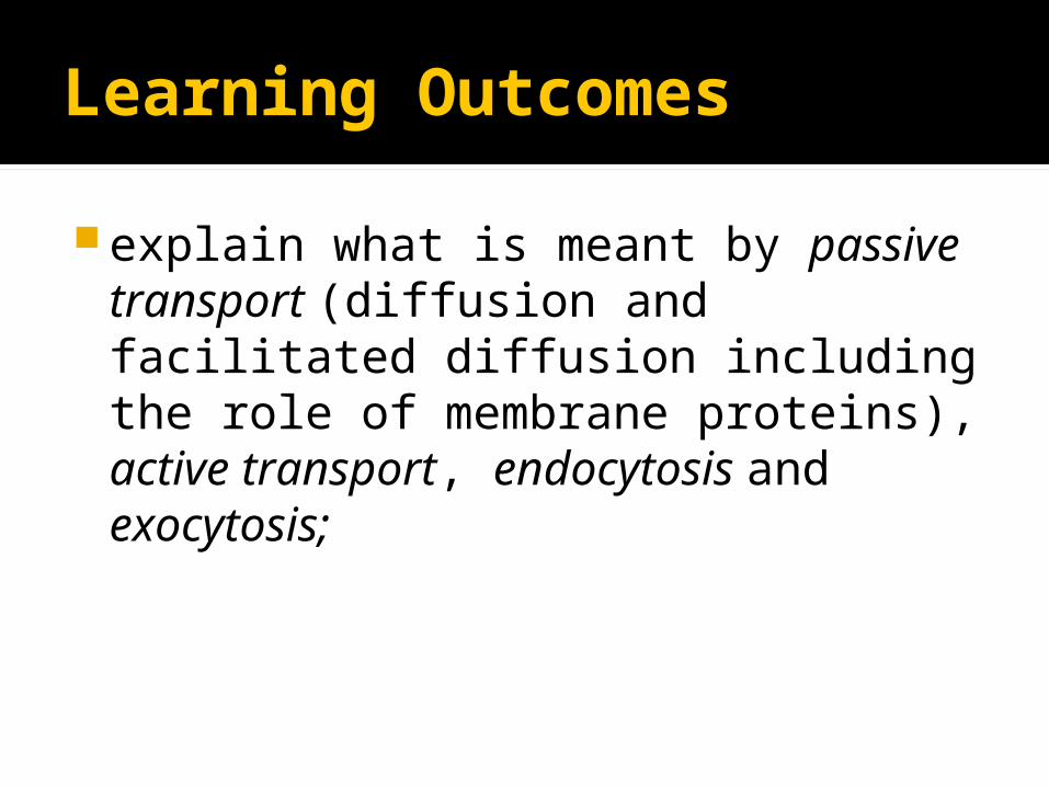
Learning Outcomes
explain what is meant by passive transport (diffusion and facilitated diffusion including the role of membrane proteins), active transport, endocytosis and exocytosis;
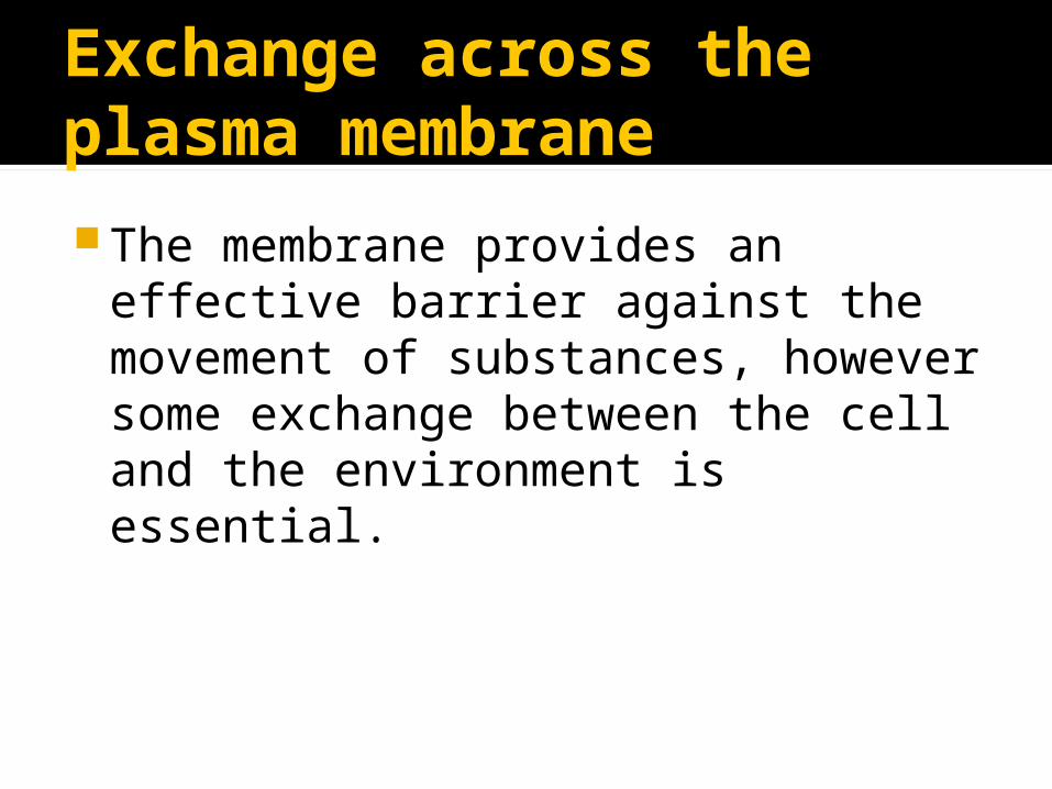
Exchange across the plasma membrane
The membrane provides an effective barrier against the movement of substances, however some exchange between the cell and the environment is essential.
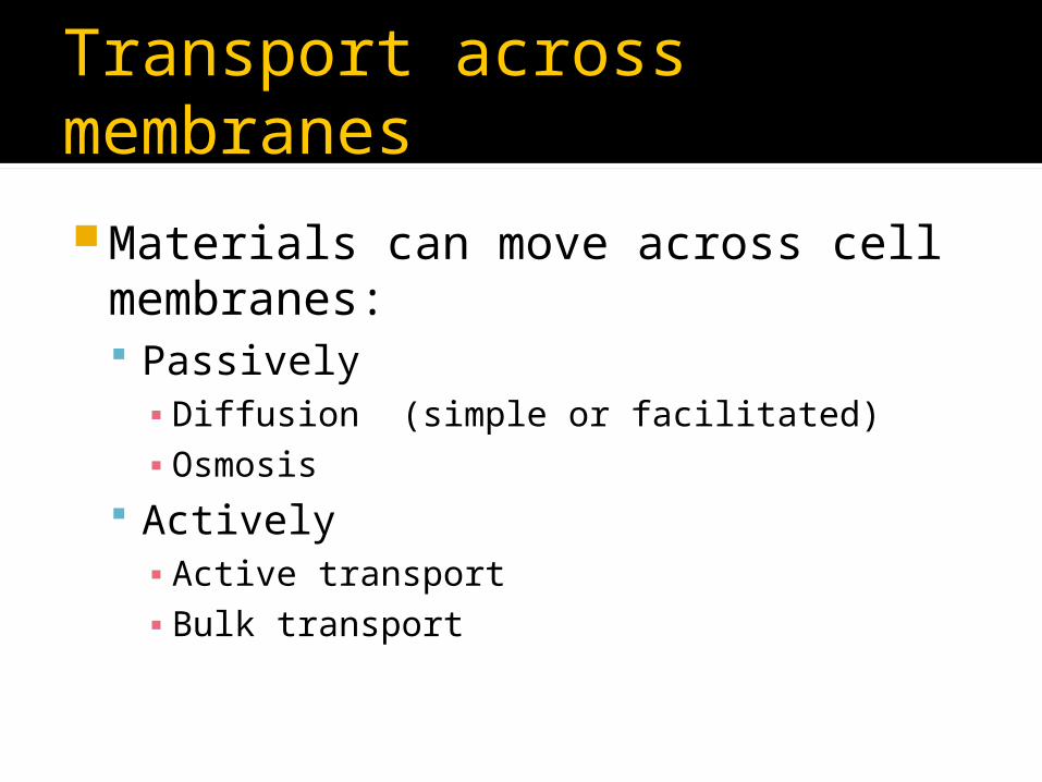
Transport across membranes
Materials can move across cell membranes: Passively▪ Diffusion (simple or facilitated)▪ Osmosis
Actively▪ Active transport▪ Bulk transport

Diffusion
Net movement of molecules or ions from a region of high concentration to a region of low concentration
Occurs along a concentration gradient
Result = equilibrium (molecules or ions evenly spread out within a given space or volume)

Factors affecting the rate of diffusion Concentration gradient
Greater the difference in concentration the greater the rate of diffusion
Temperature At higher temperature kinetic energy particles
increases Diffusion is faster
Surface area Greater the surface area, more particles can
cross Increases rate of diffusion
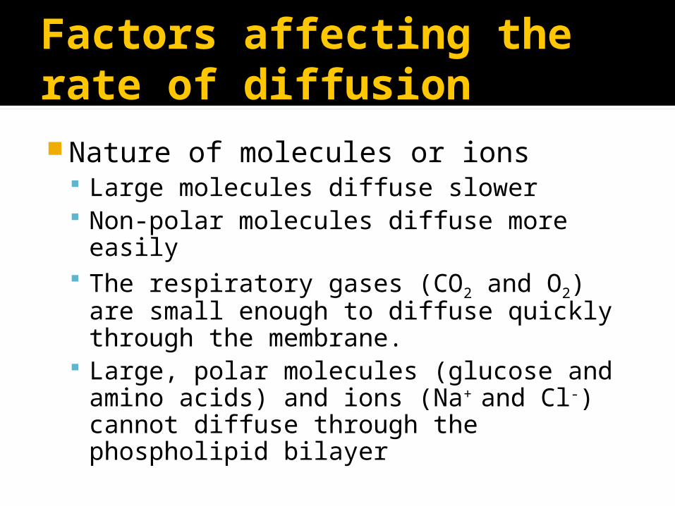
Factors affecting the rate of diffusionNature of molecules or ions
Large molecules diffuse slower Non-polar molecules diffuse more easily The respiratory gases (CO2 and O2) are
small enough to diffuse quickly through the membrane.
Large, polar molecules (glucose and amino acids) and ions (Na+ and Cl-) cannot diffuse through the phospholipid bilayer
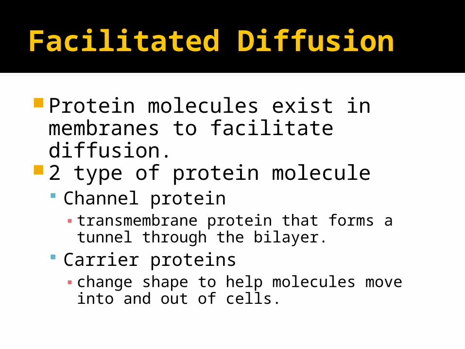
Facilitated Diffusion
Protein molecules exist in membranes to facilitate diffusion.
2 type of protein molecule Channel protein ▪ transmembrane protein that forms a tunnel
through the bilayer. Carrier proteins ▪ change shape to help molecules move into
and out of cells.

Facilitated Diffusion
Active Transport
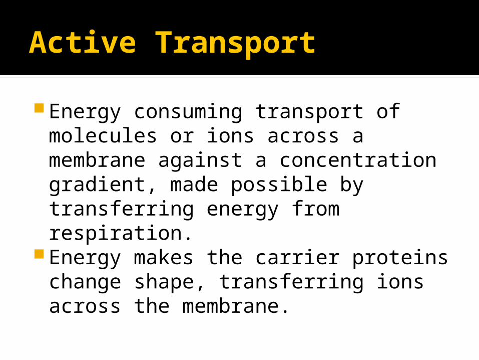
Energy consuming transport of molecules or ions across a membrane against a concentration gradient, made possible by transferring energy from respiration.
Energy makes the carrier proteins change shape, transferring ions across the membrane.
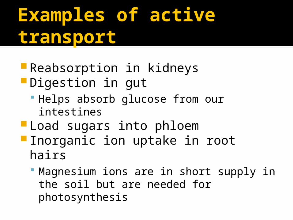
Examples of active transport
Reabsorption in kidneysDigestion in gut
Helps absorb glucose from our intestinesLoad sugars into phloem Inorganic ion uptake in root hairs
Magnesium ions are in short supply in the soil but are needed for photosynthesis

Bulk transport
This is the method of transporting large quantities of materials into cells (endocytosis) or out of cells (exocytosis) Endocytosis - Engulfing of material by cell
membrane to form a endocytic vacuole.▪ 2 forms▪ Phagocytosis the uptake of solid material▪ Pinocytosis the uptake of liquid
Exocytosis - Process by which materials are removed from cells
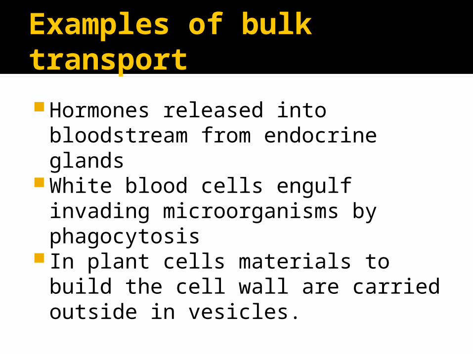
Examples of bulk transport
Hormones released into bloodstream from endocrine glands
White blood cells engulf invading microorganisms by phagocytosis
In plant cells materials to build the cell wall are carried outside in vesicles.
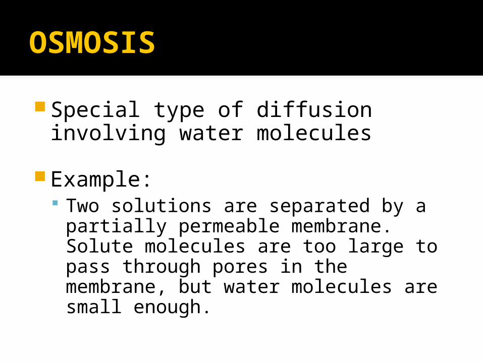
OSMOSIS
Special type of diffusion involving water molecules
Example: Two solutions are separated by a
partially permeable membrane. Solute molecules are too large to pass through pores in the membrane, but water molecules are small enough.
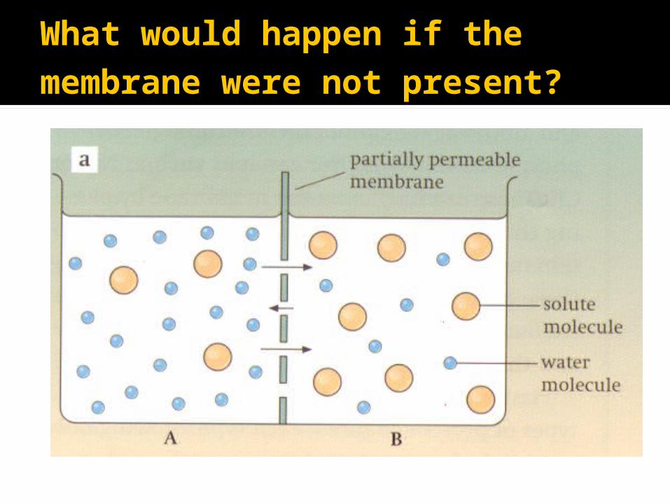
What would happen if the membrane were not present?

Net movement of solute molecules from B to A by diffusion
Net movement of water molecules from A to B by diffusion
Equilibrium – concentrations of water molecules and solute molecules in A would equal that in B.

What will happen if the membrane is present?
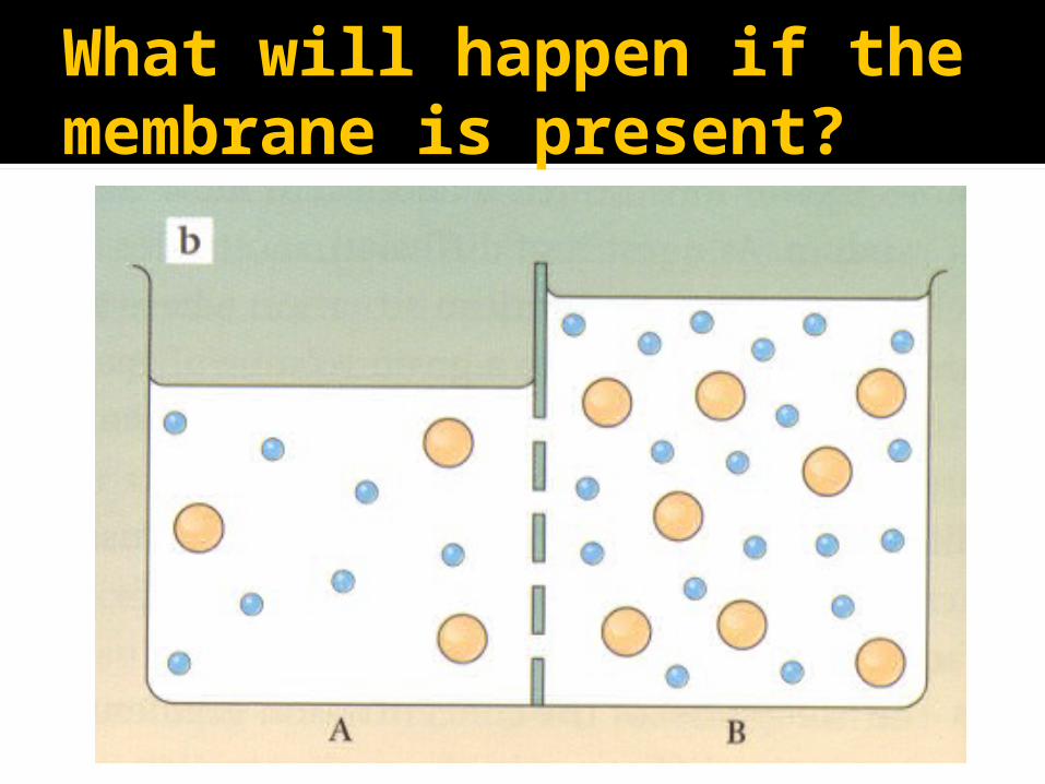
What will happen if the membrane is present?
Solute molecules too large to pass through membrane
Water molecules pass easily from A to B Net movement of water from A to B until
equilibrium is reached, i.e. solution A has the same concentration of water molecules as solution B.
The level of liquid A will fall and the level of liquid B will rise
Equilibrium is brought about by the movement of water molecules alone.
Definition of osmosis
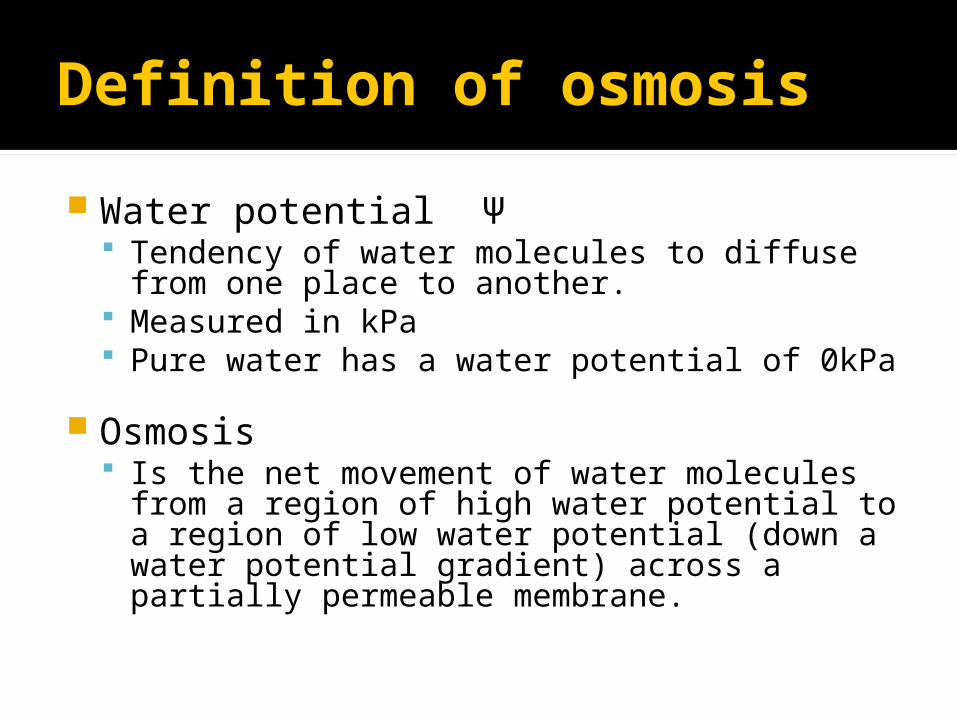
Water potential Ψ Tendency of water molecules to diffuse from
one place to another. Measured in kPa Pure water has a water potential of 0kPa
Osmosis Is the net movement of water molecules from a
region of high water potential to a region of low water potential (down a water potential gradient) across a partially permeable membrane.
Water potential
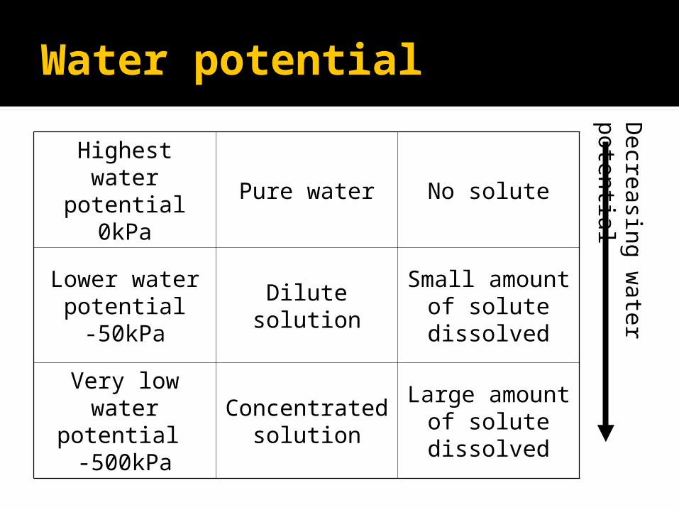
Highest water potential
0kPaPure water No solute
Lower water potential-50kPa
Dilute solutionSmall amount
of solute dissolved
Very low water
potential -500kPa
Concentrated solution
Large amount of solute dissolved
Decre
asin
g w
ate
r p
ote
ntia
l

Some Important Terms
Hypotonic a region of ▪ higher water potential.▪ Lower solute concentration
Hypertonic a region of ▪ lower water potential▪ Higher solute concentration
Isotonic a region where there are equal water potentials
on either side of a membrane.

Determining Water Potential in Potato tubers
Salt Soluntion(mol-1)
Starting Mass (g) Finishing mass (g) Change in mass (g)%age change in
mass
0.1
0.2
0.3
0.4
0.5
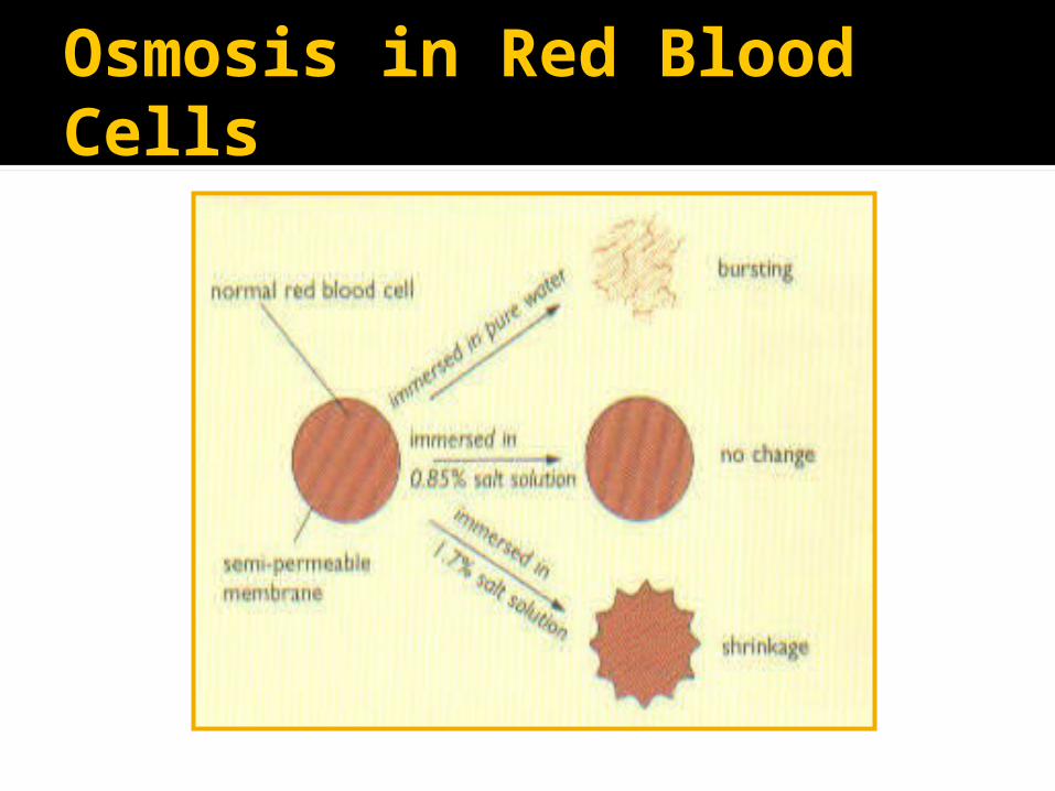
Osmosis in Red Blood Cells
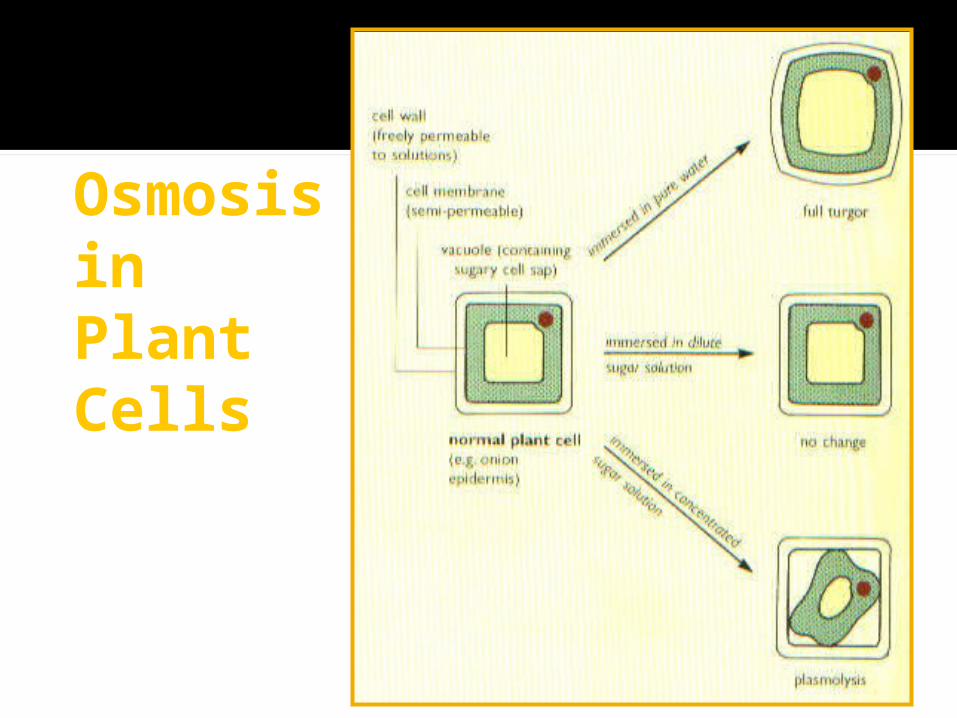
Osmosis in Plant Cells

Important Terms
Turgid the term used to describe a plant cell
where the protoplast exerts a pressure on the cell wall.
Plasmolysed the term used to describe a plant cell
where the protoplast has shrunk away from the cell wall due to loss of water by osmosis.

Osmosis in red onion cells
Cell Division, Cell Diversity and Cellular
Organisation
OCR AS BiologyUnit F211: Cells, exchange
and transportModule 1: Cells
Time to think about things?
Define growthHow do organisms increase in size?How do organisms reproduce?

Cell Division
The answers to all of these questions link into cell
division!Cell Division allows for
reproduction and growth of organisms.
Learning Outcomes
state that mitosis occupies only a small percentage of the cell cycle and that the remaining percentage includes the copying and checking of genetic information
Chromosomes
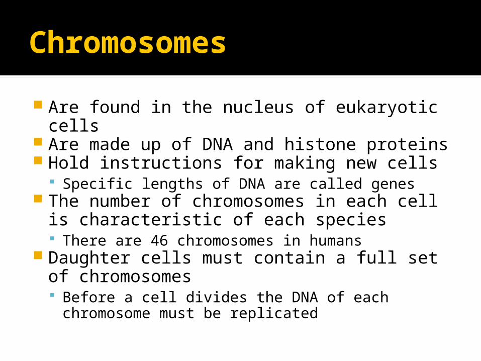
Are found in the nucleus of eukaryotic cells
Are made up of DNA and histone proteins Hold instructions for making new cells
Specific lengths of DNA are called genes The number of chromosomes in each cell
is characteristic of each species There are 46 chromosomes in humans
Daughter cells must contain a full set of chromosomes Before a cell divides the DNA of each
chromosome must be replicated
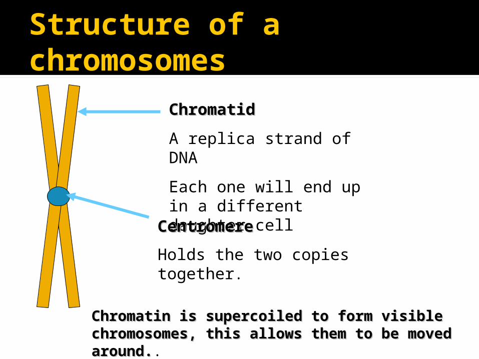
Structure of a chromosomes
ChromatidChromatid
A replica strand of DNA
Each one will end up in a different daughter cell
CentromereCentromere
Holds the two copies together.
Chromatin is supercoiled to form visible Chromatin is supercoiled to form visible chromosomes, this allows them to be moved chromosomes, this allows them to be moved around.around..

The Cell Cycledescribes the events that take place as one parent cell divides to produce new daughter cells which then each grow to full size.
M Nuclear division (mitosis) Cytokinesis (cleavage of cytoplasm)
Interphase G1▪ Biosynthesis – proteins are made and organelles
replicate,
S▪ Synthesis of new DNA▪ Replication of chromosomes
G2 - growth

The Cell Cycle (recap)
The cell cycle has 3 main phases Interphase▪ cell grows to normal size▪ carries out normal biochemical functions▪ DNA replication takes place
Nuclear division▪ mitosis, this has 4 stages; ▪ prophase, metaphase, anaphase and telophase
Cell division▪ cytoplasm divides by cytokinesis

Th
e C
ell C
ycle

Learning Outcomes
Recap state that mitosis occupies only a small
percentage of the cell cycle and that the remaining percentage includes the copying and checking of genetic information
explain the meaning of the term homologous pair of chromosomes;
describe, with the aid of diagrams and photographs, the main stages of mitosis (behaviour of the chromosomes, nuclear envelope, cell membrane and centrioles);
Nuclear Division
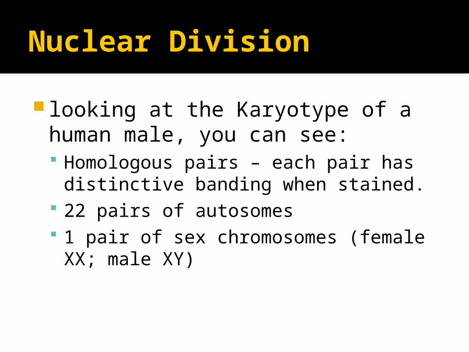
looking at the Karyotype of a human male, you can see: Homologous pairs – each pair has
distinctive banding when stained. 22 pairs of autosomes 1 pair of sex chromosomes (female XX;
male XY)

Karyotype
Human body cells are diploid (2n), meaning that they have 2 sets of chromosomes. Human gametes are haploid (n), where n is the number of chromosomes in a single set

Learning Outcomes
describe, with the aid of diagrams and photographs, the main stages of mitosis (behaviour of the chromosomes, nuclear envelope, cell membrane and centrioles);
Mitosis
Nucleus of a cell divides resulting in two nuclei which are genetically identical to the parent nucleus.
4 stages Prophase Metaphase Anaphase Telophase
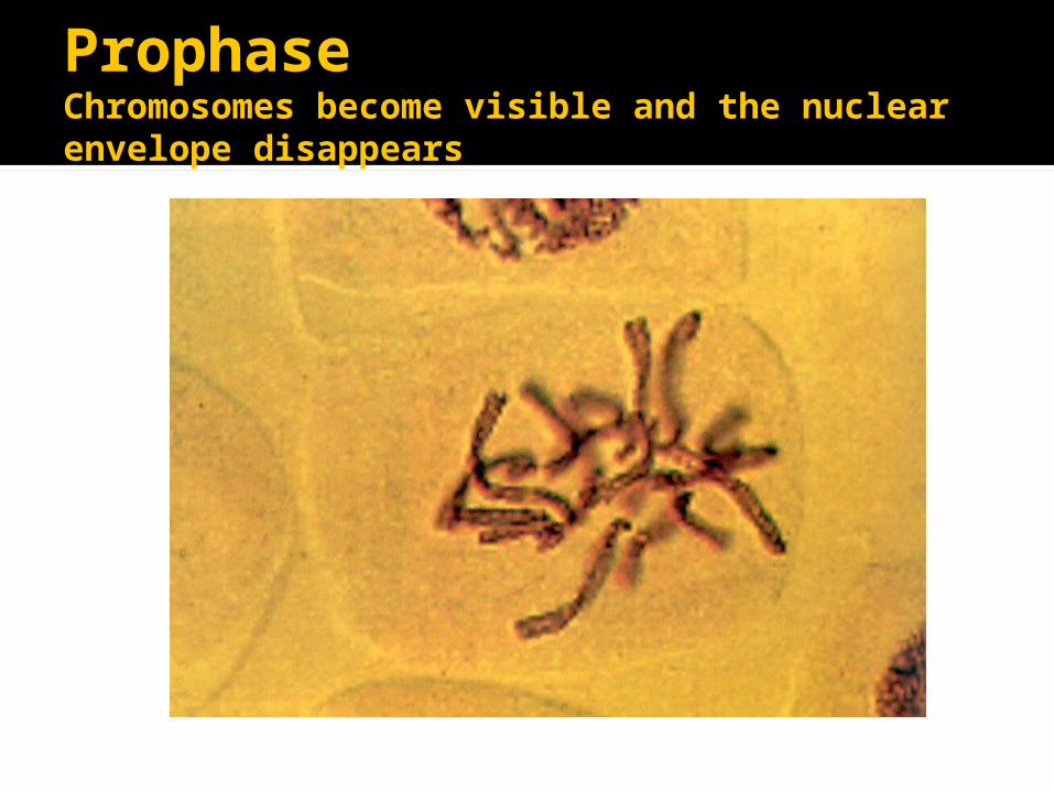
ProphaseChromosomes become visible and the nuclear envelope disappears
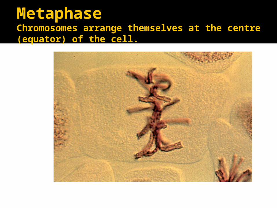
MetaphaseChromosomes arrange themselves at the centre (equator) of the cell.
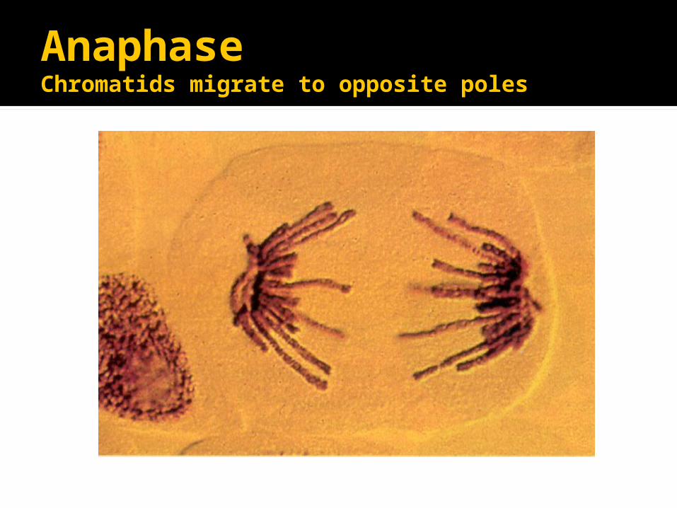
AnaphaseChromatids migrate to opposite poles
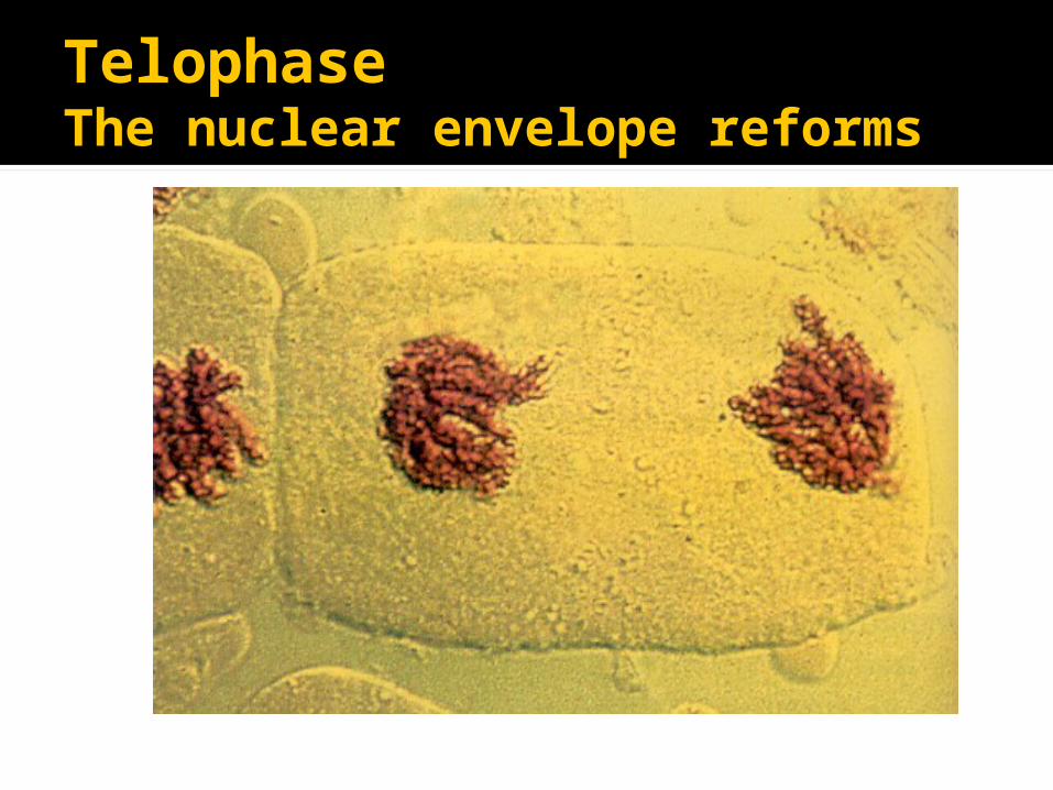
TelophaseThe nuclear envelope reforms
Mitosis Pupil Activities
HANDOUTS Looking at the diagrams – identify the stage of
mitosis, and describe what is happening Bioviewers
Slide set 55 – Plant Mitosis▪ Read the information provided ▪ Make a line drawing of each cell (all 8 slides)▪ Write a description of each drawing you make
Microscopes Calculating the mitotic index Collect a worksheet and a prepared microscope
slide
As there are not enough slides for everyone to do
the microscope work – these activities can be completed in any order

Learning Outcomes
explain the significance of mitosis for growth, repair and asexual reproduction in plants and animals;
state that cells produced as a result of meiosis are not genetically identical (details of meiosis are not required);
Requirements of Nuclear Division
There are two requirements of nuclear division Growth (Mitosis); ▪ where a diploid zygote grows into a multi-
cellular adult, all daughter cells have the same number of chromosomes as the parent cell.
sexual reproduction (Meiosis); ▪ the number of chromosomes is halved so that
gametes are haploid
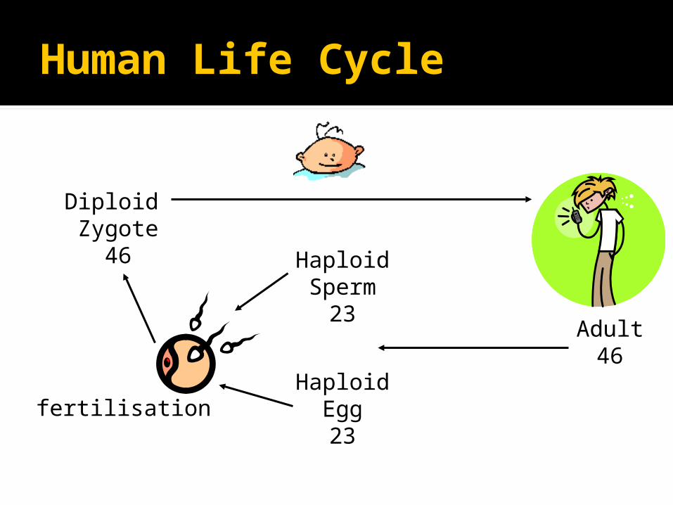
Human Life Cycle
Adult46
HaploidSperm
23
HaploidEgg23
Diploid Zygote
46
fertilisation

Meiosis
Chromosome number is halved so human gametes have 23 chromosomes
This reduction in chromosome number ensures that when gametes fuse to form a zygote (fertilisation) the diploid chromosome number is restored.

Meiosis
Learning Outcomes
outline, with the aid of diagrams and photographs, the process of cell division by budding in yeast;

Cell Division
In most cells, the nucleus divides first then the cytoplasm (cytokinesis).
This is mitosis and can be used for: Growth and repair Replacement of cells Asexual reproduction
In bacteria, cell division takes place by binary fission DNA replicates Cell divides

Cell division in yeast
Yeast is a single celled fungus It reproduces asexually by budding
Cell produces a swelling (bud) Nucleus divides into two – the bud gains
a nucleus The bud breaks off leaving a bud scar
Sometimes buds produce their own buds before separating.

Learning outcomes
To define the term stem cell Explain the meaning of the terms tissue,
organ and organ system; explain, with the aid of diagrams and
photographs, how cells are organised into tissues, using as examples squamous and ciliated epithelia, xylem and phloem
discuss the importance of cooperation between cells, tissues, organs and organ systems

Stem Cell
Stem cells are omnipotent or totipotent
The are cells which contain a full set of genetic information, and are capable of becoming any one of the different cell types found in a fully grown organism.

Stem Cells
There are a small number of stem cells found in adult mammals in the bone marrow, these are responsible for the formation of bone cells and blood cells.
Stem cells are currently being used in medical research.
Differentiation
Unspecialised cells which show totipotency include Stem cells in animals Meristematic cells in plants
These cells divide and then specialise, this is differentiation.
Differentiation and organisation
There is a physical limit to the size a single cell can reach, therefore multicellular organisms need specialised cells.
These specialised cells are organised into tissues which carry out a specific function.
Tissues are organised into organs.

Differentiation of cells
Cells differentiate in a number of ways, by changing: The number of a particular organelle▪ Muscle and liver cells contain many mitochondria
The shape of the cell▪ Red blood cells are a biconcave shape
Some of the cell contents▪ Red blood cells do not contain a nucleus
Differentiation means to specialise to carry out a particular role or function.
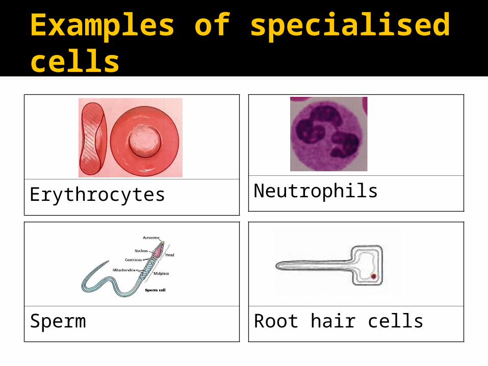
Examples of specialised cells
Erythrocytes Neutrophils
Sperm Root hair cells
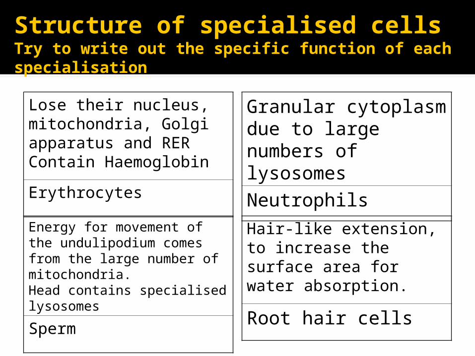
Lose their nucleus, mitochondria, Golgi apparatus and RERContain Haemoglobin
Erythrocytes
Granular cytoplasm due to large numbers of lysosomes
Neutrophils
Energy for movement of the undulipodium comes from the large number of mitochondria. Head contains specialised lysosomes
Sperm
Hair-like extension, to increase the surface area for water absorption.
Root hair cells
Structure of specialised cellsTry to write out the specific function of each specialisation

Tissues, organs and systems Tissue
a group of similar specialised cells in a many celled organism, that carries out a specific function or several related functions
Organ group of different tissues forming a distinct
structure and functioning together▪ animal – lungs, heart, kidneys▪ plant – roots, stems, leaves
System collection of organs with a particular function▪ cardiovascular and digestive systems

Tissues
A tissue is a group of similar, specialised cells which carry out a specific function, or several related functions. Animal tissues ▪ squamous and ciliated epithelium
Plant tissues▪ Xylem and phloem
Animal Tissues
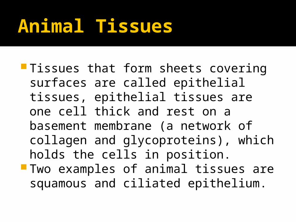
Tissues that form sheets covering surfaces are called epithelial tissues, epithelial tissues are one cell thick and rest on a basement membrane (a network of collagen and glycoproteins), which holds the cells in position.
Two examples of animal tissues are squamous and ciliated epithelium.

Ciliated Epithelium
This tissue is found lining the ends of the bronchioles in the lungs.

Squamous Epithelium
This tissue covers many surfaces in the human body including the inner lining of cheeks and lining the walls of the alveoli in the lungs.
In the alveoli, the thinness of cells allows rapid diffusion of gases between alveoli and blood.

Squamous Epithelium
Plant Tissues
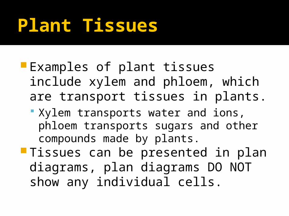
Examples of plant tissues include xylem and phloem, which are transport tissues in plants. Xylem transports water and ions,
phloem transports sugars and other compounds made by plants.
Tissues can be presented in plan diagrams, plan diagrams DO NOT show any individual cells.
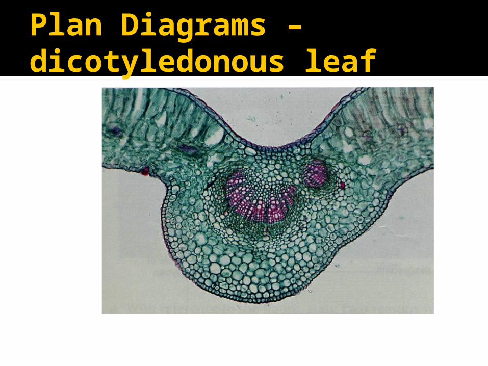
Plan Diagrams – dicotyledonous leaf
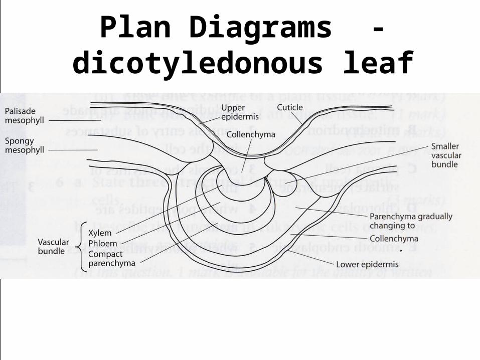
Plan Diagrams - dicotyledonous leaf