Embed Size (px)
Citation preview

Occipital Intermittent Rhythmic Delta Activity(OIRDA) and Occipital Seizures in an Elderly PatientYaşlı Bir Olguda Oksipital Nöbetler ve Oksipital İntermittentRitmik Delta Aktivitesi (OİRDA)Sabire YILDIRIM,1 Murat ÇABALAR,2 Çiğdem ÖZKARA,3 Orhan YAĞIZ,1
Filiz MANGA GÜNAYDIN,4 Sinan BAHADIR5
ÖzetOksipital intermitten ritmik delta aktivitesi (OİRDA), daha çok çocuklarda görülen ve her zaman patolojik anlamı olmayan bir elektroense-falografi (EEG) patterni olarak kabul edilir. Halüsinasyon, baş ağrısı ve düşme atakları ile başvuran 70 yaşında erkek hastanın muayenesinde dalgınlık ve konsantrasyon kaybı vardı. Kraniyal manyetik rezonans görüntülemede yaygın kronik iskemik değişiklikler, serebral atrofi ve özel-likle posterior bölgelerde lökoriazis görüldü. Rutin EEG’de oksipital bölgelerde OİRDA vardı. Fenitoin, okskarbazepin ve topiramat ile hastanın şikayetlerinin devam etmesi üzerine topiramat kesilerek levetirasetam eklendi. Tedavi sonrası hastanın halüsinasyonları ve düşme atakları azaldı, EEG’de ise OİRDA büyük ölçüde kayboldu.
Anahtar sözcükler: Epilepsi; migren; oksipital intermittent ritmik delta aktivitesi.
SummaryOccipital intermittent rhythmic delta activity (OIRDA) is considered as an electroencephalography (EEG) pattern which is usually seen in children but does not always have pathological significance. The physical examination of a 70-year-old patient who was admitted with complaints of hallucination, headache and falling episodes, revealed just a deficiency of vigilance and concentration. Magnetic resonance imaging of the patient revealed generalized chronic ischemic changes, cerebral atrophy and leukoaraiosis especially prominent in the poste-rior regions. There was OIRDA presence in the occipital regions in routine EEG examination. Since the patient’s symptoms continued despite phenytoin, oxcarbamazepine and topiramate, we replaced topiramate with levetiracetam. Patient’s visual illusions and falling episodes di-minished following treatment, and also OIRDA highly disappeared in the EEG.
Key words: Epilepsy; migraine; occipital intermittent rhythmic delta activity.
1Department of Neurology, Istanbul Training and Research Hospital, Istanbul2Department of Neurology, Bakirkoy Dr. Sadi Konuk Training and Research Hospital, Istanbul3Department of Neurology, Istanbul University Cerrahpasa Faculty of Medicine, Istanbul4Department of Neurology, Kocaeli Derince Training and Research Hospital, Izmit5Department of Neurosurgery, Bafra State Hospital, Samsun, all in Turkey
Epilepsi 2013;19(3):132-136 DOI: 10.5505/epilepsi.2013.27146
© 2013 Türk Epilepsi ile Savaş Derneği© 2013 Turkish Epilepsy Society
Introduction
Occipital intermittent rhythmic delta activity (OIRDA) is de-scribed as an intermittent, sawtooth shaped, unilateral or bilateral, symmetrical, medium or high amplitude, paroxys-mal and regular delta frequency activity occurring in the oc-
cipital region during electroencephalography (EEG) moni-torization. It increases with hyperventilation and decreases with eye opening and sleep.[1,3] Though OIRDA is suggested to be an interictal EEG finding that is mostly specific to chil-dren, it may not always be associated with epilepsy.[2]
CASE REPORT / OLGU SUNUMU
Submitted (Geliş) : 16.06.2013Accepted (Kabul) : 25.08.2013Correspondence (İletişim) : Murat CABALAR, M.D.e-mail (e-posta) : [email protected]
132
133
In this report, we present a case of an elderly patient with occipital lobe epilepsy that shows intermittent rhythmic delta activity in the occipital regions.
Case Report
A 70-year-old male was admitted to our clinic with com-plaints of colorful bright lights, headache and falling epi-sodes. His medical history revealed that his complaints were present for 6 months. He was having a wide variety of com-plex hallucinations such as moving shadows during day-time, colorful lights, red walls and bypassing cars. He also described headaches and loss of interest to environment. He was also reported to have 10-15 second-lasting episodes associated with loss of consciousness, falling down and ton-ic contractions 2-3 times, daily. He slept post-ictally and had no recall of the seizures.
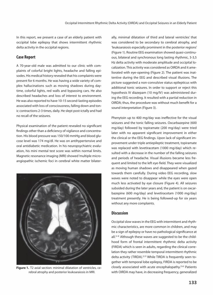
Physical examination of the patient revealed no significant findings other than a deficiency of vigilance and concentra-tion. His blood pressure was 150/100 mmHg and blood glu-cose level was 174 mg/dl. He was on antihypertensive and oral antidiabetic medication. In his neuropsychiatric evalu-ation, his mini mental test score was within normal limits. Magnetic resonance imaging (MRI) showed ‘multiple micro-angiopathic ischemic foci in cerebral white matter bilater-
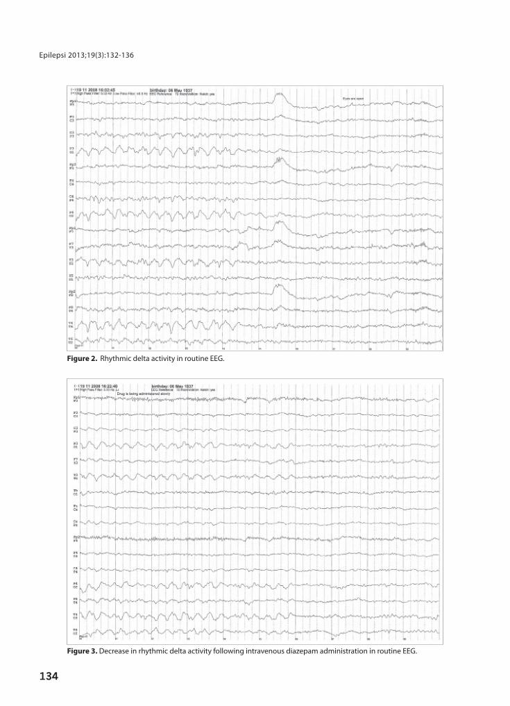
ally, minimal dilatation of third and lateral ventricles’ that was considered to be secondary to cerebral atrophy, and ‘leukoaraiosis especially prominent in the posterior regions’ (Figure 1). Routine EEG examination showed quasi-continu-ous, bilateral and synchronous long lasting rhythmic, 3-3,5 Hz delta activity with moderate amplitude and occipital lo-calization. This activity was considered as OIRDA and it ame-liorated with eye-opening (Figure 2). The patient was inat-tentive during the EEG and described visual illusions. The picture suggested a non-convulsive status epilepticus with additional tonic seizures. In order to support or reject this hypothesis IV diazepam (10 mg/IV) was administered dur-ing the EEG recording. It resulted with a partial reduction in OIRDA; thus, the procedure was without much benefit for a sound interpretation (Figure 3).
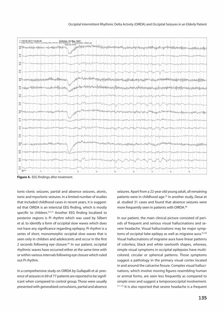
Phenytoin up to 400 mg/day was ineffective for the visual seizures and the tonic falling seizures. Oxcarbazepine (600 mg/day) followed by topiramate (200 mg/day) were tried later with no apparent significant improvement in either the clinical or the EEG findings. Upon lack of significant im-provement under triple antiepileptic treatment, topiramate was replaced with levetiracetam (1000 mg/day) which re-sulted with a decrease in the number of the falling seizures and periods of headache. Visual illusions became less fre-quent and limited to the left eye-field. They were visualized as moving human shadows and disappeared when gazed towards them carefully. During video EEG recording, slow waves were noted to disappear while the eyes were open much less activated by eye closure (Figure 4). All seizures subsided during the later years and, the patient is on oxcar-bazepine (600 mg/day) and levetiracetam (1000 mg/day) treatment presently. He is being followed-up for six years without any more complaints.
Discussion
Occipital slow waves in the EEG with intermittent and rhyth-mic characteristics, are more common in children, and may be a sign of epilepsy or have no pathological significance at all.[2-4] Although these waves are suggested to be the child-hood form of frontal intermittent rhythmic delta activity (FIRDA) which is seen in adults, regarding the clinical corre-lation they rather resemble temporal intermittent rhythmic delta activity (TIRDA).[2,3] While TIRDA is frequently seen to-gether with temporal lobe epilepsy, FIRDA is reported to be closely assosciated with acute encephalopathy.[5,6] Patients with OIRDA may have, in decreasing frequency, generalized
Figure 1. T2 axial section: minimal dilatation of ventricles, ce-rebral atrophy and posterior leukoaraiosis in MRI.
Occipital Intermittent Rhythmic Delta Activity (OIRDA) and Occipital Seizures in an Elderly Patient
Epilepsi 2013;19(3):132-136
134
Figure 2. Rhythmic delta activity in routine EEG.
Figure 3. Decrease in rhythmic delta activity following intravenous diazepam administration in routine EEG.
Occipital Intermittent Rhythmic Delta Activity (OIRDA) and Occipital Seizures in an Elderly Patient
135
tonic-clonic seizures, partial and absence seizures, atonic, tonic and myoclonic seizures. In a limited number of studies that included childhood cases in recent years, it is suggest-ed that OIRDA is an interictal EEG finding, which is mostly specific to children.[2,3,7] Another EEG finding localized to posterior regions is Pi rhythm which was used by Silbert et al. to identify a form of occipital slow waves which does not have any significance regarding epilepsy. Pi rhythm is a series of short, monomorphic occipital slow waves that is seen only in children and adolescents and occur in the first 2 seconds following eye closure.[4] In our patient, occipital rhythmic waves have occurred either at the same time with or within various intervals following eye closure which ruled out Pi rhythm.
In a comprehensive study on OIRDA by Gullapalli et al. pres-ence of seizures in 69 of 77 patients are reported to be signif-icant when compared to control group. Those were usually presented with generalized convulsions, partial and absence
seizures. Apart from a 22-year old young adult, all remaining patients were in childhood age.[3] In another study, Desai et al. studied 31 cases and found that absence seizures were more frequently seen in patients with OIRDA.[8]
In our patient, the main clinical picture consisted of peri-ods of frequent and serious visual hallucinations and se-vere headache. Visual hallucinations may be major symp-toms of occipital lobe epilepy as well as migraine aura.[9,10] Visual hallucinations of migraine aura have linear patterns of colorless, black and white sawtooth shapes, whereas, simple visual symptoms in occipital epilepsies have multi-colored, circular or spherical patterns. Those symptoms suggest a pathology in the primary visual cortex located in and around the calcarine fissure. Complex visual halluci-nations, which involve moving figures resembling human or animal forms, are seen less frequently as compared to simple ones and suggest a temporooccipital involvement.[11-13] It is also reported that severe headache is a frequent
Figure 4. EEG findings after treatment.

Epilepsi 2013;19(3):132-136
136
symptom in occipital lobe epilepsies.[14] Visual hallucina-tions were in the form of both simple and complex visual symptoms in our patient. Hallucinations seen in occipital lobe epilepsies do not last longer than 1-4 minutes, where-as, hallucinations of migraine aura may last as long as 20-150 minutes. Status epillepticus in this case, characterized mainly by visual seizures is differentiated from migraine status by history, presence of tonic seizures and detailed characteristics of visual images. However, it is not easy to accept the present case as a reliable model for status epilepticus with occipital seizures, since no epileptogenic complexes, associating the clinical visual episodes were present in the EEG.[15] In nonconvulsive status epilepti-cus, continous atypical spike-and-wave, polyspike-and-wave and rarely rhythmic bilateral 3Hz spike-and-wave complexes are commonly expected.[16] Although, ‘atypi-cal’ spike-and-wave activity may necessarily not always contain well-formed spikes or sharp-waves during a non-convulsive status epilepticus, OIRDA in the EEG associating visual illusions and headache episodes seems to provide a more cautious interpretation for this neurological condi-tion with such unique features.
In conclusion, we intended to draw attention to the pres-ence of OIRDA in an elderly patient with occipital lobe epi-lepsy, although this finding is reported to be more common in childhood.
References
1. Bora I, Yeni SN. EEG atlasi. Spesifik olmayan EEG bulgulari. Istan-
bul: Nobel Tip Kitabevi; 2012. p. 367-99.
2. Guilhoto LM, Manreza ML, Yacubian EM. Occipital intermittent
rhythmic delta activity in absence epilepsy. Arq Neuropsiquiatr
2006;64(2A):193-7. [CrossRef]
3. Gullapalli D, Fountain NB. Clinical correlation of occipital
intermittent rhythmic delta activity. J Clin Neurophysiol
2003;20(1):35-41. [CrossRef]
4. Silbert PL, Radhakrishnan K, Johnson J, Klass DW. The signifi-
cance of the phi rhythm. Electroencephalogr Clin Neurophysiol
1995;95(2):71-6. [CrossRef]
5. Buoni S, Zannolli R, Di Bartolo RM, Macucci F, Migliorini L, San-
soni R, et al. Occipital intermittent rhythmic delta activity only
following eye closure in atypical CNS Salmonellosis. Clin Neu-
rophysiol 2005;116(8):1768-70. [CrossRef]
6. Geyer JD, Bilir E, Faught RE, Kuzniecky R, Gilliam F. Significance
of interictal temporal lobe delta activity for localization of the
primary epileptogenic region. Neurology 1999;52(1):202-5.
7. Watemberg N, Linder I, Dabby R, Blumkin L, Lerman-Sagie T.
Clinical correlates of occipital intermittent rhythmic delta ac-
tivity (OIRDA) in children. Epilepsia 2007;48(2):330-4. [CrossRef]
8. Desai J, Mitchell WG, Rosser T, Ramos-Platt L, Ahsan N, Langille
MM, et al. Clinical associations of occipital intermittent rhyth-
mic delta activity. J Child Neurol 2012;27(4):503-6. [CrossRef]
9. Panayiotopoulos CP. Elementary visual hallucinations in
migraine and epilepsy. J Neurol Neurosurg Psychiatry
1994;57(11):1371-4. [CrossRef]
10. Panayiotopoulos CP. Visual phenomena and headache in oc-
cipital epilepsy: a review, a systematic study and differentiation
from migraine. Epileptic Disord 1999;1(4):205-16.
11. Schäffler L, Karbowski K. Epileptic activity of the occipital lobe.
Clinico-electroencephalographic contribution. [Article in Ger-
man] Fortschr Neurol Psychiatr 1988;56(9):286-99. [Abstract]
12. Schulze-Bonhage A. Differential diagnosis of visual aura in
migraine and epilepsy. [Article in German] Klin Monbl Augen-
heilkd 2001;218(9):595-602. [Abstract] [CrossRef]
13. George D, Bernard P. Complex hallucinations and panic attacks
in a 13-year-old with migraines: the alice in wonderland syn-
drome. Innov Clin Neurosci 2013;10(1):30-2.
14. Tokay T, Komsuoglu SS. Epileptik auralar. Düşünen Adam
2004;17(3):162-67.
15. Walker MC, Smith SJ, Sisodiya SM, Shorvon SD. Case of simple
partial status epilepticus in occipital lobe epilepsy misdiagnosed
as migraine: clinical, electrophysiological, and magnetic reso-
nance imaging characteristics. Epilepsia 1995;36(12):1233-6.
16. Kaya D, Yalınay P, Arman F. Nonkonvulsif status epilepti-
kusta tanı, klinik özellikler ve tedaviye direnç sıklığı. Epilepsi
2010;16(3):153-160.