Embed Size (px)
Citation preview

OBSERVATIONS CONCERNING THE CONTRALATERALLUNG IN PULMONARY TUBERCULOSIS TREATED
BY ARTIFICIAL PNEUMOTHORAX
•
BY
RAY W. MATSON, RALPH C. MATSON AND MARR BISAILLON
REPRINTED FROMTRE AMERICAN REVIEW OF TUBERCCTIOSii
Vox- X, No. 5, JANUARY, 1925

Reprinted from DIE AMERICAN REVIEW OF TUBERCULOSISVol. X, No. 5, January, 1925
OBSERVATIONS CONCERNING THE CONTRALATERALLUNG IN PULMONARY TUBERCULOSIS TREATED
BY ARTIFICIAL PNEUMOTHORAXiRAY W. MATSON, RALPH C. MATSON AND MARR BISAILLON
The indications and contraindications for collapse therapy seem to befairly well defined, as far as the "worse diseased lung" is concerned, atleast in the minds of most of the men who are practising the treatment.There is, however, a wide divergence of opinion as far as the oppositelung is concerned, particularly as to the amount and character of diseasewhich will permit a collapse with safety and a successful issue.
Considering this great diversity of opinion regarding the indicationsand contraindications for artificial pneumothorax as far as the contra-lateral lung is concerned, we have deemed it pertinent to present ourobservations which have been carried on in the period, 1911 to 1923,during which time 600 cases of pulmonary tuberculosis were subjectedto artificial-pneumothorax treatment.
This study has to do only with the chronic types of tuberculosis,particularly with the fibrocaseous and fibrocaseous cavernous groups,423 cases, for the reason that they lend themselves to longer continuedobservations, and therefore more accurate conclusions, than the acuteforms of tuberculosis and those cases in which the collapse was done onlyas a palliative procedure, to control severe symptoms, hem optysis, etc.
The 423 cases in which collapse was attempted or carried out wereclassified into the groups given in table 1. According to the N. T. A.Classification, 358 cases were far advanced and 65 cases were moderatelyadvanced.
TAELE 1
CASES
Chronic fibrocaseous tuberculosis, progressive, with little or no demonstrableexcavation
Chronic fibrocaseous cavernous tuberculosis, progressive Chronic fibrocaseous cavernous tuberculosis with cavity manifestations in the
foreground
194177
52
I Read at the Twentieth Annual Meeting of the National Tuberculosis Association,Atlanta, Georgia, May 8, 1924.
562

OBSERVATIONS ON CONTRALATERAL LUNG 563
The end-results of pneumothorax therapy are so intimately associatedwith and dependent upon the behavior of the contralateral lung, that, ifaccurate deductions based upon a comparison of end-results are to bemade, it is essential that a classification of the status of the oppositelung be adopted. In this way one will be able to estimate, with a fairerdegree of accuracy, the prognostic significance of the various types ofcontralateral lung disease before subjecting the patient to collapsetherapy. While we realize that many types of pathological change maycoexist in the opposite lung, those cases which we subjected to pneumo-thorax usually presented a dominant type which we classified as shown intable 2.
TABLE 2
CASES
Essentially negative Deep peribronchial infiltration Disseminated bronchogenic caseous extensions Active fibrocaseous infiltration Quiescent fibrocaseous infiltration
DESCRIPTION OF CLASSIFICATION
I. Essentially negative: It is frequently difficult, if not impossible, todemonstrate hidden or obscure foci of infection in the contralateral lung,but in the cases which we have classified as Essentially Negative the physi-cal and roentgenological findings were well within the limits of normaldisparity.
2. Deep peribronchial infiltration: The physical examination is as arule negative, aside from questionable alterations in breath-tones, withan entire absence of moisture. The diagnosis of this lesion is basedlargely upon roentgenological findings, and is characterized by a markedincrease in. the shadows of all or parts of the bronchial tree, especially ofthose shadows radiating from the hilum toward the superficies.
These shadows usually present an irregular, beady or varicose arboriza-tion, with fuzzy or clear-cut borders, depending upon the age and activ-ity of the lesion.
In view of the presence of an active tuberculosis in the other lung,roentgenological findings of this character justify the diagnosis of deepperibronchial infiltrations, undoubtedly tuberculous.
9710340
13449

564 R. W. MATSON, R. C. MATSON AND M. BISAILLON
The peribronchial type of tuberculous infiltration per se, from a stand-point of pneumothorax therapy, does not require great consideration aslong as it remains confined to the peribronchiaistructures.
These infiltrations are not always benign in character, for in lesions of
this type there is a tendency to invasion of lung parenchyma, and whenthis occurs it becomes a matter of serious import, especially when hap-pening during the course of pneumothorax therapy.
Invasion of lung parenchyma is usually readily demonstrable byphysical diagnostic methods and its evolution may be watched by thestudy of serial plates.
3. Disseminated bronchogenic caseous extensions: These lesions, soconstant in the study of gross pathological changes, usually occur as a
result of the aspiration of bacilli-laden sputum into the contralaterallung, and are characterized by the presence of localized caseous pneu-monic, caseous bronchopneumonic and disseminated confluent tubercu-
lous infiltrations, frequently associated with a tuberculous bronchitis.These bronchogenic extensions usually take place into the dependentportions of the lung or into the perihilar region. Roentgenologically,
they are revealed as disseminated patches of increased density, cor-responding to the distribution of the lesions and varying in characteraccording to the activity or inactivity of the process. If deep-seated,they may escape physical diagnostic procedures, but if superficial theyare readily recognized by the continual presence of moisture and altera-tions in breath-tones.
4 and 5. Active and quiescent fibrocaseous infiltrations: These lesions,in contradistinction to the bronchogenic infections above described,usually occupy the upper half of the lung. They are readily recognizedby the usual physical diagnostic and roentgenological procedures,although there is a frequent disproportion between physical and roent-genological findings, and to place sole reliance upon one or the other alone
will frequently lead one astray. The activity or inactivity of the processis determined by a correlation of these findings. As a rule, the presenceof moisture indicates activity, although its absence by no means indicates
quiescence.Moisture, when present, must be carefully studied in order to deter-
mine its pathological significance. An effort should be made to elicitand to differentiate those Ales characteristic of fresh invasion or destruc-tive lesions, from those associated with an obsolete or retrogressiveprocess. Riles which occur in the form of fine showers on deep inspira-

OBSERVATIONS ON CONTRALATERAL LUNG 565
tion or on expiratory cough almost invariably indicate fresh invasion.Destructive lesions are usually associated with abundant riles whichare more or less numerous, according to the rapidity of the process.They possess a resonating or metallic quality and they vary in characteraccording to the character of the excavation from which they arise.Retrogressive or stationary lesions are more often associated with rilesof a nonresonating character, are less numerous and are best elicited on,the first inspiration following expiratory cough.
Retrogressive and healed lesions are accompanied by fibrosis which
causes dilatation of terminal bronchioles. Even excavated areas mayheal and become lined with a mucosa; thus, the tuberculous process, assuch, really no longer exists.
These changes may be associated with abundant riles which are more
or less persistent, but one should be familiar with their pathologicalsignificance and not be confused by their presence, as they are evidencesof an obsolete or healed lesion. When these changes are encountered
over a small localized area, collapse of the opposite lung is not alwayscontraindicated.
R6Ies arising as a result of adhesive pleuritis and atelectasis can bereadily confused with races of great significance. Riles associated withan adhesive pleuritis occur as superficial crackles, best heard on forcedrespiratory movement, and they do not appear in the form of showers on
expiratory cough. Atelectatic crackles are usually heard along the mar-gin of the lung. These crackles disappear after a few inspirations and
are never persistently present in the same location day after day.The fibrocaseous lesions present fairly characteristic roentgenological
markings. The roentgenograph will supply evidence tending to provethe age of the lesion and, in a measure, also the activity or inactivity ofthe process. Early infiltrative lesions may escape roentgenologicalrecognition, and are at times best recognized stethoscopically.
The more active and progressive lesions are characterized by patch-like spots with indefinite irregular borders, whereas the quiescent andretrogressive processes are represented by more linear markings with moresharply circumscribed borders.
As a rule, auscultation will elicit exaggeration in breath-tones, both in
the active and quiescent lesions. One must carefully differentiate,however, that type of exaggerated breathing heard over fresh invasionfrom the compensatory type of breathing so frequently heard in thecontralateral lung.

566 R. W. MATSON, IL C. MATSON AND M. BISAILLON
SELECTION OF MATERIAL AND CONTROL OF TREATMENT
The recognition, interpretation and classification of these variouslesions in the opposite lung are of paramount importance in the selectionof material, and great caution should prevail in their treatment.
Early in our work we were loath to subject to pneumothorax therapyany case presenting more than minimal traces of disease in the contra-lateral lung, except as a last resort.
As a result of the observations made during the treatment of some ofthese last-resort cases, we realized that not infrequently excellent results,occasionally leading to complete recovery, could be obtained, even in thepresence of extensive disease in the opposite lung. The results, however,were dependent upon many factors, such as the character of collapse, flexi-bility of mediastinum, type of opposite lung lesion, and vigilance exer-cised as to changes taking place therein.
We have observed that a diseased lung varies greatly in its reactionto collapse of the other side; and we have learned that certain types of
contralateral lung disease can be approached with relative safety, whileother types are approached with great reluctance and apprehension.
As a general rule the fibrocaseous lesions involving the apex or upperportion of the lung react much more favorably than the fibrocaseouslesions located around the root of the lung, or the bronchogenic caseous
extensions scattered throughout the lower portions of the lung.This difference in reaction may be due to the fact that upper-lobe
lesions are more firmly immobilized by fibrosis and pleuritic thickening,and are usually favored with better drainage, while lower-lobe lesions areunfavorably influenced by being subjected to greater amplitude ofrespiratory motion, which inhibits fixation and favors aspiration anddissemination, which are further enhanced by poorer drainage.
Furthermore, certain types of lesions, especially those in the perihilarregion, are kept in constant motion by cardiac activity, which the fluoro-scope will readily reveal as a constant tugging and pulling that are syn-
chronous with each heart-beat.All patients reported in this series received sanatorium treatment,
many of them having been under observation for long periods of time,
and had exhausted various climatic, dietetic, and hygienic procedures.The delay in treatment in many cases was very unfortunate, becausewhen we were obliged to resort to an artificial pneumothorax, we wereunable to introduce gas on account of the presence of adhesions. Of

OBSERVATIONS ON CONTRALATERAL LUNG 567
recent years we have felt that, if after a brief period of observation,one to three months, any doubt exists as to the favorable outcome bythe usual sanatorium regimen, collapse therapy should be immediatelyinstituted if no contraindications exist. This is particularly imperativein cavity cases with abundant expectoration, in which there exists con-stant danger of infection, not only of the opposite lung by aspiration, butof the bowel through deglutition, and of the larynx through lodgment ofsputum.
Any procedure which has for its object the reduction of sputum quan-tity, thus lessening the danger of these complications, is justifiable; andwe have felt that our tardiness in utilizing pneumothorax treatment wasresponsible for many failures, through the development of these variouscomplications.
Notwithstanding the danger of delay, if the contralateral lung lesionis very active, it is better to allow the activity of the process to subsidesomewhat before resorting to collapse, but this furthermore enhancesthe danger of the formation of adhesions. If the disease in the contra-lateral lung is not too active, this danger can be minimized by interpos-ing a thin layer of gas, thus separating the worse diseased lung fromthe chest-wall to the extent of an inch or two, and then maintaining thischaracter of collapse while observing the behavior of the opposite lung.Frequently we have noted the beneficial effects of these small degreesof collapse, not only in the worse diseased lung, but also in the contra-lateral lung, particularly in the fibrocaseous lesions, involving the upperportion. By this manner of approach we have, in many cases, beenenabled to bring about a gradual satisfactory collapse of the worse dis-eased lung with an uninterrupted improvement of the opposite lungprocess, even leading to complete recovery. In some cases, collapse hasbeen carried out for years without any appreciable influence on the oppo-site lung process. In other cases, in spite of our watchfulness and care,progression of disease took place in the opposite lung. In still othercases the contralateral lung lesion remained stationary for years,later on becoming rapidly progressive and demanding a pneumothorax.
During the period of sanatorium treatment all patients were taught amethod of chart-keeping, whereby the temperature and pulse wererecorded in a graphic manner, and the charts were continued during thewhole course of their pneumothorax treatment. They were also madeacquainted with the extent and character of their disease, results of allexaminations and the principles of pneumothorax treatment, thus

568 R. W. MATSON, R. C. MATSON AND M. BISAILLON
enabling them to render more intelligent cooperation. All or mostcases were discharged to ambulant care at the earliest possible momentcompatible with safety.
Having established the indications for collapse therapy as far as theworse diseased lung is concerned, there still remains the important taskof estimating the integrity of the opposite lung, for, after instituting apneumothorax, one will frequently be confronted with the problem ofhaving to decide whether or not unfavorable symptoms are emanatingtherefrom.
A solution of this problem will be greatly facilitated by always main-taining accurate records of the status of the contralateral lung. Theserecords should contain the exact topographical distribution of physicalfindings recorded before each inflation, and as often as necessary duringthe interval. Inks, if present, should be studied and the followingpoints carefully noted: Whether they appear only on expiratory coughor on first inspiration following, or whether they are present on ordinarybreathing. Their character, amount and exact anatomic distributionshould be recorded, so that the record can he compared with all previousexaminations.
The roentgenological control must be carried out with great frequency.It is our custom to make a fluoroscopic examination from which an
orthodiagram is prepared, before each inflation, and during the earlyphases of the collapse it is imperative to resort to the fluoroscope morefrequently, in order to determine better the proper interval for inflation.
Each individual patient is a law to himself as far as reaction of the lungto inflation and absorbability of gas are concerned, and difficulties willbe encountered sooner or later if a fixed plan is adopted as to the timeinterval and as to the amount of gas introduced. By the frequentrecourse to the fluoroscope, one will be better able to adapt oneself tothese varying factors which each patient may present.
Unless the use of the fluoroscope is resorted to freely, the integrity ofthe contralateral lung may be threatened after the first few inflationsthrough undue stress placed upon it by great displacement of mediasti-nal contents, which might otherwise easily escape detection. One shouldnot depend solely upon manometric pressure readings and physical find-ings to determine the character of collapse.
In the roentgenological control of the contralateral lung the fluoroscopealone is inadequate, and one must depend upon frequent use of films for

OBSERVATIONS ON CONTRALATERAL LUNG 569
the reason that early fresh invasions escape fluoroscopic detection, evenif roentgenologically present.
Too much reliance should not be placed upon the examination of asingle film in any case, presenting actual or threatened invasion of theopposite lung; but the last film should be minutely compared with theprevious films, for it is possible only in this manner to evaluate the exactchanges taking place. In the interpretation of the markings in thecontralateral lung, one must differentiate between actual pathologicalchanges and changes due to circulatory stasis, which are especiallymarked in cases presenting a bulging mediastinum.
In doubtful cases the differentiation is facilitated by permitting aslight reexpansion of the collapsed lung, thus allowing the mediastinumto assume a more normal position, when the shadows due to circulatorydisturbance rapidly disappear.
Disease in the contralateral lung is best revealed on films taken at theheight of deep inspiration, but changes in the position of the mediasti-num are best seen in films taken at the end of expiration. By compari-son of the films taken in these two positions it is interesting to notethe great excursion which can taken place in a labile mediastinum.
X-ray films have the distinct advantage of supplying a permanentrecord.
During the early phases of collapse therapy, films should be made afterevery third or fifth inflation if one resorts to small amounts of gas atshort intervals, and later on, after the first two or three months, filmsshould be made at monthly intervals. Ambulant cases are "filmed" atleast once every three months throughout the whole course of theirtreatment.
In the selection of material, and in the control of treatment, one alwayshas to contend with the transmission of auscultatory findings from theworse diseased lung to the opposite side. From a physical diagnosticstandpoint alone, it will be exceedingly difficult to differentiate thesetransmitted auscultatory phenomena from those due to actual disease,but an endeavor should be made to find out their source, by localizingtheir point of maximum intensity and, in the case of transmittedphenomena, one will be led to the opposite lung. A decision will befurther facilitated by a study of X-ray films, which will reveal an entireabsence of disease in the contralateral lung that could account for thecharacter of the auscultatory findings. The flexibility of the mediasti-num will also have an important bearing on adventitious sounds elicited

570 R. W. MATSON, R. C. MATSON AND M. BISAILLON
in the contralateral lung, for occasionally rales, when present in theopposite lung before inflation, will change in character, or disappearimmediately afterward, in the presence of a flexible mediastinum.
ANALYSIS OF CLINICAL PHENOMENA
In the presence of actual or threatened invasion of the opposite lung,one is continually confronted with the task of having to exclude the con-tralateral lung as a source of any unfavorable clinical signs or symptoms,the most serious of which are the following: fever, increased expectora-tion and hemoptysis.
Febrile reaction following inflation, or the regular occurrence of anephemeral exacerbation of temperature within twenty-four hours afterinflation in the presence of a satisfactory collapse, should lead one tosuspect the opposite Jung, particularly in the absence of an exudate or ofadhesions.
Recurrence of fever following an afebrile period occurs most commonlyunder the following conditions: appearance of exudate in pneumothoraxcavity, insufficient collapse, that is, reexpansion of lung, progression orextension of disease in contralateral lung, extrapulmonary tuberculouscomplications, especially enteritis, and acute nontuberculous respira-tory infections.
Fluoroscopy alone will suffice to reveal an exudate and aspiration willusually cause subsidence of fever.
Difficulty will at times . arise in differentiating fever, due to progressionor extension of disease in the contralateral lung, from fever due toinsufficient collapse, that is, reexpansion of lung.
A solution of this question is exceedingly important, for to be misledat this juncture might convert a possible successful end-result into a fail-ure. If the fever is due to reexpansion, a delay in inflation will permitfurther progression of disease in the collapsed lung, and it also involvesthe danger of an early obliterative pneumothorax. If the fever is due toprogression or extension of disease in the contralateral lung, furthercompression would only aggravate this disease already present. Inarriving at a decision, the contralateral lung must be subjected to asearching examination, the results of which must be minutely comparedwith the records of previous examinations (films and physical). If,after this comparison, there is no evidence of fresh invasion or progres-sion of disease in the opposite lung, then further collapse is justifiable.
Fever due to extrapulmonary tuberculous complications is usually
OBSERVATIONS ON CONTRALATF,RAL LUNG 571
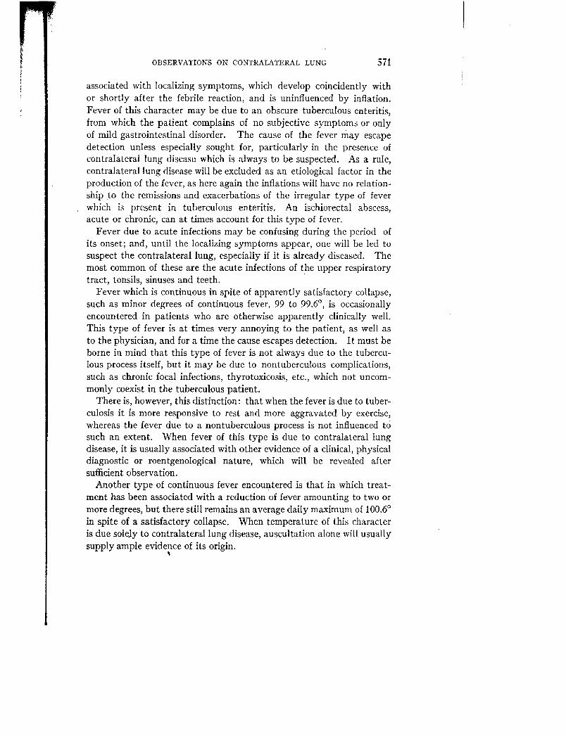
associated with localizing symptoms, which develop coincidently withor shortly after the febrile reaction, and is uninfluenced by inflation.Fever of this character may be due to an obscure tuberculous enteritis,from which the patient complains of no subjective symptoms or onlyof mild gastrointestinal disorder. The cause of the fever May escapedetection unless especially sought for, particularly in the presence ofcontralateral lung disease which is always to be suspected. As a rule,contralateral lung disease will be excluded as an etiological factor in theproduction of the fever, as here again the inflations will have no relation-ship to the remissions and exacerbations of the irregular type of feverwhich is present in tuberculous enteritis. An ischiorectal abscess,acute or chronic, can at times account for this type of fever.
Fever due to acute infections may be confusing during the period ofits onset; and, until the localizing symptoms appear, one will be led tosuspect the contralateral lung, especially if it is already diseased. Themost common of these are the acute infections of the upper respiratorytract, tonsils, sinuses and teeth.
Fever which is continuous in spite of apparently satisfactory collapse,such as minor degrees of continuous fever, 99 to 99.6°, is occasionallyencountered in patients who are otherwise apparently clinically well.This type of fever is at times very annoying to the patient, as well asto the physician, and for a time the cause escapes detection. It must beborne in mind that this type of fever is not always due to the tubercu-lous process itself, but it may be due to nontuberculous complications,such as chronic focal infections, thyrotoxicosis, etc., which not uncom-monly coexist in the tuberculous patient.
There is, however, this distinction: that when the fever is due to tuber-culosis it is more responsive to rest and more aggravated by exercise,whereas the fever due to a nontuberculous process is not influenced tosuch an extent. When fever of this type is due to contralateral lungdisease, it is usually associated with other evidence of a clinical, physicaldiagnostic or roentgenological nature, which will be revealed aftersufficient observation.
Another type of continuous fever encountered is that in which treat-ment has been associated with a reduction of fever amounting to two ormore degrees, but there still remains an average daily maximum of 100.6°in spite of a satisfactory collapse. When temperature of this characteris due solely to contralateral lung disease, auscultation alone will usuallysupply ample evidence of its origin.

572 R. W. MATSON, R. C. MATSON AND M. BISAILLON
INFLUENCE or CONTRALATERAL LUNG DISEASE ON SPUTUM
Of equal importance in eliminating the contralateral lung as a factor inthe causation of fever is its elimination as a source of bacilli-laden sputum,in the presence of a collapse of the worse diseased lung.
The persistence of bacilli-laden sputum, with an essentially negativecontralateral lung and in the presence of an apparently satisfactory col-lapse, indicates that diseased tissue is not sufficiently collapsed, eitherbecause of the presence of adhesions, particularly apical, which are notalways revealed by X-ray films, or because of the presence of rigid-wallexcavations. However, the persistence of bacilli-laden sputum in quanti-
• ties of 20 or 25 cc. daily, in the presence of a diseased contralateral lungand with an apparently satisfactory collapse of the worse diseased side,should lead one to strongly suspect the contralateral lung as a source,particularly when auscultation reveals moisture in it.
Under these circumstances one will be obliged to decide whether toallow of moderate reexpansion, thus relieving the contralateral lung, orto further collapse the worse diseased lung. When confronted withthis problem, it is essential that the twenty-four-hour quantity of sputumshould be measured carefully each day, and its relationship to inflationand physical findings in each lung closely observed.
The influence of inflations on the temperature curve should be observedas well, for, in the presence of contralateral lung disease, inflations aresometimes followed by an exacerbation of temperature, which, whenassociated with an increase in sputum amount, points more strongly tothe contralateral lung as a source.
If the contralateral lung is responsible for the increased expectoration,the physical examination will usually reveal an increase in the amountof Ales, as well as an increase in their distribution, when compared withprevious examinations.
Stereoscopic films should be made, and these should be carefully com-pared with all previous films, and if the contralateral lung is to blame forthe sputum increase corroborative evidence will usually be manifest asa progression of disease.
The influence of the patient's bed posture during coughing paroxysmsand lung drainage should be carefully observed, as much valuable evi-dence may be supplied that may tend to show the side from which the spu-tum emanates, as some positions will be associated with freedom fromcough and expectoration, due to retention of secretion in dependent por-

OBSERVATIONS ON CONTRALATERAL LUNG 573
tions of the lung, whereas other positions will provoke coughingparoxysms with abundant expectoration due to free drainage.
A final decision can best be reached only after a correlation of allfindings.
HEMOPTYSIS
Severe hemoptysis occurs at times in the course of pneumothoraxtreatment, and always directs attention to the contralateral lung, partic-ularly if it is diseased. In a series of 480 collapsed cases (all types oftuberculosis) severe hemoptysis was present preceding or shortly before
. pneumothorax treatment was begun in 82 cases, but after pneumothoraxtreatment was instituted hemoptysis occurred in only 10 cases.
It is notoriously difficult to determine from which side bleeding ishaving its source, particularly when one is dealing with a diseased con-tralateral lung, but the accurate determination of this point is impera-tive, for if the bleeding is proceeding from the collapsed lung, furthercollapse is demanded; while if from the opposite lung, further collapseis strongly contraindicated. The determination of the bleeding side iscomplicated by reason of the hesitancy in submitting the patient to asearching physical examination in the presence of active bleeding. Muchvaluable information will be obtained from the patient's own statementregarding certain subjective phenomena which he has experienced imme-diately before or very early during the hemoptysis. These sensationsare commonly described as a sense of tightness, constriction, pain,"rattling," or "fluttering," the exact location of which the patient willindicate with absolute confidence and assurance. These statementsmay be misleading, but they are, nevertheless, deserving of consideration.
As a result of bleeding, aspiration of blood occurs into dependent por-tions of the lung, and it is always more extensive on the bleeding side.The presence of this blood will furnish auscultatory evidence in the formof Odes which are quite characteristic, and may be readily heard inportions of the lung into which aspiration has taken place. These /Aleshave been variously described as "medium" or "small bubbling," or"crackles." At first they are nonresonating and only become resonatinglater on, with the development of consolidation.

C
tC
4a
si
CI
es;tcai
Ieti.le./a
Of"4\7]ce
titca
opofpei
574 R. W. MATSON, R. C. MATSON AND M. BISAILLON
COMPARATIVE SIGNIFICANCE OF THE VARIOUS TYPES OF CONTRALATERAL
LUNG LESIONS BASED UPON END-RESULTS
A valuable aid in the selection of material and in determining theindications for collapse will be furnished by a comparison of end-resultsin the various types of contralateral lung lesions, whereby one will beable to judge the prognostic significance of disease in the contralaterallung, with collapse of the worse diseased side.
TABLE a'
End-results in 423 cases of fibrocaseous and fibrocaseous cavernous tuberculosisril;.1 I'l CLINICALLY
mutt. ARRESTED DEAD
STATUS OF CONTRALATERAL LUNG afa'
CHARACTER ORCOLLAPSE
1,77,
r4d :2' 14 AI ti t
.'1.
Rz Ez mZ ,.4; oZ ii:. Z a,
Satisfactory 56 29 52 11 20 8 14Essentially negative 97 Partial 25 10 40 1 4 6 24
No free space 16 4 25 1 6 9 56
Deep peribronchial infiltration 103 Partialctory4933
243
459
127
2421
1020
2060
No free space 21 1 4 3 13 9 43
Disseminated bronchogenic Satisfactory 15 4 26 4 26 5 33caseous extension 40 Partial 21 1 4 5 24 14 66
No free space 4 0 0 0 0 3' 75
Pictive fibrocaseous infiltration 134 Partialc tory5059
244
486
1112
2220
1127
2246
No free space 25 0 0 3 12 19 76
t.ticscent fibrocaseous infil- Satisfactory 21 10 50 5 24 4 20tration 49 Partial 16 4 25 6 37 6 37
No free space 12 2 16 2 16 4 33
423 423 120 78 155
A study of the table of end-results (table 3), with a view of determiningthe significance of the contralateral lung lesions, will reveal that of ,the423 cases subjected to artificial-pneumothorax treatment, 326 cases haddemonstrable disease in the opposite lung, and in 223 of these it amountedto well-defined fibrocaseous infiltrations or disseminated bronchogenicextensions. It will be seen that only 23 per cent of the cases includedcinthis series presented an essentially negative contralateral lung.

OBSERVATIONS ON CONTRALATERAL LUNG 575
The "No Free Pleural Space" cases have been included in this seriesfor the reason that they serve as invaluable controls; for, aside from the
fact that they received no pneumothorax treatment, their treatment wasidentically the same in every other respect.
The inclusion of the "No Free Pleural Space" cases is also importantbecause a study of table 3 will reveal that satisfatory end-results areas much dependent upon the presence of free pleural space and the
character of collapse attained as they are upon the presence or absenceof disease in the opposite lung.
Of the 423 cases, 97 had an "Essentially Negative" opposite lung. Of;this number, 56 secured a satisfactory collapse, of which 52 per cent areclinically well, 20 per cent are arrested and 14 per cent are dead.
Of the 423 cases, 103 had a deep peribronchial infiltration in the oppo-site lung. Of this number, 49 received a satisfactory collapse, of which45 per cent are clinically well, 24 per cent are arrested and 20 per centare dead.
It will be noted in comparing the deep peribronchial infiltration with
the essentially negative contralateral lung, that in the presence of asatisfactory collapse there are 7 per cent less clinically well and 6 per
cent more dead in the former than there are in the latter group.Of the 423 cases, 40 cases had disseminated bronchogenic Gaseous
extensions in the opposite lung. Of this number, 15 received a satisfac-tory collapse, of which 26 per cent are clinically well, 26 per cent arearrested and 33 per cent are dead.
Thus, from a prognostic standpoint, this type of contralateral lunglesion is of serious import; for, when'compared with the essentially nega-tive contralateral lung group, it will be seen that there are 26 per centless clinically well and 19 per cent more dead in the former than in thelatter group.
Of the 423 cases, 134 had an active fibrocaseous infiltration in theopposite lung. Of this number, 50 received a satisfactory collapse, ofwhich 48 per cent are clinically well, 22 per cent are arrested and 22 percent are dead.
In comparing this type of contralateral lung with the essentially nega-
tive opposite lung, it will be observed that there are only 4 per cent fewercases clinically well, but there are 8 per cent more dead.
Of the 423 cases, 49 had a quiescent fibrocaseous infiltration in the
opposite lung. Of this number, 21 received a satisfactory collapse,of which 50 per cent are clinically well, 24 per cent are arrested and 20
per cent are dead.

576 R. W. MATSON, R. C. MATSON AND M. BISAILLON
In comparing the active with-the quiescent fibrocaseous infiltrations, itwill be seen that there are 2 per cent more clinically well, 2 per cent more
arrested and 2 per cent less dead in the quiescent than in the activefibrocaseous groups.
This difference in end-results is not as great as one would expect in acomparison of an active with a quiescent fibrocaseous opposite lung, butit is probable that activity undoubtedly existed in some cases which wewere obliged to classify as quiescent because of our inability to demon-strate activity, even though it was suspected. However, these figuresare entirely consistent when one compares the end-results (table 4) inboth the active and quiescent fibrocaseous opposite lung lesions, withthe essentially negative contralateral lung.
TABLE 4
Comparison of end-results in active and quiescent fibrocaseous contralateral lung with essentiallynegative contralateral lung in presence of a satisfactory collapse
STATUS OF CONTRALATERAL LUNG CLINICALLYWELL ARRESTED DEAD
per cent per cent per cent
Essentially negative 52 20 14Quiescent fibrocaseous infiltration 50 24 20Active fibrocaseous infiltration 48 22 22
As previously stated, the disseminated bronchogenic caseous exten-sions,'_occupying the perihilar and lower portions of the lung, offer amuch more unfavorable outlook than the fibrocaseous lesions usuallyoccupying the upper portions of the lung.
A study of end-results in the following table (table 5) will reveal thatthe disseminated bronchogenic caseous extensions are the most serioustype of contralateral lung lesions with which we have had to deal.
TABLE 5
Comparison of end-results: essentially negative with certain types of contralateral lung diseasein the presence of a satisfactory collapse
STATUS OF CONTRALATERAL LUNG CLINICALLYWELL DEAD
per cent per cent
Essentially negative 52 14Active fibrocaseous infiltration 48 22Quiescent fibrocaseous infiltration 50 20Disseminated bronchogenic caseous extensions 26 33

OBSERVATIONS ON CONTRALATERAL LUNG 577
From a study of the above statistics, as regards the percentage clini-cally well and dead in the presence of a satisfactory collapse, it might beheld by some that results equally satisfactory are attained by the usualsanatorium regimen without pneumothorax.
However, the value of pneumothorax therapy in this series will beclearly shown by a comparison of the end-results in cases satisfactorilycollapsed (table 5) with those having "No Free Pleural Space," whichserve as controls (table 6).
TABLE 6
End-results in noncollapsed cases (no free pleural space)
STATUS OF CONTRALATERAL LUNG CLINICALLYWELL DEAD
per tent per cent
Essentially negative 25 56Active fibrocaseous infiltrations 0 76Quiescent fibrocaseous infiltrations 16 33Disseminated bronchogenic caseous extensions 0 75
It will be observed that in this series, from a study of table 3, a patient'sopportunity for recovery was much better with any type of contralaterallung disease in the presence of a satisfactory collapse than in the case ofthe patient who had an essentially negative lung, in which collapse wasnot possible on account of our inability to find a free pleural space.
OBSERVATIONS ON PROGRESSION AND EXTENSION OF DISEASE INCONTRALATERAL LUNG
The term "Progression of Disease" refers to those cases in which therewas exacerbation of disease in a contralateral lung already diseased,whereas the term "Extension of Disease" refers to those in which invasionof an essentially negative contralateral lung took place.
In compiling our statistics we have included in our figures only thoseinstances of progression or extension of disease in the contralateral lung,in which these complications occurred during the actual period of pneu-mothorax treatment.
Undoubtedly progression and extension of disease took place in theterminal phases of many cases after pneumothorax treatment was aban-doned, particularly in the progressively fatal cases, but they were notincluded for the reason that the development of these complications wasnot associated-with or related to the period of collapse therapy.

578 R. W. MATSON, R. C. MATSON AND M. BISAILLON
No doubt, minor degrees of progression and extension of disease tookplace in some cases, but it was of a character which escaped physicaldiagnostic or roentgenological methods of recognition.
It is also true that many cases experienced alternating periods of
exacerbation and quiescence of activity of contralateral lung diseasewhile under collapse, but the periods of exacerbation were temporary andwere not of a character demanding discontinuance of collapse therapy.
Whether these exacerbations occurred in spite of, or were dependentupon collapse, is impossible to say, because changes of this nature arenot infrequent throughout the course of any tuberculous process.
When progression of disease in the opposite lung is of a mild nature, aprolongation of intervals and the introduction of small amounts of gas
are necessary, thus permitting of some reexpansion of the collapsed lung,which will frequently bring about a subsidence of the activity in thecontralateral lung and a coincident improvement.
Severe and rapid progression of disease in the contralateral lungdemands temporary cessation of pneumothorax treatment. Under
circumstances of this nature, one will be confronted with a complicatedproblem to decide upon future procedure. A decision will be reachedafter a brief period of observation, and one will be guided in this decision
by the behavior of both lungs during this period.When reexpansion of the collapsed lung is not followed by reactivation
of disease therein, one will be justified in considering the feasibility ofbilateral collapse. Should this procedure be adopted, the primarilycollapsed lung should be permitted to reexpand as much as possible
without evidence of reactivation of disease in it, and at the same time itmust still be under control, so that recollapse can be instituted as indica-tions require. If no reactivation occurs, one can then proceed cautiouslywith the contralateral lung collapse. The inflations should then be madealternately, the intervals and amounts of gas being determined by theindications and contraindications in each lung. We have carried out
bilateral collapse in 14 cases, 2 of which are clinically well and 12 aredead. The prognosis in cases so severe as to demand a bilateral pneu-mothorax is very grave, but the procedure will often add .much comfort
and prolongation of life to the patient.Out of the 423 cases included in this study, 345 cases were collapsed.
Of the 345 collapsed cases, 264 had demonstrable disease in the opposite
lung, 82 cases having well-defined deep peribronchial infiltration, but
in 182 cases the disease amounted to fibrocaseous infiltrations or dis-

OBSERVATIONS ON CONTRALATERAL LUNG 579
seminated bronchogenic caseous extensions. In these 264 cases progres-sion of disease in the contralateral lung took place in 24 cases as shownin table 7.
TABLE 7Progression of disease in The colaralaleral long in collapsed cases
STATUS OF CONTRALATERAL LANG NUMBEROF CASES
PROGRESSIONNUMBERCASES
CHARACTEROF COLLAPSE
Deep peribronchial infiltrations 82 2 2 partial
Disseminated bronchogenic caseous extensions 36 3{ 1 satisfactory
2 partial
Active fibrocaseous infiltrations 109 17{ 7 satisfactory
10 partialtiiescent fibrocaseous infiltrations 37 2 2 partial
INFLUENCE OF PARTIAL COLLAPSE ON CONTRALATERAL LUNG
Conclusions drawn from a statistical analysis of the behavior of thecontralateral lung in the presence of a partial collapse of the worsediseased side are unreliable for the reason that the more the partial
collapse partakes of the character of a satisfactory collapse, the more the
end-results will approach those of a satisfactory one and, contrariwise,the more limited or localized the partial collapse, the more the end-resultswill approach those of the "No Free Space" cases. Furthermore, theend-results in pneumothorax therapy in cases under partial collapse are,for the most part, so little influenced by the behavior of the contralaterallung and so dependent upon the type of disease in the worse diseasedlung and the character of collapse, that any deductions based upon thevarious types of contralateral lung disease would certainly lead toerroneous conclusions.
There can be no constant uniformity in end-results in cases under par-tial collapse, for the reason that there is no uniformity in the characterof partial collapse. It would be misleading to compare the end-results ofa case with an active fibrocaseous contralateral lung with a partial col-lapse which approached a satisfactory collapse, with a case with an
essentially negative contralateral lung in which the pneumothorax wasvery limited or transitory in character.
Even with these facts in mind, reference to table 3 will reveal certain
illogical discrepancies in the partial-pneumothorax group which mightlead to erroneous conclusions unless one was familiar with the circum-
stances surrounding these cases.

580 R. W. MATSON, R. C. MATSON AND M. BISAILLON
For example, in the "Deep Peribronchial Infiltration" group underpartial collapse, there were 33 cases, 60 per cent of which are dead,
whereas of 21 "No Free Pleural Space" cases, with the same type ofcontralateral lung, only 43 per cent are dead, which would lend support
to the opinion that in the "Deep Peribronchial" cases the partial pneu-mothorax was not only of no value but was actually detrimental. An
examination of the individual case-records of this group shows that thepartial collapse was of value, for 16 were temporarily improved and only4 were progressively fatal, whereas all the "No Free Pleural Space"control cases were progressively fatal.
Furthermore, even though these 16 improved cases were ultimatelyfatal, they should not be altogether chargeable to partial-pneumothorax
failures, since 3 cases died of intercurrent disease not related to tuber-culosis. Three more patients in this group who voluntarily discontinuedtreatment, believing themselves cured, later suffered a relapse of theirdisease which proved fatal. Three additional cases, which were steadilyimproving under sanatorium regimen, left the sanatorium against adviceand lived under very unfavorable social and economic conditions,which undoubtedly provoked a reactivation of disease.
As further evidence of the favorable influence of a partial pneumo-thorax in this group of cases, 32 out of 33 cases under partial compression
had a tubercle-bacillus-positive smear before pneumothorax treatmentand only 14 had a positive smear on discontinuation of pneumothoraxtreatment, whereas of the 20 "No Free Pleural Space" cases, 19 had apositive smear before sanatorium treatment and 14 had a positive smearupon discontinuation of treatment.
Out of 81 cases with an essentially negative contralateral Iung, exten-sion was noted in 4 instances, 3 of which were satisfactory collapses andthe extension was of such a character that it required discontinuation ofpneumothorax treatment.
Pleurisy with effusion in the contralateral lung occurred in 5 cases, fourof which had an active fibrocaseous infiltration in the contralateral lungand one had a disseminated bronchogenic caseous extension into thecontralateral lung.
CONCLUSIONS
I. A classification of the status of the contralateral lung is necessaryif accurate-deductions based upon end-results are to be made.

OBSERVATIONS ON CONTRALATERAL LUNG 581
2. The end-results of pneumothorax therapy are less dependent uponthe status of the contralateral lung than they are upon the character ofcollapse and type of disease in the worse diseased lung.
3. The various types of contralateral lung lesions vary greatly in their
prognostic significance.4. Fibrocaseous infiltrations involving the upper portion of the lung
offer a more favorable prognosis than the bronchogenic extensions intothe lower portions of the lung.
5. The mere presence of disease in the contralateral lung does notcontraindicate collapse of the worse diseased side.
6. RA.les in contralateral lung disease must be carefully studied in orderto determine their pathological significance.
7. The absence of Wes in a diseased contralateral lung by no meansindicates absence of activity.
8. Cavity cases with abundant expectoration should be subjected toearly pneumothorax treatment in order to prevent aspiration infection ofthe opposite lung.
9. Very active contralateral lung disease should be allowed to subside
before instituting pneumothorax on the worse diseased side.
10. In the presence of contralateral lung disease great caution shouldprevail in collapse of the worse diseased lung.
11. In some cases, the worse diseased lung should be held merelyseparated from the chest wall pending observation of the behavior of thecontralateral lung.
12. Flexibility or rigidity of the mediastinum plays an importantrole in the behavior of the contralateral lung during collapse.
13. Excellent results are occasionally attained by cautious collapse,even in the presence of extensive contralateral disease.
14. Satisfactory end-results are frequently proportionate to the degreeof watchfulness accorded the contralateral lung.
15. The status of the contralateral Iung should be observed by fre-quent physical and roentgenological examination, and accurate recordsshould always be maintained.
16. Auscultatory phenomena transmitted from the worse diseased tothe contralateral lung should be differentiated from those findings due to
active disease in the contralateral lung.17. The contralateral lung, when diseased, should always be suspected
when unfavorable clinical symptoms arise.
582 R. W. MATSON, R. C. MATSON AND M. BISAILLON
18. In this series, in the presence of a satisfactory collapse of the worsediseased lung, end-results were much better with any type of contralaterallung lesion than those of the "No Free Pleural Space" cases, with anessentially negative contralateral lung.
19. Out of 345 collapsed cases, progression of disease in the contra-lateral lung, demanding discontinuance of pneumothorax treatment,
took place in 24 cases.20. Heretofore, possibly too much conservatism has been exercised in
the selection of material presenting contralateral lung disease.