Embed Size (px)
Citation preview

Objects and Landmarks: Hippocampal Place Cells Respond Differentlyto Manipulations of Visual Cues Depending on Size, Perspective,
and Experience
Kristin M. Scaplen,1 Arune A. Gulati,1 Victoria L. Heimer-McGinn,1,2 andRebecca D. Burwell1,2*
ABSTRACT: Human navigation studies show that landmarks are usedfor orientation, whereas objects contribute to the contextual representa-tion of an environment. What constitutes a landmark? Classic rodentstudies show that hippocampal place fields are controlled by distal,polarizing cues. Place fields, however, are also influenced by local cues.One difficulty in examining mechanisms by which distal and local cuesinfluence the activity of hippocampal cells is that distant cues are neces-sarily processed visually, whereas local cues are generally multimodal.Here, we compared the effects of 90� rotations under different cue con-ditions in which cues were restricted to the visual modality. Three two-dimensional visual cue conditions were presented in a square open field:a large vertical cue on one wall, a large floor cue in a corner abuttingtwo walls, and a smaller complex floor cue in a corner adjacent to twowalls. We show that rotations of large distal cues, whether on the wallor floor, were equally effective in controlling place fields. Rotations ofthe smaller floor cues were significantly more likely to result in remap-ping, whether or not animals were also exposed to the distal polarizingcues. Responses of distal and local cues were affected differently byextended experience. Our data provide evidence that hippocampal placecell responses to visual cues are influenced by perspective, salience ofthe cue, and prior experience. The hippocampus processes visual cueseither as stable landmarks useful for orientation and navigation or as non-stationary objects or features of the local environment available for asso-ciative learning or binding items in context. VC 2014 Wiley Periodicals, Inc.
KEY WORDS: spatial; navigation; orientation; memory; context
INTRODUCTION
The hippocampus is critical for learning and memory in the mamma-lian brain and its role in spatial learning and navigation is welldescribed. Seminal rodent physiology studies discovered hippocampalplace cells, later identified as complex spiking CA1 and CA3 pyramidalcells, that exhibit location-specific firing when the animal is exploring anenvironment (O’Keefe and Dostrovsky, 1971; Fox and Ranck, 1981).
The firing patterns of active place cells are specific toa particular environment (O’Keefe and Conway,1978; Muller and Kubie, 1987), persist over time,and are relatively insensitive to direction, except inlinearly organized tasks (Muller et al., 1987; but seeMarkus et al., 1995). For these reasons, place cellswere originally proposed to be the neural correlates ofspatial memory. The concert of place cell activity wasthought to provide an allocentric representation of thespatial environment (O’Keefe and Nadel, 1978).
Early experiments demonstrated the importance ofvisual information in defining spatial environmentsand guiding navigation. In particular, rotations or dis-tortions of distal extramaze or polarizing intramazevisual cues tend to elicit concordant place field rota-tions or distortions (O’Keefe and Conway, 1978;Muller and Kubie, 1987; Fenton et al., 2000a,b).Local or proximal multimodal cues also modulateplace cell activity, but are less likely to be used for ori-entation or defining space (Shapiro et al., 1997;Tanila et al., 1997; see also Knierim, 2002). Onelimitation of comparing the effects of distal polarizingcues and local cues on hippocampal cell activity isthat local cues are generally multimodal, whereas dis-tal cues are restricted to the visual modality.
Other issues about the use of visual cues for orien-tation and navigation have to do with size, portability,and perspective. Studies of the parahippocampal andretrosplenial cortices suggest that the spatially-definingproperties of visual objects are related at least in partto size and portability (Mullally and Maguire, 2011,2013; Auger et al., 2012). Others argue the salienceof visual cues, or likelihood to be used for orientation,is not an inherent property, but rather a combinationof cue characteristics, the environment, and theobserver’s point of view, or perspective (Caduff andTimpf, 2008). Whether such properties of visual stim-uli influence hippocampal activity has not been tested.
The present study examined the effect of purely vis-ual cues differing in size and proximity on hippocam-pal place cells. There is evidence that rats are moreprepared to process visual information presented on,or low to, the ground (Lashley, 1938; Minini andJeffery, 2006; but see Wallace et al., 2013). Thisnotion is consistent with findings that retinal ganglion
1 Department of Neuroscience, Brown University, Providence, RhodeIsland; 2 Department of Cognitive, Linguistics and Psychological Science,Brown University, Providence, Rhode IslandGrant sponsor: NSF Awards; Grant numbers: IOB-0522220, EFRI-0937848,and IOB-1146334 (to R.D.B.) and 1T32NS062443 to (K.M.S.).*Correspondence to: Rebecca Burwell, Department of Cognitive, Linguis-tics and Psychological Sciences, Brown University, 190 Thayer Street,Providence, Rhode Island 02912. E-mail: [email protected] for publication 11 July 2014.DOI 10.1002/hipo.22331Published online 18 July 2014 in Wiley Online Library(wileyonlinelibrary.com).
VC 2014 WILEY PERIODICALS, INC.
HIPPOCAMPUS 24:1287–1299 (2014)

cells are concentrated in the superior retina of rats, resemblinga visual streak (Fukuda, 1977; Dreher et al., 1985; Salinas-Navarro et al., 2009). Indeed, recent research suggests that hip-pocampal place fields may be more strongly modulated by theappearance of the floors than the appearance of the walls (Jeff-ery and Anderson, 2003; Anderson et al., 2006; Ji and Wilson,2007). We compared the responses of hippocampal place fieldsto 90� rotations of three two-dimensional (2D) visual cue con-ditions presented on the wall or floor of a square open field: alarge vertical cue on one wall, a large floor cue in a cornerabutting two walls, or smaller floor cues in a corner adjacent totwo walls. In the real world, landmarks are defined as large fea-tures of the landscape that can be easily seen from a distance.Objects can also be large, but are less likely to be classified aslandmarks if they are smaller. Thus, we have termed the largerstimuli as Landmarks and the smaller stimuli as Objects. Wehypothesized that salience could be dynamically determineddepending on the environment. We predicted that larger, distalcues would be used for orientation. Thus 90� rotations of thesecues would elicit concordant place field rotations. Smaller, localcues would be less likely to be used for orientation and,instead, would be processed by the hippocampus for other pur-poses, for example associative or configural learning. As such,90� rotations of these cues would more likely to elicit placefield remapping.
We found that hippocampal place fields responded similarly torotations of the large polarizing cues whether located on the wallor floor. We also found that place fields responded to rotationsof the pair of small stimuli, but not in a way that suggested thesestimuli would be utilized for orientation and navigation. In theabsence of any experience with large distal cues, small objectstimuli were more likely to be used for orientation.
MATERIALS AND METHODS
Subjects
Eleven male Long Evans rats (Charles River Laboratories,Wilmington, MA) used in this experiment and were 2 to 4months old (275–325 g) at the time of implantation. Eightrats were exposed to all three conditions, and three rats wereexposed to a single condition. Before behavioral training, ratswere brought to 85 to 90% of free feeding weight. After sur-gery, rats were housed individually in large PlexiglasVR cages ona 12 hr light/12 hr dark schedule with ad libitum access towater. All testing occurred during the light phase. Experimentswere performed with the approval of Brown University’s insti-tutional animal care committee and in accordance with allNIH guidelines for the care and use of animals in research.
Surgery, Electrode Preparation, and Localization
Microdriver assemblies housing between four and eight tetr-odes were constructed in-house. Each independently driveable
tetrode was constructed from four twisted 12 mm nichromewires (California Fine Wire Company, Grover Beach, CA).Electrode tips were cut and plated with platinum chloride(Sigma-Aldrich, St. Louis, MO) before surgery to reduce impe-dances to 100 to 300 kX at 1 kHz.
All surgeries were performed under aseptic conditions.Thirty minutes before the beginning of surgery, rats were giventhe anticholinergic glycopyrrolate (0.5 mg/kg SC), the antiepi-leptic diazepam (2 mg/kg I.P.), and analgesics butorphanol tar-trate (0.5 mg/kg SC) and carprofen (5.0 mg/kg SC). Rats werethen deeply anesthetized with vaporized isoflurane, placed in astereotaxic apparatus, and secured with blunt ear bars. Ratswere implanted with a microdrive assembly using a stereotaxicapparatus. One or two rows of four tetrodes were insertedabove the right hippocampus in the coronal plane with theintention of recording along the proximodistal CA1 subfieldaxis (AP 3.8 mm, ML 2.5–3.0 mm, DV 2.0–2.5 mm relativeto bregma, 2–2.5 mm diameter)(Leutgeb et al., 2004, 2005).Jeweler’s screws, grip cement, and dental cement (HenrySchein, Melville, NY) were used to secure the microdrive tothe skull. One or two screws in the skull of the contralateralhemisphere were connected to the microdrive ground.
At the end of the experiment, rats were given an overdose ofBeuthanasia-D (100 mg/kg, i.p.) and electrode tip placementswere marked with a small lesion. Rats were perfused with0.9% Saline followed by 10% formalin and the brains wereextracted and prepared for histology. The locations of electrodetips were reconstructed with a light microscope.
Behavioral Apparatus
The behavioral recording chamber (Fig. 1A) was a 1 msquare open field arena with modular white PlexiglasVR walls(46 cm height, 0.6 cm thick) that rested on top of a Floor Pro-jection Maze (Furtak et al., 2009; Jacobson et al., 2014). Thisapparatus was a custom built 112 3 147.3 376 cm aluminumframe tabletop (80/20, Inc., Columbia City, IN) holding a1.27 cm thick top of clear PlexiglasVR covered by flexible fabricprojection screen material (Dual Vision Fabric, Da-Lite ScreenCompany, Warsaw, IN) protected by a thin protectivePlexiglasVR floor. This design allows for rear projection ofimages to the maze floor by an ultra-short throw projector(NEC WT615, NEC Display Solutions, Ltd.).
The maze was interfaced with three computers, one for track-ing, one for data acquisition, and one for behavioral control.Tracking was accomplished by a CinePlex Digital Video Record-ing and Tracking System (Plexon, Inc., Dallas, TX) with a Sting-rayTM camera (640 3 480 resolution, 40 frames per second)interfaced with a computer running Windows XP or Windows7. The digital video camera was positioned 140 cm above themaze floor and provided a live-image aerial view of the chamber.Behavioral control, including dropping of pellets and control ofscreen projection to the floor of the maze, was accomplished bya custom program written in MedState Notation and runningon a Windows XP or Windows 7 under the control of MED-PC IV (Med-Associates, Inc., Burlington, VT).
1288 SCAPLEN ET AL.
Hippocampus

Behavior and Recording Procedures
Before implantation, rats were trained to collect randomlyscattered 45 mg dustless pellets (BioServ, Frenchtown, NJ) fortwo 10-min sessions in an 81 cm square arena located in a sep-arate behavior room. Pellets were dropped from above at ran-domly selected intervals of 10, 15, or 17 s. The habituationarena comprised three white and one black PlexiglasVR walls(46 cm tall, 0.6 cm thick) and a white PlexiglasVR floor. Thehabituation arena was not encircled by curtains, thereby allow-ing the animal to have visual access to distal extramaze cues.By the end of the second session, all rats were continuallyexploring and foraging the entirety of the arena.
There were three cue conditions in this experiment (Figs.1B–D). Two cue conditions were termed “Landmark” condi-tions because the cues were large, highly-salient, peripherallylocated, polarizing cues. For the remaining cue condition weused the term, “Object,” because the cue comprised smallerstimuli, though still highly salient. These cues were locatedclose, but not adjacent, to the walls. The rationale was thatproperties of size and portability appear to determine whetheror not an item is used for navigation (Auger et al., 2012). Inthe Landmark Wall condition, the walls were opaque whiteexcept for a black polarizing rectangle ( �1,084 cm2) centered
on the east wall accompanied by a projected grey floor with nomarkings. The Landmark Floor condition consisted of a pro-jected grey floor and a dark grey triangle that occupied �1/3of the maze floor (2,080 cm2) in the northeast corner abuttingtwo walls. The walls were opaque white. The Object Floorcondition consisted of a grey floor with a dark grey polygon(345 cm2) and an ellipse (355 cm2) located in the northeastcorner of the maze. These objects were located 5 cm from eachother and �4 cm from the wall. Again, the walls were opaquewhite. Note that each condition included an image projectedto the floor under the control of the MedState Notation pro-gram, either a plain grey floor for the Landmark Wall condi-tion, or salient cues on a grey background floor for theLandmark Floor and Object Floor conditions.
Following 5 to 7 days of postsurgical recovery, rats receivedadditional habituation sessions in the recording arena, but norecordings were obtained during these sessions. In each session,rats collected scattered pellets that dropped at randomlyselected intervals of 10, 15, or 17 s until they were habituatedto the recording apparatus, automated food dispenser andwhite noise. Eight rats received daily habituation sessions toeach of the three cue conditions. To address the issue ofwhether there was interaction among distal and local cues, twoadditional rats were habituated and tested only in the ObjectFloor condition.
Rats were transported to and from the recording room dur-ing the habitation phase and experimental phase in a blackrubber container covered with a towel to minimize any expo-sure to extramaze cues. Before being placed into the recordingchamber and after being removed, subjects were disoriented tominimize the impact of vestibular cues by rotation of the hold-ing container. Habituation sessions were terminated when ratsno longer exhibited thigmotaxic behavior and collected all scat-tered food pellets. Rats generally required two to three habitua-tion foraging sessions in each cue condition.
Once habituated, rats were screened daily for evidence ofsingle unit activity. Rats continued to run one 10-min sessionper cue condition (three total) per day until single unit activityappeared at depths of 2.0 mm or lower relative to the cortex.Tetrodes were lowered in steps of 26 to 53 mm at the end ofeach screening day if neuronal activity was not observed. Oncesingle unit activity was evident, unit data was recorded and 90�
rotations were performed in each cue condition. After each 10-min foraging session the rat was removed from the recordingchamber, and placed back in its home cage, located outside therecording room. The wireless headstage was turned off but notunplugged during these intersession intervals in an effort tomaintain the unit activity across sessions and cue conditions.All data recorded on the same day were saved to one dataacquisition file or merged offline (PlexUtil, Plexon Inc. Dallas,TX) to optimize offline unit isolation across sessions and cueconditions.
The microdrive was connected to a 31-channel wirelessheadstage (Triangle BioSystems Inc., Durham, NC) with a 23
gain headstage that passed the signal to a high-gain amplifier(total 5 10,0003; Plexon, Inc., Dallas, TX). Recordings were
FIGURE 1. Behavioral apparatus and cue manipulation condi-tions. (A) Schematic of Floor Projection Maze with a depiction ofthe Landmark Wall cue. Floor cues are projected to the PlexiglasVRmaze floor using a short throw projector. The maze was completelyenclosed by a white curtain, thereby masking extramaze visual cues.A speaker and automatic food dispenser were located directly abovethe maze, near the video camera; the speaker played 70 db whitenoise to mask auditory cues. Top down views are shown for an ani-mal in the Landmark Wall Condition (B), the Landmark FloorCondition (C), and the Object Floor Condition (D).
HIPPOCAMPAL PLACE CELL RESPONSES TO LANDMARKS AND OBJECTS 1289
Hippocampus

band-passed filtered between 0.8 Hz and 6 kHz for single unitsand between 3.3 Hz and 89 Hz for local field potentials(LFP). The signal was then processed by a Multichannel Acqui-sition Processor (Plexon, Inc., Dallas, TX), which allowed forreal-time thresholding and waveform discrimination. Spikewaveforms above a threshold set by the experimenter weretime-stamped and digitized at 40kHz for 1 ms.
Two LEDs, �2 cm apart located on top of the head stagewere used for reflective color marker tracking with CinePlex(Plexon, Inc., Dallas, TX). This tracking mode optimized LEDtracking in the presence of an illuminated background. Time-stamped x and y coordinates of the rat’s position were providedin real time to a Multichannel Acquisition Processor (MAP,Plexon Inc., Dallas TX). The third computer controlled allbehavioral equipment with a DIG-716P2 Smart Control Out-put Interface (Med Associates Inc, St. Albans, VT) includingfood pellet delivery, auditory white noise and the display offloor images with custom written software in Med PC. Sort-Client (Plexon, Inc., Dallas, TX) collected rat head position,single unit data and timestamps in real time for later offlineanalysis.
Analysis
Single units were isolated offline based on the relative ampli-tudes of signals recorded simultaneously by the four wires ofeach tetrode (Offline Sorter, Plexon, Inc., Dallas, TX). Finalsorted files were converted to NEX files and individual sessionfiles containing position and unit timestamps were exportedfrom the final sorted file based on event start and stop time-stamps (Nex Technologies, Littleton, MA). Custom writtenMatlab programs (Mathworks Inc., Natick, MA) were used toread NEX files (readNexFile.m; author Benjamin Kraus), fixbad position coordinates, and filter data for speed (>3 cm/s)(adapted from FixMyPOS.m. author R. Jonathan Robitsek). Ingeneral, position coordinates were obtained by averaging thecoordinates of the two LEDs to obtain one position coordinatefor each timestamp. In the event that tracking of one LED wasmissing or out of range, the position coordinates for theremaining LED were used instead.
Custom Matlab programs were used to calculate spatial infor-mation content, construct speed filtered spike position, firingrate, and threshold firing rate maps as well as Gaussian smoothed(3 3 3 pixels) firing rate maps. Correlation coefficients were alsocalculated for spatial analysis. Place fields were identified usingthreshold firing rate maps that filtered the firing rate maps forpixels in which the firing rate of the cell was 33 greater than thegrand average firing rate of that cell. At least five adjacent andcontiguous pixels were required for the identification of a placefield (Burwell and Hafeman, 2003). Spatial information contentwas calculated for all hippocampal cells in all three sessions percondition using unsmoothed data and the formula:
Information content 5PiðRi=RÞlog2ðRi=RÞ
where i is the spatial bin number, Pi is the probability foroccupancy of bin i, Ri is the mean firing rate for bin i and R is
the mean firing rate (Skaggs et al., 1993). Cells were includedin the analysis if spike counts were at least 50 and spatial infor-mation content scores were at least 0.25 in the rotated 10-minsession and either the first or second standard 10-min session.
In order to compare spatial firing patterns across sessions ineach condition, firing rate maps for the rotated and secondstandard session were rotated clockwise in 90� increments andcorrelation coefficients were calculated in comparison to thefirst standard session. In the event that a cell did not start fir-ing until the rotated session, the second standard session wascompared to the rotated session for correlations. Place fieldresponses within a condition were classified by visually inspect-ing the place fields between the standard and cue-rotated ses-sions as rotation, remap, or no change and confirmed with acorrelation approach. Place fields were classified as having pre-dictably rotated if the field rotated at least once in concert withthe rotation of the relevant cue. Correlation coefficients forrotation responses were highest for the location concordantwith the rotated cue and exceeded all other correlations by aminimum of 0.15. Place fields were classified as havingremapped if the field appeared or disappeared, if the highestcorrelation coefficient was for a location discordant with therotated cue, or if the place field shifted to an unpredictablelocation with the rotation of the relevant cue and the correla-tion coefficient was similar across all 90 degree increments.Finally, place fields were classified as having not changed if theplace field remained in the same location following the rotationof the relevant cue and correlation coefficients were highest forthe 0� rotation increment.
RESULTS
A total of 249 hippocampal CA1 cells were recorded fromeleven rats. This data set included 172 cells recorded fromeight animals that experienced multiple conditions, and 77cells that were recorded from three animals that experiencedonly the Object Floor condition. Of the cells recorded in themultiple-condition subjects, 104 cells were recorded in at leasttwo of the three cue conditions on the same day. On thesedays, the order of the cue conditions was randomized. Toavoid duplicate recordings of the same cell in the same condi-tion, electrodes were advanced in increments of at least 26mm between recording days until new cells were evident. Atotal of 106 cells were recorded in the Landmark Wall condi-tion, 106 cells in the Landmark Floor condition, and 105cells in the Object Floor condition. Of these, 67, 62, and 57cells exhibited spatial firing patterns and met analysis criterionin the Landmark Wall, Landmark Floor, and Object Floorconditions, respectively. Of the 77 hippocampal CA1 cellsrecorded from three rats subjected only to the Object Floorcondition, 30 cells exhibited spatial firing patterns and metanalysis criterion. The responses of these cells are discussedseparately below. Figure 2 shows the location of electrode tipsin CA1.
1290 SCAPLEN ET AL.
Hippocampus
Place Fields Rotate Following Landmark Floorand Landmark Wall Manipulations, but RemapFollowing Object Floor Manipulations
In the Landmark Wall and Floor conditions, the predomi-nant response to 90� cue rotation was a place field rotation in
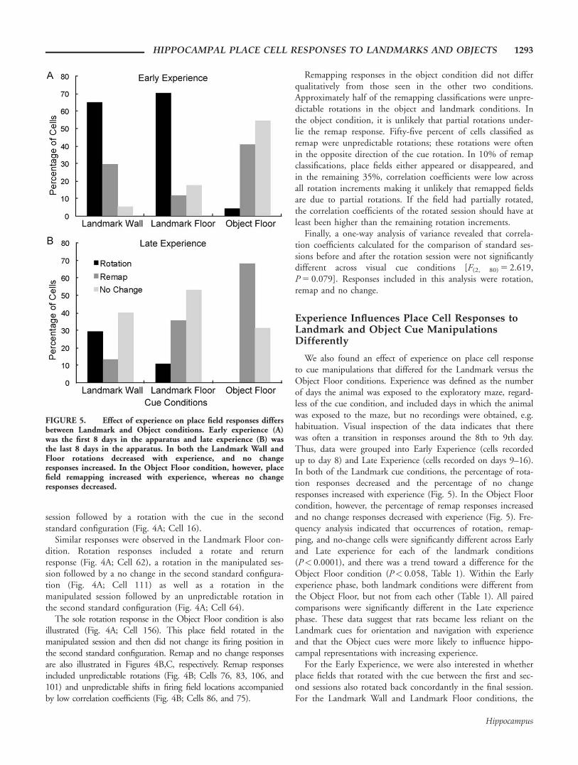
concordance with the visual cue (Fig. 3A). Of the 67 cells inthe Landmark Wall condition, responses of 52.24% were classi-fied as rotation, 22.40% as remap, and 25.37% as no change.The results were similar for the 62 cells in the Landmark Floorcondition; responses of 43.55% of cells were classified as rota-tion, 22.58% as remap, and 33.87% as no change. A v2 analy-sis revealed that responses in the Landmark Wall and Floorconditions were not significantly different (v2
(2) 5 1.296,P 5 0.523). In contrast, in the Object Floor condition, the pre-dominant response to 90 degree cue rotations was a place fieldremap. Of the 57 cells in the Object Floor condition, only1.75% of responses (one cell) were classified as rotation,whereas 57.90% and 40.35% were classified as remap and nochange, respectively. A v2 analysis revealed that responses inthe Object Floor condition were significantly different fromresponses in the Landmark Wall condition (v2
(2) 5 39.210,P < 0.0001) and the Landmark Floor condition(v2
(2) 5 31.761, P < 0.0001). A one-way analysis of variancerevealed the average speed of animals did not significantly dif-fer across conditions (mean 6 SE was 23 6 0.80 for LandmarkFloor, 22 6 0.84 for Landmark Wall, and 21 6 0.85 for ObjectFloor, (F(2,163) 5 1.333, P 5 0.267)), and thus cannot accountfor the differences in responses to cues.
Figure 4 illustrates exemplar responses in each cue condition.Place cell responses were defined as a rotation response if theplace field rotated in concordance with the cue between thefirst standard and rotation sessions, between the rotation ses-sion and the second standard, or both. In the Landmark Wallcondition, rotation responses included a rotation in the manip-ulated session, followed by a return to the original firing fieldlocation (Fig. 4A; Cell 89), a rotation in the manipulated ses-sion followed by a no change in the second standard configura-tion (Fig. 4A; Cell 143), and a no change in the manipulated
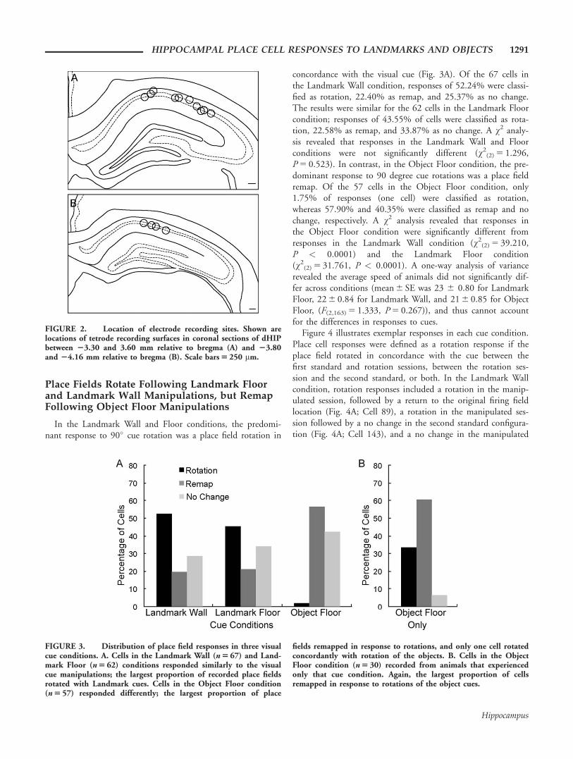
FIGURE 2. Location of electrode recording sites. Shown arelocations of tetrode recording surfaces in coronal sections of dHIPbetween 23.30 and 3.60 mm relative to bregma (A) and 23.80and 24.16 mm relative to bregma (B). Scale bars 5 250 mm.
FIGURE 3. Distribution of place field responses in three visualcue conditions. A. Cells in the Landmark Wall (n 5 67) and Land-mark Floor (n 5 62) conditions responded similarly to the visualcue manipulations; the largest proportion of recorded place fieldsrotated with Landmark cues. Cells in the Object Floor condition(n 5 57) responded differently; the largest proportion of place
fields remapped in response to rotations, and only one cell rotatedconcordantly with rotation of the objects. B. Cells in the ObjectFloor condition (n 5 30) recorded from animals that experiencedonly that cue condition. Again, the largest proportion of cellsremapped in response to rotations of the object cues.
HIPPOCAMPAL PLACE CELL RESPONSES TO LANDMARKS AND OBJECTS 1291
Hippocampus

FIGURE 4. Smoothed rate maps showing exemplar place field responses. Rotation (A),remap (B), and no change (C) responses are shown for the Landmark Wall (left), LandmarkFloor (middle), and Object Floor (right) conditions. Above each rate map series is a schematicshowing the direction of cue rotation. Abbreviations: peak firing field rate (pf), informationcontent score for the first standard session (ic), the correlation coefficient (cc) for the rotationand second standard sessions. [Color figure can be viewed in the online issue, which is avail-able at wileyonlinelibrary.com.]
1292 SCAPLEN ET AL.
Hippocampus
session followed by a rotation with the cue in the secondstandard configuration (Fig. 4A; Cell 16).
Similar responses were observed in the Landmark Floor con-dition. Rotation responses included a rotate and returnresponse (Fig. 4A; Cell 62), a rotation in the manipulated ses-sion followed by a no change in the second standard configura-tion (Fig. 4A; Cell 111) as well as a rotation in themanipulated session followed by an unpredictable rotation inthe second standard configuration (Fig. 4A; Cell 64).
The sole rotation response in the Object Floor condition is alsoillustrated (Fig. 4A; Cell 156). This place field rotated in themanipulated session and then did not change its firing position inthe second standard configuration. Remap and no change responsesare also illustrated in Figures 4B,C, respectively. Remap responsesincluded unpredictable rotations (Fig. 4B; Cells 76, 83, 106, and101) and unpredictable shifts in firing field locations accompaniedby low correlation coefficients (Fig. 4B; Cells 86, and 75).
Remapping responses in the object condition did not differqualitatively from those seen in the other two conditions.Approximately half of the remapping classifications were unpre-dictable rotations in the object and landmark conditions. Inthe object condition, it is unlikely that partial rotations under-lie the remap response. Fifty-five percent of cells classified asremap were unpredictable rotations; these rotations were oftenin the opposite direction of the cue rotation. In 10% of remapclassifications, place fields either appeared or disappeared, andin the remaining 35%, correlation coefficients were low acrossall rotation increments making it unlikely that remapped fieldsare due to partial rotations. If the field had partially rotated,the correlation coefficients of the rotated session should have atleast been higher than the remaining rotation increments.
Finally, a one-way analysis of variance revealed that correla-tion coefficients calculated for the comparison of standard ses-sions before and after the rotation session were not significantlydifferent across visual cue conditions [F(2, 80) 5 2.619,P 5 0.079]. Responses included in this analysis were rotation,remap and no change.
Experience Influences Place Cell Responses toLandmark and Object Cue ManipulationsDifferently
We also found an effect of experience on place cell responseto cue manipulations that differed for the Landmark versus theObject Floor conditions. Experience was defined as the numberof days the animal was exposed to the exploratory maze, regard-less of the cue condition, and included days in which the animalwas exposed to the maze, but no recordings were obtained, e.g.habituation. Visual inspection of the data indicates that therewas often a transition in responses around the 8th to 9th day.Thus, data were grouped into Early Experience (cells recordedup to day 8) and Late Experience (cells recorded on days 9–16).In both of the Landmark cue conditions, the percentage of rota-tion responses decreased and the percentage of no changeresponses increased with experience (Fig. 5). In the Object Floorcondition, however, the percentage of remap responses increasedand no change responses decreased with experience (Fig. 5). Fre-quency analysis indicated that occurrences of rotation, remap-ping, and no-change cells were significantly different across Earlyand Late experience for each of the landmark conditions(P< 0.0001), and there was a trend toward a difference for theObject Floor condition (P< 0.058, Table 1). Within the Earlyexperience phase, both landmark conditions were different fromthe Object Floor, but not from each other (Table 1). All pairedcomparisons were significantly different in the Late experiencephase. These data suggest that rats became less reliant on theLandmark cues for orientation and navigation with experienceand that the Object cues were more likely to influence hippo-campal representations with increasing experience.
For the Early Experience, we were also interested in whetherplace fields that rotated with the cue between the first and sec-ond sessions also rotated back concordantly in the final session.For the Landmark Wall and Landmark Floor conditions, the
FIGURE 5. Effect of experience on place field responses differsbetween Landmark and Object conditions. Early experience (A)was the first 8 days in the apparatus and late experience (B) wasthe last 8 days in the apparatus. In both the Landmark Wall andFloor rotations decreased with experience, and no changeresponses increased. In the Object Floor condition, however, placefield remapping increased with experience, whereas no changeresponses decreased.
HIPPOCAMPAL PLACE CELL RESPONSES TO LANDMARKS AND OBJECTS 1293
Hippocampus

majority of rotation fields returned to the original location(71% and 63%, respectively). In the Object Floor condition,the one field that showed a rotation response did not return tothe same position. With experience, fields were less likely torotate or return to the original configuration.
A number of cells were recorded in multiple conditions onthe same day, and we were interested in how the same cellresponded across different conditions (Fig. 6). Overall, therewas more correspondence across the two Landmark conditions(50%) followed by the two Floor conditions (33%). Responsesof cells across the Landmark Wall and the Object floor showedthe least correspondence (12.5%).
Eight cells were recorded on the same day in both the Land-mark Wall and Landmark Floor conditions. Of these, half ofthe cells responded similarly. Three cells (47, 49, and 58)exhibited rotation responses in both conditions and one cell(41) exhibited no change in both conditions. The remainingfour cells (50, 52, 54, and 57) all remapped in the LandmarkWall condition and rotated in the Landmark Floor condition.Nine cells were recorded in the Landmark Floor and ObjectFloor conditions. Of these, one third of the cells respondedsimilarly. These cells (41, 43, and 44) exhibited no change inboth conditions. The remaining six cells had different responsein the Landmark and Object Floor conditions. The place fieldsof five of these cells (47, 49, 50, 52, and 57) rotated in theLandmark Floor condition, but did not change in the ObjectFloor condition. The remaining cell (58) rotated in the Land-mark Floor condition, but remapped in the Object Floor con-dition. Finally, a total of eight cells were recorded in theLandmark Wall and Object Floor condition. Of these, one cellhad similar responses in both conditions. This cell (41) didnot change its place field location in either condition. Of theremaining seven cells, three cells (47, 49, and 51) rotated inthe Landmark Wall, but did not change their response in theObject Floor condition. One cell (58) rotated in the LandmarkWall and remapped in the Object Floor condition, and threecells (50, 52, and 57) remapped in the Landmark Wall condi-tion and did not change in the Object Floor condition.
The order in which multiple cue conditions were presentedto animals on the same day did not affect the place fieldresponse of the same cell. That is to say, cells recorded in thesame animal on the same day that rotated, remap or did notchange in one condition were equally likely to rotate, remap ornot change in subsequent conditions. Animals presented withthe Object Floor condition before the Landmark Floor or Wallcondition, had cells that rotated in the Landmark conditionsand those same cells remapped or did not change in the ObjectFloor condition (Fig. 7).
To determine if exposure to Landmark conditions influencedwhether the Object Floor cues were used for orientation, threena€ıve animals were implanted and experienced only the ObjectFloor condition. Similar to animals that experienced all threecue conditions, the predominant response to 90� degree Objectcue rotation was a place field remap (Fig. 3B). Rotationresponses, however, were also evident. These animals weretested in a different space in which extraneous cues were bettercontrolled. This is consistent with the observation of the lownumber of “no change” responses. It is also possible thatbecause the animal’s experience in the recording room was lim-ited to one condition as opposed to three, the reduced dailyexperience impeded their ability to notice uncontrolled extrane-ous cues that the other animals exposed to all three conditionswere using. Of the 30 cells recorded, responses of 60% wereclassified as remap, 33.3% as rotation and 6.7% as no change.When these cells were added to the Object Floor cells in Fig-ure 3A, the distribution of cells showing Rotation, Remapping,and No Change responses still differed significantly from distri-butions in the Landmark Wall condition (v2
(2) 5 42.659, P <0.0001) and the Landmark Floor condition ((v2
(2) 5 34.928, P< 0.0001).
These data suggest that rats are more likely to use Objectcues for orientation if they have not experienced peripherallylocated cues (i.e. Landmark cues). Given that coherent rota-tions were not the dominant response, Object cues seem toafford qualitatively different stimulus control over place fieldsthan do Landmark cues.
TABLE 1.
Effects of Experience on Responses to Stimulus Conditions
Within cue condition LW LF OF
Early vs. Late v2(2) 5 26.79;
P< 0.0001
v2(2) 5 22.39;
P< 0.0000
v2(1) 5 3.59;
P< 0.058
Across cue conditions LW vs. LF LW vs. OF LF vs. OF
Early v2(3) 5 33.41;
P< 0.0000
v2(2) 5 5.15;
P< 0.076
v2(2) 5 26.40;
P< 0.0000
v2(2) 5 23.60;
P< 0.0000
Late v2(3) 5 29.33;
P< 0.0000
v2(2) 5 7.08;
P< 0.029
v2(2) 5 25.69;
P< 0.0000
v2(2) 5 8.71;
P< 0.01
v2 analysis of frequencies of cells showing rotation, remapping, or no change in response to cue manipulations. Degrees of freedom are in parentheses. For theearly versus late analysis for the Object Floor Condiition, rotations were not included as there was only one occurance.LF, Landmark Floor; LW, Landmark Wall; OF, Object Floor.
1294 SCAPLEN ET AL.
Hippocampus

DISCUSSION
This study explored the properties of visual cues used by thehippocampus for orientation and navigation. Ours is the firststudy to employ cues restricted to the visual domain to exam-ine the influence of stimulus properties on the use of cues forspatial orientation. We provide evidence for the hypothesis thathippocampal place cells are biased to employ large, distallylocated cues for this purpose. Moreover, such cues can belocated on the wall or the floor in the same way that either atall rock or a pond viewed at a distance can be used for orient-ing and navigating. We manipulated three types of 2D visualcues and examined responses of hippocampal place cells. Rota-tion of large, peripherally located, 2D cues, whether placed ona wall or on the floor adjacent to walls, were more likely toresult in concordant rotations of hippocampal place fields, sug-gesting that both types of cues are used for orientation. Wealso found that place fields responded to manipulations ofsmaller 2D objects on the floor, but not in a way that sug-
gested these stimuli were normally used for orienting. Even ifthe rats experienced only 2D objects, and not larger peripher-ally located cues, manipulation of the 2D object cues was morelikely to result in place field remapping and less likely to resultin concordant rotation. Our data suggest that the hippocampusprocesses visual cues for at least two purposes. Large, distalcues are more likely to be processed as stable landmarks and tobe used for orientation and navigation. In contrast, smaller,more proximal cues are more likely to be processed as non-stationary objects or features in the environment.
The control of hippocampal place cell activity by polarizing2D cues on the wall of an apparatus is well documented(O’Keefe and Conway, 1978; Muller and Kubie, 1987; Fentonet al., 2000a,b), and other studies have used the color of theentire floor as a context indicator (Jeffery and Anderson, 2003;Anderson et al., 2006; Ji and Wilson, 2007). To our knowl-edge, this is the first demonstration that large, polarizing 2Dvisual cues on the floor of an arena and on the wall of an arenaare equally effective in controlling hippocampal place fields. Inaddition, although a few studies have examined the effects of
FIGURE 6. Cells responded similarly for Landmark conditionsand differently across Landmark and Object conditions. Shownare smoothed rate maps showing exemplar responses of cellsrecorded during early (D3–8) experience in all visual cue condi-tions. The peak firing field rate (pf), Information content scorefor the first standard session (ic) and the Correlation Coefficient(cc) for the rotated session and second standard are listed aboveeach map. A. Schematic of condition order and rotation direction.
B. Cell 47 and 49 rotated with cues in the Landmark conditions,but did not change in response to rotations of cues in the Objectcondition. C. Cells 52 and 57 rotated with cues in the LandmarkFloor condition, remapped in the Landmark Wall condition, anddid not change in response to rotations of cues in the ObjectFloor Condition. [Color figure can be viewed in the online issue,which is available at wileyonlinelibrary.com.]
HIPPOCAMPAL PLACE CELL RESPONSES TO LANDMARKS AND OBJECTS 1295
Hippocampus

FIGURE 7. Order of conditions does not impact place fieldresponse. Shown are smoothed rate maps for cells recorded in allcue conditions. Rate maps and schematics are organized in theorder the animal was run in each condition. The peak firing fieldrate (pf), Information content score for the first standard session(ic) and the Correlation Coefficient (cc) for the rotated session
and second standard are listed. Firing rate maps were smoothedfor illustrative purposes. Cells rotated (A–C) or remapped (D)fields in the last, but not previous, conditions, indicating thatorder of conditions did not impact place field responses. [Colorfigure can be viewed in the online issue, which is available atwileyonlinelibrary.com.]
1296 SCAPLEN ET AL.
Hippocampus

manipulation of 3D objects on hippocampal place fields (Cres-sant et al., 1997, 1999; Burke et al., 2011), ours is the firststudy to assess the effects of 2D floor-based cues on hippocam-pal place fields. Finally, we provide the first evidence that hip-pocampal responses to visual cues on the floor are modulateddifferentially consistent with whether or not cues might bebeneficial for navigation.
Why do large floor-based cues adjacent to the perimeter ofan arena and smaller cues away from the perimeter control hip-pocampal place fields in such different ways? A key feature of avisual cue that results in its use as a landmark for orientation issaliency, and size tends to correlate with saliency. Cue sizealone, however, does not determine the degree of stimulus con-trol over place field location (Muller and Kubie, 1987). Caduffand Timpf (2008) argued that saliency is not an inherent prop-erty, but a combined evaluation of the actual feature or cue,the environment, and the observer’s point of view. Other worksuggests that relationship between the position of objects andthe location of the rat impact the perception of object stability(Cressant et al., 1997, 1999). In our study, the 2D objectswere differently shaped and were located away from walls suchthat the appearance would change based on the rat’s vantagepoint. For example, the rat was able to walk between theobjects and the wall. In contrast, the two larger cues located onthe wall or on the floor adjacent to the wall would remain rela-tively unchanged as the rat moves about the maze. Thus, ourdata are consistent with other findings that an interaction ofenvironmental space, characteristics of cues, including size, andvantage point influence the nature of stimulus control overplace field responses.
Remapping was predominant in the object cue conditiondemonstrating that these cues were able to control spatial firingpatterns. It may be, however, that in absence of a more salientlandmark, our object cues were in competition with uncon-trolled extraneous cues. To address this possibility we recordedcells from animals that were exposed only to the Object Floorcondition (Fig. 3B). In that experiment we carefully controlledthe possibility of modulation by extraneous stable cues. Judgingby the low number of cells that exhibited no change responses,we think we were successful in this regard. A relatively largerproportion of cells showed rotation responses, but the predom-inant response of place fields to rotation of object floor cueswas to remap.
A number of studies have addressed the influence of experi-ence on landmark stability. In one study, place cell responses tocue manipulations were examined in a double rotation task inwhich sets of distal visual cues and local multimodal sensorycues were counter rotated by 90 degrees resulting in cue con-flict between local and distal cues (Shapiro et al., 1997). Afterrepeated trials, the percentage of place field rotations concord-ant with distal cues decreased and the percentage of remapresponses increased. The authors suggested that rats learned toencode distinct representations for the organization of stimuliin the double rotation condition and the standard environ-ment. A more consistent finding, however, is that when exter-nal visual cues are non-stationary or unavailable, place cells rely
on idiothetic cues to update spatial representations (Knierimet al., 1995; Sharp et al., 1995; Jeffery et al., 1997, 1998).Consistent with this idea, our data show that an experience-dependent decrease in rotations that are concordant to land-mark cues is accompanied by an increase in no changeresponses. Because the animals were disoriented before place-ment in the arena, idiothetic cues would not have been useful.It is more likely that the animals learned over time that thelandmark cues were not reliable for updating spatial positionand began to rely on extraneous uncontrolled, but stable, envi-ronmental cues. Thus with experience, extraneous uncontrolledcues in the environment may have gained stimulus control overplace fields as the landmark cues were losing stimulus control.
The effects of the 2D object cues on place fields alsochanged with experience, but in a qualitatively different way.As the animal gained more experience in the maze, manipula-tion of the object cues on the floor resulted in increased placefield remapping. Although somewhat speculative, one interpre-tation is that as increased experience led to a heightened aware-ness for stationary extramaze cues, it may have becomeincreasingly apparent to the animals that the objects were mov-ing. This observation may have increased the attention of theanimals to those cues, thus increasing the associability of thecues. Similarly, if the animals viewed the objects as portable,this may have negatively impacted the use of the objects fororientation. The object cues were rarely used for orientation,but the increased remap response with experience suggests thatthe object cues were increasingly available for associative learn-ing and for incorporation into the contextual representation.Data from rat (Biegler and Morris, 1993) and human (Burgesset al., 2004) also suggest that geometric stability is a prerequi-site for a cue to be included into an animal’s representation ofallocentric space, but that the lack of geometric stability doesnot prevent a cue from acquiring associative strength. Eventhough the pair of object cues in the current study was locatedtoward the walls, the physical space between the objects andbetween the objects and walls may have decreased the geomet-ric stability of the cue and its likelihood to be used fororientation.
Two lines of evidence in the present study suggest that thehippocampus represents different classes of cues that could befunctionally described as landmarks and objects. First, manipu-lation of the large distal cues tended to produce concordantrotations, whereas manipulation of the pair of object cuestended to result in place field remapping. Indeed, the singleconcordant rotation in response to rotation of the object cuesmay have been an unpredictable rotation that happened to lineup with the cue rotation. Second, the influence of experienceon place cell responses to cue manipulations was qualitativelydifferent for the large distally located cues as compared withthe pair of object cues located away from the wall. For thelarge distal cues, numbers of rotations concordant with land-mark manipulation decreased with experience and the numberof no change responses increased. In contrast, for the pair ofobject cues, responses of no change decreased with experienceand remap responses increased. Taken together, the evidence
HIPPOCAMPAL PLACE CELL RESPONSES TO LANDMARKS AND OBJECTS 1297
Hippocampus

suggests that the hippocampus processes at least two classes ofcues for two purposes: landmarks are processed for navigationalrelevance and objects are processed for associative properties.
Whether cues are classified as landmarks or objects in thehippocampus or whether the hippocampus relies on otherstructures for this process is an open question. Hippocampallesions do not disrupt the activity of head direction cells, inthe postsubiculum and anterior thalamus or their stabilityacross multiple days in both novel and familiar environments(Golob and Taube, 1997). That animals can still orient inboth without hippocampal processing of landmark or objectssuggests that classification of these cues occurs elsewhere. Onepossibility is that the later and medial entorhinal areas (LEAand MEA, respectively) determine the navigational relevance ofobject and landmark cues. In fact, many studies have shownthat the MEA is involved in processing landmarks for spatialnavigation or orientation (Hafting et al., 2005; Hargreaveset al., 2007; Savelli et al., 2008; but see Clark and Taube,2011). Recent lesion, recording, and immediate early genestudies suggest that the LEA is involved in processing objectand object place information and is therefore a reasonable can-didate for object processing (Deshmukh and Knierim, 2011;Van Cauter et al., 2013; Wilson et al., 2013). Additionalresearch is required to further elucidate the contributions ofthe medial and lateral entorhinal cortices, as well as landmarkand object processing.
To conclude, here we have shown that cues restricted to thevisual modality can impact the behavior of hippocampal placecells. Consistent with studies in other brain regions, our datasuggest that whether such cues are used for orientation dependson size, proximity, and portability. Large, distally locatedobjects are more likely to be used as landmarks for orientationand navigation. Smaller, more proximal, portable objects areless likely to be used for navigation, but appear to be availablefor contextual and associative learning.
REFERENCES
Dreher B, Sefton AJ, Ni SY, Nisbett G. 1985. The morphology, num-ber, distribution and central projections of Class I retinal ganglioncells in albino and hooded rats. Brain Behav Evol 26:10–48.
Fukuda Y. 1977. A three-group classification of rat retinal ganglioncells: Histological and physiological studies. Brain Res 119:327–334.
Salinas-Navarro M, Mayor-Torroglosa S, Jimenez-Lopez M, Aviles-Trigueros M, Holmes TM, Lund RD, Villegas-Perez MP, Vidal-Sanz M. 2009. A computerized analysis of the entire retinal gan-glion cell population and its spatial distribution in adult rats.Vision Res 49:115–126.
Anderson MI, Killing S, Morris C, O’Donoghue A, Onyiagha D,Stevenson R, Verriotis M, Jeffery KJ. 2006. Behavioral correlates ofthe distributed coding of spatial context. Hippocampus 16:730–742.
Auger SD, Mullally SL, Maguire EA. 2012. Retrosplenial cortex codesfor permanent landmarks. PLoS One 7:e43620.
Biegler R, Morris RG. 1993. Landmark stability is a prerequisite forspatial but not discrimination learning. Nature 361:631–633.
Burgess N, Spiers HJ, Paleologou E. 2004. Orientational manoeuvresin the dark: Dissociating allocentric and egocentric influences onspatial memory. Cognition 94:149–166.
Burke SN, Maurer AP, Nematollahi S, Uprety AR, Wallace JL, BarnesCA. 2011. The influence of objects on place field expression andsize in distal hippocampal CA1. Hippocampus 21:783–801.
Burwell RD, Hafeman DM. 2003. Positional firing properties of post-rhinal cortex neurons. Neuroscience 119:577–588.
Caduff D, Timpf S. 2008. On the assessment of landmark saliencefor human navigation. Cognit Process 9:249–267.
Clark BJ, Taube JS. 2011. Intact landmark control and angular pathintegration by head direction cells in the anterodorsal thalamus afterlesions of the medial entorhinal cortex. Hippocampus 21:767–782.
Cressant A, Muller RU, Poucet B. 1997. Failure of centrally placedobjects to control the firing fields of hippocampal place cells.J Neurosci 17:2531–2542.
Cressant A, Muller RU, Poucet B. 1999. Further study of the control ofplace cell firing by intra-apparatus objects. Hippocampus 9:423–431.
Deshmukh SS, Knierim JJ. 2011. Representation of non-spatial andspatial information in the lateral entorhinal cortex. Front BehavNeurosci 5:69.
Fenton AA, Csizmadia G, Muller RU. 2000a. Conjoint control ofhippocampal place cell firing by two visual stimuli. II. A vector-field theory that predicts modifications of the representation of theenvironment. J Gen Physiol 116:211–221.
Fenton AA, Csizmadia G, Muller RU. 2000b. Conjoint control of hip-pocampal place cell firing by two visual stimuli. I. The effects of mov-ing the stimuli on firing field positions. J Gen Physiol 116:191–209.
Fox SE, Ranck JB, Jr.. 1981. Electrophysiological characteristics of hippo-campal complex-spike cells and theta cells. Exp Brain Res 41:399–410.
Furtak SC, Cho CE, Kerr KM, Barredo JL, Alleyne JE, Patterson YR,Burwell RD. 2009. The Floor Projection Maze: A novel behavioralapparatus for presenting visual stimuli to rats. J Neurosci Methods181:82–88.
Golob EJ, Taube JS. 1997. Head direction cells and episodic spatialinformation in rats without a hippocampus. Proc Natl Acad SciUSA 94:7645–7650.
Hafting T, Fyhn M, Molden S, Moser MB, Moser EI. 2005. Microstruc-ture of a spatial map in the entorhinal cortex. Nature 436:801–806.
Hargreaves EL, Yoganarasimha D, Knierim JJ. 2007. Cohesiveness ofspatial and directional representations recorded from neural ensem-bles in the anterior thalamus, parasubiculum, medial entorhinalcortex, and hippocampus. Hippocampus 17:826–841.
Jacobson TK, Ho JW, Kent BW, Yang F-C, Burwell RD. 2014. Auto-mated visual cognitive tasks for recording neural activity using afloor projection maze. J Vis Exp 84:9.
Jeffery KJ. 1998. Learning of landmark stability and instability by hip-pocampal place cells. Neuropharmacology 37:677–687.
Jeffery KJ, Anderson MI. 2003. Dissociation of the geometric andcontextual influences on place cells. Hippocampus 13:868–872.
Jeffery KJ, Donnett JG, Burgess N, O’Keefe JM. 1997. Directionalcontrol of hippocampal place fields. Exp Brain Res 117:131–142.
Ji D, Wilson MA. 2007. Coordinated memory replay in the visualcortex and hippocampus during sleep. Nat Neurosci 10:100–107.
Knierim JJ. 2002. Dynamic interactions between local surface cues,distal landmarks, and intrinsic circuitry in hippocampal place cells.J Neurosci 22:6254–6264.
Knierim JJ, Kudrimoti HS, McNaughton BL. 1995. Place cells, headdirection cells, and the learning of landmark stability. J Neurosci15:1648–1659.
Lashley KS. 1938. The mechanism of vision: XV. Preliminary studiesof the rat’s capacity for detail vision. J Gen Psychol 18:123–193.
Leutgeb JK, Leutgeb S, Treves A, Meyer R, Barnes CA, McNaughtonBL, Moser MB, Moser EI. 2005. Progressive transformation ofhippocampal neuronal representations in "morphed" environments.Neuron 48:345–358.
1298 SCAPLEN ET AL.
Hippocampus

Leutgeb S, Leutgeb JK, Treves A, Moser MB, Moser EI. 2004. Dis-tinct ensemble codes in hippocampal areas CA3 and CA1. Science305:1295–1298.
Markus EJ, Qin YL, Leonard B, Skaggs WE, McNaughton BL,Barnes CA. 1995. Interactions between location and task affect thespatial and directional firing of hippocampal neurons. J Neurosci15:7079–7094.
Minini L, Jeffery KJ. 2006. Do rats use shape to solve "shape discrim-inations"? Learn Mem 13:287–297.
Mullally SL, Maguire EA. 2011. A new role for the parahippocampalcortex in representing space. J Neurosci 31:7441–7449.
Mullally SL, Maguire EA. 2013. Exploring the role of space-definingobjects in constructing and maintaining imagined scenes. BrainCognit 82:100–107.
Muller RU, Kubie JL. 1987. The effects of changes in the environ-ment on the spatial firing of hippocampal complex-spike cells.J Neurosci 7:1951–1968.
Muller RU, Kubie JL, Ranck JB Jr. 1987. Spatial firing patterns ofhippocampal complex-spike cells in a fixed environment.J Neurosci 7:1935–1950.
O’Keefe J, Dostrovsky J. 1971. The hippocampus as a spatial map.Preliminary evidence from unit activity in the freely-moving rat.Brain Res 34:171–175.
O’Keefe J, Conway DH. 1978. Hippocampal place units in the freely mov-ing rat: Why they fire where they fire. Exp Brain Res 31:573–590.
O’Keefe J, Nadel L. 1978. The Hippocampus as a Cognitive Map.Oxford: Clarendon.
Savelli F, Yoganarasimha D, Knierim JJ. 2008. Influence of boundaryremoval on the spatial representations of the medial entorhinal cor-tex. Hippocampus 18:1270–1282.
Shapiro ML, Tanila H, Eichenbaum H. 1997. Cues that hippocampalplace cells encode: Dynamic and hierarchical representation of localand distal stimuli. Hippocampus 7:624–642.
Sharp PE, Blair HT, Etkin D, Tzanetos DB. 1995. Influencesof vestibular and visual motion information on the spatialfiring patterns of hippocampal place cells. J Neurosci 15:173–189.
Skaggs WE, McNaughton BL, Gothard KM, Markus EJ. 1993. Aninformation-theoretic approach to deciphering the hippocampalcode. In: Hanson SJ, Cowan JD, Giles CL, editors. Advances inNeural Information Processing Systems 5. San Mateo, CA: MorganKaufmann Publishers. pp 1030–1037.
Tanila H, Shapiro ML, Eichenbaum H. 1997. Discordance of spatialrepresentation in ensembles of hippocampal place cells. Hippocam-pus 7:613–623.
Van Cauter T, Camon J, Alvernhe A, Elduayen C, Sargolini F, Save E.2013. Distinct roles of medial and lateral entorhinal cortex in spa-tial cognition. Cereb Cortex 23:451–459.
Wallace DJ, Greenberg DS, Sawinski J, Rulla S, Notaro G, Kerr JN.2013. Rats maintain an overhead binocular field at the expense ofconstant fusion. Nature 498:65–69.
Wilson DI, Langston RF, Schlesiger MI, Wagner M, Watanabe S,Ainge JA. 2013. Lateral entorhinal cortex is critical for novelobject-context recognition. Hippocampus 23:352–366.
HIPPOCAMPAL PLACE CELL RESPONSES TO LANDMARKS AND OBJECTS 1299
Hippocampus